ABSTRACT
Injuries to the distal biceps occur at the tendinous insertion at the radial tuberosity. Distal biceps injuries range from tendinosis to partial tears to non-retracted and retracted complete tears. Acute and chronic complete tears result from a tendinous avulsion at the radial tuberosity. Acute tears result from a strong force exerted on an eccentric biceps contraction, leading to tendon injury.
Distal biceps tendon injuries are uncommon (1.2 per 100,000 patients in one study).1 An underlying degenerative component is involved in all distal biceps tendon tears and tendinosis.2 Partial tears can be caused by the same mechanism or by no particular inciting event.3 Magnetic resonance imaging (MRI) is the optimal imaging modality for distal tendon tears because of its excellent specificity and sensitivity in the detection of complete tears.4,5 Imaging also accurately diagnoses and characterizes partial tears and tendinosis.5 On MRI, fast spin-echo intermediate-weighted and T2-weighted or short tau inversion recovery (STIR) sequences are normally obtained to assess tendon integrity. Along with standard axial and sagittal views, the FABS (flexed elbow, abducted shoulder, supinated forearm) view is an important tool in the diagnosis of distal biceps tendon tears.6 The FABS view is obtained with the patient prone with the shoulder abducted 180° (above the head), with the elbow flexed to 90°, and the forearm supinated. This position allows a longitudinal view of along the entire length of the distal tendon.
Complete distal biceps tears can usually be diagnosed by history and physical examinations. However, imaging can be helpful when intact brachialis function can compensate for a completely torn tendon. MRI is also useful in the setting of a complete tear to locate the torn tendon stump, and assess the degree of retraction for tendon retrieval7,8 and quality of the tendon stump for repair. For associated rupture of the lacertus, the degree of proximal tendon retraction can be significant (Figures 1A, 1B).
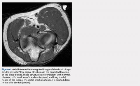
Given that distal biceps tendon rupture occurs as an avulsion at the tendon-bone interface (Figure 2), complete distal biceps tendon tears typically demonstrate no tendon at the insertion on the radial tuberosity with a fluid-filled tendon gap with edema and/or hemorrhage7,9 or an ill-defined T2-hyperintense mass at the expected site of the tendon.7 Complete tears without rupture of the lacertus fibrosis (bicipital aponeurosis) will have a small amount of retraction because the intact aponeurosis tethers the torn tendon stump (Figures 3A-3C). Chronic complete tears demonstrate heterogeneous signal intensity and fluid signal at the tendon, as well as muscle belly atrophy.9 A small percentage of distal biceps brachii tendons are bifid 10 (Figure 4). When injured, 75% have complete rupture of the short head with 17% of these having additional complete rupture of the long head, whereas 50% of those with complete rupture of the short head have partial tear or tendinosis of the long head.Continue to: Partial distal bicep tears...


