DALLAS – Deadly invasive fungal infections are increasingly being identified in soft-tissue and intra-abdominal wounds in troops deployed to Iraq and Afghanistan, prompting the creation of hospital-based clinical practice guidelines.
When the first case emerged in July 2009, the medical team initially suspected significant bacterial infection in a young, previously healthy Marine with extensive soft-tissue injuries. By the time appropriate antifungal therapies were initiated in week 3, it was too late. An autopsy revealed that the 22-year-old soldier’s death was from systemic angioinvasive Aspergillus terreus (Surg. Infect. [Larchmt.] 2011;12:397-400).
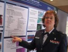
"We were not expecting the degree of mold infestation early on," Col. Debra Malone, MC, USAF, trauma research director of the Walter Reed National Military Medical Center (NMMC) in Bethesda, Md., said in an interview. "It’s not necessarily that things were missed – it’s that the disease itself didn’t present early on.
"Sometimes it can take 2, 3, 6 weeks for certain molds to actually grow. Now we don’t wait for them to grow. If we have the clinical suspicion, we treat right away with very powerful antimold medications."
The NMMC has also developed a 24-hour invasive fungal infection (IFI) pathology protocol and hospital-based clinical practice guidelines to improve outcomes for trauma-related IFI, which has a reported mortality rate of 25% in previously immunocompetent patients.
From July 2009 to November 2011, IFI was suspected or identified on pathology in 75 of 2,755 trauma patients admitted to the NMMC, Dr. Malone reported at the annual meeting of the Surgical Infection Society.
Although the index case occurred in the heavily agricultural Helmand Province in Afghanistan, IFI cases are cropping up throughout Iraq and Afghanistan, particularly in troops with injuries caused by improvised explosive devices (IEDs).
"It’s not uncommon for the bomb to cause a big crater in the ground, and all that dirt or whatever has been displaced," she said. "We think it’s been driven up into the body, deep into the open wounds, and that the mold spores are sitting up there, just percolating."
The typical patient is an 18-year-old with devastating soft-tissue IED blast injuries who is resuscitated and has wounds debrided to normal tissue. However, 5-7 days later, there is evidence of infected tissue on successive washouts, and the patient is hypertensive and in shock.
Frozen-section histopathology was initially used for diagnosis because of its quick turnaround, but resulted in a 33% negative predictive value in their lab and is no longer used, Dr. Malone said. This led to the 24-hour IFI protocol utilizing permanent sections. Periodic acid-Schiff and Gomori methenamine silver staining have proved efficacious for Aspergillus species, but not as efficacious for identifying Mucorales species. Calcofluor white staining is also used and may allow morphology-based speciation.
In all, 42 (56%) of the 75 patients met histologic criteria for IFI. Notably, IFI was suspected clinically in the remaining patients but never proved by histologic criteria. More than a fourth (28%) of wounds infected with Mucor were coinfected with non-Mucor species. In addition, the vast majority of wounds will be coinfected with bacteria, according to the investigators, led by Dr. Carlos Rodriguez, an attending trauma and critical care surgeon at NMMC.
Therapy should include aggressive debridement, with minimization of blood loss. Surgery should be repeated every 24-48 hours until the wounds are clean, with compromised tissue sent from every surgery for histopathology and culture. Changes in surgical practice as related to the treatment of nonnecrotic tissue abnormalities may have led to improvement in tissue salvage.
Broad-spectrum antifungal medications should be tailored based on serial tissue specimen culture results. Focused antifungal therapies should be continued for 2-3 weeks after wound closure. Liposomal amphotericin B and the triazoles – voriconazole (Vfend) and posaconazole (Noxafil) – have been used as initial therapies. Vancomycin (Vancocin) and meropenem (Merrem IV) also should be considered because of the high risk of coinfection with bacteria.
Adjunctive therapies include minimization of immunosuppression, antimicrobial beads in open wounds, and negative-pressure wound therapy with a 0.025% Dakin’s solution (sodium hypochlorite). Intra-abdominal washings with amphotericin B, voriconazole, or bacitracin plus gentamicin can be utilized on a case-by-case basis.
Among the 75 patients, the time from injury to IFI diagnosis decreased from 10 days (range 7-12) in 2009 to 3 days (range 2-6 days) in 2011. The mortality rate was 4%; mean ICU length of stay, 13 days; and mean hospital stay, 48 days. All IFI patients were male; their mean age was 24 years, and their mean Injury Severity Score was 23.

