User login
Survival similar with hearts donated after circulatory or brain death
in the first randomized trial comparing the two approaches.
“This randomized trial showing recipient survival with DCD to be similar to DBD should lead to DCD becoming the standard of care alongside DBD,” lead author Jacob Schroder, MD, surgical director, heart transplantation program, Duke University Medical Center, Durham, N.C., said in an interview.
“This should enable many more heart transplants to take place and for us to be able to cast the net further and wider for donors,” he said.
The trial was published online in the New England Journal of Medicine.
Dr. Schroder estimated that only around one-fifth of the 120 U.S. heart transplant centers currently carry out DCD transplants, but he is hopeful that the publication of this study will encourage more transplant centers to do these DCD procedures.
“The problem is there are many low-volume heart transplant centers, which may not be keen to do DCD transplants as they are a bit more complicated and expensive than DBD heart transplants,” he said. “But we need to look at the big picture of how many lives can be saved by increasing the number of heart transplant procedures and the money saved by getting more patients off the waiting list.”
The authors explain that heart transplantation has traditionally been limited to the use of hearts obtained from donors after brain death, which allows in situ assessment of cardiac function and of the suitability for transplantation of the donor allograft before surgical procurement.
But because the need for heart transplants far exceeds the availability of suitable donors, the use of DCD hearts has been investigated and this approach is now being pursued in many countries. In the DCD approach, the heart will have stopped beating in the donor, and perfusion techniques are used to restart the organ.
There are two different approaches to restarting the heart in DCD. The first approach involves the heart being removed from the donor and reanimated, preserved, assessed, and transported with the use of a portable extracorporeal perfusion and preservation system (Organ Care System, TransMedics). The second involves restarting the heart in the donor’s body for evaluation before removal and transportation under the traditional cold storage method used for donations after brain death.
The current trial was designed to compare clinical outcomes in patients who had received a heart from a circulatory death donor using the portable extracorporeal perfusion method for DCD transplantation, with outcomes from the traditional method of heart transplantation using organs donated after brain death.
For the randomized, noninferiority trial, adult candidates for heart transplantation were assigned to receive a heart after the circulatory death of the donor or a heart from a donor after brain death if that heart was available first (circulatory-death group) or to receive only a heart that had been preserved with the use of traditional cold storage after the brain death of the donor (brain-death group).
The primary end point was the risk-adjusted survival at 6 months in the as-treated circulatory-death group, as compared with the brain-death group. The primary safety end point was serious adverse events associated with the heart graft at 30 days after transplantation.
A total of 180 patients underwent transplantation, 90 of whom received a heart donated after circulatory death and 90 who received a heart donated after brain death. A total of 166 transplant recipients were included in the as-treated primary analysis (80 who received a heart from a circulatory-death donor and 86 who received a heart from a brain-death donor).
The risk-adjusted 6-month survival in the as-treated population was 94% among recipients of a heart from a circulatory-death donor, as compared with 90% among recipients of a heart from a brain-death donor (P < .001 for noninferiority).
There were no substantial between-group differences in the mean per-patient number of serious adverse events associated with the heart graft at 30 days after transplantation.
Of 101 hearts from circulatory-death donors that were preserved with the use of the perfusion system, 90 were successfully transplanted according to the criteria for lactate trend and overall contractility of the donor heart, which resulted in overall utilization percentage of 89%.
More patients who received a heart from a circulatory-death donor had moderate or severe primary graft dysfunction (22%) than those who received a heart from a brain-death donor (10%). However, graft failure that resulted in retransplantation occurred in two (2.3%) patients who received a heart from a brain-death donor versus zero patients who received a heart from a circulatory-death donor.
The researchers note that the higher incidence of primary graft dysfunction in the circulatory-death group is expected, given the period of warm ischemia that occurs in this approach. But they point out that this did not affect patient or graft survival at 30 days or 1 year.
“Primary graft dysfunction is when the heart doesn’t fully work immediately after transplant and some mechanical support is needed,” Dr. Schroder commented to this news organization. “This occurred more often in the DCD group, but this mechanical support is only temporary, and generally only needed for a day or two.
“It looks like it might take the heart a little longer to start fully functioning after DCD, but our results show this doesn’t seem to affect recipient survival.”
He added: “We’ve started to become more comfortable with DCD. Sometimes it may take a little longer to get the heart working properly on its own, but the rate of mechanical support is now much lower than when we first started doing these procedures. And cardiac MRI on the recipient patients before discharge have shown that the DCD hearts are not more damaged than those from DBD donors.”
The authors also report that there were six donor hearts in the DCD group for which there were protocol deviations of functional warm ischemic time greater than 30 minutes or continuously rising lactate levels and these hearts did not show primary graft dysfunction.
On this observation, Dr. Schroder said: “I think we need to do more work on understanding the ischemic time limits. The current 30 minutes time limit was estimated in animal studies. We need to look more closely at data from actual DCD transplants. While 30 minutes may be too long for a heart from an older donor, the heart from a younger donor may be fine for a longer period of ischemic time as it will be healthier.”
“Exciting” results
In an editorial, Nancy K. Sweitzer, MD, PhD, vice chair of clinical research, department of medicine, and director of clinical research, division of cardiology, Washington University in St. Louis, describes the results of the current study as “exciting,” adding that, “They clearly show the feasibility and safety of transplantation of hearts from circulatory-death donors.”
However, Dr. Sweitzer points out that the sickest patients in the study – those who were United Network for Organ Sharing (UNOS) status 1 and 2 – were more likely to receive a DBD heart and the more stable patients (UNOS 3-6) were more likely to receive a DCD heart.
“This imbalance undoubtedly contributed to the success of the trial in meeting its noninferiority end point. Whether transplantation of hearts from circulatory-death donors is truly safe in our sickest patients with heart failure is not clear,” she says.
However, she concludes, “Although caution and continuous evaluation of data are warranted, the increased use of hearts from circulatory-death donors appears to be safe in the hands of experienced transplantation teams and will launch an exciting phase of learning and improvement.”
“A safely expanded pool of heart donors has the potential to increase fairness and equity in heart transplantation, allowing more persons with heart failure to have access to this lifesaving therapy,” she adds. “Organ donors and transplantation teams will save increasing numbers of lives with this most precious gift.”
The current study was supported by TransMedics. Dr. Schroder reports no relevant financial relationships.
A version of this article first appeared on Medscape.com.
FROM THE NEW ENGLAND JOURNAL OF MEDICINE
FDA class 1 recall for some Abiomed Impella heart pumps
“If a purge leak occurs, the system will experience low purge pressures, prompting alarms and requiring evaluation,” the U.S. Food and Drug Administration says in an advisory posted on its website.

“If the leak issue is not resolved, persistent low purge pressure and purge flow may lead to pump stop and loss of therapy. In patients who are critical, failure of the pump’s support can lead to further deterioration and worsening of their already critical condition and may even lead to serious injury or death,” the FDA says.
The FDA has identified this as a class I recall, the most serious type, because of the potential for serious injury or death.
To date, Abiomed says it has received 179 complaints; there have been three injuries and no deaths related to this problem.
The Impella 5.5 with SmartAssist System is used for up to 14 days to support the ventricles in the setting of ongoing cardiogenic shock that occurs less than 48 hours after acute myocardial infarction, open-heart surgery, or when the heart is not functioning well owing to cardiomyopathy.
All the devices that are being recalled were distributed from September 2021 to March 2023. Detailed product information is available on the FDA website.
Abiomed has sent an urgent medical device recall letter to customers asking them to review their inventory to check for any recalled product and to contact Abiomed customer support to coordinate return of the product.
Customers are advised not to use affected products unless no other product is available. The letter includes “best practices” for situations in which no other option is available and the device must be used until a replacement is available.
Customers with questions about this recall should contact Abiomed’s clinical support center at 1-800-422-8666.
A version of this article was first published on Medscape.com.
“If a purge leak occurs, the system will experience low purge pressures, prompting alarms and requiring evaluation,” the U.S. Food and Drug Administration says in an advisory posted on its website.
“If the leak issue is not resolved, persistent low purge pressure and purge flow may lead to pump stop and loss of therapy. In patients who are critical, failure of the pump’s support can lead to further deterioration and worsening of their already critical condition and may even lead to serious injury or death,” the FDA says.
The FDA has identified this as a class I recall, the most serious type, because of the potential for serious injury or death.
To date, Abiomed says it has received 179 complaints; there have been three injuries and no deaths related to this problem.
The Impella 5.5 with SmartAssist System is used for up to 14 days to support the ventricles in the setting of ongoing cardiogenic shock that occurs less than 48 hours after acute myocardial infarction, open-heart surgery, or when the heart is not functioning well owing to cardiomyopathy.
All the devices that are being recalled were distributed from September 2021 to March 2023. Detailed product information is available on the FDA website.
Abiomed has sent an urgent medical device recall letter to customers asking them to review their inventory to check for any recalled product and to contact Abiomed customer support to coordinate return of the product.
Customers are advised not to use affected products unless no other product is available. The letter includes “best practices” for situations in which no other option is available and the device must be used until a replacement is available.
Customers with questions about this recall should contact Abiomed’s clinical support center at 1-800-422-8666.
A version of this article was first published on Medscape.com.
Noncardiac mortality is not increased by revascularization in a meta-analysis: New data refute recent study
In response to a randomized trial that associated elective revascularization for ischemia with an increase in noncardiac mortality versus medical therapy alone, a meta-analysis with a far larger dataset challenges this assertion, suggesting the initial conclusion is due to a type 1 error.
, reports William Wijns, MD, PhD, professor of interventional cardiology, National University of Ireland, Galway.
The larger pool of data from the meta-analysis was considered compelling by several experts at the annual meeting of the European Association of Percutaneous Cardiovascular Interventions, where it was presented.
“I think these data will close once and forever this controversy,” said Davide Capodanno, MD, PhD, a professor of cardiology and interventional cardiologist at the University of Catania (Italy).
Evidence for an unexpected increased risk of noncardiac mortality was drawn from the ISCHEMIA-EXTEND study, which was published earlier this year. Numerous prior studies comparing percutaneous intervention (PCI) to medical therapy for relief of ischemia had shown no such safety signal.
The ISCHEMIA-EXTEND study provided long-term follow up of patients enrolled in ISCHEMIA, a study that randomized patients with stable coronary disease and moderate or severe ischemia to PCI or a conservative approach. After 3.2 years of follow up, there was no reduction in risk of cardiovascular events or all-cause death. While this lack of benefit was a disappointing result from the perspective of interventional cardiology, there was also no increase in these risks.
In ISCHEMIA-EXTEND, the more than 5,000 patients originally randomized were followed for an additional 2.5 years (total 5.7 years). During this extended period, the estimated 7-year risk of cardiovascular mortality was 22% lower in the group randomized to PCI (hazard ratio, 0.78; 95% confidence interval, 0.63-0.96) but the noncardiac mortality was increased by 44% (HR, 1.44; 95% CI, 1.08-1.91). Because of the counterbalancing effects on survival, all-cause mortality was similar in the two groups.
The newly completed meta-analysis was undertaken to address this surprising result not least because the increased rates of noncardiac death did not have a plausible explanation, according to Dr. Wijns.
When the patients from the 18 randomized trials were compared, noncardiac death occurred in 4.68% of the 8,665 patients assigned to elective revascularization and in 4.17% of the 8,243 patients assigned to medical therapy alone at an average follow up of 5.7 years.
This difference was not significant overall (HR, 1.09; 95% CI, 0.94-1.26; P = .26) or after sensitivity analyses. For example, there was no difference (P = .52) between an invasive or conservative approach after controlling for length of follow up.
There was also no heterogeneity (I2 = 0%) among the studies when ISCHEMIA-EXTEND was excluded.
Absence of negative effect ‘is confirmed’
On the basis of a Bayesian meta-analysis designed to account for residual uncertainty (relative risk, 1.08, 95% CI, 0.90-1.30) and the consistency of results among all studies with the exception of ISCHEMIA-EXTEND (RR, 1.0; 95% CI, 0.84-1;18; P = .7), “the absence of a negative effect of revascularization on noncardiac death was confirmed,” Dr. Wijns reported.
Based on the preponderance of evidence assembled in this meta-analysis, the “noncardiac mortality excess risk observed following revascularization relative to medical therapy was confined to a single large trial and is likely due to a type 1 error,” Dr. Wijns reported. He noted that this study is “the first large-scale meta-analysis study designed to systematically evaluate potential differences in noncardiac mortality between treatment strategies for chronic coronary syndromes.”
Eliano P. Navarese, MD, PhD, an associate professor of interventional cardiology at Nicolaus Copernicus University, Bydgoszcz, Poland, was the lead author of this study and Dr. Wijns was a coinvestigator. The study was published simultaneously in the Journal of the American College of Cardiology at the time of the EuroPCR meeting.
In the late-breaking session where these data were presented, there was a general consensus among invited panelists that the data are convincing. For example, Michael Joner, MD, PhD, director of early clinical trials, German Heart Centre, Munich, agreed that these data “resolve the issue.”
Bernard de Bruyne, MD, PhD, an interventional cardiologist associated with the Cardiovascular Center Aalst, Kraainem, Belgium, also agreed that these data argue convincingly against the concern raised by publication of ISCHEMIA-EXTEND, but he added that this controversy has raised an important issue.
“We should always be reporting all-cause mortality, not just cardiovascular mortality, in our clinical trials,” he said, emphasizing that extending all-cause survival, not just preventing cardiovascular-related events, should be recognized as the goal of invasive strategies.
In an editorial accompanying the publication, Dr. Harvey D. White, MD, Te Whatu Ora-Health New Zealand, Auckland, writes similarly that the current findings, “alert us to the importance of adjudicating causes of death in clinical trials.
“The current trial-level meta-analysis may seem to dispel concerns about increases in noncardiac and cardiovascular deaths seen in some revascularization trials, but paradoxically, it has raised the need for more and careful analysis of causes of death,” Dr. White notes. He feels the signal of increased noncardiac or noncardiovascular death in ISCHEMIA EXTEND and the REVIVED trials is something “that we should pay attention to and explore the possibility that increased radiation doses with PCI may cause increased rates of cancer.”
Further study, including longer follow-up, other datasets, and quality of life data including cognitive function and “patient-focused outcomes such as day alive out of hospital,” is needed, he concludes.
Dr. Navarese has received research grants from Abbott and Amgen and lecture fees/honoraria from Amgen, AstraZeneca, Bayer, Pfizer, and Sanofi-Regeneron. Dr. Wijns reports financial relationships with Argonauts, Corrib Core Laboratory, and Rede Optimus Research. Dr. Capodanno reports financial relationships with Amgen, Daiichi Sankyo, and Sanofi. Dr. de Bruyne and Dr. Joner report financial relationships with multiple pharmaceutical and device manufacturers. Prof. White, as the John Neutze scholar, is supported by the Green Lane Research and Educational Fund. Prof. White has received grant support paid to the institution and fees for serving on steering committees of multiple trials sponsored by various companies.
FROM EUROPCR 2023
Ticagrelor, DAPT equal in preventing repeat revascularization
PHOENIX – Post hoc analysis of the randomized TWILIGHT trial comparing ticagrelor alone with ticagrelor plus aspirin in high-risk patients after percutaneous coronary intervention (PCI) shows both regimens were similarly effective in preventing repeat revascularization after 1 year.
In TWILIGHT, the main findings of which were previously published in the New England Journal of Medicine, 7,119 high-risk PCI patients on standard dual antiplatelet therapy (DAPT) of ticagrelor plus aspirin for 3 months were randomized to continuation of DAPT or to ticagrelor plus placebo for 12 months.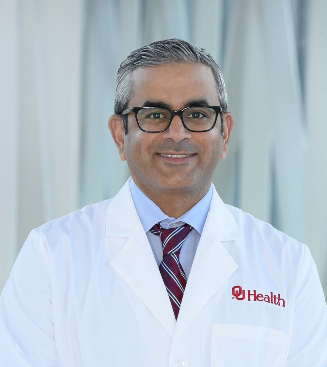
The new post hoc analysis included 6,759 patients and shows the rates of clinically driven revascularization were similar between the two groups: 7.1% and 6.6% for the ticagrelor monotherapy and ticagrelor-based DAPT groups, respectively (P = .363).
The findings were presented at the Society for Cardiovascular Angiography & Interventions annual scientific sessions.
Three key findings come from the post hoc analysis, Usman Baber, MD, director of the cardiac catheterization lab and associate professor at the University of Oklahoma Health Sciences Center, Oklahoma City, who presented the findings, said in an interview.
“The first is that, over the 1-year follow-up of our trial, we found that a repeat revascularization event occurred in 6.7% of patients,” he said. “We found that a slight majority of these repeat revascularization events were due to events at the target lesion or target vessel; and we found that most of the repeat revascularization events actually occurred in patients without a concomitant acute coronary syndrome. In other words, these were essentially stable patients when they were getting their repeat revascularization.”
The second major finding was that these high-risk patients who had repeat revascularization were at three times greater risk for major adverse cardiac and cerebrovascular events (MACCE), based on a multivariable adjusted model, Dr. Baber said.
“And then third is that he said.
Repeat revascularization
The goal of the analysis was to focus on clinically driven repeat revascularization as an outcome, Dr. Baber said. The analysis also aimed to understand the association between repeat revascularization and subsequent risk.
Secondary endpoints included target lesion revascularization (TLR); target vessel revascularization (TVR); MACCE, including clinically driven revascularization; and net adverse clinical events (NACE), a composite of MACCE or Bleeding Academic Research Consortium (BARC) 2, 3, or 5 bleeding.
The outcomes of all those endpoints, except for NACE, were similar, Dr. Baber said. “Overall, ticagrelor monotherapy, as expected, reduced rates of bleeding as compared with ticagrelor plus aspirin,” he said. The rates of NACE were 12.2% versus 14.6%, respectively (P = .004). For BARC 2,3, or 5 bleeding, the rates were 3.4% versus 7.1% (P < .001).
The findings validated repeat revascularization as a meaningful endpoint, Dr. Baber said. “Certainly, we don’t elevate repeat revascularization as an endpoint to the same level as death or stroke, but certainly this analysis and some others prior to it highlight the fact that when these patients come back for repeat revascularization, even if they’re stable, they clearly are at elevated risk for future ischemic events,” he said.
One limitation of the analysis is that the data are from a clinical trial, “which renders the findings not as generalizable to the broader patients in a clinical practice,” he said. However, the TWILIGHT data are validated and adjusted for multiple risk factors.
“When patients come in and they have a repeat revascularization, should there be a consideration to placing them on more intensive antithrombotic therapy?” he asked. “Right now, if patients have a repeat revascularization event and they’re stable, guidelines and clinical practice usually calls for continuing clopidogrel, but again our study and others like it indicate these patients are at a higher thrombotic risk, so maybe there’s a rationale for at least a short course of a more potent antiplatelet agent in such patients.”
The post hoc findings confirm those of the primary TWILIGHT trial, Lorenzo Azzalini, MD, PhD, MSc, director of interventional cardiology research at the University of Washington Medical Center, Seattle, said in an interview.
“It’s not surprising to find no difference between the two therapies with regard to unplanned revascularization,” he said. “It’s considered that only stent thrombosis can only actually be mitigated by the drugs being investigated in the trial; all the other ischemic endpoints reflect more chronic ischemia—TLR or known TVR—upon which ticagrelor and aspirin do not play any role.”
However, he added, “I still think this study provides useful information to the community in a period of intense scrutiny on the relative benefits and merits of PCI versus CABG [coronary artery bypass graft], and this study confirms that shortening DAPT to 3 months and then continuing with just ticagrelor does not bring any penalty in terms of ischemic events or repeat revascularization.”
TWILIGHT enrolled high-risk patients, but not “very-high-risk” patients, Dr. Azzalini noted. The enrollment criteria excluded patients on chronic anticoagulation, who had a prior stroke or liver sclerosis, or were on dialysis.
“Future trials should focus more on very-high-risk patients because these are the patients that we deal with on a daily basis in our clinical practice and we need data to inform our decisions,” he said. “I’m not sure I could use the science contained in this study and extrapolate them to patients on dialysis because these patients really have a high risk of restenosis on follow-up.”
Dr. Baber disclosed relationships with Amgen and Abbott. Dr. Azzalini had no relevant disclosures.
AT SCAI 2023
ECMO signals benefit for cardiogenic shock after MI in halted trial
Data support new randomized trial
At the time that it was halted, a multicenter randomized trial was associating venoarterial extracorporeal membrane oxygenation (VA-ECMO) with an intriguing signal of benefit for patients in cardiogenic shock undergoing percutaneous intervention (PCI) for acute myocardial infarction.
Stopped early because of the pandemic, the EURO SHOCK trial has data on only 35 patients, but all-cause mortality at 30 days was nearly 30% lower in the VA-ECMO arm than in the standard-therapy arm, reported Manel Sabate, MD, PhD, chief of the interventional cardiology unit, Clinic University Hospital, Barcelona.
When patients were followed out to 12 months, the numerical survival advantage appeared to persist.
Yet, because of the early trial termination, “there really are no definite conclusions to be drawn from these results,” acknowledged Dr. Sabate, who noted that less than 10% of the planned enrollment had been reached. In addition, the survival benefit in the VA-ECMO arm was achieved at the cost of a higher rate of complications.
Despite the small numbers, results from the halted trial were presented as a late-breaker at the annual meeting of the European Association of Percutaneous Cardiovascular Interventions. They were also simultaneously published in EuroIntervention.
The interest is based on an important unmet need, said Dr. Sabate. Cardiogenic shock occurs in about 10% of acute MI patients. Of those that continue on to revascularization, the 30-day mortality can approach 50%.
Meanwhile, the potential of mechanical circulatory support to maintain perfusion during cardiogenic shock makes it one of the most attractive, if unproven, approaches for improving outcomes.
Major multicenter trial terminated
The EURO SHOCK trial had a planned enrollment of 428 patients when it was initiated; 15 centers in six European countries participated. Recruitment and the trial were brought to a halt by the COVID-19 pandemic.
When trial recruitment was stopped, 18 patients had been assigned to standard supportive care and 17 patients to VA-ECMO. The primary endpoint of the trial was all-cause mortality at 30 days. Mortality at 12 months along with bleeding complications, cerebrovascular events, and readmission for heart failure, were among secondary endpoints.
At 30 days, the mortality rate was 61.1% among patients randomized to standard care, versus 43.8% among patients randomized to VA ECMO (hazard ratio, 0.56; 95% confidence interval, 0.21-01.45; P = 0.22).
At 12 months, the numerical advantage of VA-ECMO persisted (81.5% vs. 51.8%) with a similar nonsignificant signal for potential benefit despite the small sample size (HR, 0.52; 95% CI, 0.21-1.26; P = 0.14).
There were also numerically lower rates of cardiovascular death, ischemic stroke, recurrent MI, and acute kidney injury among patients in the VA-ECMO group relative to those in the standard-care group, Dr. Sabate reported.
However, VA-ECMO was associated with more vascular complications (21.4% vs. 0%) and bleeding events (35.7% vs. 5.6%).
Furthermore, although quality of life data were limited, Dr. Sabate noted that about half of patients in the VA-ECMO group reported problems with mobility, self-care, or usual activities on the basis of the EQ-5D-3L questionnaire at 30 days. None of the patients in the standard-care group reported any such difficulties.
When standard care was compared with VA-ECMO, rates of readmission for heart failure over 12 months (8.0% vs. 6.9%) were not different.
To be enrolled in this study, patients being treated for MI had to be in cardiogenic shock for at least 30 minutes following primary PCI. The median time from onset of cardiogenic shock to VA-ECMO in the active treatment arm was 4.8 hours.
Patient enrollment was challenging
Even independent of the COVID-19 pandemic, enrolling patients proved to be difficult. The 35 patients enrolled represented about 10% of the 333 patients screened at the participating centers. Unwitnessed out-of-hospital cardiac arrest, cardiogenic shock from a cause other than MI, and recovery from cardiogenic shock after the PCI was performed were among reasons for the high rate of exclusions.
The difficulty of identifying and engaging appropriate candidates for VA-ECMO, along with a substantial crossover rate, should be among lessons for investigators planning the next trial, said Dr. Sabate, who pointed out that 5 of the 17 patients assigned to VA-ECMO were never treated due to complications or patient refusal.
Dr. Sabate said.
Davide Capodanno, MD, PhD, a professor of cardiology and interventional cardiologist at the University of Catania (Italy), agreed.
“It was a good decision to publish these results,” he said. Noting that there were challenges in conducting the trial unrelated to COVID-19, Dr. Capodanno acknowledged the promise of mechanical ventilatory support for a relatively common and life-threatening complication.
“This study must be read for the lessons it will provide for future trials,” he said.
Dr. Sabate reported he has no potential conflicts of interest. Dr. Capodanno reported financial relationships with Amgen, Daiichi Sankyo, and Sanofi.
Data support new randomized trial
Data support new randomized trial
At the time that it was halted, a multicenter randomized trial was associating venoarterial extracorporeal membrane oxygenation (VA-ECMO) with an intriguing signal of benefit for patients in cardiogenic shock undergoing percutaneous intervention (PCI) for acute myocardial infarction.
Stopped early because of the pandemic, the EURO SHOCK trial has data on only 35 patients, but all-cause mortality at 30 days was nearly 30% lower in the VA-ECMO arm than in the standard-therapy arm, reported Manel Sabate, MD, PhD, chief of the interventional cardiology unit, Clinic University Hospital, Barcelona.
When patients were followed out to 12 months, the numerical survival advantage appeared to persist.
Yet, because of the early trial termination, “there really are no definite conclusions to be drawn from these results,” acknowledged Dr. Sabate, who noted that less than 10% of the planned enrollment had been reached. In addition, the survival benefit in the VA-ECMO arm was achieved at the cost of a higher rate of complications.
Despite the small numbers, results from the halted trial were presented as a late-breaker at the annual meeting of the European Association of Percutaneous Cardiovascular Interventions. They were also simultaneously published in EuroIntervention.
The interest is based on an important unmet need, said Dr. Sabate. Cardiogenic shock occurs in about 10% of acute MI patients. Of those that continue on to revascularization, the 30-day mortality can approach 50%.
Meanwhile, the potential of mechanical circulatory support to maintain perfusion during cardiogenic shock makes it one of the most attractive, if unproven, approaches for improving outcomes.
Major multicenter trial terminated
The EURO SHOCK trial had a planned enrollment of 428 patients when it was initiated; 15 centers in six European countries participated. Recruitment and the trial were brought to a halt by the COVID-19 pandemic.
When trial recruitment was stopped, 18 patients had been assigned to standard supportive care and 17 patients to VA-ECMO. The primary endpoint of the trial was all-cause mortality at 30 days. Mortality at 12 months along with bleeding complications, cerebrovascular events, and readmission for heart failure, were among secondary endpoints.
At 30 days, the mortality rate was 61.1% among patients randomized to standard care, versus 43.8% among patients randomized to VA ECMO (hazard ratio, 0.56; 95% confidence interval, 0.21-01.45; P = 0.22).
At 12 months, the numerical advantage of VA-ECMO persisted (81.5% vs. 51.8%) with a similar nonsignificant signal for potential benefit despite the small sample size (HR, 0.52; 95% CI, 0.21-1.26; P = 0.14).
There were also numerically lower rates of cardiovascular death, ischemic stroke, recurrent MI, and acute kidney injury among patients in the VA-ECMO group relative to those in the standard-care group, Dr. Sabate reported.
However, VA-ECMO was associated with more vascular complications (21.4% vs. 0%) and bleeding events (35.7% vs. 5.6%).
Furthermore, although quality of life data were limited, Dr. Sabate noted that about half of patients in the VA-ECMO group reported problems with mobility, self-care, or usual activities on the basis of the EQ-5D-3L questionnaire at 30 days. None of the patients in the standard-care group reported any such difficulties.
When standard care was compared with VA-ECMO, rates of readmission for heart failure over 12 months (8.0% vs. 6.9%) were not different.
To be enrolled in this study, patients being treated for MI had to be in cardiogenic shock for at least 30 minutes following primary PCI. The median time from onset of cardiogenic shock to VA-ECMO in the active treatment arm was 4.8 hours.
Patient enrollment was challenging
Even independent of the COVID-19 pandemic, enrolling patients proved to be difficult. The 35 patients enrolled represented about 10% of the 333 patients screened at the participating centers. Unwitnessed out-of-hospital cardiac arrest, cardiogenic shock from a cause other than MI, and recovery from cardiogenic shock after the PCI was performed were among reasons for the high rate of exclusions.
The difficulty of identifying and engaging appropriate candidates for VA-ECMO, along with a substantial crossover rate, should be among lessons for investigators planning the next trial, said Dr. Sabate, who pointed out that 5 of the 17 patients assigned to VA-ECMO were never treated due to complications or patient refusal.
Dr. Sabate said.
Davide Capodanno, MD, PhD, a professor of cardiology and interventional cardiologist at the University of Catania (Italy), agreed.
“It was a good decision to publish these results,” he said. Noting that there were challenges in conducting the trial unrelated to COVID-19, Dr. Capodanno acknowledged the promise of mechanical ventilatory support for a relatively common and life-threatening complication.
“This study must be read for the lessons it will provide for future trials,” he said.
Dr. Sabate reported he has no potential conflicts of interest. Dr. Capodanno reported financial relationships with Amgen, Daiichi Sankyo, and Sanofi.
FROM EUROPCR 2023
Circulatory support for RV failure caused by pulmonary embolism
A new review article highlights approaches for mechanical circulatory support in patients with high-risk acute pulmonary embolism (PE).
Pulmonary embolism with hemodynamic significance is widely underdiagnosed, and the mortality rate can be as high as 30%, but new therapeutic developments offer promise. “Over the past few years, a renewed interest in mechanical circulatory support (MCS; both percutaneous and surgical) for acute RVF has emerged, increasing viable treatment options for high-risk acute PE,” wrote the authors of the review, which was published online in Interventional Cardiology Clinics.
Poor outcomes are often driven by RVF, which is tricky to diagnose and manage, and it stems from a sudden increase in pulmonary vascular resistance (PVR) following PE. “The mechanism for increased PVR in acute PE is multifactorial, including direct blood flow impedance, local hypoxia-induced vasoconstriction, and platelet/thrombin-induced release of vasoactive peptides. The cascade of events that then leads to RVF includes decreased RV stoke volume, increased RV wall tension, and RV dilation,” the authors wrote.
The authors noted that diuretics help to correct changes to RV geometry and can improve left ventricle filling, which improves hemodynamics. Diuretics can be used in patients who are hypotensive and volume overloaded, but vasopressors should be employed to support blood pressure.
When using mechanical ventilation, strategies such as low tidal volumes, minimization of positive end expiratory pressure, and prevention of hypoxemia and acidemia should be employed to prevent an increase of pulmonary vascular resistance, which can worsen RV failure.
Pulmonary vasodilators aren’t recommended for acute PE, but inhaled pulmonary vasodilators may be considered in hemodynamically unstable patients.
Surgically implanted right ventricle assistance device are generally not used for acute RV failure in high-risk PE, unless the patient has not improved after medical management.
Percutaneous devices
Percutaneous mechanical circulatory support devices can be used for patients experiencing refractory shock. The review highlighted three such devices, including the Impella RP, tandem-heart right ventricular assist devices (TH-RVAD) or Protek Duo, and venoarterial extracorporeal membrane oxygenation (VA-ECMO), but they are not without limitations. “Challenges to using these devices in patients with acute PE include clot dislodgement, vascular complications, infections, device migration, and fracture of individual elements,” the authors wrote.
The Impella RP is easy to deploy and bypasses the RV, but it can’t provide blood oxygenation and may cause bleeding or hemolysis. TH-RVAD oxygenates the blood and bypasses the RV, but suffers from a large sheath size. VA-ECMO oxygenates the blood but may cause bleeding.
There are important differences among the mechanical support devices, according to Jonathan Ludmir, MD, who was asked to comment. “In reality, if someone has a large pulmonary embolism burden, to put in the Impella RP or the Protek Duo would be a little bit risky, because you’d be sometimes putting the device right where the clot is. At least what we do in our institution, when someone is in extremis despite using [intravenous] medications like vasopressors or inotropes, VA-ECMO is kind of the go to. This is both the quickest and probably most effective way to support the patient. I say the quickest because this is a procedure you can do at the bedside.”
Benefits of PERT
One message that the review only briefly mentions, but Dr. Ludmir believes is key, is employing a pulmonary embolism response team. “That’s been looked at extensively, and it’s a really key part of any decision-making. If someone presents to the emergency room or someone inside the hospital has an acute pulmonary embolism, you have a team of people that can respond and help assess the next step. Typically, that involves a cardiologist or an interventional cardiologist, a hematologist, vascular surgeon, often a cardiac surgeon, so it’s a whole slew of people. Based on the patient assessment they can quickly decide, can this patient just be okay with a blood thinner like heparin? Does this patient need something more aggressive, like a thrombectomy? Or is this a serious case where you involve the shock team or the ECMO team, and you have to stabilize the patient on mechanical circulatory support, so you can accomplish what you need to do to get rid of the pulmonary embolism,” said Dr. Ludmir, who is an assistant professor of medicine at Corrigan Minehan Heart Center at Massachusetts General Hospital and Harvard Medical School, both in Boston.
“Every case is individualized, hence the importance of having a team of a variety of different backgrounds and thoughts to approach it. And I think that’s kind of like the key takeaway. Yes, you have to be familiar with all the therapies, but at the end of the day, not every patient is going to fit into the algorithm for how you approach pulmonary embolism,” said Dr. Ludmir.
Dr. Ludmir has no relevant conflicts of interest.
FROM INTERVENTIONAL CARDIOLOGY CLINICS
Noninferior to DES, novel bioadaptable stent may improve target vessel physiology
Stent is not a “me-too” device
Moving in a very different direction from past coronary stent designs, at 12 months in a randomized controlled trial.
“The device restored vessel motion, which we think is the reason that we saw plaque stabilization and regression,” reported Shigero Saito, MD, director of the catheterization laboratory at Shonan Kamakura (Japan) General Hospital.
The principal features of the bioadaptable design are cobalt-chromium metal helical strands to provide indefinite scaffolding support coupled with a biodegradable sirolimus-containing poly(D,L-lacti-co-glycolic acid) (PLGA) topcoat and a biodegradable poly-L-lactic acid (PLLA) bottom coat to “uncage” the vessel once these materials are resorbed, said Dr. Saito.
Twelve-month data from the randomized BIOADAPTOR trial, presented as a late breaker at the annual meeting of the European Association of Percutaneous Cardiovascular Interventions, provide the first evidence that this uncaging of the vessel is an advantage.
Compared head-to-head in a contemporary drug-eluting stent (DES) in a randomized trial, the bioadaptable stent, as predicted in prior studies, “improved hemodynamics and supported plaque stabilization and positive remodeling,” said Dr. Saito.
In BIOADAPTOR, 445 patients in Japan, Germany, Belgium, and New Zealand were randomized to the novel stent, called DynamX, or to the Resolute Onyx. The trial has a planned follow-up of 5 years.
While the primary endpoint at 12 months was noninferiority for target lesion failure (TLF), it was a series of secondary imaging endpoints that suggest an important impact of uncaging the vessel. This includes better vessel function potentially relevant to resistance to restenosis.
As a result of numerically lower TLF in the DynamX group (1.8% vs. 2.8%), the new device easily demonstrated noninferiority at a high level of significance (P < .001). A numerical advantage for most events, including cardiovascular death (0% vs. 0.9%) and target-vessel myocardial infarction (1.4% vs. 1.9%), favored the novel device, but event rates were low in both arms and none of these differences were statistically significant.
However, the secondary imaging analyses at 12 months suggested major differences between the two devices from “uncaging” the vessel.
These differences included a highly significant improvement at 12 months in vessel pulsatility (P < .001) within the DynamX stent relative to the Onyx stent in all measured segments (proximal, mid, and distal).
In addition, compliance remained suppressed relative to both the proximal (P < .001) and distal (P < .001) vessels of patients fitted with Onyx device. Conversely, there was no significant relative difference in this measure among those fitted with the DynamX device.
At 12 months, the plaque volume change behind the stent of noncalcified lesions increased 9% in the Onyx group but was reduced 4% in the DynamX group (P = .028).
While there was a 13% gain overall in percent diameter stenosis within the stent of patients receiving the DynamX device, it was consistently lower than that observed in the Onyx group. This difference was only a trend overall (12.7% vs. 17.3%; P = .051), but the advantage reached significance, favoring DynamX, for the left anterior descending (LAD) artery (12.1% vs. 19.0%; P = .006), small vessels (13.0% vs. 18.3%; P = .045), and long lesions (13.0% vs. 22.9%; P = .008).
The same relative advantage for DynamX was seen on late lumen loss at 6 months. In this case, the overall advantage of DynamX (0.09 vs. 0.25; P = .038) did reach significance, and there was an advantage for the LAD (–0.02 vs. 0.24; P = .007) and long lesions (–0.06 vs. 0.38; P = .016). The difference did not reach significance for small vessels (0.08 vs. 0.26; P = .121).
All of these advantages on the secondary endpoints can be directly attributed to the effect of uncaging the vessel, according to Dr. Saito, who said this new design “addresses the shortcomings” of both previous drug-eluting and biodegradable stents.
Pointing out that the nonplateauing of late events has persisted regardless of stent design after “more than 20 years of innovation in design and materials,” Dr. Saito said all current stents have weaknesses. While biodegradable stents have not improved long-term outcomes relative to DES “as a result of loss of long-term vessel dynamic support,” DES are flawed due to “permanent caging of the vessel and loss of vessel motion and function.”
This novel hybrid design, employing both metal and biodegradable components, “is a completely different concept,” said Ron Waksman, MD, associate director, division of cardiology, Medstar Hospital Center, Washington. He was particularly impressed by the improvements in pulsatility and compliance in target vessels along with the favorable effects on plaque volume.
“The reduction in plaque volume is something we have not seen before. Usually we see the opposite,” Dr. Waksman said.
“Clearly, the Bioadaptor device is not a me-too stent,” he said. He was not surprised that there was no difference in hard outcomes given both the small sample size and the fact that the advantages of uncaging the vessel are likely to accrue over time.
“We need to look at what happens after 1 year. We still have not seen the potential of this device,” he said, adding he was “impressed” by the features of this novel concept. However, he suggested the advantages remain theoretical from the clinical standpoint, advising Dr. Saito that “you still need to demonstrate the clinical benefits.”
Dr. Saito reports a financial relationship with Elixir Medical, which funded the BIOADAPTOR trial. Dr. Waksman reports financial relationships with 19 pharmaceutical companies including those that manufacture cardiovascular stents.
Stent is not a “me-too” device
Stent is not a “me-too” device
Moving in a very different direction from past coronary stent designs, at 12 months in a randomized controlled trial.
“The device restored vessel motion, which we think is the reason that we saw plaque stabilization and regression,” reported Shigero Saito, MD, director of the catheterization laboratory at Shonan Kamakura (Japan) General Hospital.
The principal features of the bioadaptable design are cobalt-chromium metal helical strands to provide indefinite scaffolding support coupled with a biodegradable sirolimus-containing poly(D,L-lacti-co-glycolic acid) (PLGA) topcoat and a biodegradable poly-L-lactic acid (PLLA) bottom coat to “uncage” the vessel once these materials are resorbed, said Dr. Saito.
Twelve-month data from the randomized BIOADAPTOR trial, presented as a late breaker at the annual meeting of the European Association of Percutaneous Cardiovascular Interventions, provide the first evidence that this uncaging of the vessel is an advantage.
Compared head-to-head in a contemporary drug-eluting stent (DES) in a randomized trial, the bioadaptable stent, as predicted in prior studies, “improved hemodynamics and supported plaque stabilization and positive remodeling,” said Dr. Saito.
In BIOADAPTOR, 445 patients in Japan, Germany, Belgium, and New Zealand were randomized to the novel stent, called DynamX, or to the Resolute Onyx. The trial has a planned follow-up of 5 years.
While the primary endpoint at 12 months was noninferiority for target lesion failure (TLF), it was a series of secondary imaging endpoints that suggest an important impact of uncaging the vessel. This includes better vessel function potentially relevant to resistance to restenosis.
As a result of numerically lower TLF in the DynamX group (1.8% vs. 2.8%), the new device easily demonstrated noninferiority at a high level of significance (P < .001). A numerical advantage for most events, including cardiovascular death (0% vs. 0.9%) and target-vessel myocardial infarction (1.4% vs. 1.9%), favored the novel device, but event rates were low in both arms and none of these differences were statistically significant.
However, the secondary imaging analyses at 12 months suggested major differences between the two devices from “uncaging” the vessel.
These differences included a highly significant improvement at 12 months in vessel pulsatility (P < .001) within the DynamX stent relative to the Onyx stent in all measured segments (proximal, mid, and distal).
In addition, compliance remained suppressed relative to both the proximal (P < .001) and distal (P < .001) vessels of patients fitted with Onyx device. Conversely, there was no significant relative difference in this measure among those fitted with the DynamX device.
At 12 months, the plaque volume change behind the stent of noncalcified lesions increased 9% in the Onyx group but was reduced 4% in the DynamX group (P = .028).
While there was a 13% gain overall in percent diameter stenosis within the stent of patients receiving the DynamX device, it was consistently lower than that observed in the Onyx group. This difference was only a trend overall (12.7% vs. 17.3%; P = .051), but the advantage reached significance, favoring DynamX, for the left anterior descending (LAD) artery (12.1% vs. 19.0%; P = .006), small vessels (13.0% vs. 18.3%; P = .045), and long lesions (13.0% vs. 22.9%; P = .008).
The same relative advantage for DynamX was seen on late lumen loss at 6 months. In this case, the overall advantage of DynamX (0.09 vs. 0.25; P = .038) did reach significance, and there was an advantage for the LAD (–0.02 vs. 0.24; P = .007) and long lesions (–0.06 vs. 0.38; P = .016). The difference did not reach significance for small vessels (0.08 vs. 0.26; P = .121).
All of these advantages on the secondary endpoints can be directly attributed to the effect of uncaging the vessel, according to Dr. Saito, who said this new design “addresses the shortcomings” of both previous drug-eluting and biodegradable stents.
Pointing out that the nonplateauing of late events has persisted regardless of stent design after “more than 20 years of innovation in design and materials,” Dr. Saito said all current stents have weaknesses. While biodegradable stents have not improved long-term outcomes relative to DES “as a result of loss of long-term vessel dynamic support,” DES are flawed due to “permanent caging of the vessel and loss of vessel motion and function.”
This novel hybrid design, employing both metal and biodegradable components, “is a completely different concept,” said Ron Waksman, MD, associate director, division of cardiology, Medstar Hospital Center, Washington. He was particularly impressed by the improvements in pulsatility and compliance in target vessels along with the favorable effects on plaque volume.
“The reduction in plaque volume is something we have not seen before. Usually we see the opposite,” Dr. Waksman said.
“Clearly, the Bioadaptor device is not a me-too stent,” he said. He was not surprised that there was no difference in hard outcomes given both the small sample size and the fact that the advantages of uncaging the vessel are likely to accrue over time.
“We need to look at what happens after 1 year. We still have not seen the potential of this device,” he said, adding he was “impressed” by the features of this novel concept. However, he suggested the advantages remain theoretical from the clinical standpoint, advising Dr. Saito that “you still need to demonstrate the clinical benefits.”
Dr. Saito reports a financial relationship with Elixir Medical, which funded the BIOADAPTOR trial. Dr. Waksman reports financial relationships with 19 pharmaceutical companies including those that manufacture cardiovascular stents.
FROM EUROPCR 2023
Distal radial access doesn’t harm hand function at 1 year
Outcomes equal to proximal approach
In what may be the first randomized trial to compare coronary intervention access using the distal or proximal radial arteries, researchers have found no significant differences between the two in hand function a year after the procedure.
The distal radial artery (DRA) access point is just below the thumb on the inside of the wrist. The proximal radial artery (PRA) entry is in the inside lower forearm above the wrist.
“There has been growing interest in the use of distal radial access given its ease of hemostasis, lower incidence of radial artery occlusions, as well as the more ergonomic favorable setup for a left radial access, which is typically utilized in patients with prior CABG who undergo a cardiac catheterization when used as alternative to femoral artery access,” Karim Al-Azizi, MD, of Texas A&M University, an interventional cardiologist and associate program director of the cardiology fellowship at Baylor Scott & White Health, in Plano, Tex., said in an interview.
Dr Al-Azizi presented the late-breaking 1-year results of the DIPRA–for Distal vs. Proximal Radial Artery–study at the Society for Cardiovascular Angiography & Interventions annual scientific sessions. The 30-day results of the DIPRA trial were presented in 2022 at this meeting.
Dr. Al-Azizi said DIPRA is the first randomized, controlled trial comparing hand function outcomes with the two approaches. “I think the biggest question for most investigators and most practitioners is that, is this safe on the hand? Are we doing the right thing by going into the radial artery in the anatomical snuff box in proximity to the radial nerve and would that affect motor function?” he said. “And it does not seem like it from a head-to-head comparison of proximal versus distal access.”
The DIPRA study randomized 300 patients 1:1 to cardiac catheterization through either the distal or proximal access. Of those, 216 completed 1-year follow-up, 112 randomized to DRA and 104 to PRA.
The study used three metrics to evaluate hand function: hand-grip strength; pinch test, which measured the strength of a pinch between the thumb and index finger; and QuickDASH, an abbreviated version of the Disabilities of the Arm, Shoulder, and Hand questionnaire, in which participants self-evaluate their hand function. Study protocol mandated that operators use ultrasound guidance for DRA access.
The 1-year results of all three measures showed no significant difference in change of hand function from baseline between the two groups. The composite average score change was –0.07 (–0.41, 0.44) for the DRA patients and –0.03. (–0.36, 0.44) for the PRA group (P = .59).
One-year change for the specific hand function measures for DRA and PRA, respectively, were: hand grip, 0.7 (–3, 4.5) vs. 1.3 (–2, 4.3) kg (P = .57); pinch grip, –0.1 (–1.1, 1) vs. –0.3 (–1, 0.7) kg (P = .66); and none for change in the QuickDASH score (–6.6, 2.3 vs. –4.6, 2.9) points (P = .58).
Outcomes at intervention were also similar. Bleeding incidence was 0% and 1.4% (P = .25) in the respective groups. Successful RA access was achieved in 96.7% and 98% (P = .72).
Baseline characteristics were balanced between the two groups: 75% were male; mean age was 66.6 ± 9.6 years; 32% had diabetes; 77% had hypertension; and 19% had a previous percutaneous coronary intervention.
One key strength of the DIPRA study Dr. Al-Azizi noted is that it included some investigators who were at the early stage of the learning curve with the procedure. A limitation is that it didn’t evaluate hand numbness or tingling, but hand sensory testing is “very subjective,” he said. “To avoid confusion, we decided to go with the more repeatable questionnaire rather than a sensation or sensory test,” he added.
The next step for his research team is to conduct a meta-analysis of studies that have evaluated DRA and PRA, Dr. Al-Azizi said.
‘Slow to the party’
U.S. interventional cardiologists have been “slow to the party” in adopting radial artery access for PCI, said David A. Cox, MD, of Sanger Heart and Vascular Institute in Charlotte, N.C., and SCAI communications committee chair. Even now uptake is low, compared with the rest of the world, he said.
“I can tell you what patients care about: Did you have to stick my groin?” he said at a SCAI press conference. “What they just want to know is that there are no issues with hand function.”
Some patients who need fine motor hand function would still opt for femoral access, he said.
“Are we looking at the right metric?” he asked Dr. Al-Azizi. “It took a long time to get American doctors to stick the radial, so why would I want to learn distal radial artery if I’m really pretty good at proximal and if it’s not inferior?”
Dr. Al-Azizi noted that previous studies showed a trend toward a lower incidence of radial artery occlusion (RAO) with DRA access. It also better preserves the renal arteries for dialysis and CABG, he said.
“The metric that would move the needle,” Dr. Cox noted, “is if you had radial artery occlusion rates vs. snuff box occlusion rates, and we don’t have that rate.”
Dr. Al-Azizi has no relevant financial disclosures. Dr. Cox disclosed financial relationships with Medtronic.
Outcomes equal to proximal approach
Outcomes equal to proximal approach
In what may be the first randomized trial to compare coronary intervention access using the distal or proximal radial arteries, researchers have found no significant differences between the two in hand function a year after the procedure.
The distal radial artery (DRA) access point is just below the thumb on the inside of the wrist. The proximal radial artery (PRA) entry is in the inside lower forearm above the wrist.
“There has been growing interest in the use of distal radial access given its ease of hemostasis, lower incidence of radial artery occlusions, as well as the more ergonomic favorable setup for a left radial access, which is typically utilized in patients with prior CABG who undergo a cardiac catheterization when used as alternative to femoral artery access,” Karim Al-Azizi, MD, of Texas A&M University, an interventional cardiologist and associate program director of the cardiology fellowship at Baylor Scott & White Health, in Plano, Tex., said in an interview.
Dr Al-Azizi presented the late-breaking 1-year results of the DIPRA–for Distal vs. Proximal Radial Artery–study at the Society for Cardiovascular Angiography & Interventions annual scientific sessions. The 30-day results of the DIPRA trial were presented in 2022 at this meeting.
Dr. Al-Azizi said DIPRA is the first randomized, controlled trial comparing hand function outcomes with the two approaches. “I think the biggest question for most investigators and most practitioners is that, is this safe on the hand? Are we doing the right thing by going into the radial artery in the anatomical snuff box in proximity to the radial nerve and would that affect motor function?” he said. “And it does not seem like it from a head-to-head comparison of proximal versus distal access.”
The DIPRA study randomized 300 patients 1:1 to cardiac catheterization through either the distal or proximal access. Of those, 216 completed 1-year follow-up, 112 randomized to DRA and 104 to PRA.
The study used three metrics to evaluate hand function: hand-grip strength; pinch test, which measured the strength of a pinch between the thumb and index finger; and QuickDASH, an abbreviated version of the Disabilities of the Arm, Shoulder, and Hand questionnaire, in which participants self-evaluate their hand function. Study protocol mandated that operators use ultrasound guidance for DRA access.
The 1-year results of all three measures showed no significant difference in change of hand function from baseline between the two groups. The composite average score change was –0.07 (–0.41, 0.44) for the DRA patients and –0.03. (–0.36, 0.44) for the PRA group (P = .59).
One-year change for the specific hand function measures for DRA and PRA, respectively, were: hand grip, 0.7 (–3, 4.5) vs. 1.3 (–2, 4.3) kg (P = .57); pinch grip, –0.1 (–1.1, 1) vs. –0.3 (–1, 0.7) kg (P = .66); and none for change in the QuickDASH score (–6.6, 2.3 vs. –4.6, 2.9) points (P = .58).
Outcomes at intervention were also similar. Bleeding incidence was 0% and 1.4% (P = .25) in the respective groups. Successful RA access was achieved in 96.7% and 98% (P = .72).
Baseline characteristics were balanced between the two groups: 75% were male; mean age was 66.6 ± 9.6 years; 32% had diabetes; 77% had hypertension; and 19% had a previous percutaneous coronary intervention.
One key strength of the DIPRA study Dr. Al-Azizi noted is that it included some investigators who were at the early stage of the learning curve with the procedure. A limitation is that it didn’t evaluate hand numbness or tingling, but hand sensory testing is “very subjective,” he said. “To avoid confusion, we decided to go with the more repeatable questionnaire rather than a sensation or sensory test,” he added.
The next step for his research team is to conduct a meta-analysis of studies that have evaluated DRA and PRA, Dr. Al-Azizi said.
‘Slow to the party’
U.S. interventional cardiologists have been “slow to the party” in adopting radial artery access for PCI, said David A. Cox, MD, of Sanger Heart and Vascular Institute in Charlotte, N.C., and SCAI communications committee chair. Even now uptake is low, compared with the rest of the world, he said.
“I can tell you what patients care about: Did you have to stick my groin?” he said at a SCAI press conference. “What they just want to know is that there are no issues with hand function.”
Some patients who need fine motor hand function would still opt for femoral access, he said.
“Are we looking at the right metric?” he asked Dr. Al-Azizi. “It took a long time to get American doctors to stick the radial, so why would I want to learn distal radial artery if I’m really pretty good at proximal and if it’s not inferior?”
Dr. Al-Azizi noted that previous studies showed a trend toward a lower incidence of radial artery occlusion (RAO) with DRA access. It also better preserves the renal arteries for dialysis and CABG, he said.
“The metric that would move the needle,” Dr. Cox noted, “is if you had radial artery occlusion rates vs. snuff box occlusion rates, and we don’t have that rate.”
Dr. Al-Azizi has no relevant financial disclosures. Dr. Cox disclosed financial relationships with Medtronic.
FROM SCAI 2023
Anticipating FDA action, SCAI drafts guidance for adoption of renal denervation
Anticipating Food and Drug Administration approval of at least one investigational device for renal denervation (RDN) as a treatment for hypertension (HTN) refractory to medical therapies, the Society for Cardiovascular Angiography and Interventions is asking its members and the public to provide input on a draft position statement to guide use of the procedure.
SCAI is requesting feedback by June 14.
“With the anticipated FDA approval of renal denervation, there will be a need for SCAI to formulate an official position around clinical competence and training standards, best practices, and institutional and operator requirements for RDN,” Herbert D. Aronow, MD, MPH, chair of the statement writing committee, said in an interview.
RDN is an endoscopic procedure that disrupts the sympathetic nerves near the renal arteries. A number of studies, including sham-controlled, randomized trials, have shown that RDN can achieve short-term reductions in blood pressure for patients for whom HTN medications don’t work. Two devices are awaiting FDA premarket approval: the Paradise uRDN system, by ReCor Medical; and the Symplicity Spyral device, by Medtronic.
However, the trajectory of RDN has been uneven, said Dr. Aronow, president of the Society for Vascular Medicine and medical director for heart and vascular services and Benson Ford Chair in cardiology at Henry Ford Health, Detroit.
“Despite supportive early animal and human data, the first sham-controlled randomized trial of RDN was negative on its primary endpoint,” he said. “Modifications to patient inclusion/exclusion criteria, refinements in denervation technology and protocols, and selection of more appropriate study endpoints resulted in a series of positive randomized, sham-controlled trials. These second-generation trials found that RDN, when compared with sham therapy, substantially reduced ambulatory and office blood pressures.”
The draft is available for review at the SCAI website. Comments may be submitted via a link to a questionnaire.
“Through the open comment process, we are hoping to gain broad perspective from stakeholders, including clinicians, hospitals, payers, professional societies, industry and patients,” Dr. Aronow said. “By incorporating this feedback, we hope to enhance the quality of the document before we submit it for publication.”
The bulk of the SCAI draft position statement is devoted to patient selection and procedural and technical considerations. “We believe it will serve as a road map for the successful launch of RDN programs around the United States,” Dr. Aronow said.
Patient selection considerations
The draft statement notes that RDN for all patients with uncontrolled HTN “would not currently be practical.” The average age of patients for whom RDN showed effectiveness in the cited clinical trials was less than 60 years. The effectiveness of RDN for patients in whom arterial stiffness is a primary driver of HTN “is less certain.”
Patients who may benefit most from RDN are those with limited medical treatment options. Initially, RDN was tried on patients who had continued to experience resistant HTN despite taking three or more medications, including a diuretic, the statement noted. But even nonadherent patients may derive some potential benefit from RDN.
The statement also added that RDN isn’t a panacea; about one-third of trial patients didn’t respond to the procedure. The most reliable predictor of response may be higher levels of baseline systolic blood pressure, otherwise known as Wilder’s principle. The statement listed other potential markers of success, including higher nocturnal blood pressure and wider swings in nocturnal blood pressure.
Procedural and technical considerations
The statement also provided direction on a protocol for RDN procedures. The preprocedure evaluation should include noninvasive imaging to rule out disqualifying secondary causes of HTN, such as renal artery stenosis or fibromuscular dysplasia.
Patient characteristics should drive the selection of imaging modality, and availability as well as local expertise should be taken into account. The statement gave CT angiography or magnetic resonance angiography the edge over duplex ultrasound.
The statement also noted a number of anatomic considerations, citing preclinical analyses that “consistently reinforce” circumferential, perivascular RDN to ablate the renal nerves. In planning the procedure, consideration should be given to accessory renal arteries.
Additionally, operators should have training in obtaining access, and they should be familiar with different catheters and console devices as well as troubleshooting.
Training, competency, and institutional requirements
Interventional cardiologists who want to perform RDN should demonstrate proficiency in a number of specific skill sets germane to the procedure, from arterial vascular access and hemostasis to recognizing and treating potential renovascular complications.
For institutions that want to offer RDN, the statement offered a number of requirements. One is to designate a primary physician stakeholder who’s well versed in HTN management to oversee the long-term management of RDN patients.
The institution must have a dedicated HTN program and a multidisciplinary team to manage treated patients. Requirements for RDN referral centers range from operators experienced with FDA-approved RDN devices to an infrastructure that includes CT or MR angiography to identify appropriate candidates.
“Renal denervation has been a long time coming, and it’s a great example of how academicians, clinicians and industry leaders can partner to move the cardiovascular field forward, addressing a major public health issue for which alternative solutions are greatly needed,” Dr. Aronow said.
Dr. Aronow has served as an unpaid council member for Medtronic and as a paid moderator for ReCor.
A version of this article first appeared on Medscape.com.
Marijuana linked to higher PAD risk
But death, intervention rates same
PHOENIX – although there was no greater risk of death from myocardial infarction or other cardiac causes or need for revascularization.

The researchers noted, however, that the study population was young, with an average age of 37.4 years, and that the study period, from 2016 to 2019, predates the legalization of recreational marijuana in a number of states.
Nonetheless, even in this young study population, marijuana users’ risk of developing peripheral artery disease (PAD) was 3.68 times greater (P < .001) than that of nonusers. PAD at a young age could precede worse outcomes later in life, the study authors said.
“Basically, marijuana users were at increased risk of being diagnosed with peripheral artery disease, but there was no increased risk for them requiring any intervention, such as a peripheral vascular intervention, nor were they at increased risk of death from what we found,” said Hirva Vyas, DO, an internal medicine resident at Hackensack University Medical Center in New Jersey, who presented the results at the Society for Cardiovascular Angiography & Interventions annual scientific sessions.
The study used data on 623,768 marijuana users from the National Inpatient Sample, a nationwide database of inpatient visits covered by all public and commercial payers, then extracted a diagnosis for PAD from all 30 million–plus patient encounters to compare PAD rates between marijuana users and nonusers. Marijuana users were more likely to be White and to have elective rather than emergency admissions (P < .001). The researchers used diagnostic codes to identify marijuana users and PAD patients.
Recreational marijuana is legal in 22 states and the District of Columbia, according to ProCon.org. Since 2019, the last year of the study, 11 states have legalized marijuana for recreational use. “It’s a data point that we studied at one point in time, only from 2016 to 2019,” Dr. Vyas said in an interview.
“As we’ve seen over the past 4-5 years, legalization has skyrocketed and recreational use has become more and more favorable not only among younger folks but older folks,” study coauthor Harsh Jain, MD, a second-year internal medicine resident at Montefiore Medical Center in New York, said in the interview. “It would be really refreshing to see how these data change as we look at endpoints from 2019 to 2023.”
Because of the young age of the study population, Dr. Jain said, these findings may not accurately represent the true cardiovascular risks of marijuana use, especially later in life.
“One of the biggest secondary endpoints that we wanted to study was the development of chronic conditions that lead to multiple rehospitalizations, the most significant one of which would be the development of heart failure,” Dr. Jain said. “However, it was difficult to stratify because, again, many of these patients were very young and so they did not carry the diagnosis for heart failure, so we couldn’t complete that subset analysis.”
The goal is to extend the study period out to 2023, Dr. Jain said. “We know that these are very crude and rudimentary data findings that we presented so far, but we’re hoping that the final paper gives us a chance to flesh out all the details of our study and also gives us a chance to expand going forward,” he said.
The findings are in line with other research into the effects of marijuana and cardiovascular disease, said Carl “Chip” Lavie, MD, medical director for cardiac rehabilitation and prevention at the John Ochsner Heart and Vascular Institute in New Orleans who’s published a number of studies on PAD and substance use, including marijuana.
“It is known that cannabis is associated with more vasoconstriction, has sympathomimetic effects, causes endothelial dysfunction and increased platelet aggregation, and is known to increase the risk of acute myocardial infarction, especially in the hour or so after use,” he said in written comments sent to this news organization.
“It is also well known to be a cause of thromboangiitis obliterans, which is in the PAD family,” he added. “Based on these mechanisms, one would expect an increased PAD and, especially, PAD events. The 3.7-fold increased risk is supportive of this increased PAD.”
One study strength, Dr. Lavie pointed out, is that it’s one of the few studies that found an association between marijuana and PAD, which hasn’t been studied as well as other cardiovascular endpoints. “However,” he said, “the limitation is this is just an inpatient sample, and it is all based on coding – e.g., a patient could have PAD and it may not have been coded.”
But death, intervention rates same
But death, intervention rates same
PHOENIX – although there was no greater risk of death from myocardial infarction or other cardiac causes or need for revascularization.
The researchers noted, however, that the study population was young, with an average age of 37.4 years, and that the study period, from 2016 to 2019, predates the legalization of recreational marijuana in a number of states.
Nonetheless, even in this young study population, marijuana users’ risk of developing peripheral artery disease (PAD) was 3.68 times greater (P < .001) than that of nonusers. PAD at a young age could precede worse outcomes later in life, the study authors said.
“Basically, marijuana users were at increased risk of being diagnosed with peripheral artery disease, but there was no increased risk for them requiring any intervention, such as a peripheral vascular intervention, nor were they at increased risk of death from what we found,” said Hirva Vyas, DO, an internal medicine resident at Hackensack University Medical Center in New Jersey, who presented the results at the Society for Cardiovascular Angiography & Interventions annual scientific sessions.
The study used data on 623,768 marijuana users from the National Inpatient Sample, a nationwide database of inpatient visits covered by all public and commercial payers, then extracted a diagnosis for PAD from all 30 million–plus patient encounters to compare PAD rates between marijuana users and nonusers. Marijuana users were more likely to be White and to have elective rather than emergency admissions (P < .001). The researchers used diagnostic codes to identify marijuana users and PAD patients.
Recreational marijuana is legal in 22 states and the District of Columbia, according to ProCon.org. Since 2019, the last year of the study, 11 states have legalized marijuana for recreational use. “It’s a data point that we studied at one point in time, only from 2016 to 2019,” Dr. Vyas said in an interview.
“As we’ve seen over the past 4-5 years, legalization has skyrocketed and recreational use has become more and more favorable not only among younger folks but older folks,” study coauthor Harsh Jain, MD, a second-year internal medicine resident at Montefiore Medical Center in New York, said in the interview. “It would be really refreshing to see how these data change as we look at endpoints from 2019 to 2023.”
Because of the young age of the study population, Dr. Jain said, these findings may not accurately represent the true cardiovascular risks of marijuana use, especially later in life.
“One of the biggest secondary endpoints that we wanted to study was the development of chronic conditions that lead to multiple rehospitalizations, the most significant one of which would be the development of heart failure,” Dr. Jain said. “However, it was difficult to stratify because, again, many of these patients were very young and so they did not carry the diagnosis for heart failure, so we couldn’t complete that subset analysis.”
The goal is to extend the study period out to 2023, Dr. Jain said. “We know that these are very crude and rudimentary data findings that we presented so far, but we’re hoping that the final paper gives us a chance to flesh out all the details of our study and also gives us a chance to expand going forward,” he said.
The findings are in line with other research into the effects of marijuana and cardiovascular disease, said Carl “Chip” Lavie, MD, medical director for cardiac rehabilitation and prevention at the John Ochsner Heart and Vascular Institute in New Orleans who’s published a number of studies on PAD and substance use, including marijuana.
“It is known that cannabis is associated with more vasoconstriction, has sympathomimetic effects, causes endothelial dysfunction and increased platelet aggregation, and is known to increase the risk of acute myocardial infarction, especially in the hour or so after use,” he said in written comments sent to this news organization.
“It is also well known to be a cause of thromboangiitis obliterans, which is in the PAD family,” he added. “Based on these mechanisms, one would expect an increased PAD and, especially, PAD events. The 3.7-fold increased risk is supportive of this increased PAD.”
One study strength, Dr. Lavie pointed out, is that it’s one of the few studies that found an association between marijuana and PAD, which hasn’t been studied as well as other cardiovascular endpoints. “However,” he said, “the limitation is this is just an inpatient sample, and it is all based on coding – e.g., a patient could have PAD and it may not have been coded.”
AT SCAI 2023