User login
Next-generation sequencing can shed light on neuropathy etiology
AUSTIN, TEX. – Patients with peripheral neuropathy may benefit from genetic testing to determine of the cause of their neuropathy even if they do not have a family history of the condition, according to new research.
The same research identified more than 80 genetic variants in patients with neuropathy who lacked any other known genetic mutations, potentially representing not-yet-identified pathogenic mutations.
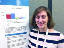
Sasa Zivkovic, MD, PhD, of the University of Pittsburgh Medical Center (UPMC), and associates shared a poster of their findings at the annual meeting of the American Association for Neuromuscular and Electrodiagnostic Medicine.
The researchers conducted next-generation sequencing (NGS) on 85 adult patients with peripheral neuropathy at the UPMC Neuromuscular Clinic during May 2017–Feb. 2019. The targeted NGS panel included 70 genes. The patients, aged 60 years on average, were primarily from Allegheny County, Pa., and had neuropathy either suspected to be hereditary or of unknown etiology.
Among the 19% of patients (n = 16) who tested positive for a known pathogenic mutation, half had Charcot-Marie-Tooth disease type 1A (CMT1A). Two patients – 13% of those with pathogenic variants – had hereditary neuropathy with liability to pressure palsies, and two had CMT1X. The remaining four patients had CMT1B, CMT2B1, CMT2E, and hereditary sensory and autonomic neuropathy mutations.
Another 4% of the overall patient sample (n = 3) had likely pathogenic mutations in genes associated with CMT2S, CMT4C and CMT4F. A third of the patients (32%) tested negative for the full NGS panel, and, comprising the largest proportion of patients, 46% had variants of unknown significance.
“The high occurrence of variants of unknown significance has uncertain significance but some variations may represent unrecognized pathogenic mutations,” the authors noted.
They identified 81 of these variants, with the DST, PLEKHG5, and SPG11 genes most commonly affected, each found in six patients. Four patients had a variant in the next most commonly affected gene, SBF2. The following variants occurred in three people each: BICD2, NEFL3, PRX, SCN11A, SCN9A, SLC52A2, and WNK1.
Among the 73 patients who underwent electrodiagnostic testing, 44 had sporadic axonal neuropathy, 17 had sporadic demyelinating neuropathy, and 11 had mixed neuropathies; the 1 remaining patient was not accounted for. Positive genetic testing occurred in a third (32%) of those with familial neuropathy (n = 28) and in 12% of those with sporadic neuropathy (n = 57).
No external funding was noted, and the authors had no disclosures.
SOURCE: Zivkovic S et al. AANEM 2019. Abstract 160. Targeted genetic testing in the evaluation of neuropathy .
REPORTING FROM AANEM
Primary periodic paralysis attacks reduced with long-term dichlorphenamide
AUSTIN, TEX. – Dichlorphenamide continues to reduce attacks from primary periodic paralysis (PPP) through 1 year with mild or moderate paresthesia and cognition-related adverse events, according to new research.
“These adverse events rarely resulted in discontinuation from the study and were sometimes managed by dichlorphenamide dose reductions,” concluded Nicholas E. Johnson, MD, of Virginia Commonwealth University, Richmond, and colleagues. “Reduction in dose was frequently associated with resolution of these events, suggesting a potential intervention to hasten resolution.” Dr. Johnson presented the findings in an abstract at the annual meeting of the American Association for Neuromuscular and Electrodiagnostic Medicine.
Dichlorphenamide (Keveyis) was approved by the Food and Drug Administration in 2015 for treating primary hyperkalemic and hypokalemic periodic paralysis and similar variants. The original hyperkalemic/hypokalemic PPP trial was a phase 3 randomized, double-blind, placebo-controlled trial that lasted 9 weeks and assessed the efficacy of dichlorphenamide in reducing PPP attacks and its adverse events. In the dichlorphenamide group, 47% experienced paresthesia, compared with 14% in the placebo group, and 19% experienced cognitive disorder, compared with 7% in the placebo.
In a 52-week open-label extension, participants who had been receiving the placebo switched to receiving 50 mg of dichlorphenamide twice daily. The intervention group continued with the dose they had been receiving when the 9-week double-blind phase ended. (During the initial intervention, they took either 50 mg twice daily or the dose they had at baseline for those taking it before the study began.)
The researchers then tracked rates of attacks and their severity over the next year – through week 61 after baseline – to compare these endpoints both within the intervention groups and between them.
Among the 63 predominantly white (84.1%) male (61.9%) adults who began the trial, 36 received dichlorphenamide and 27 received placebo. Just over two-thirds (68.3%) had hypokalemic PPP. Among the 47 patients (74.6%) who completed the open-label extension phase, 26 had been in the original dichlorphenamide group and 21 had been in the placebo group.
The median weekly attack rate in the dichlorphenamide group dropped from 1.75 at baseline to 0.06 at week 61 (median decrease 1.00, 93.8%; P less than .0001). In the placebo group that switched over to dichlorphenamide at week 9, the median weekly attack rate dropped from 3.00 at baseline to 0.25 at week 61 (median decrease 0.63, 75%; P = .01).
The median attack rate weighted for severity in the dichlorphenamide group dropped from 2.25 at baseline to 0.06 at week 61 (median decrease 2.25, 97.1%; P less than .0001). In the placebo group, it dropped from 5.88 to 0.50 (median decrease 1.69, 80.8%; P = .01).
No significant difference in median weekly attack rates and severity-weighted attack rates was found between the intervention groups through week 61.
Across all patients during the extension, 39.7% patients experienced at least one paresthesia adverse event, none of which were determined to be severe and resulting in one discontinuation.
A quarter of the participants (25.4%) experienced at least one cognition-related adverse event, and four patients (6.3%) discontinued because of these side effects. Most (14.3%) were mild with 7.9% reporting moderate and 3.2% reporting severe effects.
Dr. Johnson has received research support from or consulted with a variety of pharmaceutical companies including Strongbridge Biopharma, the manufacturer of the drug. Other authors consulted for several pharmaceutical companies, and one author is an employee of Strongbridge Biopharma.
SOURCE: Johnson NE et al. AANEM 2019. Abstract 102. Long-term efficacy and adverse event characterization of dichlorphenamide for the treatment of primary periodic paralysis.
REPORTING FROM AANEM
Patients with Charcot-Marie-Tooth disease describe wide range of care
AUSTIN, TEX. – Patients with Charcot-Marie-Tooth disease (CMT) receive a range of supportive care that includes physical therapy, surgery, medications, orthoses, and walking aids, according to patient-reported data presented at the annual meeting of the American Association of Neuromuscular and Electrodiagnostic Medicine. Patients describe approaches to CMT management that are broadly consistent with guidelines, researchers said.
“The range of different CMT treatments was wide,” reported Tjalf Ziemssen, MD, PhD, a researcher at Technische Universität Dresden in Germany, and colleagues. “ These results indicate that pain may have a substantial impact on people with CMT.”
The data also suggest that “lower-limb problems and mobility issues have a considerable impact on people with CMT,” they said.
CMT is a rare, progressive neuropathy that leads to distal muscle weakness, muscle atrophy, and sensory loss. There is no cure, and patients rely on supportive care. Until recently, few studies have assessed the impact of CMT on patients’ lives.
An ongoing, international, 2-year observational study is collecting data from adults with CMT. Patients report data via an app called CMT & Me.
To examine patient-reported treatment patterns and care standards for CMT in the United States and the United Kingdom, Dr. Ziemssen and colleagues analyzed data through Aug. 5, 2019, about 9.5 months into the study. Their interim analysis included data from 439 patients, including 222 patients in the United Kingdom and 217 in the United States.
More than 70% of participants visit a family doctor each year, and a similar proportion visit a neurologist. About 40% visit physical therapists, orthotists, or podiatrists. Other health care professionals seen by patients include occupational therapists (20%), orthopedic surgeons (nearly 20%), and pain specialists (about 15%).
About 70% of participants had received rehabilitation therapy such as physical therapy or occupational therapy, and about 70% had used medications, most frequently nonopioid analgesics (about 50%) and antidepressants (about 30%).
More than 80% used orthoses or walking aids, most commonly ankle or leg braces, insoles, or walking sticks.
In addition, about half of respondents had undergone a surgery for CMT. The most common procedures were osteotomy, hammertoe correction, and plantar fascia release.
Together, patients saw about a dozen types of health care professionals. “Small proportions of participants had visited each professional, which suggests that the care requirements of CMT patients are varied,” the researchers said.
The study was sponsored by Pharnext. Dr. Ziemssen and coauthors received compensation for participating in the study. Other coauthors are employees of Pharnext or Vitaccess, the company that developed the app used in the study.
SOURCE: Ziemssen T et al. AANEM 2019. Abstract 83. Treatment of Charcot-Marie-Tooth Disease in the United Kingdom and United States.
REPORTING FROM AANEM 2019
Researchers identify common reasons for misdiagnosis of ALS
AUSTIN – Lack of upper motor neuron signs on examination, presence of sensory symptoms, and absence of tongue fasciculations are common causes of amyotrophic lateral sclerosis (ALS) misdiagnosis, according to an investigation presented at the annual meeting of the American Association of Neuromuscular and Electrodiagnostic Medicine.
Because its initial presenting symptoms vary, ALS can be difficult for clinicians to diagnose. A diagnostic error may prompt clinicians and patients to pursue ineffective and potentially harmful medical or surgical interventions. Research suggests that surgery, for example, hastens the progression of ALS.
Catherine Rodriguez, a medical student at University of Missouri in Columbia, and colleagues conducted a study to identify the clinical factors and types of cognitive errors that can result in misdiagnosis of ALS. The researchers analyzed electronic medical records of 88 patients with a diagnosis of ALS who were receiving treatment at the University of Missouri Hospital during 2011-2017 with at least 1 year of follow-up. They collected demographic information and clinical characteristics (e.g., ALS Functional Rating Scale and site of symptom onset) for each patient. If a patient received an incorrect diagnosis, Ms. Rodriguez and colleagues recorded the number of physicians he or she had seen, the incorrect diagnosis, the treatment, the type of diagnostic error, the clinical factors contributing to the misdiagnosis, and the type of physician who gave the incorrect diagnosis.
The investigators classed diagnostic errors according to the four categories of cognitive bias of the Patient Safety Network. The categories are availability heuristic (i.e., the diagnosis of a current patient is biased by the clinician’s experience with previous cases), anchoring heuristic (i.e., relying on the initial impression despite the emergence of evidence that may contradict it), framing effects (i.e., subtle cues and collateral information bias the diagnosis), and blind obedience (i.e., undue reliance on test results or expert opinion). Ms. Rodriguez and colleagues used Fisher’s exact test to perform a statistical analysis of the data.
Thirty-four (39%) of the 88 patients were female, and the populations average age was about 60 years. Eighty patients (91%) were white, six (7%) were black, and two (2%) were Hispanic. Twenty patients (23%) received an incorrect diagnosis. Common incorrect diagnoses included spinal abnormality, Bell’s palsy, myasthenia gravis, ulnar neuropathy, autoimmune motor neuropathy, and stroke.
The investigators observed significant differences in the reasons for misdiagnosis, depending on patient characteristics. Veterans were misdiagnosed because of the availability heuristic, while nonveterans were misdiagnosed because of the anchoring heuristic. Lower-limb onset was most commonly misdiagnosed because of the anchoring heuristic. Bulbar onset was most commonly misdiagnosed because of the availability heuristic. Surgical intervention was the most common treatment for an incorrect diagnosis.
The data serve as a reminder of the prevalence of cognitive biases, said Ms. Rodriguez. “Common things are common, so we tend to stick with those [diagnoses]. Especially with ALS, nobody wants to give anyone that diagnosis.” Clinicians should “recognize that incorrect diagnoses have equally bad outcomes for those patients,” she concluded.
The study was supported by a University of Missouri School of Medicine Summer Research Fellowship Program.
SOURCE: Rodriguez C et al. AANEM 2019. Abstract 10. Diagnostic errors and the implications for amyotrophic lateral sclerosis patients.
Which patients are most likely to have a positive RNS test for myasthenia gravis?
AUSTIN, TEX. – , according to research presented at the annual meeting of the American Association of Neuromuscular and Electrodiagnostic Medicine.
Low-frequency RNS is a common test that neurologists perform to evaluate a patient for myasthenia gravis. The effects of various clinical factors on the diagnostic yield of this test are unknown, however.
Myasthenia gravis is “mostly a clinical diagnosis,” study first author Tingting Hua, a medical student at the University of Missouri in Columbia, said in an interview. “RNS is just one of the helpful diagnostic tests for it. If we can find out in what kind of populations of patients this test is more helpful, maybe that would help cut down unnecessary tests in patients for whom it’s not necessarily helpful.”
Ms. Hua and her colleagues conducted research to assess the effects of clinical, serologic, and demographic factors on the diagnostic yield of RNS. They retrospectively analyzed patients with an established diagnosis of myasthenia gravis and at least 1 year of follow-up. The variables that the investigators examined were demographic characteristics, MGFA class, RNS study results, antibody test results, thymoma status, and treatments received.
Ms. Hua and her colleagues included 65 patients in their analysis. Thirty-one patients were female. Fifty-five patients were white, eight were black, and two were categorized as “unknown.” Of this population, 32 patients (49.2%) were in MGFA Class I, 14 (21.5%) were in MGFA Class IIa, 13 (20.0%) were in MGFA Class IIb, and the remaining 6 (9.2%) were in MGFA Classes IIIa through V. Twenty-seven patients (42%) had positive RNS studies. Twenty-one patients (32%) were seropositive for myasthenia gravis antibodies.
Eleven patients underwent RNS in an inpatient setting, and 54 were tested in an outpatient setting. Acetylcholine receptor (AChR) binding antibody titer ranged from 0.12 nmol/L to 118 nmol/L. The RNS results were significantly more likely to be positive for seropositive patients, compared with seronegative patients. Patients with MGFA Class III or higher also had higher likelihood of positive RNS results, compared with patients in lower classes. Finally, the diagnostic yield was highest for patients with MGFA Class III or higher who were tested in an inpatient setting.
The study was supported by a Missouri School of Medicine Summer Research Fellowship.
SOURCE: Hua T et al. AANEM 2019, Abstract 9.
REPORTING FROM AANEM 2019
Study examines utility of repeat outpatient electrodiagnostic testing
AUSTIN, TEX. – During a 3-year period, 5.7% of patients referred to an electromyography laboratory returned for at least one additional electrodiagnostic study, according to research presented at the annual meeting of the American Association of Neuromuscular and Electrodiagnostic Medicine. A preliminary analysis suggests that repeat testing for the same indication does not change symptom or disease management in about one-third of cases.
Physicians may request repeat electrodiagnostic studies to monitor previous, new, or progressing symptoms in the same or different body segments. “While the utility of [electrodiagnostic] studies for clinical care has been established, testing is associated with some patient risk, time, and cost,” the researchers wrote. “To date, there have been no known studies investigating the utility of repeat [electrodiagnostic] testing in the outpatient setting.”
To study referral patterns and outcomes following repeat electrodiagnostic testing, Aimee K. Boegle, MD, PhD, an instructor in neurology at Beth Israel Deaconess Medical Center in Boston, and colleagues examined all outpatient electromyography and nerve conduction studies performed between 2015 and 2017 in the neurology department at their institution. The investigators excluded patients who underwent inpatient electrodiagnostic studies from their analysis.
Approximately 4,800 patients underwent electrodiagnostic testing, 276 of whom underwent testing more than once.
Among patients who underwent two studies, 55% were referred by a different physician for the second study. Median neuropathy was the most common referring and final diagnosis among patients who underwent repeat electrodiagnostic testing, Dr. Boegle said. This finding was not surprising because carpal tunnel syndrome is among the most common reasons for referral overall.
Median neuropathy was the referring diagnosis in 31% and the final diagnosis in 30%, cervical radiculopathy in 15% and 14%, ulnar neuropathy in 14% and 17%, lumbosacral radiculopathy in 12% and 10%, and polyneuropathy in 8% and 10%.
The neurology and orthopedics departments made the most referrals for repeat electrodiagnostic studies (49.4% and 29.3%, respectively), followed by primary care physicians/internal medicine (13%).
About 24% of the returning patients underwent testing for the same indication as their initial referral.
A preliminary analysis of 26 patients who underwent a repeat study for the same indication found no change in treatment in 34%. When a study prompted intervention, a conservative course of management such as a splint or physical therapy was used in 42%. About 8% received a pharmacologic intervention, such as a medication change or steroid injections. Another 8% received a surgical intervention and about 8% received further work-up.
The researchers had no relevant disclosures.
SOURCE: Boegle AK et al. AANEM 2019, Abstract 85.
REPORTING FROM AANEM 2019
Neurologists consider flu shot safe in most patients with autoimmune neuromuscular disorders
AUSTIN, TEX. – (CIDP), according to a survey presented at the annual meeting of the American Association of Neuromuscular and Electrodiagnostic Medicine. They are more conservative in recommending immunization for patients with a history of Guillain-Barré syndrome, however. Temporally associated disease relapses may be a risk factor for relapse with subsequent immunization, according to the investigators.
Influenza vaccination of patients with autoimmune neuromuscular disorders such as myasthenia gravis, CIDP, or Guillain-Barré syndrome is controversial, and no clear guideline helps clinicians to decide whether vaccination for such patients is appropriate. Tess Litchman, a medical student at Yale University, New Haven, Conn., and colleagues conducted a web-based survey of neurologists throughout the United States to examine current practices for recommending influenza vaccination for patients with myasthenia gravis, CIDP, and Guillain-Barré syndrome.
The researchers received 184 survey responses, with the highest proportions of responses coming from California (8.8%), Connecticut (8.8%), and Texas (8.3%). On average, respondents had been in practice for 15.5 years. Their reported practice specialties were neuromuscular medicine in 50%, general neurology in 20%, mixed specialties in 20%, and other in 10%.
Across practice settings, neurologists followed 6,448 patients with myasthenia gravis, 2,310 patients with CIDP, and 1,907 patients with Guillain-Barré syndrome. Approximately 83% of respondents reported recommending influenza vaccination for all of their patients with myasthenia gravis, 59% reported recommending vaccination for all of their patients with CIDP, and 43% of respondents reported recommending vaccination for all of their patients with Guillain-Barré syndrome. About 2%, 8%, and 15% of respondents reported that they do not recommend influenza vaccination for any of their patients with myasthenia gravis, CIDP, and Guillain-Barré syndrome, respectively.
A temporal association between disease relapse and influenza vaccination was reported in 1.5% of patients with myasthenia gravis, 3.7% of patients with CIDP, and 8.7% of patients with Guillain-Barré syndrome. Recurrent relapses occurred in 87% (26 of 30) of patients with myasthenia gravis, 92% (23 of 25) of patients with CIDP, and 74% (26 of 35) of patients with Guillain-Barré syndrome who received another influenza vaccination.
“According to existing guidelines per the Centers for Disease Control and Prevention Advisory Committee on Immunization Practices, all patients with myasthenia gravis and CIDP should be vaccinated, and patients with Guillain-Barré syndrome who did not develop the syndrome due to a flu shot should be vaccinated,” said Richard J. Nowak, MD, director of the program in clinical and translational neuromuscular research at Yale and one of the senior investigators on the study. “This survey demonstrates that clearer guidelines and education from a professional academic neurology society is an unmet need and would be helpful to better inform the neurology community about the possible risks and benefits of immunization in myasthenia gravis, CIDP, and Guillain-Barré syndrome patients. We hope to utilize these initial results to stimulate a larger scale study, and identify whether this topic represents a knowledge gap in the community or an area in which we can improve on the best-practice standard.”
Dr. Nowak had no relevant disclosures. The study was supported by the department of neurology at Yale University; there was no external funding.
SOURCE: Litchman T et al. AANEM 2019, Abstract 16.
REPORTING FROM AANEM 2019
Placebo response in negative rituximab BeatMG trial provides important lessons
AUSTIN, TEX. – Among patients with acetylcholine receptor (AChR) antibody-positive generalized myasthenia gravis, rituximab and placebo may have a similar glucocorticoid-sparing effect regardless of disease severity, according to research presented at the annual meeting of the American Association of Neuromuscular and Electrodiagnostic Medicine.
B Cell Targeted Treatment In Myasthenia Gravis (BeatMG) was a 52-week, randomized, double-blind, placebo-controlled clinical trial. The phase 2 study’s primary outcomes were safety and the glucocorticoid-sparing effect assessed by the percentage of patients who reduced their mean daily prednisone dose by at least 75% and maintained clinical stability. Secondary outcomes were change in Myasthenia Gravis Composite (MGC) score and change in Quantitative Myasthenia Gravis (QMG) score from baseline to 52 weeks.
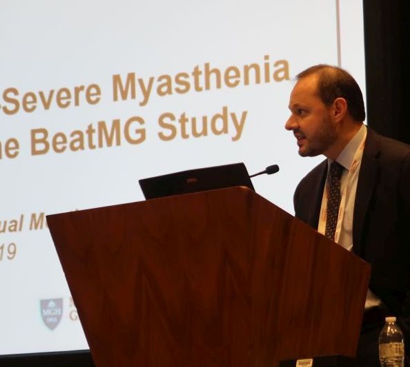
Investigators randomized 52 participants 1:1 to rituximab (Rituxan) or placebo. Patients were taking at least 15 mg/day of prednisone and were a mean of about age 50 years. About two-thirds were treated with glucocorticoids alone at baseline, and nearly two-thirds had a Myasthenia Gravis Foundation of America (MGFA) clinical classification of II. “It was a mildly symptomatic group of individuals in terms of disease severity,” said Richard Nowak, MD, assistant professor of neurology and director of the Yale Myasthenia Gravis Clinic in New Haven, Conn.
During the study, 60% of patients who received rituximab had a 75% or greater reduction in their mean daily prednisone dose and maintained clinical stability. “However, what surprised us is that we had a significantly high placebo response rate of 56%,” he said. The difference between groups was not significant.
Patients who received rituximab had “directionally favorable reductions” in MGC and QMG scores over 52 weeks, compared with patients who received placebo. Nevertheless, “after correcting for baseline differences, there was no significant difference between the two groups,” Dr. Nowak said.
Rituximab had good safety and tolerability, and the placebo group had a threefold higher rate of clinical relapse requiring IV immunoglobulin or plasmapheresis. “While the placebo arm did achieve a similar rate of reduction in their steroid dose, at 52 weeks the patients may have been doing less well, reflected by the higher rate of relapse,” he said.
Subgroup analysis
To explore whether rituximab might benefit patients who were treatment resistant or had more symptomatic disease, Dr. Nowak and his colleagues conducted a post hoc subgroup analysis of 20 participants – 10 in the rituximab arm and 10 in the placebo arm – who were MGFA class III-IV. In each group, 70% were on glucocorticoid treatment alone, and 90% were MGFA class III.
As in the overall study, the glucocorticoid-sparing effect was not significant (60% of the rituximab group vs. 50% of the placebo group).
Mean change in QMG score was –3.9 in the rituximab group and –0.5 in the placebo group, and mean change in MGC score was –7.0 in the rituximab group and –4.8 in the placebo group. These secondary outcomes again show “directional favorability” with rituximab, Dr. Nowak said. The researchers saw similar trends for scores that assess quality of life and activities of daily living.
In the subgroup analysis, myasthenia gravis relapses requiring rescue therapy occurred in 20% of patients in the rituximab arm and in 30% of the placebo arm. Overall, the researchers did not see a difference in treatment response between those with moderate to severe disease and those with mild disease, Dr. Nowak said.
Suggestions from the data
The post hoc subgroup analysis should be interpreted with caution, and the study does not provide firm conclusions, Dr. Nowak noted. In addition, the trial population does not reflect all patients with myasthenia gravis. Dr. Nowak routinely uses rituximab in patients with muscle-specific kinase antibody-positive disease, he said. For acetylcholine receptor antibody-positive generalized myasthenia gravis, which has approved therapies available, Dr. Nowak presents rituximab as an option for some patients. Further research may clarify where B-cell depletion therapy may fit into the treatment paradigm.
Nonetheless, BeatMG and the subgroup analysis may help physicians better understand the disease and the role of various therapies. Investigators successfully lowered prednisone dose “at a pretty high rate in the placebo arm,” he said. “It suggests that many of our patients potentially may be on higher than required prednisone doses.”
Finally, the BeatMG findings emphasize the need for placebo-controlled trials to understand potential therapies. “We need to pause when we see a lot of retrospective and uncontrolled studies that are very promising,” Dr. Nowak said.
The study was supported by the National Institute of Neurological Disorders and Stroke. Genentech provided the study drug and placebo through an investigator-sponsored study agreement. Dr. Nowak has received research support from Alexion Pharmaceuticals, Genentech, Grifols, and Ra Pharmaceuticals. He has served as a paid consultant for Alexion, Momenta, Ra, Roivant, Shire, Grifols, and CSL Behring.
SOURCE: Nowak R et al. AANEM 2019. Unnumbered Abstract: Rituximab in patients with moderate to severe myasthenia gravis: a subgroup analysis of the BeatMG study
REPORTING FROM AANEM 2019
Congenital myasthenic syndrome diagnosed best with repetitive stimulation and jitter analysis
AUSTIN, TEX. – suggests newly presented research.
“In case RS is negative, SFEMG [single fiber electromyography] alone is not very specific and cannot distinguish CMS from mitochondrial myopathies, even in the presence of impulse blocking,” Vitor Marques Caldas, MD, a neurologist at the Syrian Libanes Hospital in Brasilia, Brazil, and a PhD student at the University of São Paulo, told attendees at the annual meeting of the American Association for Neuromuscular and Electrodiagnostic Medicine. “An isolated SFEMG test can lead to a misdiagnosis of myasthenia syndrome if not interpreted in the right clinical context.”
The researchers sought to understand the relative sensitivity and specificity of low-frequency RS versus jitter analysis using disposable concentric needle electrodes (CNE).
The study involved 69 patients, of whom 19 had mitochondrial myopathy, 18 had congenital myopathy, 18 had CMS, and 14 were asymptomatic controls. The control group all tested normal with both RS and jitter analysis.
The 18 participants with CMS, average age 24 years, received low-frequency RS in at least six different muscles: two distal muscles (abductor digiti minimi and tibialis anterior), two proximal muscles (deltoid and trapezius) and two facial muscles (nasalis and orbicularis oculi). They also underwent jitter analysis of their orbicularis oculi muscle under voluntary activation using CNE.
These patients had heterogeneous genetic profiles: 11 had the CHRNE gene mutation, 2 had the RAPSN gene mutation, 2 had the COLQ gene mutation, 2 had the DOK-7 gene mutation, and 1 had the COL13A1 mutation.
All but two patients with congenital CMS tested positive (88.9%) with RS: one female with CHRNE mutation and one male with RAPSN mutation. Using mean jitter, all but one patient tested positive (94.4%): a female with DOK-7 mutation who had tested abnormal on RS.
All patients with CMS tested positive with at least one of the two tests, but only 83.3% tested positive with both tests, resulting in a sensitivity of 83.3%, a specificity of 100%, and overall accuracy of 95.6% using both tests.
Among the 19 patients with mitochondrial myopathy, 5 had abnormal jitter analysis.
When the researchers looked only at participants with abnormal jitter analysis but normal RS, two of these were patients with CMS, but another seven had congenital or mitochondrial myopathies. Using abnormal jitter alone therefore resulted in a sensitivity of 100% but a specificity of only 86%, for overall 86.5% accuracy.
“It’s important to notice that if you have an abnormal jitter, we have to look at the clinical symptoms of the patients,” Dr. Marques Caldas said in an interview. “Jitter abnormalities are not enough to distinguish between myasthenic disorder and a myopathic disorder.”
The research used no external funding, and Dr. Marques Caldas had no disclosures.
SOURCE: Caldas VM et al. AANEM 2019. Unnumbered Abstract: Sensitivity of neurophysiologic tests regarding the neuromuscular junction in patients with congenital myasthenic syndromes.
REPORTING FROM AANEM 2019
Late response to eculizumab may occur in minority of myasthenia gravis patients
AUSTIN – according to a secondary analysis presented at the annual meeting of the American Association of Neuromuscular and Electrodiagnostic Medicine.
Evidence for the sustained effectiveness of eculizumab, a terminal complement inhibitor, in adult patients with antiacetylcholine receptor antibody-positive refractory generalized myasthenia gravis was provided by the 6-month, double-blind, placebo-controlled REGAIN study and its open-label extension. James F. Howard Jr., MD, distinguished professor of neuromuscular disease at the University of North Carolina in Chapel Hill and colleagues sought to analyze response profiles in REGAIN and its open-label extension.
The findings raise the possibility that complement inhibition with eculizumab should not be abandoned rapidly. “We accept that [eculizumab] works quickly,” Dr. Howard said. “There is an impression that if you don’t respond within the first 3 months, you’re not going to respond. I think these data would suggest otherwise.”
The investigators analyzed participants’ Myasthenia Gravis–Activities of Daily Living (MG-ADL) and Quantitative Myasthenia Gravis (QMG) scores, which had been recorded throughout REGAIN and the extension. They defined early and late responses as improvement in MG-ADL score (i.e., a decrease of three or more points) or QMG score (i.e., a decrease of five or more points) occurring at 12 weeks or earlier or after 12 weeks, respectively, after initiation of eculizumab therapy. Patients randomized to eculizumab in REGAIN initially were treated with an IV induction dose of 900 mg/week before receiving 1,200 mg every 2 weeks thereafter.
Dr. Howard and colleagues included 98 patients in their analysis. Approximately 32% of patients achieved their first response within the first week of treatment, and 15% responded at week 2. About 16% of patients had a late response.
Responses to treatment on the MG-ADL scale had occurred in 67.3% by week 12 and in 84.7% by the end of the extension. Treatment with eculizumab resulted in QMG responses in 56.1% by week 12 and 71.4% by the end of the extension. The investigators observed response over multiple consecutive assessments for the vast majority of patients.
At week 130, the least-squares mean percentage changes from baseline in MG-ADL score were −61.9% and −47.5% in early and late MG-ADL responders, respectively. The least-squares mean percentage changes from baseline in QMG score were −40.8% and −55.5% in early and late QMG responders, respectively.
The investigators observed significant baseline differences between early versus late QMG responders in mean duration of myasthenia gravis (10.46 years vs. 5.46 years) and mean QMG score (18.6 vs. 15.1).
“I can’t explain [this finding]. It may simply be due to the low numbers of patients,” Dr. Howard said. “Whether this is going to hold up in postmarketing analysis remains to be seen. I’m not convinced that that is meaningful.”
Study funding was provided by Alexion Pharmaceuticals (the developer of eculizumab), the Centers for Disease Control and Prevention, and the National Institutes of Health. Dr. Howard reported receiving research support and consulting fees or honoraria from Alexion and several other pharmaceutical companies. Several other authors reported financial relationships with Alexion and other pharmaceutical companies; two authors are employees of Alexion.
Howard J et al. AANEM 2019. Unnumbered Abstract.
REPORTING FROM AANEM 2019


