User login
For MD-IQ on Family Practice News, but a regular topic for Rheumatology News
Decision aids benefit OA patients considering joint replacement
Osteoarthritis patients who received patient decision aids (PtDA) in addition to usual educational resources had shorter wait times and fewer surgeries, and were more likely to reach good decision quality about joint replacement surgery, although the overall effect was not statistically significant, reported Dawn Stacey, Ph.D., of the University of Ottawa (Ont.) and her coauthors.
In a randomized, controlled trial of 343 patients undergoing orthopedic screening for hip or knee replacement surgery at an academic hospital or a community hospital, being given a PtDA (intervention) was associated with reduced wait times, compared with controls who received usual care (hazard ratio = 1.25; 95% CI, 0.99–1.60; P = .0653). Mean age of the patients was 66 years; about half had high school education or less and half had household incomes at or below the median income ($58,000) for couples aged 65 or older in Canada.
Surgery rates were 73.2% in the intervention group, compared with 80.5% in controls (relative risk = 0.91; 95% CI, 0.81-1.03), the investigators found. Good decision quality was reached by 56.1% of intervention patients and 44.5% of controls (RR = 1.25; 95% CI, 1.00–1.56, P = .050). However, the overall effect was not significant, Dr. Stacey and coauthors reported.
Most patients in the study had more severe osteoarthritis and were appropriate for considering surgery, but the PtDA may have had “greater effect and discrimination” when it was given to patients with earlier stages of osteoarthritis and may have helped them to consider their nonsurgical options, the investigators wrote.
Read the full study in Osteoarthritis and Cartilage.
Dual ACL, tibial osteotomy surgery helped knee OA patients
Gait biomechanics in patients with medial knee osteoarthritis were significantly improved 5 years after concomitant high tibial osteotomy and anterior cruciate ligament reconstruction, according to a prospective cohort study conducted by Kendal Marriott and associates.
In the 33 patients who underwent the dual surgeries, three-dimensional gait analysis showed that the peak knee adduction moment had decreased significantly in the surgical limb 5 years after surgery, compared with a nonsignificant increase in the nonsurgical limb. A significant decrease in the peak knee flexion moment was seen in both the surgical and nonsurgical limbs. Progress plateaued 2-5 years post surgery, but improvements gained were maintained at the 5-year postsurgery mark.
As concomitant high tibial osteotomy and anterior cruciate ligament reconstruction is relatively rare, “longer term follow-up and comparisons with other treatment strategies are both warranted and required to better evaluate the clinical effect of this seemingly biomechanically efficacious procedure,” the investigators concluded.
Find the full study in the American Journal of Sports Medicine (doi: 10.1177/0363546515591995).
Optimized analgesia, exercise cut pain in severe knee OA
A 6-week protocol of optimized analgesia followed by a 12-week exercise program significantly improved pain and functional limitations for patients with knee osteoarthritis, researchers reported in Arthritis Care and Research.
The study was the first to explore how to achieve sufficient pain relief for patients with severely painful knee OA to participate in exercise therapy, and it achieved that goal for 78% of patients, said Joyce van Tunen of the Amsterdam Rehabilitation Center and associates. “The newly developed intervention protocol was feasible, which means that patients were able to participate in exercise therapy despite their severe pain at baseline,” the researchers said. “Although the results are promising, they need to be confirmed in a randomized controlled trial.”

Guidelines recommend combining pharmacologic and nonpharmacologic modalities to improve osteoarthritis outcomes, and past studies have suggested that acetaminophen, NSAIDs, and glucosamine therapy might augment the benefits of exercise in OA, they said.
The study included 49 patients with severe knee OA whose pain scored at least 7 on a 10-point scale. Patients received standardized analgesia with acetaminophen and then were stepped up to NSAIDs, weak opioids, and intra-articular steroid injections if their pain did not improve to a 5 or lower. Patients had 2 weeks to adapt to each new prescription without further changes, and their doses were cut if they maintained pain scores of 4 or less for a month or longer. The protocol was based on the World Health Organization analgesic ladder and the Beating osteoARThritis strategy for stepped care in hip and knee OA, the investigators noted (Arthritis Care Res. 2015 Aug 3 doi: 10.1002/acr.22682).
After 6 weeks, patients continued with analgesia and started a 12-week exercise program of two 60-minute sessions per week. The first 6 weeks of the program focused on muscle strength, while the last 6 weeks aimed to maximize strength while adding functional and aerobic exercises. Patients also were asked to do physical therapy exercises at home on the days they did not take part in supervised exercise, the researchers said.
At the end of the 18-week intervention, 72% of patients reported improvement on a combined global scale, the study showed. Average pain scores improved by 30% (P < .001) and activity limitations improved by 17% (P < .001). Fully 78% of patients were able to follow the exercise program, and these patients improved their physical limitations by an extra 10% (P = .004), compared with patients who could not complete the program. Patients who were not able to finish the exercise program also had significantly worse radiographic signs of OA, compared with the others (P = .03), and tended to be younger; had higher body mass indices; and reported more pain, anxiety, and depression, the researchers said. These patients might need surgical interventions or therapy to help them learn to better cope with pain, they added.
Most of the patients had used analgesics irregularly and at suboptimal doses at baseline, in part because they feared adverse effects. However, they experienced no serious side effects from the analgesia protocol or the exercise program, the investigators noted.
The Dutch Arthritis Association funded the work. The investigators reported having no conflicts of interest.
Therapy of osteoarthritis often remains a frustration to both the patient and physician. In clinical trials as well as the clinical setting, no single nonsurgical therapeutic approach has consistently been of significant benefit.
 |
| Dr. Roy Altman |
Summaries of the medical literature are reflected by unimpressive P values, effect sizes, and other statistical methods. Yet the clinician has to use the tools available, so that several partially effective programs are often employed in the clinical setting, most often in combination – sometimes called multimodal therapy. Therapeutic guidelines are based on the literature but usually only allude to a multimodal approach.
In this study, the investigators examined a combined nonpharmacologic and pharmacologic program over an 18-week period. They easily demonstrated significant benefit of the multimodal approach. No study is perfect, and this one has multiple drawbacks, not the least of which is the lack of any control or comparison group to determine if the benefits seen are from one or both programs. Despite the drawbacks in their study design, they have contributed to filling that gap in the literature. The program of medications to reduce pain enough to increase the physical rehabilitation is logical and reflects a real-world setting.
Dr. Roy D. Altman is professor emeritus in the division of rheumatology and immunology at the University of California, Los Angeles. He reported having no relevant disclosures.
Therapy of osteoarthritis often remains a frustration to both the patient and physician. In clinical trials as well as the clinical setting, no single nonsurgical therapeutic approach has consistently been of significant benefit.
|
| Dr. Roy Altman |
Summaries of the medical literature are reflected by unimpressive P values, effect sizes, and other statistical methods. Yet the clinician has to use the tools available, so that several partially effective programs are often employed in the clinical setting, most often in combination – sometimes called multimodal therapy. Therapeutic guidelines are based on the literature but usually only allude to a multimodal approach.
In this study, the investigators examined a combined nonpharmacologic and pharmacologic program over an 18-week period. They easily demonstrated significant benefit of the multimodal approach. No study is perfect, and this one has multiple drawbacks, not the least of which is the lack of any control or comparison group to determine if the benefits seen are from one or both programs. Despite the drawbacks in their study design, they have contributed to filling that gap in the literature. The program of medications to reduce pain enough to increase the physical rehabilitation is logical and reflects a real-world setting.
Dr. Roy D. Altman is professor emeritus in the division of rheumatology and immunology at the University of California, Los Angeles. He reported having no relevant disclosures.
A 6-week protocol of optimized analgesia followed by a 12-week exercise program significantly improved pain and functional limitations for patients with knee osteoarthritis, researchers reported in Arthritis Care and Research.
The study was the first to explore how to achieve sufficient pain relief for patients with severely painful knee OA to participate in exercise therapy, and it achieved that goal for 78% of patients, said Joyce van Tunen of the Amsterdam Rehabilitation Center and associates. “The newly developed intervention protocol was feasible, which means that patients were able to participate in exercise therapy despite their severe pain at baseline,” the researchers said. “Although the results are promising, they need to be confirmed in a randomized controlled trial.”
Guidelines recommend combining pharmacologic and nonpharmacologic modalities to improve osteoarthritis outcomes, and past studies have suggested that acetaminophen, NSAIDs, and glucosamine therapy might augment the benefits of exercise in OA, they said.
The study included 49 patients with severe knee OA whose pain scored at least 7 on a 10-point scale. Patients received standardized analgesia with acetaminophen and then were stepped up to NSAIDs, weak opioids, and intra-articular steroid injections if their pain did not improve to a 5 or lower. Patients had 2 weeks to adapt to each new prescription without further changes, and their doses were cut if they maintained pain scores of 4 or less for a month or longer. The protocol was based on the World Health Organization analgesic ladder and the Beating osteoARThritis strategy for stepped care in hip and knee OA, the investigators noted (Arthritis Care Res. 2015 Aug 3 doi: 10.1002/acr.22682).
After 6 weeks, patients continued with analgesia and started a 12-week exercise program of two 60-minute sessions per week. The first 6 weeks of the program focused on muscle strength, while the last 6 weeks aimed to maximize strength while adding functional and aerobic exercises. Patients also were asked to do physical therapy exercises at home on the days they did not take part in supervised exercise, the researchers said.
At the end of the 18-week intervention, 72% of patients reported improvement on a combined global scale, the study showed. Average pain scores improved by 30% (P < .001) and activity limitations improved by 17% (P < .001). Fully 78% of patients were able to follow the exercise program, and these patients improved their physical limitations by an extra 10% (P = .004), compared with patients who could not complete the program. Patients who were not able to finish the exercise program also had significantly worse radiographic signs of OA, compared with the others (P = .03), and tended to be younger; had higher body mass indices; and reported more pain, anxiety, and depression, the researchers said. These patients might need surgical interventions or therapy to help them learn to better cope with pain, they added.
Most of the patients had used analgesics irregularly and at suboptimal doses at baseline, in part because they feared adverse effects. However, they experienced no serious side effects from the analgesia protocol or the exercise program, the investigators noted.
The Dutch Arthritis Association funded the work. The investigators reported having no conflicts of interest.
FROM ARTHRITIS CARE AND RESEARCH
Key clinical point: Patients with knee osteoarthritis and severe pain improved significantly with an optimized standard protocol of analgesia and exercise therapy.
Major finding: Average pain scores improved by 30% (P < .001) and activity limitations improved by 17% (P < .001).
Data source: Single-center prospective study of 49 patients with knee OA and severe pain.
Disclosures: The Dutch Arthritis Association funded the work. The investigators reported having no conflicts of interest.
New analysis suggests functional decline for most people with knee OA
Accounting for missing data revealed functional declines over time in patients with newly diagnosed knee osteoarthritis, according to researchers.
The findings contradict more optimistic results from other longitudinal studies of knee OA, said Britt Elin Øiestad, Ph.D., of Oslo and Akershus University College of Applied Sciences and her associates. “The adjusted analyses showed either stable or worsening physical function, which is more in line with what is observed in the clinic,” the investigators said.
Their analysis included data from 802 participants in either the Multicenter Osteoarthritis Study (MOST) or the Osteoarthritis Initiative (OAI) who developed symptomatic knee OA during the course of the studies.
Approximately two-thirds of affected patients were women. Patients averaged about 63-66 years of age, had average body mass index of about 30-31 kg/m2, and had not undergone total knee replacement surgery at baseline or total hip replacement at any time. The researchers used a multiple imputation method to account for missing physician visits and a local regression smoothing curve to predict clinical status just prior to total knee replacement surgery (Arthritis Care Res. 2015 Aug 3. doi: 10.1002/acr.22674). Patients in MOST who developed knee OA showed no significant change over 4 years in scores on the Western Ontario and McMaster Universities Osteoarthritis Index (WOMAC) Physical Function (pf) subscale, while their Five Times Sit to Stand Test and 20-Meter Walk Test results rose by 1.5 seconds over 5-6 years (P less than .003), the investigators said. Adjusted results from OAI were similar, revealing significant worsening over time in WOMAC-pf and 20-Meter Walk Test results. “In crude results in which we did not impute missing values or pre–total knee replacement physical function status, the trajectory of physical function was more favorable,” the researchers added. “We found that imputing missing values and predicting pre–total knee replacement function reduced some of the bias seen in the unadjusted analyses, which incorrectly suggested improvement in physical function in people with knee osteoarthritis.”
The Research Council of Norway and the National Institutes of Health funded the study. The authors did not report funding sources or conflicts of interest.
FROM ARTHRITIS CARE & RESEARCH
Key clinical point:A new analysis shows either stable or worsening physical function after new-onset knee osteoarthritis, contradicting more optimistic results from other longitudinal studies of knee OA.
Major finding: In adjusted analyses, patients showed stable to significantly worse results over time in WOMAC-pf, Five Times Sit to Stand, and the 20-Meter Walk Test. Crude data suggested a more positive trajectory.
Data source: Longitudinal analysis of 802 patients in the Multicenter Osteoarthritis Study or the Osteoarthritis Initiative.
Disclosures: The Research Council of Norway and the National Institutes of Health funded the study. The authors did not report funding sources or conflicts of interest.
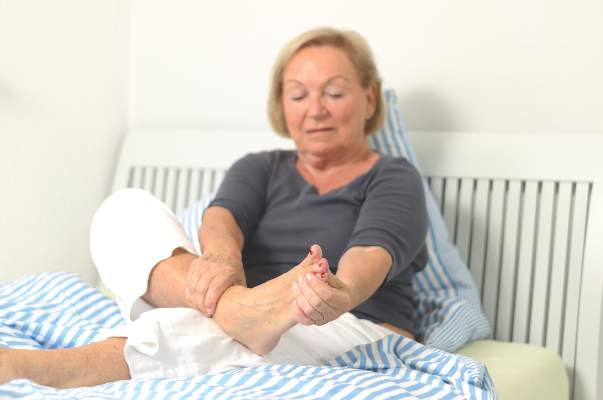
Study reveals distinct forms of foot osteoarthritis
Osteoarthritis of the foot takes one of two forms: isolated disease of the first metatarsophalangeal joint or more extensive disease of that and several other joints of the midfoot, researchers reported online in Arthritis Care & Research.
The latter, known as polyarticular foot osteoarthritis (OA), disproportionately affects women, is associated with worse pain and disability, compared with localized disease, and tends to co-occur with nodal hand OA, said Trishna Rathod of Keele University, Staffordshire, England, and her associates.
Studies of OA phenotypes have yielded targeted treatments (Ann Intern Med. 2009;150[10]:661-9andOsteoarthritis Cartilage. 2014;22[suppl S431]) for other joints, but had not yet characterized foot OA, the researchers said. Therefore, they surveyed 533 adults who reported foot pain in the prior year and scored radiographs of their first metatarsophalangeal, first and second cuneometatarsal, navicular first cuneiform, and talonavicular joints. The patients did not have psoriatic or rheumatoid arthritis and averaged 65 years of age (Arthritis Care Res. [Hoboken] 2015 Aug. 3. doi: 10.1002/acr.22677).
In all, 15% of patients had polyarticular foot OA, 22% had isolated OA of the first metatarsophalangeal joint, and 64% had no or minimal foot arthritis, the investigators reported. About 77% of patients with polyarticular disease were women, compared with approximately half of those in the other groups (P = .001). After the researchers controlled for age and sex, polyarticular disease was significantly linked to nodal hand OA (P = .04), higher body mass index (P = .002), worse scores on a 10-point foot pain scale (6 vs. 4.9 and 5.2 for the other classes; P = .02), and worse pain and disability scores on the Manchester Foot Pain and Disability Index (P = .002 and .007), they said.
“As is the case for OA at other small joint sites, particularly the hands, patterning of individual joint involvement in radiographic foot OA is polyarticular and strongly symmetrical,” the researchers concluded. “Patterns of joint involvement in radiographic foot OA have indicated a distinction between individuals with isolated [first] metatarsophalangeal joint OA and those with a more widespread form of OA ... which also includes one or both first metatarsophalangeal joints.”
The Arthritis Research UK Programme Grant and the West Midlands North CLRN supported the work. The investigators declared no competing interests.
FROM ARTHRITIS CARE & RESEARCH
Key clinical point:Osteoarthritis of the foot manifests as either isolated disease of the first metatarsophalangeal joint or as more debilitating disease of that joint and several others of the midfoot.
Major finding: Affected patients had either polyarticular foot OA or isolated OA of the first metatarsophalangeal joint, and the former was associated with significantly worse pain and functional limitations.
Data source: Prospective observational cohort study of 533 adults who reported foot pain in the previous year.
Disclosures: The Arthritis Research UK Programme Grant and the West Midlands North CLRN supported the work. The investigators declared no competing interests.

Pain, quality of life measures improve more in OA than RA after knee arthroplasty
Total knee arthroplasty provides osteoarthritis patients with greater improvement in pain and health-related quality of life than it does for rheumatoid arthritis patients, possibly relating to the lower pain and younger age of RA patients at the time of surgery, according to a study based on patients’ responses to semiannual questionnaires.
The study included 834 patients diagnosed with RA and 315 patients diagnosed with osteoarthritis (OA), who had a primary total knee arthroplasty (TKA) between Jan. 1, 1999, and June 30, 2012. The patients were probed on their demographic characteristics, disease duration, mental health, functional status, health-related quality of life (HRQoL), pain, and usage of pain medication. All study participants participated in at least three consecutive sampling intervals: a 6-month preoperative period, a 6-month immediate postoperative period, and a subsequent 6-month “recovery” period. Of the patients who underwent a TKA, 144 (11%) did not complete all three sampling intervals.

At baseline, compared with OA patients, RA patients had significantly less severe scores for measures of pain, lesser usage of pain medications, and significantly more severe scores for measures of disease activity.
After recovering from a TKA, the RA and OA patients improved in almost all outcome measures of pain, function, and HRQoL. The surgery had a larger beneficial effect in OA patients than in RA patients for all measures of pain and HRQoL indices, except for the RA disease activity index (RADAI)/total joint count. In contrast to the OA patients, RA patients showed greater improvements in joint involvement.
For both groups, all outcome measures of pain and function worsened a year before TKA and improved immediately after the surgery; however, the improvement leveled off in the 6-12 months after the procedure.
“After adjusting for preoperative variables, post TKA, a diagnosis of RA (vs. OA) (P = .03), income (P < .01), and anxiety (P = .03) were most useful in predicting the reduction in [visual analog scale] pain scores,” noted Dr. Anand Dusad of the Veterans Affairs Nebraska–Western Iowa Health Care System, Omaha, and his colleagues.
“In summary, using a large cohort of arthritis patients, we have shown that TKA is performed in patients with severe disease and leads to marked improvements in pain function and HRQoL,” according to the researchers.
Read the full study published online July 20 in Arthritis & Rheumatology (doi:10.1002/art.39221).
Total knee arthroplasty provides osteoarthritis patients with greater improvement in pain and health-related quality of life than it does for rheumatoid arthritis patients, possibly relating to the lower pain and younger age of RA patients at the time of surgery, according to a study based on patients’ responses to semiannual questionnaires.
The study included 834 patients diagnosed with RA and 315 patients diagnosed with osteoarthritis (OA), who had a primary total knee arthroplasty (TKA) between Jan. 1, 1999, and June 30, 2012. The patients were probed on their demographic characteristics, disease duration, mental health, functional status, health-related quality of life (HRQoL), pain, and usage of pain medication. All study participants participated in at least three consecutive sampling intervals: a 6-month preoperative period, a 6-month immediate postoperative period, and a subsequent 6-month “recovery” period. Of the patients who underwent a TKA, 144 (11%) did not complete all three sampling intervals.
At baseline, compared with OA patients, RA patients had significantly less severe scores for measures of pain, lesser usage of pain medications, and significantly more severe scores for measures of disease activity.
After recovering from a TKA, the RA and OA patients improved in almost all outcome measures of pain, function, and HRQoL. The surgery had a larger beneficial effect in OA patients than in RA patients for all measures of pain and HRQoL indices, except for the RA disease activity index (RADAI)/total joint count. In contrast to the OA patients, RA patients showed greater improvements in joint involvement.
For both groups, all outcome measures of pain and function worsened a year before TKA and improved immediately after the surgery; however, the improvement leveled off in the 6-12 months after the procedure.
“After adjusting for preoperative variables, post TKA, a diagnosis of RA (vs. OA) (P = .03), income (P < .01), and anxiety (P = .03) were most useful in predicting the reduction in [visual analog scale] pain scores,” noted Dr. Anand Dusad of the Veterans Affairs Nebraska–Western Iowa Health Care System, Omaha, and his colleagues.
“In summary, using a large cohort of arthritis patients, we have shown that TKA is performed in patients with severe disease and leads to marked improvements in pain function and HRQoL,” according to the researchers.
Read the full study published online July 20 in Arthritis & Rheumatology (doi:10.1002/art.39221).
FROM ARTHRITIS & RHEUMATOLOGY
Evidence builds for lithium chloride’s ability to slow cartilage degradation
Treatment with lithium chloride (LiCl) inhibited the mechanical degradation of articular cartilage induced by the inflammatory cytokine interleukin 1-beta, and long-term exposure to LiCl did not appear to adversely affect joint health or induce arthritis in rats, according to research published in Journal of Orthopaedic Research.
In order to investigate the effect of LiCl on the biomechanical properties of healthy and interleukin 1-beta–treated cartilage, Dr. Clare Thompson of the Queen Mary University of London and her associates examined the effect of long-term dietary lithium on cartilage health in vivo (using a rat model and bovine cartilage explants) and in vitro (using human articular chondrocytes). They noted that chondrocyte viability, matrix catabolism, and the biomechanical properties of bovine cartilage explants treated with LiCl for 12 days were not significantly altered following treatment. In addition, long-term exposure (9 months) to dietary lithium did not induce osteoarthritis in rats, as determined by histological staining, while LiCl did not induce the expression of catabolic enzymes in human articular chondrocytes.
“This study suggests that, in addition to the treatment of bipolar disorder, lithium has the potential to reduce matrix catabolism in OA and the associated loss of mechanical functionality that occurs with disease progression. However, it must be emphasized that lithium is a powerful drug, the use of which must be carefully monitored in humans to prevent the development of negative side effects.”
Read the full article in the Journal of Orthopaedic Research (doi:10.1002/jor.22913).
Usual physiotherapy remains best approach in knee OA
ROME – There was no advantage to individually prescribed exercises for knee osteoarthritis over usual physiotherapy in a multicenter, longitudinal, randomized study reported at the European Congress of Rheumatology.
Indeed, the results of the United Kingdom–based Benefits of Effective Exercise for knee Pain (BEEP) study showed that all of three of the interventions tested improved patients’ pain and physical function to a similar degree over the 18-month follow-up period.
“Clearer identification of those who respond to exercise, rather than changing the characteristics of exercise programs, is needed in future research,” suggested the presenting author Emma Healey, Ph.D., of Keele University, Staffordshire, England.
The aim of the BEEP was to see if changing the characteristics of exercise programs could improve patients’ outcomes when compared with usual physiotherapy. A total of 65 general practices, five National Health Service physiotherapy services, and 47 physiotherapists took part in the study and recruited 526 adults aged 45 years or older with knee osteoarthritis (OA) from a total of 1,530 who had been screened.
Three different interventions were compared: usual physiotherapy care consisting of up to four treatment sessions over 12 weeks (176 patients), an individually tailored and supervised exercise (ITE) program consisting of six to eight sessions over 12 weeks (178 patients), and a targeted exercise adherence (TEA) program consisting of 8-10 sessions over 6 months (172 patients). Data were collected at 3, 6, 9 and 18 months via postal questionnaires.
Participants in all groups received an advice booklet outlining the benefits of exercise and exercises to perform. Exercises were focused on the lower limb and selected from a template in the usual care group but individually prescribed and supervised in the other two groups. Patients in the TEA group also had exercises aimed at improving their overall fitness. An exercise diary was completed by those in the ITE group and an ‘adherence-enhancing toolkit’ was used by the TEA group.
The primary outcome measure used was change in the Western Ontario and McMaster Universities Osteoarthritis Index (WOMAC) pain and function scales at 6 months. On a scale of 0-20, no clinically or statistically significant differences were seen between the groups, with pain scores of 6.4, 6.4, and 6.2 for the usual care, ITE, and TEA groups, respectively. A similar pattern was seen for function scores (21.4, 22.3, 21.5, respectively) assessed on a scale of 0-68. These findings didn’t change over time, with all patients doing well with longer follow-up, Dr. Healey observed.
Clinical effectiveness was also evaluated according to Outcome Measures in Rheumatology–Osteoarthritis Research Society International (OMERACT-OARSI) responder criteria, but again no differences between the groups were found, with around half of the study population fitting responder criteria at 6 months.
Although patients’ self-reported adherence to their exercise was high at the 3-month assessment (75%-77%), it gradually declined over the course of the follow-up period. “Exercise behavior was back to baseline levels by 18 months,” Dr. Healey noted. Self-reported adherence appeared to remain higher for longer in the TEA group, but differences between treatment groups were again not statistically significant upon closer evaluation.
Usual physiotherapy had an edge over the other interventions in terms of both effectiveness measured in quality-adjusted life-years and knee OA–related resource use at 18 months’ follow-up, according to an economic evaluation.
“Economic analysis suggests usual care is ‘treatment of choice,’ ” Dr. Healey said.
The research was funded by the National Institute for Health Research and Arthritis UK. Dr. Healey reported having no financial disclosures.
AT THE EULAR 2015 CONGRESS
Key clinical point: Usual physiotherapy care for knee osteoarthritis remains the standard for practice and is the most cost-effective option.
Major finding: WOMAC pain and function scores at 6 months were 6.4, 6.4, and 6.2 and 21.4, 22.3, and 21.5 for the usual care, individually tailored exercise, and targeted exercise adherence groups, respectively.
Data source: Multicenter, longitudinal study of more than 500 patients aged 45 years and older with knee osteoarthritis pain randomized to receive usual physiotherapy or one of two tailored exercise programs.
Disclosures: The research was funded by the National Institute for Health Research and Arthritis UK. Dr. Healey reported having no financial disclosures.
Erosive hand disease likely ‘more severe form of OA’
ROME – Erosive hand osteoarthritis (OA) is probably a more severe form of the disease, rather than a separate clinical entity, a team of Norwegian researchers has suggested.
Dr. Alexander Mathiessen and associates from Diakonhjemmet Hospital in Oslo found that synovial inflammation was more common in patients with erosive disease. However, when they stratified the 293 patients studied according to the degree of structural joint damage, they found that the differences disappeared.
“Modern imaging techniques such as MRI and ultrasound have shown high prevalence of synovitis in hand osteoarthritis,” they explained in a poster presentation at the European Congress of Rheumatology.
Although erosive hand OA is often considered a more inflammatory phenotype, with more pain and disability and a more aggressive disease course, “it has been debated whether erosive hand OA is an inflammatory subset with more synovitis than conventional OA, or just a severe form of the disease” they observed.
Although a recent study (Ann. Rheum. Dis. 2013;72:930-4) had found a higher frequency of inflammation in patients with erosive hand OA versus nonerosive hand OA, the study had not adjusted for the severity of structural damage. Dr. Mathiessen and coworkers therefore set out to examine whether the higher prevalence of synovitis that had been seen in patients with erosive hand OA was linked to the extent of joint disease according to the Kellgren-Lawrence (KL) scale.
The team used data from the Musculoskeletal Pain in Ullensaker Study (MUST) cohort (BMC Musculoskelet. Disord. 2013;14:201), an observational study comprising 630 participants with self-reported OA. Their analysis used data on 293 patients who reported having hand OA and who fulfilled American College of Rheumatology criteria, with no other inflammatory joint disease.
The majority (76%) of patients with hand OA studied were women, with a mean age of 64.9 years. There were over 4,000 joints examined using both ultrasonography and radiography of which 359 (7.9%) were erosive.
“We focused mainly on the proximal and distal interphalangeal joints, since radiographic erosions occur in these joints mainly,” the researchers said.
Just fewer than 30% (n = 86) participants had at least one erosive interphalangeal joint. The median number of these finger joints involved was five, ranging from 0 to 15.
Grey scale (GS) and power Doppler (PD) synovitis was seen in 18.9% and 1.8% of patients with erosive hand OA and 11.1% and 0.4% of those with nonerosive hand OA, respectively (P < .001 for both comparisons).
Patients with erosive disease were more likely to have greater joint damage on the KL scale than patients with nonerosive disease, with 41.7% versus 4.5%, respectively, having a KL grade of 3-4 and 26.7% versus 64% having a KL grade of 0-1.
The team reported that the prevalence of both GS and PD synovitis increases with more structural joint damage irrespective of erosive status and that there was a similar level of joint inflammation when data were stratified according to KL grade.
Somewhat paradoxically, they said, “erosive joints actually have less inflammation than nonerosive joints.”
The investigators did not report having any disclosures.
AT THE EULAR 2015 CONGRESS
Key clinical point: These data suggest that erosive hand osteoarthritis is a more severe form of the disease, rather than a separate entity as has been suggested.
Major finding: Patients with erosive and nonerosive disease had similar levels of joint inflammation after stratifying for structural damage.
Data source: 293 patients with self-reported OA of the hands and without coexisting anti-inflammatory conditions who underwent ultrasound assessment.
Disclosures: The investigators did not report having any disclosures.
FDA will strengthen heart attack, stroke risk warnings for all NSAIDs
The Food and Drug Administration has taken new action to strengthen existing warning labels about the increased risk of heart attack or stroke with the use of prescription and over-the-counter nonaspirin nonsteroidal anti-inflammatory drugs.
In a July 9 drug safety communication, the agency did not provide the exact language that will be used on NSAID labels, but said that they “will be revised to reflect” information describing that:
• The risk of heart attack or stroke can occur as early as the first weeks of using an NSAID.
• The risk may increase with longer use and at higher doses of the NSAID.

• The drugs can increase the risk of heart attack or stroke even in patients without heart disease or risk factors for heart disease, but patients with heart disease or risk factors for it have a greater likelihood of heart attack or stroke following NSAID use.
• Treatment with NSAIDs following a first heart attack increases the risk of death in the first year after the heart attack, compared with patients who were not treated with NSAIDs after their first heart attack.
• NSAID use increases the risk of heart failure.
The new wording will also note that although newer information may suggest that the risk for heart attack or stroke is not the same for all NSAIDs, it “is not sufficient for us to determine that the risk of any particular NSAID is definitely higher or lower than that of any other particular NSAID.”
*The update to NSAID labels follows the recommendations given by panel members from a joint meeting of the FDA’s Arthritis Advisory Committee and the Drug Safety and Risk Management Advisory Committee in February 2014 in which there was a split vote (14-11) that was slightly in favor of rewording the warning labeling for NSAIDs in regard to the drug class’s current labeling, which implies that the cardiovascular thrombotic risk is not substantial with short treatment courses. At that meeting, the panelists also voted 16-9 that there were not enough data to suggest that naproxen presented a substantially lower risk of CV events than did either ibuprofen or selective NSAIDs, such as cyclooxygenase-2 inhibitors.
The FDA made its decision based on a comprehensive review of the data presented during that meeting.
*Correction, 7/16/2015: An earlier version of this story misstated the FDA panels’ recommendation for labeling changes.
The Food and Drug Administration has taken new action to strengthen existing warning labels about the increased risk of heart attack or stroke with the use of prescription and over-the-counter nonaspirin nonsteroidal anti-inflammatory drugs.
In a July 9 drug safety communication, the agency did not provide the exact language that will be used on NSAID labels, but said that they “will be revised to reflect” information describing that:
• The risk of heart attack or stroke can occur as early as the first weeks of using an NSAID.
• The risk may increase with longer use and at higher doses of the NSAID.
• The drugs can increase the risk of heart attack or stroke even in patients without heart disease or risk factors for heart disease, but patients with heart disease or risk factors for it have a greater likelihood of heart attack or stroke following NSAID use.
• Treatment with NSAIDs following a first heart attack increases the risk of death in the first year after the heart attack, compared with patients who were not treated with NSAIDs after their first heart attack.
• NSAID use increases the risk of heart failure.
The new wording will also note that although newer information may suggest that the risk for heart attack or stroke is not the same for all NSAIDs, it “is not sufficient for us to determine that the risk of any particular NSAID is definitely higher or lower than that of any other particular NSAID.”
*The update to NSAID labels follows the recommendations given by panel members from a joint meeting of the FDA’s Arthritis Advisory Committee and the Drug Safety and Risk Management Advisory Committee in February 2014 in which there was a split vote (14-11) that was slightly in favor of rewording the warning labeling for NSAIDs in regard to the drug class’s current labeling, which implies that the cardiovascular thrombotic risk is not substantial with short treatment courses. At that meeting, the panelists also voted 16-9 that there were not enough data to suggest that naproxen presented a substantially lower risk of CV events than did either ibuprofen or selective NSAIDs, such as cyclooxygenase-2 inhibitors.
The FDA made its decision based on a comprehensive review of the data presented during that meeting.
*Correction, 7/16/2015: An earlier version of this story misstated the FDA panels’ recommendation for labeling changes.