User login
For MD-IQ on Family Practice News, but a regular topic for Rheumatology News
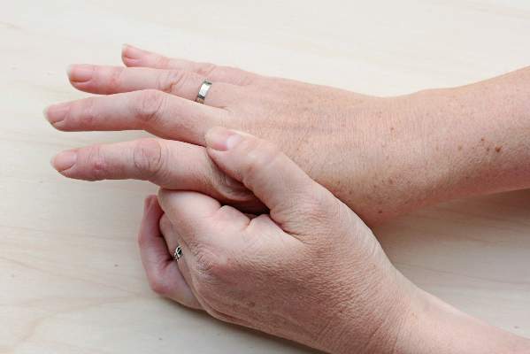
Inflammatory features linked to erosive development in hand OA
Among adults with hand osteoarthritis, ultrasonographic evidence of persistent joint-level inflammation increased the odds of subsequent erosions by as much as 11 times, said authors of a 2-year prospective study.
“These observations implicate a role for inflammation in the pathogenesis of erosive osteoarthritis and might render new therapeutic options that can halt erosive development,” said Dr. Marion C. Kortekaas and her associates at Leiden (the Netherlands) University Medical Center. The findings appeared online in Arthritis & Rheumatology.
The pathogenesis of erosive hand OA remains poorly understood, despite its high clinical burden, the researchers noted. To assess potential risk factors for erosive development, they used standard ultrasonographic methods to examine the interphalangeal joints of 56 consecutive patients who presented to a rheumatology outpatient clinic with hand OA based on American College of Rheumatology criteria. They also scored radiographs for osteophytes or joint-space narrowing with the OARSI method and used the Verbruggen-Veys method to identify and exclude joints that were already eroded (E phase) or remodeled (R phase) at baseline (Arthritis Rheum. 2015 Sep 28. doi: 10.1002/art.39438). At baseline, 18 patients had ultrasonographic evidence of erosions in a total of 51 interphalangeal joints, the investigators said. At the 2.3-year follow-up, a total of 38 interphalangeal joints from 26 patients showed erosive development.
After accounting for age, gender, body mass index, and baseline cartilage and bone abnormalities, all ultrasonographic features of inflammation that were at least grade 1 at baseline and follow-up predicted erosive development. Persistent power Doppler signal was the strongest risk factor, yielding an odds ratio of 11.4 in the adjusted model (95% confidence interval, 2.7-49.1). Other significant risk factors included moderate to severe baseline synovial thickening (OR, 8.8; 95% CI, 2.4-32.3) and power Doppler signal (OR, 7.1; 95% CI, 1.9-26.9).
“The present study confirms that inflammatory US features found at baseline are associated with erosive development on the joint level in hand OA,” the investigators wrote. “These associations are already found after 2 years of follow-up, which is important for future prospective trials.”
The researchers reported having no funding sources or conflicts of interest.
FROM ARTHRITIS & RHEUMATOLOGY
Key clinical point: Ultrasonographic evidence of inflammation significantly predicted the development of erosions in hand osteoarthritis.
Major finding: All inflammatory ultrasonographic features that were at least grade 1 at baseline and follow-up significantly predicted erosive development.
Data source: Single-center, prospective, ultrasonographic and radiographic study of 56 patients who met ACR criteria for hand OA.
Disclosures: The investigators reported having no funding sources or conflicts of interest.

Recall issued on U.S. Compounding sterile products
U.S. Compounding Inc. is issuing a recall on all sterile products distributed between March 14, 2015, and Sept. 9, 2015, according to a safety alert from the Food and Drug Administration.
The product recall applies to all aseptically compounded and packaged USC sterile products distributed to hospitals, patients, providers, and clinics because of FDA concerns over lack of sterility assurance. Because of the risk to any patients using a compromised product, USC is proceeding voluntarily with the recall.
Patients or providers who received sterile compounded products from USC within the recall date and have not expired should stop using the products immediately, quarantine the product until proper disposal is possible, and contact USC as soon as possible to coordinate a plan to return the product.
Patients should also contact their physicians if they have experienced any issues relating to the recalled product, and physicians should contact patients to inform them of the recall and to advise them to stop using the product.
The USC recall does not apply to any nonsterile compounded medication produced or distributed by USC, according to the FDA alert.
Find the full safety alert on the FDA website.
PsA, PsC do not affect total hip replacement outcomes
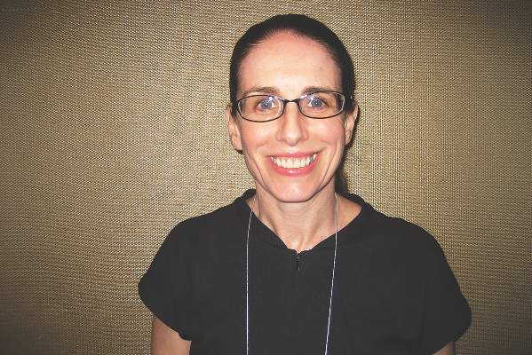
Neither psoriatic arthritis (PsA) nor cutaneous psoriasis (PsC) is an independent predictor of poor postoperative pain or function following a total hip arthroplasty, according to the results of a case-control study by Dr. Lisa A. Mandl and her colleagues.
The study’s participants underwent surgery between May 1, 2007, and Dec. 31, 2010, in a center that performs more than 4,300 THAs annually. All subjects lived for at least 2 years after their operations. The researchers compared pre- and postoperative data from patients in the following three categories: those with PsA, those with PsC without evidence of inflammatory arthritis, and those with osteoarthritis (OA). Patients with OA comprised the control group, which excluded any patient who self-reported a history of PsA, rheumatoid arthritis, lupus erythematosus, or any other systematic rheumatic disease, or who had documentation of skin psoriasis. The researchers acquired postoperative self-report data from 47 PsA patients, 106 PsC patients, and 864 OA patients. Seventeen percent of patients submitted information on their status at 1 year, 69% at 2 years, and 14% at 3-5 years.
The primary outcomes of interest were postoperative pain and function, which were assessed via the Hip Osteoarthritis Outcome Score (HOOS), from which the Western Ontario and McMaster Universities Osteoarthritis Index (WOMAC) was derived.
There were no statistically significant differences in postoperative WOMAC pain or function scores between the three groups of patients (P = .78 and .96, respectively). The mean pain scores were 14.9, 6.1, and 15.8 for patients with PsA, PsC, and OA, respectively. These patients’ mean function scores were 16.3, 19.6, and 18.8 for the PsA, PsC and OA groups, respectively.
Overall levels of satisfaction with the surgery were similar among the three groups (P = .54). Ninety-three percent of the PsA patients, 79% of the PsC patients, and 84% of the OA patients were “very satisfied” with their total hip arthroplasty. Between 1% and 3% of each group reported being “very dissatisfied” with their surgery. The researchers found that extent of skin disease was not associated with worse postoperative pain or function.
“Further work needs to be done to better understand the interplay of disease activity and quality of life on the outcomes of [total hip arthroplasty] in PsA and PsC,” they wrote.
Read the report in Arthritis & Rheumatology (doi: 10.1002/art.39431).
FROM ARTHRITIS & RHEUMATOLOGY

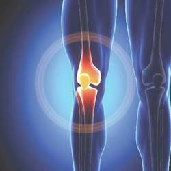
Knee OA patients see most benefit from small doses of fish oil
The anti-inflammatory properties of fish oil have been demonstrated, but for patients with knee osteoarthritis, high doses of fish oil were no more effective than was low-dose fish oil, according to Dr. Catherine L. Hill and her associates.
Just over 200 patients with knee OA were included in the study, with half receiving a full anti-inflammatory dose of 15 mL of fish oil/sunola oil per day containing 4.5 g of omega-3 fatty acids, and the other group receiving 15 mL of fish oil/sunola oil per day containing 0.45 g omega-3 fatty acids.

After 1 year, Western Ontario and McMaster Universities Arthritis Index scores were similar in both groups, but after 2 years, WOMAC pain and function scores were better in the low-dose fish oil group.
There was no difference in usage of analgesics or NSAIDs between the two groups during the 2-year study period. Cartilage volume loss was similar in both groups. Weight gain was greater in the high-dose fish oil group, but analysis showed this had no effect on pain scores in the high-dose group.
“One possible explanation could be that sunola oil with or without low-dose fish may confer a beneficial effect, but this unanticipated finding requires confirmation in further trials,” the investigators said.
Find the study here in Annals of the Rheumatic Diseases (doi: 10.1136/annrheumdis-2014-207169).
The anti-inflammatory properties of fish oil have been demonstrated, but for patients with knee osteoarthritis, high doses of fish oil were no more effective than was low-dose fish oil, according to Dr. Catherine L. Hill and her associates.
Just over 200 patients with knee OA were included in the study, with half receiving a full anti-inflammatory dose of 15 mL of fish oil/sunola oil per day containing 4.5 g of omega-3 fatty acids, and the other group receiving 15 mL of fish oil/sunola oil per day containing 0.45 g omega-3 fatty acids.
After 1 year, Western Ontario and McMaster Universities Arthritis Index scores were similar in both groups, but after 2 years, WOMAC pain and function scores were better in the low-dose fish oil group.
There was no difference in usage of analgesics or NSAIDs between the two groups during the 2-year study period. Cartilage volume loss was similar in both groups. Weight gain was greater in the high-dose fish oil group, but analysis showed this had no effect on pain scores in the high-dose group.
“One possible explanation could be that sunola oil with or without low-dose fish may confer a beneficial effect, but this unanticipated finding requires confirmation in further trials,” the investigators said.
Find the study here in Annals of the Rheumatic Diseases (doi: 10.1136/annrheumdis-2014-207169).

New cartilage, better function with stem cells for knee OA
Mesenchymal stem cell transplants appeared to regenerate cartilage and improve clinical outcomes after 2 years in patients with knee osteoarthritis in a small South Korean study.
There were 24 treated knees among the 11 men and 9 women in the study, whose average age was 58 years and mean body mass index was 26.6 kg/m2.
At baseline, 21 lesions (87.5%) were grade 2 or 3 on the MRI Osteoarthritis Knee Score scale for size of cartilage-loss area, and 23 lesions (95.9%) were grade 2 or 3 for percentage of full-thickness cartilage loss. A grade of 2-3 indicates moderate to severe disease.
At MRI follow-up 2 years after transplant, only five lesions (20.8%) were grade 2 or 3 for cartilage-loss area and five were grade 2 or 3 for full-thickness cartilage loss, reported Dr. Yong Sang Kim of the Yonsei Sarang Hospital in Seoul and his associates (Osteoarthritis Cartilage. 2015. doi: 10.1016/j.joca.2015.08.009).
There were clinical improvements, too. At baseline, the mean International Knee Documentation Committee score improved from 38.7 at baseline to 67.3 at follow-up and the mean Tegner activity scale score improved from 2.5 at baseline to 3.9 at follow-up. The results were all statistically significant and independent of age, sex, body mass index, and size and location of the cartilage lesions.
“This study showed encouraging clinical outcomes of [mesenchymal stem cell] implantation with fibrin glue as a scaffold in OA knees,” and it adds to the growing body of literature supporting mesenchymal stem cells (MSCs) in osteoarthritis. “MSC implantation with fibrin glue as a scaffold seems to be effective for repairing cartilage lesions in OA knees,” the investigators wrote.
The cells were derived from fat liposuctioned from the subjects’ buttocks and delivered to their damaged knees in a fibrinogen-thrombin gel under arthroscopic guidance. The cartilage lesions, which were most often on the medial femoral condyle, were debrided beforehand. Patients’ knees were immobilized for 2 weeks after transplant. Weight bearing was allowed at 4 weeks, and sports at 3 months.
It’s not fully known why MSCs help knee OA. Perhaps they release factors that stimulate cartilage formation by resident chondrocytes or other cells in the joint, and inhibit joint inflammation. The investigators derived their cells from fat – instead of bone marrow – because stem cells from fat are easier to get, easy to purify, and may be more chondrogenic. Fat is also richer in stem cells than marrow.
The next step is to identify predictors of good outcomes. “Patient characteristics or cartilage lesion variables may serve as important selection criteria for stem cell–based repair strategies,” Dr. Kim and his associates said.
The researchers had no disclosures.
FROM OSTEOARTHRITIS AND CARTILAGE
Key clinical point: Mesenchymal stem cell transplants give promising 2-year results in knee osteoarthritis, but larger studies with longer follow-up periods are needed.
Major finding: At baseline, 23 lesions (95.9%) were grade 2 or 3 for percentage of full-thickness cartilage loss; at MRI follow-up 2 years after transplant, only five lesions (20.8%) were grade 2 or 3.
Data source: A 2-year follow-up of 24 treated knees.
Disclosures: The investigators had no disclosures.

Muscle power tops strength for knee OA assessment
Leg extensor muscle power is an independent determinant of pain and quality of life in knee osteoarthritis, according to a cross-sectional analysis of data from a trial.
“Compared to strength, muscle power may be a more clinically important measure of muscle function within this population. New trials to systematically examine the impact of muscle power training interventions on disease severity in knee OA are particularly warranted,” concluded the investigators, led by Kieran Reid, Ph.D., of Tufts University in Boston (Arthritis Rheumatol. 2015 Aug 28. doi: 10.1002/art.39336).

The conclusion comes from their analysis of baseline data for 190 subjects participating in a trial comparing Tai Chi to standard physical therapy for knee OA. Their mean age was 60 years, and they had a mean body mass index of 32.7 kg/m2. The majority of participants were women.
On univariate analysis, greater muscle power – defined as the product of dynamic muscular force and muscle contraction velocity – was significantly associated with pain (r = –0.17, P less than .02), as assessed by the Western Ontario and McMaster Osteoarthritis Index. It was also significantly and positively associated with Short Form 36 physical component scores (r = 0.16, P less than .05), a measure of health-related quality of life.
After adjusting for multiple covariates, muscle power was a significant independent predictor of pain (P less than or equal to .05) and physical component scores (P less than or equal to .04). Strength, a far more common assessment in knee OA, was not an independent determinant of either pain or quality of life (P greater than or equal to .06).
To measure leg extension muscle power, subjects were asked to do two sets of five bilateral leg press repetitions, one set at 40% of their maximum strength and one set at 70%. Each repetition within a set was performed as quickly as possible, with 30 seconds rest between each repetition.
Knee OA patients “have substantial and accelerated impairments in muscle power,” and might benefit from training to improve it. Power generated at 70% resistance in the study seemed to be “a marginally greater determinant of pain and quality of life in knee OA” than power at 40%, suggesting that higher intensity training might be more helpful.
The investigators had several ideas about how power relates to knee pain. Power, rather than strength alone, helps keep the knee stable when in motion. Impairment could contribute to “inadequate control of tibial translation during ambulation, leading to damage, and ultimately knee pain.” Also, impairment probably limits the ability to dissipate knee joint loads, increasing “the risk of articular contact stress, which leads to pain,” they said.
“Muscle strength, the maximal force generating capacity of skeletal muscle, has been the focus of numerous investigations. However, studies examining muscle strength in the etiology of knee OA have presented inconsistent and controversial findings.” Dynamic leg extensor power “represents a more functionally relevant assessment of muscle performance and ... a more clinically important correlate of disease burden in knee OA,” they said.
The study was supported by grants from the National Institutes of Health. The investigators have no disclosures.
Leg extensor muscle power is an independent determinant of pain and quality of life in knee osteoarthritis, according to a cross-sectional analysis of data from a trial.
“Compared to strength, muscle power may be a more clinically important measure of muscle function within this population. New trials to systematically examine the impact of muscle power training interventions on disease severity in knee OA are particularly warranted,” concluded the investigators, led by Kieran Reid, Ph.D., of Tufts University in Boston (Arthritis Rheumatol. 2015 Aug 28. doi: 10.1002/art.39336).
The conclusion comes from their analysis of baseline data for 190 subjects participating in a trial comparing Tai Chi to standard physical therapy for knee OA. Their mean age was 60 years, and they had a mean body mass index of 32.7 kg/m2. The majority of participants were women.
On univariate analysis, greater muscle power – defined as the product of dynamic muscular force and muscle contraction velocity – was significantly associated with pain (r = –0.17, P less than .02), as assessed by the Western Ontario and McMaster Osteoarthritis Index. It was also significantly and positively associated with Short Form 36 physical component scores (r = 0.16, P less than .05), a measure of health-related quality of life.
After adjusting for multiple covariates, muscle power was a significant independent predictor of pain (P less than or equal to .05) and physical component scores (P less than or equal to .04). Strength, a far more common assessment in knee OA, was not an independent determinant of either pain or quality of life (P greater than or equal to .06).
To measure leg extension muscle power, subjects were asked to do two sets of five bilateral leg press repetitions, one set at 40% of their maximum strength and one set at 70%. Each repetition within a set was performed as quickly as possible, with 30 seconds rest between each repetition.
Knee OA patients “have substantial and accelerated impairments in muscle power,” and might benefit from training to improve it. Power generated at 70% resistance in the study seemed to be “a marginally greater determinant of pain and quality of life in knee OA” than power at 40%, suggesting that higher intensity training might be more helpful.
The investigators had several ideas about how power relates to knee pain. Power, rather than strength alone, helps keep the knee stable when in motion. Impairment could contribute to “inadequate control of tibial translation during ambulation, leading to damage, and ultimately knee pain.” Also, impairment probably limits the ability to dissipate knee joint loads, increasing “the risk of articular contact stress, which leads to pain,” they said.
“Muscle strength, the maximal force generating capacity of skeletal muscle, has been the focus of numerous investigations. However, studies examining muscle strength in the etiology of knee OA have presented inconsistent and controversial findings.” Dynamic leg extensor power “represents a more functionally relevant assessment of muscle performance and ... a more clinically important correlate of disease burden in knee OA,” they said.
The study was supported by grants from the National Institutes of Health. The investigators have no disclosures.
FROM ARTHRITIS & RHEUMATOLOGY
Key clinical point: It might be time to learn how to assess muscle power in your knee OA patients.
Major finding: After adjusting for multiple covariates, muscle power was a significant independent predictor of pain (P less than or equal to .05) and physical component scores (P less than or equal to .04).
Data source: Cross-sectional analysis of 190 knee OA patients.
Disclosures: The study was supported by grants from the National Institutes of Health. The investigators have no disclosures.

VIDEO: Consistency is key to monitoring patients on opioids
ORLANDO – Take a consistent approach with all patients on opioid therapy, regardless of a patient’s perceived potential for abuse, Dr. Melissa B. Weimer advised.
“This is an area of medicine where I feel we need to apply universal precautions to all patients,” noted Dr. Weimer, assistant professor of medicine, Oregon Health and Science University, Portland. “We’re treating all patients as at some level of potential harm from this medication.”
In an interview at a meeting held by the American Pain Society and Global Academy for Medical Education, Dr. Weimer outlined a strategy that employs the same protocol to monitor therapy, even when the abuse potential is considered to be low.
Global Academy and this news organization are owned by the same company. Dr. Weimer reported no financial disclosures.
The video associated with this article is no longer available on this site. Please view all of our videos on the MDedge YouTube channel
EXPERT ANALYSIS FROM PAIN CARE FOR PRIMARY CARE

VIDEO: Use fiduciary duty to set pain medication boundaries
ORLANDO – Physicians should use the concept of fiduciary duty to set appropriate boundaries with patients taking pain medications, explained Dr. Louis Kuritzky.
Often, patients want treatments that are not in their best interests, noted Dr. Kuritzky of the department of community health and family medicine at the University of Florida, Gainesville.
In an interview at a meeting held by the American Pain Society and Global Academy for Medical Education, Dr. Kuritzky outlined how physicians can take a fiduciary duty approach to set boundaries with patients in a dispassionate manner.
Global Academy and this news organization are owned by the same company. Dr. Kuritzky reported a financial relationship with Lilly.
The video associated with this article is no longer available on this site. Please view all of our videos on the MDedge YouTube channel
EXPERT ANALYSIS FROM PAIN CARE FOR PRIMARY CARE
ESC: Celecoxib safety study may soothe cardio concerns
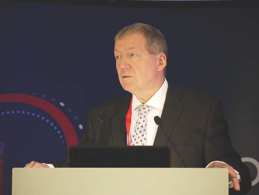
LONDON – Celecoxib was associated with very low cardiovascular event rates, and its use posed no more risk than other painkillers commonly used to treat elderly individuals with arthritic conditions but no heart disease in a large, pragmatic, family practice–based study.
Results of the Standard Care Versus Celecoxib Outcome Trial (SCOT) reported at the annual congress of the European Society of Cardiology also showed that celecoxib was no more likely than nonselective nonsteroidal anti-inflammatory drugs (nsNSAIDs) to cause ulcer-related upper gastrointestinal (GI) tract complications.

In fact, the rates of both cardiovascular and GI events were so low overall that it made the trial difficult to complete, said study investigator Dr. Tom MacDonald, professor of clinical pharmacology and pharmacoepidemiology at the University of Dundee (Scotland), which sponsored the study.
The on-treatment and intention-to-treat (ITT) cardiovascular event rates were 0.9% and 1.1% per 100 patient-years, he observed, adding that he would have expected the event rate to be around 2%-3% in the population studied. GI complication rates were even lower, with just 12 on-treatment and 15 ITT events reported during the entire follow-up period, which was a maximum of 6.3 years and mean of about 3 years.
“You may remember the brouhaha surrounding the use of rofecoxib and other [cyclo-oxygenase-2 inhibitors],” said Dr. MacDonald. Both coxibs and nsNSAIDs have been associated with adverse cardiovascular outcomes such as myocardial infarction (BMJ 2005;330:1366), and rofecoxib was voluntarily withdrawn in 2004 by its manufacturer from the U.S. market. A recent meta-analysis (Lancet 2013;382:769-79) has suggested that coxibs increase the risk of major cardiovascular events by about 37%.
The SCOT study (BMJ Open 2013;3:e002295) was designed to assess if celecoxib was better, worse, or the same as the other available NSAIDs in terms of its cardiovascular and gastrointestinal safety. It was originally set up because of a requirement by the European Medicines Agency, Dr. McDonald explained.
More than 9,400 patients aged 60 years or older with osteoarthritis or rheumatoid arthritis who were prescribed chronic NSAID therapy and had no existing cardiovascular disease were screened at 706 family practices in Scotland, England, Denmark, and the Netherlands. A total of 7,297 patients were included in the prospective study and were randomized to switch to treatment with celecoxib or to continue their current nsNSAID.
General practice records were linked to hospital and mortality databases to derive the primary composite endpoint of the first occurrence of hospitalization for nonfatal MI, nonfatal stroke, or cardiovascular death, as well as secondary endpoints such as time to first hospitalization or death from upper GI complications and all-cause mortality.
Randomized patients were about 68 years old, and about 40% of patients were male. Dr. MacDonald noted that, although there was no known existing cardiovascular disease at enrollment, the baseline characteristics showed that around 44% of patients had high blood pressure; a third of patients had high cholesterol; and 20%, 12%, and 38% were taking a statin, aspirin, or ulcer-healing treatments, respectively. The most common nsNSAIDs being used were diclofenac (38.7%) and ibuprofen (31%).
There was no significant difference between celecoxib or nsNSAIDs for any of the cardiovascular endpoints studied, with hazard ratios (HR) for the primary composite cardiovascular endpoints of 1.12 (95% confidence interval, 0.81-1.55; P = .5) while on celecoxib treatment and 1.04 (95% CI, 0.81-1.33; P = .75) in the ITT analysis. Similar results were obtained for all-cause mortality (HR, 1.2 and 0.92, respectively).
Dr. MacDonald reported that 50% of patients randomized to celecoxib and 30% randomized to continue nsNSAIDs withdrew from the study. The main reasons for stopping celecoxib were a lack of efficacy (11.2% vs. 2% for nsNSAIDs), adverse events (8.3% vs. 4.4%), patient request (6% vs. 2.3%), not tolerated (3.9% vs. 1.2%), or a serious adverse event (2.6% vs. 1.9%). There was, however, a lot of adverse publicity about the coxibs, he noted, and patients who had been happy on an nsNSAID might not have been happy with the switch.
The rates of serious cardiovascular adverse events (31.7% vs. 32.4%) or reactions (5.2% vs. 5.8%) were similar with celecoxib and nsNSAIDs, but there were significantly fewer serious GI adverse reactions with celecoxib than with nsNSAIDs (38 vs. 66; P = .007). Overall, the adverse reaction rate was 22% vs. 16.1%, respectively (P <.001).
“In the study population, nsNSAIDs and celecoxib both appeared acceptably safe,” Dr. MacDonald concluded. “In patients who get significant symptomatic relief from these medicines, the benefit/risk balance appears positive.”
Although the findings are perhaps reassuring, they are unlikely to change clinical practice, observed Dr. José López-Sendon, who was invited to comment on the study results after their presentation at the conference.
The study findings suggest that celecoxib may continue to be safe to use in patients without existing cardiac disease, noted Dr. López-Sendon of Hospital Universitario La Paz in Madrid, but he would not modify the guidelines that advise that the lowest effective dose be used for the shortest duration of time in low-risk patients.
The study was sponsored by the University of Dundee and funded by an investigator-initiated research grant from Pfizer. The university’s Medicines Monitoring Unit also holds research grants from Amgen, Menarini, and Novartis. Dr. MacDonald has consulted on the use of NSAIDs for AstraZeneca, NiCox, Novartis, and Pfizer. Dr. López-Sendon did not have any disclosures relevant to his comments.
LONDON – Celecoxib was associated with very low cardiovascular event rates, and its use posed no more risk than other painkillers commonly used to treat elderly individuals with arthritic conditions but no heart disease in a large, pragmatic, family practice–based study.
Results of the Standard Care Versus Celecoxib Outcome Trial (SCOT) reported at the annual congress of the European Society of Cardiology also showed that celecoxib was no more likely than nonselective nonsteroidal anti-inflammatory drugs (nsNSAIDs) to cause ulcer-related upper gastrointestinal (GI) tract complications.
In fact, the rates of both cardiovascular and GI events were so low overall that it made the trial difficult to complete, said study investigator Dr. Tom MacDonald, professor of clinical pharmacology and pharmacoepidemiology at the University of Dundee (Scotland), which sponsored the study.
The on-treatment and intention-to-treat (ITT) cardiovascular event rates were 0.9% and 1.1% per 100 patient-years, he observed, adding that he would have expected the event rate to be around 2%-3% in the population studied. GI complication rates were even lower, with just 12 on-treatment and 15 ITT events reported during the entire follow-up period, which was a maximum of 6.3 years and mean of about 3 years.
“You may remember the brouhaha surrounding the use of rofecoxib and other [cyclo-oxygenase-2 inhibitors],” said Dr. MacDonald. Both coxibs and nsNSAIDs have been associated with adverse cardiovascular outcomes such as myocardial infarction (BMJ 2005;330:1366), and rofecoxib was voluntarily withdrawn in 2004 by its manufacturer from the U.S. market. A recent meta-analysis (Lancet 2013;382:769-79) has suggested that coxibs increase the risk of major cardiovascular events by about 37%.
The SCOT study (BMJ Open 2013;3:e002295) was designed to assess if celecoxib was better, worse, or the same as the other available NSAIDs in terms of its cardiovascular and gastrointestinal safety. It was originally set up because of a requirement by the European Medicines Agency, Dr. McDonald explained.
More than 9,400 patients aged 60 years or older with osteoarthritis or rheumatoid arthritis who were prescribed chronic NSAID therapy and had no existing cardiovascular disease were screened at 706 family practices in Scotland, England, Denmark, and the Netherlands. A total of 7,297 patients were included in the prospective study and were randomized to switch to treatment with celecoxib or to continue their current nsNSAID.
General practice records were linked to hospital and mortality databases to derive the primary composite endpoint of the first occurrence of hospitalization for nonfatal MI, nonfatal stroke, or cardiovascular death, as well as secondary endpoints such as time to first hospitalization or death from upper GI complications and all-cause mortality.
Randomized patients were about 68 years old, and about 40% of patients were male. Dr. MacDonald noted that, although there was no known existing cardiovascular disease at enrollment, the baseline characteristics showed that around 44% of patients had high blood pressure; a third of patients had high cholesterol; and 20%, 12%, and 38% were taking a statin, aspirin, or ulcer-healing treatments, respectively. The most common nsNSAIDs being used were diclofenac (38.7%) and ibuprofen (31%).
There was no significant difference between celecoxib or nsNSAIDs for any of the cardiovascular endpoints studied, with hazard ratios (HR) for the primary composite cardiovascular endpoints of 1.12 (95% confidence interval, 0.81-1.55; P = .5) while on celecoxib treatment and 1.04 (95% CI, 0.81-1.33; P = .75) in the ITT analysis. Similar results were obtained for all-cause mortality (HR, 1.2 and 0.92, respectively).
Dr. MacDonald reported that 50% of patients randomized to celecoxib and 30% randomized to continue nsNSAIDs withdrew from the study. The main reasons for stopping celecoxib were a lack of efficacy (11.2% vs. 2% for nsNSAIDs), adverse events (8.3% vs. 4.4%), patient request (6% vs. 2.3%), not tolerated (3.9% vs. 1.2%), or a serious adverse event (2.6% vs. 1.9%). There was, however, a lot of adverse publicity about the coxibs, he noted, and patients who had been happy on an nsNSAID might not have been happy with the switch.
The rates of serious cardiovascular adverse events (31.7% vs. 32.4%) or reactions (5.2% vs. 5.8%) were similar with celecoxib and nsNSAIDs, but there were significantly fewer serious GI adverse reactions with celecoxib than with nsNSAIDs (38 vs. 66; P = .007). Overall, the adverse reaction rate was 22% vs. 16.1%, respectively (P <.001).
“In the study population, nsNSAIDs and celecoxib both appeared acceptably safe,” Dr. MacDonald concluded. “In patients who get significant symptomatic relief from these medicines, the benefit/risk balance appears positive.”
Although the findings are perhaps reassuring, they are unlikely to change clinical practice, observed Dr. José López-Sendon, who was invited to comment on the study results after their presentation at the conference.
The study findings suggest that celecoxib may continue to be safe to use in patients without existing cardiac disease, noted Dr. López-Sendon of Hospital Universitario La Paz in Madrid, but he would not modify the guidelines that advise that the lowest effective dose be used for the shortest duration of time in low-risk patients.
The study was sponsored by the University of Dundee and funded by an investigator-initiated research grant from Pfizer. The university’s Medicines Monitoring Unit also holds research grants from Amgen, Menarini, and Novartis. Dr. MacDonald has consulted on the use of NSAIDs for AstraZeneca, NiCox, Novartis, and Pfizer. Dr. López-Sendon did not have any disclosures relevant to his comments.
AT THE ESC CONGRESS 2015
Key clinical point: Celecoxib and nonselective NSAIDs were “acceptably safe” in a population without confirmed cardiovascular disease.
Major finding: The hazard ratio for the primary composite cardiovascular endpoint with celecoxib use was 1.12 (95% confidence interval, 0.81-1.55; P = .5), compared with NSAIDs.
Data source: The Standard Care Versus Celecoxib Outcome Trial (SCOT) of more than 7,200 elderly patients with osteoarthritis or rheumatoid arthritis and no confirmed cardiovascular disease.
Disclosures: The study was sponsored by the University of Dundee and funded by an investigator-initiated research grant from Pfizer. The university’s Medicines Monitoring Unit also holds research grants from Amgen, Menarini, and Novartis. Dr. MacDonald has consulted on the use of NSAIDs for AstraZeneca, NiCox, Novartis, and Pfizer. Dr. López-Sendon did not have any disclosures relevant to his comments.
Heart attack risk rises in first month after knee, hip arthroplasty
Total knee and hip arthroplasty were associated with a significantly increased risk of myocardial infarction in the first month after surgery, but not at 6 months after surgery, a cohort study showed.
The finding challenges other studies suggesting that total knee or hip arthroplasty surgery reduces the risk of serious cardiovascular events among individuals with osteoarthritis.

Analysis of data from 13,849 British patients who underwent a total knee arthroplasty, 6,063 patients who received total hip arthroplasty, and an equal number of matched controls showed a greater than eightfold increase in the risk of myocardial infarction in the first postoperative month in the knee arthroplasty group (hazard ratio, 8.75), and a fourfold increase in risk in the hip arthroplasty group (HR, 4.33), compared with controls.
However, the hazard ratio declined to insignificance after 6 months, in contrast to the risk of venous thromboembolism – a known complication of arthroplasty – according to a paper published online in the Aug. 31 edition of Arthritis & Rheumatology.
“The major difference between the previous study and ours is that cardiovascular events occurring shortly after total joint arthroplasty were excluded from the previous study,” wrote Na Lu and his colleagues from Boston University.
The study observed 306 cases of myocardial infarction among individuals who underwent total knee arthroplasty and 128 cases in those who underwent total hip arthroplasty (Arthritis Rheumatol. 2015; August 31 [doi:10.1002/art.39246]).
The study was partly support by the National Institute of Arthritis and Musculoskeletal and Skin Diseases. There were no other conflicts of interest declared.
Total knee and hip arthroplasty were associated with a significantly increased risk of myocardial infarction in the first month after surgery, but not at 6 months after surgery, a cohort study showed.
The finding challenges other studies suggesting that total knee or hip arthroplasty surgery reduces the risk of serious cardiovascular events among individuals with osteoarthritis.
Analysis of data from 13,849 British patients who underwent a total knee arthroplasty, 6,063 patients who received total hip arthroplasty, and an equal number of matched controls showed a greater than eightfold increase in the risk of myocardial infarction in the first postoperative month in the knee arthroplasty group (hazard ratio, 8.75), and a fourfold increase in risk in the hip arthroplasty group (HR, 4.33), compared with controls.
However, the hazard ratio declined to insignificance after 6 months, in contrast to the risk of venous thromboembolism – a known complication of arthroplasty – according to a paper published online in the Aug. 31 edition of Arthritis & Rheumatology.
“The major difference between the previous study and ours is that cardiovascular events occurring shortly after total joint arthroplasty were excluded from the previous study,” wrote Na Lu and his colleagues from Boston University.
The study observed 306 cases of myocardial infarction among individuals who underwent total knee arthroplasty and 128 cases in those who underwent total hip arthroplasty (Arthritis Rheumatol. 2015; August 31 [doi:10.1002/art.39246]).
The study was partly support by the National Institute of Arthritis and Musculoskeletal and Skin Diseases. There were no other conflicts of interest declared.
FROM ARTHRITIS & RHEUMATOLOGY
Key clinical point: Total knee and hip arthroplasty were associated with a significantly increased risk of myocardial infarction in the first month after surgery.
Major finding: Patients undergoing total knee arthroplasty have an eightfold increase in the risk of myocardial infarction in the month after surgery.
Data source: Cohort study of 13,849 total knee arthroplasty patients and 6,063 total hip arthroplasty patients and an equal number of matched controls.
Disclosures: The study was partly support by the National Institute of Arthritis and Musculoskeletal and Skin Diseases. There were no conflicts of interest declared.