User login
The urgent need to diagnose Sanfilippo syndrome at an early age
Sanfilippo syndrome is a rare inherited neurodegenerative metabolic disorder for which there are no approved therapies. Symptoms of the more severe subtypes typically begin within the first years of life, rapidly producing serious and progressive physical and cognitive deficits. The underlying pathophysiology is targetable, but the delay in diagnosis of this as well as other lysosomal storage disorders (LSDs) is slowing progress toward effective therapies.
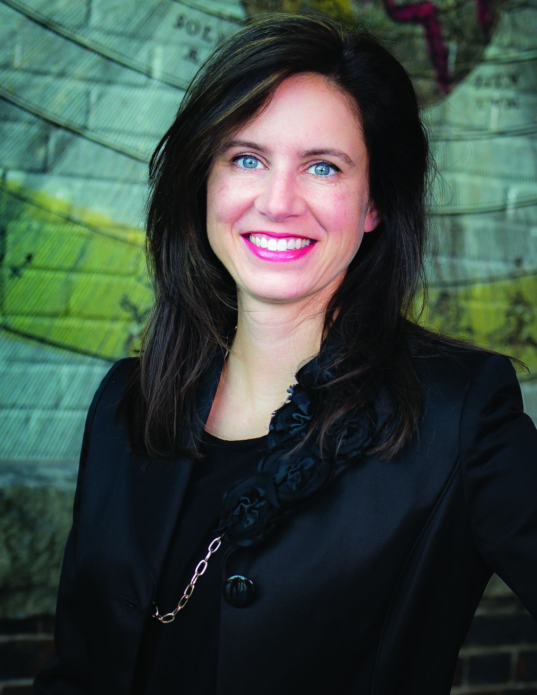
“Lack of awareness and the delays to diagnosis have been a real challenge for us. There is reason for cautious optimism about treatments now in or approaching clinical studies, but to evaluate efficacy on cognitive outcomes we need to enroll more children at a very young age, before loss of milestones,” according to Cara O’Neill, MD, a co-founder and chief science officer of Cure Sanfilippo Foundation.
Epidemiology and description
Sanfilippo syndrome, like the more than 50 other LSDs, is caused by a gene mutation that leads to an enzyme deficiency in the lysosome.1 In the case of Sanfilippo syndrome, also known as mucopolysaccharidosis (MPS III), there are hundreds of mutations that can lead to Sanfilippo by altering the function of one of the four genes essential to degradation of heparan sulfate.2 Lysosomal accumulation of heparan sulfate drives a broad spectrum of progressive and largely irreversible symptoms that typically begin with somatic manifestations, such as bowel dysfunction and recurrent ear and upper respiratory infections.
Impairment of the central nervous system (CNS) usually occurs early in life, halting physical and mental development. As it progresses, accumulation of heparan sulfate in a variety of cells leads to a cascade of abnormal cellular signaling and dysfunction. Disruption of these processes, which are critical for normal neurodevelopment, result in loss of the developmental skills already gained and eventually loss of brain tissue.3 Although life expectancy has improved with supportive care, survival into adulthood is typically limited to milder forms.4
Over the past several years, progress in this and other LSDs has yielded therapeutic targets, including those involving gene repair and enzyme replacement. Already approved for use in some LSDs, these therapies have also shown promise in the experimental setting for Sanfilippo syndrome, leading to several completed clinical trials.5
So far, none of these treatments has advanced beyond clinical trials in Sanfilippo syndrome, but there have been favorable changes in the markers of disease, suggesting that better methods of treatment delivery and/or more sensitive tools to measure clinical change might lead to evidence of disease attenuation. However, the promise of treatment in all cases has been to prevent, slow, or halt progression, not to reverse it. This point is important, because it indicates that degree of benefit will depend on enrolling patients early in life. Even if effective therapies are identified, few patients will benefit without strategies to accelerate diagnosis.
In fact, “one study6 reported that the average age of diagnosis for Sanfilippo syndrome has not improved over the past 30 years,” according to Dr. O’Neill. She indicated that this has been frustrating, given the availability of clinical trials on which progress is dependent. There is no widely accepted protocol for who and when to test for Sanfilippo syndrome or other LSDs, but Dr. O’Neill’s organization is among those advocating for strategies to detect these diseases earlier, including screening at birth.
Almost by definition, the clinical diagnosis of rare diseases poses a challenge. With nonspecific symptoms and a broad range of potential diagnoses, diseases with a low incidence are not the first ones that are typically considered. In the case of Sanfilippo syndrome, published studies indicate incidence rates at or below 1 per 70,000 live births.7 However, the incidence rates have been highly variable not only by geographical regions but even across neighboring countries where genetic risk would be expected to be similar.
In Europe, for example, epidemiologic studies suggest the lifetime risk of MPS IIIA is approximately two times greater in Germany and the Netherlands relative to France and Sweden.7 It is possible that the methodology for identifying cases might be a more important factor than differences in genetic risk to explain this variability. Many experts, including Dr. O’Neill, believe that prevalence figures for Sanfilippo syndrome are typically underestimates because of the frequency with which LSDs are attributed to other pathology.
“For these types of rare disorders, a clinician might only see a single case over a career, and the symptoms can vary in presentation and severity with many alternatives to consider in the differential diagnosis,” Dr. O’Neill explained. She cited case reports in which symptoms of Sanfilippo syndrome after a period of initial normal development has been initially attributed to autism, which is a comorbid feature of the disease, idiopathic developmental delay, or other nonprogressive disorders until further clinical deterioration leads to additional testing. The implication is that LSDs must be considered far earlier despite their rarity.
For the least common of the four clinical subtypes, MPS IIIC and MPS IIID, the median ages of diagnosis have ranged from 4.5 to 19 years of age.7 This is likely a reflection of a slower progression and a later onset of clinical manifestations.
For the more rapidly progressing and typically more severe subtypes, MPS IIIA and MPS IIIB, the diagnosis is typically made earlier. In one review of epidemiologic studies in different countries, the earliest reported median age at diagnosis was 2.5 years,7 a point at which significant disease progression is likely to have already occurred. If the promise of treatments in development is prevention of disease progression, disability in many patients might be substantial if the time to diagnosis is not reduced.
Screening and testing
Independent of the potential to enroll children in clinical trials, early diagnosis also advances the opportunities for supportive care to lessen the burden of the disease on patients and families. Perhaps even more important, early diagnosis is vital to family planning. Since the American pediatrician Sylvester Sanfilippo, MD, first described this syndrome in 1963,7 the genetic profile and many of the features of the disease have become well characterized.8
“One reason to emphasize the importance of early diagnosis is the heritability of this disorder. With prompt diagnosis, genetic counseling can be offered to families to provide them with critical information for future family planning and for cascade testing of other potentially affected siblings,” Dr. O’Neill reported. The inheritance pattern of Sanfilippo syndrome is autosomal recessive.3 In families with an affected child, the risk for any subsequent child to have the same disorder is 25%. The chance of a sibling to be unaffected and not a carrier is also 25%. There is a 50% chance of a sibling to be a carrier but asymptomatic. Of priorities, spreading awareness has been a critical mission of the Cure Sanfilippo Foundation since it was founded 8 years ago, according to Glenn O’Neill, the president. He and his wife, Dr. O’Neill, who is a pediatrician, founded the organization after their own child’s diagnosis of Sanfilippo syndrome. Creating awareness is fundamental to the mission of attracting funds for research, but support to patients and their families as well as early enrollment in clinical trials are among other initiatives being pursued by the foundation to improve care and prognosis.
These strategies include some novel ideas, including an algorithm based on artificial intelligence (AI) that can accelerate suspicion of Sanfilippo syndrome in advance of laboratory or genetic testing, according to Dr. O’Neill. She reported that the facial phenotype, which is observed in a high proportion of but not in all Sanfilippo patients, includes coarse facial features such as puffiness around the eyes, heavy eyebrows, full lips, and macrocephaly.9 Interpretation of photos for AI-based analysis is enhanced when combined with other clinical symptoms.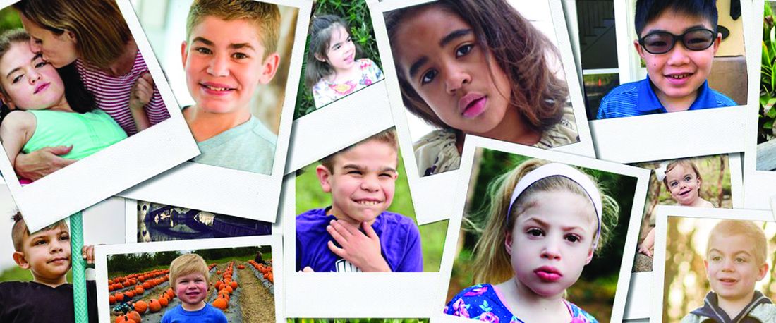
“The Foundation was involved in honing such a tool by submitting the photos that were used to teach the AI to recognize the Sanfilippo syndrome phenotype,” Dr. O’Neill said. The AI-based tool (Face2Gene.com) is available from FDNA, a company that has been involved in analyzing complex phenotypic and genomic information to guide diagnosis and therapeutic strategies for an array of diseases, not just Sanfilippo syndrome.
The preferred method for diagnosis is biochemical or genetic testing. Of these, urine testing for elevated levels of heparan sulfate glycosaminoglycans (GAG) can be useful for screening, although false-negative tests occur. Analysis of the blood can be performed to detect abnormal levels or activity of the enzymes that break down this GAG. In addition, genetic testing can be performed on blood, fibroblast, buccal swab, or saliva samples. Genetic testing of the blood is the most frequently performed.
For the four MPS III subtypes – MPS IIIA, IIIB, IIIC, and IIID – the presence of two pathogenic mutations in the SGSH (17q25.3), NAGLU (17q21.2), HGSNAT (8p11.21), and GNS (12q14.3) genes, respectively, are likely diagnostic, but enzymatic testing or GAG analysis should be performed to confirm disease status, according to Dr. O’Neill, who said that global consensus based clinical care guidelines led by the Foundation were recently accepted for publication and also include a section on the approach to diagnosis.
While laboratory testing is sensitive, urinary excretion of GAG can be variable, with the potential for ambiguous results. Typically, biochemical and genetic testing provide more reliable results for the diagnosis. They can be readily performed in utero or at the time of birth. In addition, gene panels can permit the diagnosis of multiple types of LSDs, not just Sanfilippo, making screening a cost-effective strategy to consider multiple diseases with overlapping symptoms when an LSD is suspected. Dr. O’Neill said clinical guidelines recommend confirmation of enzyme deficiency or evidence of GAG substrate accumulation as confirmatory tests when genetic testing is positive.
“Ultimately, our goal is to promote universal screening at birth for these serious genetic disorders affecting children,” Dr. O’Neill said.
“We are in a catch-22 when it comes to newborn screening. Currently our federal system requires there be an available treatment before recommending routine screening for a disease. However, it is extremely difficult to power trials with patients who are most likely to show benefit in a trial setting without that very early diagnosis. Universal newborn screening would pave the way for accelerated drug development for children,” she added.
In the meantime, Dr. O’Neill suggests that clinicians should employ a low threshold of suspicion to pursue diagnostic studies of LSDs in infants and children with developmental delays or otherwise unexplained progressive disorders.
Importantly, clinicians can now act quickly on their suspicions and order testing without concern for delays or denial by insurers through a special program, according to Dr. O’Neill. Free genetic testing, offered by the Invitae Corporation, evaluates a panel of 58 genes associated with lysosomal disorders, permitting detection of Sanfilippo syndrome and other LSDs, according to Dr. O’Neill. The Invitae testing is typically performed on 3 mL of whole blood delivered to a central testing facility.
“Results can be obtained within a few weeks or sooner. This can seem like a long wait for families, but it is much more efficient than ordering tests sequentially,” Dr. O’Neill said.
Diagnosis: Signs and symptoms
Despite the differences in progression of the MPS III subtypes, the clinical characteristics are more similar than different. In all patients, prenatal and infant development are typically normal. The initial signs of disease can be found in the newborn, such as neonatal tachypnea, through the early infancy period, such as macrocephaly. However, these are not commonly recognized until about age 1 or soon after in those with MPS IIIA and IIIB.3 Speech delay is the first developmental delay seen in most patients. In those with MPS IIIC, initial symptoms are typically detected at age 3 or later and progress more slowly.10,11 The same is likely to be true of MPS IIID, although this subtype is less well characterized than the other three.7
Although many organs can be involved, degeneration of the CNS is regarded as the most characteristic.3 In aggressive disease, this includes slower acquisition of and failure to meet developmental milestones with progressive intellectual disability, while behavioral difficulties are a more common initial compliant in children with milder disease.13,14 These behavioral changes include hyperactivity, inattention, autistic behaviors, worsening safety awareness, and in some cases aggressive behavior that can be destructive. Sleep disturbances are common.15Because of variability inherent in descriptions of relatively small numbers of patients, the characterization of each of the MPS III subgroups is based on a limited number of small studies, but most patients demonstrate behavior disorders, have coarse facial features, and develop speech delay, according to a survey conducted of published studies.7 Collectively, abnormal behavior was identified as an early symptom in 77% of those with MPS IIIA, 69% of those with IIIB, and 77% of those with IIIC.
For MPS IIIA, loss of speech was observed at a median age of 3.8 years and loss of walking ability at 10.4 years. The median survival has been reported to range between 13 and 18 years. In children with MPS IIIB, the median age of speech loss was reported to about the same age, while loss of walking ability occurred at 11 years. In one study of MPS IIIB, 24% of patients had developed dementia by age 6 years, and the reported median survival has ranged between 17 and 19 years. For MPS IIIC, the onset of clinical symptoms has been observed at a median age of 3.5 years with evidence of cognitive loss observed in 33% of children by the age of 6 years. The median survival has ranged from 19 to 34 years in three studies tracing the natural history of this MPS III subtype.
The differential diagnosis reasonably includes other types of mucopolysaccharidosis disorders with cognitive impairment, including Hurler, Hunter, or Sly syndromes, other neurodevelopmental disorders, and inborn errors of metabolism. The heterogeneity of the features makes definitive laboratory or genetic testing, rather than the effort to differentiate clinical features, appropriate for a definitive diagnosis.
Once the diagnosis is made, other examinations for the common complications of Sanfilippo syndrome are appropriate. Abdominal imaging is appropriate for detecting complications in the gastrointestinal tract, including hepatomegaly, which has been reported in more than half of patients with MPS IIIA and IIIB and in 39% of patients with IIIC.7 In patients with breathing concerns at night and/or sleep disturbance, polysomnography can be useful for identifying sleep apnea and nocturnal seizure activity. In children suspected of seizures, EEG is appropriate. In one study, 66% of patients with MPS IIIA developed seizure activity.16 This has been less commonly reported in MPS IIIB and IIIC, ranging from 8% to 13%.15
Formal hearing evaluation is indicated for any child with speech delays. Hearing loss typically develops after the newborn period in Sanfilippo and may affect peak language acquisition if not treated, according to Dr. O’Neill.
Radiographic studies for dysostosis multiplex or other skeletal abnormalities are also appropriate based on clinical presentation.
Treatment: Present and future
In the absence of treatments to improve the prognosis of Sanfilippo syndrome, current management is based on supportive care and managing organ-specific complications. However, several strategies have proven viable in experimental models and led to clinical trials. None of these therapies has reached approval yet, but several have been associated with attenuation of biomarkers of MPS III disease activity.
Of nearly 30 Sanfilippo clinical trials conducted over the past 20 years, at least 9 have now been completed.5 In addition to studying gene therapy and enzyme replacement therapy, these trials have included stem cell transplantation and substrate reduction therapy, for which the goal is to reduce synthesis of the heparan sulfate GAG to prevent accumulation.5 Of this latter approach, promising initial results with genistein, an isoflavone that breaks down heparan sulfate, reached a phase 3 evaluation.18 Although heparan sulfate levels in the CNS were non-significantly reduced over the course of the trial, the reduction was not sufficient to attenuate cognitive decline.
In other LSDs, several forms of enzyme replacement therapy are now approved. In Fabry disease, for example, recombinant alpha-galactosidase A has now been used for more than 15 years.19 Clinical benefit has not yet been demonstrated in patients with Sanfilippo syndrome because of the difficulty of delivering these therapies past the blood-brain barrier. Several strategies have been pursued. For example, intrathecal delivery of recombinant heparan-N-sulfatase reduced CNS levels of GAG heparan sulfate in one phase 2B study, but it approached but fell short of the statistical significance for the primary endpoint of predefined cognitive stabilization.20 The signal of activity and generally acceptable tolerability has encouraged further study, including an ongoing study with promising interim results of intracerebroventricular enzyme replacement in MPS IIIB, according to Dr. O’Neill.
Acceptable safety and promising activity on disease biomarkers have also been seen with gene therapy in clinical trials. In one study that showed attenuation of brain atrophy, there was moderate improvement in behavior and sleep in three of the four patients enrolled.21 Other studies using various strategies for gene delivery have also produced signals of activity against the underlying pathology, generating persistent interest in ongoing and planned clinical studies with this form of treatment.22Unmodified hematopoietic stem cell transplantation (HSCT), an approach that has demonstrated efficacy when delivered early in the course of other LSDs, such as Hurler syndrome,23 has not yet been associated with significant activity in clinical studies of MPS III, including those that initiated treatment prior to the onset of neurological symptoms.24 However, promising early results have been reported in a study of gene-modified HSCT, which overexpresses the MPS IIIA enzyme.
“The clinical trial landscape fluctuates quite a bit, so I always encourage clinicians and families to check back often for updates. Patient organizations can also be helpful for understanding the most up-to-date and emerging trial options,” Dr. O’Neill reported.
Although it is expected that the greatest benefit would be derived from treatments initiated before or very early after the onset of symptoms, based on the limited potential for reversing cognitive loss, Dr. O’Neill said that she and others are also striving to offer treatments for individuals now living with Sanfilippo syndrome.
“We have to be willing to test treatments that are symptomatic in nature. To that aim, the Cure Sanfilippo Foundation has sponsored a study of a CNS-penetrating anti-inflammatory agent in advanced-disease patients more than 4 years of age,” Dr. O’Neill said. This group of patients typically been ineligible for clinical trials in the past. Dr. O’Neill hopes to change this orientation.
“It is important to highlight that all patients deserve our efforts to improve their quality of life and alleviate suffering, regardless of how old they are or how progressed in the disease they happen to be,” she said.
However, whether the goal is enrollment before or early in disease or later in disease progression, the challenge of enrolling sufficient numbers of patients to confirm clinical activity has been and continues to be a hurdle to progress.
“Clinical studies in Sanfilippo enroll relatively small numbers of patients, often 20 or less,” said Dr. O’Neill, explaining one of the reasons why her organization has been so active in raising awareness and funding such studies. For patients and families, the Cure Sanfilippo Foundation can offer a variety of guidance and support, but information about opportunities for clinical trial participation is a key resource they provide for families and their physicians.
Conclusion
For most children with Sanfilippo syndrome, life expectancy is limited. However, the characterization of the genetic causes and the biochemistry of the subtypes has led to several viable therapeutic approaches under development. There has been progress in delivery of therapeutic enzymes to the CNS, and there is substantial optimism that more progress is coming. One issue for treatment development, is the last of a clear regulatory pathway addressing important biomarkers of pathology, such as heparan sulfate burden. Developing treatments that address this issue or impaired enzyme activity levels have promise for preventing progression, particularly if started in infancy. However, the effort to draw awareness to this disease is the first step toward accelerating the time to an early diagnosis and subsequent opportunities to enroll in clinical trials.
References
1. Sun A. Lysosomal storage disease overview. Ann Transl Med. 2018 Dec;6(24):476. doi: 10.21037/atm.2018.11.39.
2. Andrade F et al. Sanfilippo syndrome: Overall review. Pediatr Int. 2015 Jun;57(3):331-8. doi: 10.1111/ped.12636.
3. Fedele AO. Sanfilippo syndrome: Causes, consequences, and treatments. Appl Clin Genet. 2015 Nov 25;8:269-81. doi: 10.2147/TACG.S57672.
4. Lavery C et al. Mortality in patients with Sanfilippo syndrome. Orphanet J Rare Dis. 2017 Oct 23;12(1):168. doi: 10.1186/s13023-017-0717-y.
5. Pearse Y et al. A cure for Sanfilippo syndrome? A summary of current therapeutic approaches and their promise. Med Res Arch. 2020 Feb 1;8(2). doi: 10.18103/mra.v8i2.2045.
6. Kuiper GA et al. Failure to shorten the diagnostic delay in two ultrao-rphan diseases (mucopolysaccharidosis types I and III): potential causes and implication. Orphanet J Rare Dis. 2018;13:2. Doi: 10.1186/s13023-017-0733-y.
7. Zelei T et al. Epidemiology of Sanfilippo syndrome: Results of a systematic literature review. Orphanet J Rare Dis. 2018 Apr 10;13(1):53. doi: 10.1186/s13023-018-0796-4.
8. Wagner VF, Northrup H. Mucopolysaccaharidosis type III. Gene Reviews. 2019 Sep 19. University of Washington, Seattle. https://www.ncbi.nlm.nih.gov/books/NBK546574/8.
9. O’Neill C et al. Natural history of facial features observed in Sanfilippo syndrome (MPS IIIB) using a next generation phenotyping tool. Mol Genet Metab. 2019 Feb;126:S112.
10. Ruijter GJ et al. Clinical and genetic spectrum of Sanfilippo type C (MPS IIIC) disease in the Netherlands. Mol Genet Metab. 2008 Feb;93(2):104-11. doi: 10.1016/j.ymgme.2007.09.011.
11. Valstar MJ et al. Mucopolysaccharidosis type IIID: 12 new patients and 15 novel mutations. Hum Mutat. 2010 May;31(5):E1348-60. doi: 10.1002/humu.21234.
12. Nijmeijer SCM. The attenuated end of phenotypic spectrum in MPS III: from late-onset stable cognitive impairment to non-neuronopathic phenotype. Orphanet J Rare Dis. 2019;14:249. Doi10.1186/s13023-019-1232-0.
13. Nidiffer FD, Kelly TE. Developmental and degenerative patterns associated with cognitive, behavioural and motor difficulties in the Sanfilippo syndrome: An epidemiological study. J Ment Defic Res. 1983 Sep;27 (Pt 3):185-203. doi: 10.1111/j.1365-2788.1983.tb00291.x.
14. Bax MC, Colville GA. Behaviour in mucopolysaccharide disorders. Arch Dis Child. 1995 Jul;73(1):77-81. doi: 10.1136/adc.73.1.77.
15. Fraser J et al. Sleep disturbance in mucopolysaccharidosis type III (Sanfilippo syndrome): A survey of managing clinicians. Clin Genet. 2002 Nov;62(5):418-21. doi: 10.1034/j.1399-0004.2002.620512.x.
16. Valstar MJ et al. Mucopolysaccharidosis type IIIA: Clinical spectrum and genotype-phenotype correlations. Ann Neurol. 2010 Dec;68(6):876-87. doi: 10.1002/ana.22092.
17. Heron B et al. Incidence and natural history of mucopolysaccharidosis type III in France and comparison with United Kingdom and Greece. Am J Med Genet A. 2011 Jan;155A(1):58-68. doi: 10.1002/ajmg.a.33779.
18. Delgadillo V et al. Genistein supplementation in patients affected by Sanfilippo disease. J Inherit Metab Dis. 2011 Oct;34(5):1039-44. doi: 10.1007/s10545-011-9342-4.
19. van der Veen SJ et al. Developments in the treatment of Fabry disease. J Inherit Metab Dis. 2020 Sep;43(5):908-21. doi: 10.1002/jimd.12228.
20. Wijburg FA et al. Intrathecal heparan-N-sulfatase in patients with Sanfilippo syndrome type A: A phase IIb randomized trial. Mol Genet Metab. 2019 Feb;126(2):121-30. doi: 10.1016/j.ymgme.2018.10.006.
21. Tardieu M et al. Intracerebral administration of adeno-associated viral vector serotype rh.10 carrying human SGSH and SUMF1 cDNAs in children with mucopolysaccharidosis type IIIA disease: Results of a phase I/II trial. Hum Gene Ther. 2014 Jun;25(6):506-16. doi: 10.1089/hum.2013.238.
22. Marco S et al. In vivo gene therapy for mucopolysaccharidosis type III (Sanfilippo syndrome): A new treatment horizon. Hum Gene Ther. 2019 Oct;30(10):1211-1121. doi: 10.1089/hum.2019.217.
23. Taylor M et al. Hematopoietic stem cell transplantation for mucopolysaccharidoses: Past, present, and future. Biol Blood Marrow Transplant. 2019 Jul;25(7):e226-e246. doi: 10.1016/j.bbmt.2019.02.012.
24. Sivakumur P, Wraith JE. Bone marrow transplantation in mucopolysaccharidosis type IIIA: A comparison of an early treated patient with his untreated sibling. J Inherit Metab Dis. 1999 Oct;22(7):849-50. doi: 10.1023/a:1005526628598.
Sanfilippo syndrome is a rare inherited neurodegenerative metabolic disorder for which there are no approved therapies. Symptoms of the more severe subtypes typically begin within the first years of life, rapidly producing serious and progressive physical and cognitive deficits. The underlying pathophysiology is targetable, but the delay in diagnosis of this as well as other lysosomal storage disorders (LSDs) is slowing progress toward effective therapies.
“Lack of awareness and the delays to diagnosis have been a real challenge for us. There is reason for cautious optimism about treatments now in or approaching clinical studies, but to evaluate efficacy on cognitive outcomes we need to enroll more children at a very young age, before loss of milestones,” according to Cara O’Neill, MD, a co-founder and chief science officer of Cure Sanfilippo Foundation.
Epidemiology and description
Sanfilippo syndrome, like the more than 50 other LSDs, is caused by a gene mutation that leads to an enzyme deficiency in the lysosome.1 In the case of Sanfilippo syndrome, also known as mucopolysaccharidosis (MPS III), there are hundreds of mutations that can lead to Sanfilippo by altering the function of one of the four genes essential to degradation of heparan sulfate.2 Lysosomal accumulation of heparan sulfate drives a broad spectrum of progressive and largely irreversible symptoms that typically begin with somatic manifestations, such as bowel dysfunction and recurrent ear and upper respiratory infections.
Impairment of the central nervous system (CNS) usually occurs early in life, halting physical and mental development. As it progresses, accumulation of heparan sulfate in a variety of cells leads to a cascade of abnormal cellular signaling and dysfunction. Disruption of these processes, which are critical for normal neurodevelopment, result in loss of the developmental skills already gained and eventually loss of brain tissue.3 Although life expectancy has improved with supportive care, survival into adulthood is typically limited to milder forms.4
Over the past several years, progress in this and other LSDs has yielded therapeutic targets, including those involving gene repair and enzyme replacement. Already approved for use in some LSDs, these therapies have also shown promise in the experimental setting for Sanfilippo syndrome, leading to several completed clinical trials.5
So far, none of these treatments has advanced beyond clinical trials in Sanfilippo syndrome, but there have been favorable changes in the markers of disease, suggesting that better methods of treatment delivery and/or more sensitive tools to measure clinical change might lead to evidence of disease attenuation. However, the promise of treatment in all cases has been to prevent, slow, or halt progression, not to reverse it. This point is important, because it indicates that degree of benefit will depend on enrolling patients early in life. Even if effective therapies are identified, few patients will benefit without strategies to accelerate diagnosis.
In fact, “one study6 reported that the average age of diagnosis for Sanfilippo syndrome has not improved over the past 30 years,” according to Dr. O’Neill. She indicated that this has been frustrating, given the availability of clinical trials on which progress is dependent. There is no widely accepted protocol for who and when to test for Sanfilippo syndrome or other LSDs, but Dr. O’Neill’s organization is among those advocating for strategies to detect these diseases earlier, including screening at birth.
Almost by definition, the clinical diagnosis of rare diseases poses a challenge. With nonspecific symptoms and a broad range of potential diagnoses, diseases with a low incidence are not the first ones that are typically considered. In the case of Sanfilippo syndrome, published studies indicate incidence rates at or below 1 per 70,000 live births.7 However, the incidence rates have been highly variable not only by geographical regions but even across neighboring countries where genetic risk would be expected to be similar.
In Europe, for example, epidemiologic studies suggest the lifetime risk of MPS IIIA is approximately two times greater in Germany and the Netherlands relative to France and Sweden.7 It is possible that the methodology for identifying cases might be a more important factor than differences in genetic risk to explain this variability. Many experts, including Dr. O’Neill, believe that prevalence figures for Sanfilippo syndrome are typically underestimates because of the frequency with which LSDs are attributed to other pathology.
“For these types of rare disorders, a clinician might only see a single case over a career, and the symptoms can vary in presentation and severity with many alternatives to consider in the differential diagnosis,” Dr. O’Neill explained. She cited case reports in which symptoms of Sanfilippo syndrome after a period of initial normal development has been initially attributed to autism, which is a comorbid feature of the disease, idiopathic developmental delay, or other nonprogressive disorders until further clinical deterioration leads to additional testing. The implication is that LSDs must be considered far earlier despite their rarity.
For the least common of the four clinical subtypes, MPS IIIC and MPS IIID, the median ages of diagnosis have ranged from 4.5 to 19 years of age.7 This is likely a reflection of a slower progression and a later onset of clinical manifestations.
For the more rapidly progressing and typically more severe subtypes, MPS IIIA and MPS IIIB, the diagnosis is typically made earlier. In one review of epidemiologic studies in different countries, the earliest reported median age at diagnosis was 2.5 years,7 a point at which significant disease progression is likely to have already occurred. If the promise of treatments in development is prevention of disease progression, disability in many patients might be substantial if the time to diagnosis is not reduced.
Screening and testing
Independent of the potential to enroll children in clinical trials, early diagnosis also advances the opportunities for supportive care to lessen the burden of the disease on patients and families. Perhaps even more important, early diagnosis is vital to family planning. Since the American pediatrician Sylvester Sanfilippo, MD, first described this syndrome in 1963,7 the genetic profile and many of the features of the disease have become well characterized.8
“One reason to emphasize the importance of early diagnosis is the heritability of this disorder. With prompt diagnosis, genetic counseling can be offered to families to provide them with critical information for future family planning and for cascade testing of other potentially affected siblings,” Dr. O’Neill reported. The inheritance pattern of Sanfilippo syndrome is autosomal recessive.3 In families with an affected child, the risk for any subsequent child to have the same disorder is 25%. The chance of a sibling to be unaffected and not a carrier is also 25%. There is a 50% chance of a sibling to be a carrier but asymptomatic. Of priorities, spreading awareness has been a critical mission of the Cure Sanfilippo Foundation since it was founded 8 years ago, according to Glenn O’Neill, the president. He and his wife, Dr. O’Neill, who is a pediatrician, founded the organization after their own child’s diagnosis of Sanfilippo syndrome. Creating awareness is fundamental to the mission of attracting funds for research, but support to patients and their families as well as early enrollment in clinical trials are among other initiatives being pursued by the foundation to improve care and prognosis.
These strategies include some novel ideas, including an algorithm based on artificial intelligence (AI) that can accelerate suspicion of Sanfilippo syndrome in advance of laboratory or genetic testing, according to Dr. O’Neill. She reported that the facial phenotype, which is observed in a high proportion of but not in all Sanfilippo patients, includes coarse facial features such as puffiness around the eyes, heavy eyebrows, full lips, and macrocephaly.9 Interpretation of photos for AI-based analysis is enhanced when combined with other clinical symptoms.
“The Foundation was involved in honing such a tool by submitting the photos that were used to teach the AI to recognize the Sanfilippo syndrome phenotype,” Dr. O’Neill said. The AI-based tool (Face2Gene.com) is available from FDNA, a company that has been involved in analyzing complex phenotypic and genomic information to guide diagnosis and therapeutic strategies for an array of diseases, not just Sanfilippo syndrome.
The preferred method for diagnosis is biochemical or genetic testing. Of these, urine testing for elevated levels of heparan sulfate glycosaminoglycans (GAG) can be useful for screening, although false-negative tests occur. Analysis of the blood can be performed to detect abnormal levels or activity of the enzymes that break down this GAG. In addition, genetic testing can be performed on blood, fibroblast, buccal swab, or saliva samples. Genetic testing of the blood is the most frequently performed.
For the four MPS III subtypes – MPS IIIA, IIIB, IIIC, and IIID – the presence of two pathogenic mutations in the SGSH (17q25.3), NAGLU (17q21.2), HGSNAT (8p11.21), and GNS (12q14.3) genes, respectively, are likely diagnostic, but enzymatic testing or GAG analysis should be performed to confirm disease status, according to Dr. O’Neill, who said that global consensus based clinical care guidelines led by the Foundation were recently accepted for publication and also include a section on the approach to diagnosis.
While laboratory testing is sensitive, urinary excretion of GAG can be variable, with the potential for ambiguous results. Typically, biochemical and genetic testing provide more reliable results for the diagnosis. They can be readily performed in utero or at the time of birth. In addition, gene panels can permit the diagnosis of multiple types of LSDs, not just Sanfilippo, making screening a cost-effective strategy to consider multiple diseases with overlapping symptoms when an LSD is suspected. Dr. O’Neill said clinical guidelines recommend confirmation of enzyme deficiency or evidence of GAG substrate accumulation as confirmatory tests when genetic testing is positive.
“Ultimately, our goal is to promote universal screening at birth for these serious genetic disorders affecting children,” Dr. O’Neill said.
“We are in a catch-22 when it comes to newborn screening. Currently our federal system requires there be an available treatment before recommending routine screening for a disease. However, it is extremely difficult to power trials with patients who are most likely to show benefit in a trial setting without that very early diagnosis. Universal newborn screening would pave the way for accelerated drug development for children,” she added.
In the meantime, Dr. O’Neill suggests that clinicians should employ a low threshold of suspicion to pursue diagnostic studies of LSDs in infants and children with developmental delays or otherwise unexplained progressive disorders.
Importantly, clinicians can now act quickly on their suspicions and order testing without concern for delays or denial by insurers through a special program, according to Dr. O’Neill. Free genetic testing, offered by the Invitae Corporation, evaluates a panel of 58 genes associated with lysosomal disorders, permitting detection of Sanfilippo syndrome and other LSDs, according to Dr. O’Neill. The Invitae testing is typically performed on 3 mL of whole blood delivered to a central testing facility.
“Results can be obtained within a few weeks or sooner. This can seem like a long wait for families, but it is much more efficient than ordering tests sequentially,” Dr. O’Neill said.
Diagnosis: Signs and symptoms
Despite the differences in progression of the MPS III subtypes, the clinical characteristics are more similar than different. In all patients, prenatal and infant development are typically normal. The initial signs of disease can be found in the newborn, such as neonatal tachypnea, through the early infancy period, such as macrocephaly. However, these are not commonly recognized until about age 1 or soon after in those with MPS IIIA and IIIB.3 Speech delay is the first developmental delay seen in most patients. In those with MPS IIIC, initial symptoms are typically detected at age 3 or later and progress more slowly.10,11 The same is likely to be true of MPS IIID, although this subtype is less well characterized than the other three.7
Although many organs can be involved, degeneration of the CNS is regarded as the most characteristic.3 In aggressive disease, this includes slower acquisition of and failure to meet developmental milestones with progressive intellectual disability, while behavioral difficulties are a more common initial compliant in children with milder disease.13,14 These behavioral changes include hyperactivity, inattention, autistic behaviors, worsening safety awareness, and in some cases aggressive behavior that can be destructive. Sleep disturbances are common.15Because of variability inherent in descriptions of relatively small numbers of patients, the characterization of each of the MPS III subgroups is based on a limited number of small studies, but most patients demonstrate behavior disorders, have coarse facial features, and develop speech delay, according to a survey conducted of published studies.7 Collectively, abnormal behavior was identified as an early symptom in 77% of those with MPS IIIA, 69% of those with IIIB, and 77% of those with IIIC.
For MPS IIIA, loss of speech was observed at a median age of 3.8 years and loss of walking ability at 10.4 years. The median survival has been reported to range between 13 and 18 years. In children with MPS IIIB, the median age of speech loss was reported to about the same age, while loss of walking ability occurred at 11 years. In one study of MPS IIIB, 24% of patients had developed dementia by age 6 years, and the reported median survival has ranged between 17 and 19 years. For MPS IIIC, the onset of clinical symptoms has been observed at a median age of 3.5 years with evidence of cognitive loss observed in 33% of children by the age of 6 years. The median survival has ranged from 19 to 34 years in three studies tracing the natural history of this MPS III subtype.
The differential diagnosis reasonably includes other types of mucopolysaccharidosis disorders with cognitive impairment, including Hurler, Hunter, or Sly syndromes, other neurodevelopmental disorders, and inborn errors of metabolism. The heterogeneity of the features makes definitive laboratory or genetic testing, rather than the effort to differentiate clinical features, appropriate for a definitive diagnosis.
Once the diagnosis is made, other examinations for the common complications of Sanfilippo syndrome are appropriate. Abdominal imaging is appropriate for detecting complications in the gastrointestinal tract, including hepatomegaly, which has been reported in more than half of patients with MPS IIIA and IIIB and in 39% of patients with IIIC.7 In patients with breathing concerns at night and/or sleep disturbance, polysomnography can be useful for identifying sleep apnea and nocturnal seizure activity. In children suspected of seizures, EEG is appropriate. In one study, 66% of patients with MPS IIIA developed seizure activity.16 This has been less commonly reported in MPS IIIB and IIIC, ranging from 8% to 13%.15
Formal hearing evaluation is indicated for any child with speech delays. Hearing loss typically develops after the newborn period in Sanfilippo and may affect peak language acquisition if not treated, according to Dr. O’Neill.
Radiographic studies for dysostosis multiplex or other skeletal abnormalities are also appropriate based on clinical presentation.
Treatment: Present and future
In the absence of treatments to improve the prognosis of Sanfilippo syndrome, current management is based on supportive care and managing organ-specific complications. However, several strategies have proven viable in experimental models and led to clinical trials. None of these therapies has reached approval yet, but several have been associated with attenuation of biomarkers of MPS III disease activity.
Of nearly 30 Sanfilippo clinical trials conducted over the past 20 years, at least 9 have now been completed.5 In addition to studying gene therapy and enzyme replacement therapy, these trials have included stem cell transplantation and substrate reduction therapy, for which the goal is to reduce synthesis of the heparan sulfate GAG to prevent accumulation.5 Of this latter approach, promising initial results with genistein, an isoflavone that breaks down heparan sulfate, reached a phase 3 evaluation.18 Although heparan sulfate levels in the CNS were non-significantly reduced over the course of the trial, the reduction was not sufficient to attenuate cognitive decline.
In other LSDs, several forms of enzyme replacement therapy are now approved. In Fabry disease, for example, recombinant alpha-galactosidase A has now been used for more than 15 years.19 Clinical benefit has not yet been demonstrated in patients with Sanfilippo syndrome because of the difficulty of delivering these therapies past the blood-brain barrier. Several strategies have been pursued. For example, intrathecal delivery of recombinant heparan-N-sulfatase reduced CNS levels of GAG heparan sulfate in one phase 2B study, but it approached but fell short of the statistical significance for the primary endpoint of predefined cognitive stabilization.20 The signal of activity and generally acceptable tolerability has encouraged further study, including an ongoing study with promising interim results of intracerebroventricular enzyme replacement in MPS IIIB, according to Dr. O’Neill.
Acceptable safety and promising activity on disease biomarkers have also been seen with gene therapy in clinical trials. In one study that showed attenuation of brain atrophy, there was moderate improvement in behavior and sleep in three of the four patients enrolled.21 Other studies using various strategies for gene delivery have also produced signals of activity against the underlying pathology, generating persistent interest in ongoing and planned clinical studies with this form of treatment.22Unmodified hematopoietic stem cell transplantation (HSCT), an approach that has demonstrated efficacy when delivered early in the course of other LSDs, such as Hurler syndrome,23 has not yet been associated with significant activity in clinical studies of MPS III, including those that initiated treatment prior to the onset of neurological symptoms.24 However, promising early results have been reported in a study of gene-modified HSCT, which overexpresses the MPS IIIA enzyme.
“The clinical trial landscape fluctuates quite a bit, so I always encourage clinicians and families to check back often for updates. Patient organizations can also be helpful for understanding the most up-to-date and emerging trial options,” Dr. O’Neill reported.
Although it is expected that the greatest benefit would be derived from treatments initiated before or very early after the onset of symptoms, based on the limited potential for reversing cognitive loss, Dr. O’Neill said that she and others are also striving to offer treatments for individuals now living with Sanfilippo syndrome.
“We have to be willing to test treatments that are symptomatic in nature. To that aim, the Cure Sanfilippo Foundation has sponsored a study of a CNS-penetrating anti-inflammatory agent in advanced-disease patients more than 4 years of age,” Dr. O’Neill said. This group of patients typically been ineligible for clinical trials in the past. Dr. O’Neill hopes to change this orientation.
“It is important to highlight that all patients deserve our efforts to improve their quality of life and alleviate suffering, regardless of how old they are or how progressed in the disease they happen to be,” she said.
However, whether the goal is enrollment before or early in disease or later in disease progression, the challenge of enrolling sufficient numbers of patients to confirm clinical activity has been and continues to be a hurdle to progress.
“Clinical studies in Sanfilippo enroll relatively small numbers of patients, often 20 or less,” said Dr. O’Neill, explaining one of the reasons why her organization has been so active in raising awareness and funding such studies. For patients and families, the Cure Sanfilippo Foundation can offer a variety of guidance and support, but information about opportunities for clinical trial participation is a key resource they provide for families and their physicians.
Conclusion
For most children with Sanfilippo syndrome, life expectancy is limited. However, the characterization of the genetic causes and the biochemistry of the subtypes has led to several viable therapeutic approaches under development. There has been progress in delivery of therapeutic enzymes to the CNS, and there is substantial optimism that more progress is coming. One issue for treatment development, is the last of a clear regulatory pathway addressing important biomarkers of pathology, such as heparan sulfate burden. Developing treatments that address this issue or impaired enzyme activity levels have promise for preventing progression, particularly if started in infancy. However, the effort to draw awareness to this disease is the first step toward accelerating the time to an early diagnosis and subsequent opportunities to enroll in clinical trials.
References
1. Sun A. Lysosomal storage disease overview. Ann Transl Med. 2018 Dec;6(24):476. doi: 10.21037/atm.2018.11.39.
2. Andrade F et al. Sanfilippo syndrome: Overall review. Pediatr Int. 2015 Jun;57(3):331-8. doi: 10.1111/ped.12636.
3. Fedele AO. Sanfilippo syndrome: Causes, consequences, and treatments. Appl Clin Genet. 2015 Nov 25;8:269-81. doi: 10.2147/TACG.S57672.
4. Lavery C et al. Mortality in patients with Sanfilippo syndrome. Orphanet J Rare Dis. 2017 Oct 23;12(1):168. doi: 10.1186/s13023-017-0717-y.
5. Pearse Y et al. A cure for Sanfilippo syndrome? A summary of current therapeutic approaches and their promise. Med Res Arch. 2020 Feb 1;8(2). doi: 10.18103/mra.v8i2.2045.
6. Kuiper GA et al. Failure to shorten the diagnostic delay in two ultrao-rphan diseases (mucopolysaccharidosis types I and III): potential causes and implication. Orphanet J Rare Dis. 2018;13:2. Doi: 10.1186/s13023-017-0733-y.
7. Zelei T et al. Epidemiology of Sanfilippo syndrome: Results of a systematic literature review. Orphanet J Rare Dis. 2018 Apr 10;13(1):53. doi: 10.1186/s13023-018-0796-4.
8. Wagner VF, Northrup H. Mucopolysaccaharidosis type III. Gene Reviews. 2019 Sep 19. University of Washington, Seattle. https://www.ncbi.nlm.nih.gov/books/NBK546574/8.
9. O’Neill C et al. Natural history of facial features observed in Sanfilippo syndrome (MPS IIIB) using a next generation phenotyping tool. Mol Genet Metab. 2019 Feb;126:S112.
10. Ruijter GJ et al. Clinical and genetic spectrum of Sanfilippo type C (MPS IIIC) disease in the Netherlands. Mol Genet Metab. 2008 Feb;93(2):104-11. doi: 10.1016/j.ymgme.2007.09.011.
11. Valstar MJ et al. Mucopolysaccharidosis type IIID: 12 new patients and 15 novel mutations. Hum Mutat. 2010 May;31(5):E1348-60. doi: 10.1002/humu.21234.
12. Nijmeijer SCM. The attenuated end of phenotypic spectrum in MPS III: from late-onset stable cognitive impairment to non-neuronopathic phenotype. Orphanet J Rare Dis. 2019;14:249. Doi10.1186/s13023-019-1232-0.
13. Nidiffer FD, Kelly TE. Developmental and degenerative patterns associated with cognitive, behavioural and motor difficulties in the Sanfilippo syndrome: An epidemiological study. J Ment Defic Res. 1983 Sep;27 (Pt 3):185-203. doi: 10.1111/j.1365-2788.1983.tb00291.x.
14. Bax MC, Colville GA. Behaviour in mucopolysaccharide disorders. Arch Dis Child. 1995 Jul;73(1):77-81. doi: 10.1136/adc.73.1.77.
15. Fraser J et al. Sleep disturbance in mucopolysaccharidosis type III (Sanfilippo syndrome): A survey of managing clinicians. Clin Genet. 2002 Nov;62(5):418-21. doi: 10.1034/j.1399-0004.2002.620512.x.
16. Valstar MJ et al. Mucopolysaccharidosis type IIIA: Clinical spectrum and genotype-phenotype correlations. Ann Neurol. 2010 Dec;68(6):876-87. doi: 10.1002/ana.22092.
17. Heron B et al. Incidence and natural history of mucopolysaccharidosis type III in France and comparison with United Kingdom and Greece. Am J Med Genet A. 2011 Jan;155A(1):58-68. doi: 10.1002/ajmg.a.33779.
18. Delgadillo V et al. Genistein supplementation in patients affected by Sanfilippo disease. J Inherit Metab Dis. 2011 Oct;34(5):1039-44. doi: 10.1007/s10545-011-9342-4.
19. van der Veen SJ et al. Developments in the treatment of Fabry disease. J Inherit Metab Dis. 2020 Sep;43(5):908-21. doi: 10.1002/jimd.12228.
20. Wijburg FA et al. Intrathecal heparan-N-sulfatase in patients with Sanfilippo syndrome type A: A phase IIb randomized trial. Mol Genet Metab. 2019 Feb;126(2):121-30. doi: 10.1016/j.ymgme.2018.10.006.
21. Tardieu M et al. Intracerebral administration of adeno-associated viral vector serotype rh.10 carrying human SGSH and SUMF1 cDNAs in children with mucopolysaccharidosis type IIIA disease: Results of a phase I/II trial. Hum Gene Ther. 2014 Jun;25(6):506-16. doi: 10.1089/hum.2013.238.
22. Marco S et al. In vivo gene therapy for mucopolysaccharidosis type III (Sanfilippo syndrome): A new treatment horizon. Hum Gene Ther. 2019 Oct;30(10):1211-1121. doi: 10.1089/hum.2019.217.
23. Taylor M et al. Hematopoietic stem cell transplantation for mucopolysaccharidoses: Past, present, and future. Biol Blood Marrow Transplant. 2019 Jul;25(7):e226-e246. doi: 10.1016/j.bbmt.2019.02.012.
24. Sivakumur P, Wraith JE. Bone marrow transplantation in mucopolysaccharidosis type IIIA: A comparison of an early treated patient with his untreated sibling. J Inherit Metab Dis. 1999 Oct;22(7):849-50. doi: 10.1023/a:1005526628598.
Sanfilippo syndrome is a rare inherited neurodegenerative metabolic disorder for which there are no approved therapies. Symptoms of the more severe subtypes typically begin within the first years of life, rapidly producing serious and progressive physical and cognitive deficits. The underlying pathophysiology is targetable, but the delay in diagnosis of this as well as other lysosomal storage disorders (LSDs) is slowing progress toward effective therapies.
“Lack of awareness and the delays to diagnosis have been a real challenge for us. There is reason for cautious optimism about treatments now in or approaching clinical studies, but to evaluate efficacy on cognitive outcomes we need to enroll more children at a very young age, before loss of milestones,” according to Cara O’Neill, MD, a co-founder and chief science officer of Cure Sanfilippo Foundation.
Epidemiology and description
Sanfilippo syndrome, like the more than 50 other LSDs, is caused by a gene mutation that leads to an enzyme deficiency in the lysosome.1 In the case of Sanfilippo syndrome, also known as mucopolysaccharidosis (MPS III), there are hundreds of mutations that can lead to Sanfilippo by altering the function of one of the four genes essential to degradation of heparan sulfate.2 Lysosomal accumulation of heparan sulfate drives a broad spectrum of progressive and largely irreversible symptoms that typically begin with somatic manifestations, such as bowel dysfunction and recurrent ear and upper respiratory infections.
Impairment of the central nervous system (CNS) usually occurs early in life, halting physical and mental development. As it progresses, accumulation of heparan sulfate in a variety of cells leads to a cascade of abnormal cellular signaling and dysfunction. Disruption of these processes, which are critical for normal neurodevelopment, result in loss of the developmental skills already gained and eventually loss of brain tissue.3 Although life expectancy has improved with supportive care, survival into adulthood is typically limited to milder forms.4
Over the past several years, progress in this and other LSDs has yielded therapeutic targets, including those involving gene repair and enzyme replacement. Already approved for use in some LSDs, these therapies have also shown promise in the experimental setting for Sanfilippo syndrome, leading to several completed clinical trials.5
So far, none of these treatments has advanced beyond clinical trials in Sanfilippo syndrome, but there have been favorable changes in the markers of disease, suggesting that better methods of treatment delivery and/or more sensitive tools to measure clinical change might lead to evidence of disease attenuation. However, the promise of treatment in all cases has been to prevent, slow, or halt progression, not to reverse it. This point is important, because it indicates that degree of benefit will depend on enrolling patients early in life. Even if effective therapies are identified, few patients will benefit without strategies to accelerate diagnosis.
In fact, “one study6 reported that the average age of diagnosis for Sanfilippo syndrome has not improved over the past 30 years,” according to Dr. O’Neill. She indicated that this has been frustrating, given the availability of clinical trials on which progress is dependent. There is no widely accepted protocol for who and when to test for Sanfilippo syndrome or other LSDs, but Dr. O’Neill’s organization is among those advocating for strategies to detect these diseases earlier, including screening at birth.
Almost by definition, the clinical diagnosis of rare diseases poses a challenge. With nonspecific symptoms and a broad range of potential diagnoses, diseases with a low incidence are not the first ones that are typically considered. In the case of Sanfilippo syndrome, published studies indicate incidence rates at or below 1 per 70,000 live births.7 However, the incidence rates have been highly variable not only by geographical regions but even across neighboring countries where genetic risk would be expected to be similar.
In Europe, for example, epidemiologic studies suggest the lifetime risk of MPS IIIA is approximately two times greater in Germany and the Netherlands relative to France and Sweden.7 It is possible that the methodology for identifying cases might be a more important factor than differences in genetic risk to explain this variability. Many experts, including Dr. O’Neill, believe that prevalence figures for Sanfilippo syndrome are typically underestimates because of the frequency with which LSDs are attributed to other pathology.
“For these types of rare disorders, a clinician might only see a single case over a career, and the symptoms can vary in presentation and severity with many alternatives to consider in the differential diagnosis,” Dr. O’Neill explained. She cited case reports in which symptoms of Sanfilippo syndrome after a period of initial normal development has been initially attributed to autism, which is a comorbid feature of the disease, idiopathic developmental delay, or other nonprogressive disorders until further clinical deterioration leads to additional testing. The implication is that LSDs must be considered far earlier despite their rarity.
For the least common of the four clinical subtypes, MPS IIIC and MPS IIID, the median ages of diagnosis have ranged from 4.5 to 19 years of age.7 This is likely a reflection of a slower progression and a later onset of clinical manifestations.
For the more rapidly progressing and typically more severe subtypes, MPS IIIA and MPS IIIB, the diagnosis is typically made earlier. In one review of epidemiologic studies in different countries, the earliest reported median age at diagnosis was 2.5 years,7 a point at which significant disease progression is likely to have already occurred. If the promise of treatments in development is prevention of disease progression, disability in many patients might be substantial if the time to diagnosis is not reduced.
Screening and testing
Independent of the potential to enroll children in clinical trials, early diagnosis also advances the opportunities for supportive care to lessen the burden of the disease on patients and families. Perhaps even more important, early diagnosis is vital to family planning. Since the American pediatrician Sylvester Sanfilippo, MD, first described this syndrome in 1963,7 the genetic profile and many of the features of the disease have become well characterized.8
“One reason to emphasize the importance of early diagnosis is the heritability of this disorder. With prompt diagnosis, genetic counseling can be offered to families to provide them with critical information for future family planning and for cascade testing of other potentially affected siblings,” Dr. O’Neill reported. The inheritance pattern of Sanfilippo syndrome is autosomal recessive.3 In families with an affected child, the risk for any subsequent child to have the same disorder is 25%. The chance of a sibling to be unaffected and not a carrier is also 25%. There is a 50% chance of a sibling to be a carrier but asymptomatic. Of priorities, spreading awareness has been a critical mission of the Cure Sanfilippo Foundation since it was founded 8 years ago, according to Glenn O’Neill, the president. He and his wife, Dr. O’Neill, who is a pediatrician, founded the organization after their own child’s diagnosis of Sanfilippo syndrome. Creating awareness is fundamental to the mission of attracting funds for research, but support to patients and their families as well as early enrollment in clinical trials are among other initiatives being pursued by the foundation to improve care and prognosis.
These strategies include some novel ideas, including an algorithm based on artificial intelligence (AI) that can accelerate suspicion of Sanfilippo syndrome in advance of laboratory or genetic testing, according to Dr. O’Neill. She reported that the facial phenotype, which is observed in a high proportion of but not in all Sanfilippo patients, includes coarse facial features such as puffiness around the eyes, heavy eyebrows, full lips, and macrocephaly.9 Interpretation of photos for AI-based analysis is enhanced when combined with other clinical symptoms.
“The Foundation was involved in honing such a tool by submitting the photos that were used to teach the AI to recognize the Sanfilippo syndrome phenotype,” Dr. O’Neill said. The AI-based tool (Face2Gene.com) is available from FDNA, a company that has been involved in analyzing complex phenotypic and genomic information to guide diagnosis and therapeutic strategies for an array of diseases, not just Sanfilippo syndrome.
The preferred method for diagnosis is biochemical or genetic testing. Of these, urine testing for elevated levels of heparan sulfate glycosaminoglycans (GAG) can be useful for screening, although false-negative tests occur. Analysis of the blood can be performed to detect abnormal levels or activity of the enzymes that break down this GAG. In addition, genetic testing can be performed on blood, fibroblast, buccal swab, or saliva samples. Genetic testing of the blood is the most frequently performed.
For the four MPS III subtypes – MPS IIIA, IIIB, IIIC, and IIID – the presence of two pathogenic mutations in the SGSH (17q25.3), NAGLU (17q21.2), HGSNAT (8p11.21), and GNS (12q14.3) genes, respectively, are likely diagnostic, but enzymatic testing or GAG analysis should be performed to confirm disease status, according to Dr. O’Neill, who said that global consensus based clinical care guidelines led by the Foundation were recently accepted for publication and also include a section on the approach to diagnosis.
While laboratory testing is sensitive, urinary excretion of GAG can be variable, with the potential for ambiguous results. Typically, biochemical and genetic testing provide more reliable results for the diagnosis. They can be readily performed in utero or at the time of birth. In addition, gene panels can permit the diagnosis of multiple types of LSDs, not just Sanfilippo, making screening a cost-effective strategy to consider multiple diseases with overlapping symptoms when an LSD is suspected. Dr. O’Neill said clinical guidelines recommend confirmation of enzyme deficiency or evidence of GAG substrate accumulation as confirmatory tests when genetic testing is positive.
“Ultimately, our goal is to promote universal screening at birth for these serious genetic disorders affecting children,” Dr. O’Neill said.
“We are in a catch-22 when it comes to newborn screening. Currently our federal system requires there be an available treatment before recommending routine screening for a disease. However, it is extremely difficult to power trials with patients who are most likely to show benefit in a trial setting without that very early diagnosis. Universal newborn screening would pave the way for accelerated drug development for children,” she added.
In the meantime, Dr. O’Neill suggests that clinicians should employ a low threshold of suspicion to pursue diagnostic studies of LSDs in infants and children with developmental delays or otherwise unexplained progressive disorders.
Importantly, clinicians can now act quickly on their suspicions and order testing without concern for delays or denial by insurers through a special program, according to Dr. O’Neill. Free genetic testing, offered by the Invitae Corporation, evaluates a panel of 58 genes associated with lysosomal disorders, permitting detection of Sanfilippo syndrome and other LSDs, according to Dr. O’Neill. The Invitae testing is typically performed on 3 mL of whole blood delivered to a central testing facility.
“Results can be obtained within a few weeks or sooner. This can seem like a long wait for families, but it is much more efficient than ordering tests sequentially,” Dr. O’Neill said.
Diagnosis: Signs and symptoms
Despite the differences in progression of the MPS III subtypes, the clinical characteristics are more similar than different. In all patients, prenatal and infant development are typically normal. The initial signs of disease can be found in the newborn, such as neonatal tachypnea, through the early infancy period, such as macrocephaly. However, these are not commonly recognized until about age 1 or soon after in those with MPS IIIA and IIIB.3 Speech delay is the first developmental delay seen in most patients. In those with MPS IIIC, initial symptoms are typically detected at age 3 or later and progress more slowly.10,11 The same is likely to be true of MPS IIID, although this subtype is less well characterized than the other three.7
Although many organs can be involved, degeneration of the CNS is regarded as the most characteristic.3 In aggressive disease, this includes slower acquisition of and failure to meet developmental milestones with progressive intellectual disability, while behavioral difficulties are a more common initial compliant in children with milder disease.13,14 These behavioral changes include hyperactivity, inattention, autistic behaviors, worsening safety awareness, and in some cases aggressive behavior that can be destructive. Sleep disturbances are common.15Because of variability inherent in descriptions of relatively small numbers of patients, the characterization of each of the MPS III subgroups is based on a limited number of small studies, but most patients demonstrate behavior disorders, have coarse facial features, and develop speech delay, according to a survey conducted of published studies.7 Collectively, abnormal behavior was identified as an early symptom in 77% of those with MPS IIIA, 69% of those with IIIB, and 77% of those with IIIC.
For MPS IIIA, loss of speech was observed at a median age of 3.8 years and loss of walking ability at 10.4 years. The median survival has been reported to range between 13 and 18 years. In children with MPS IIIB, the median age of speech loss was reported to about the same age, while loss of walking ability occurred at 11 years. In one study of MPS IIIB, 24% of patients had developed dementia by age 6 years, and the reported median survival has ranged between 17 and 19 years. For MPS IIIC, the onset of clinical symptoms has been observed at a median age of 3.5 years with evidence of cognitive loss observed in 33% of children by the age of 6 years. The median survival has ranged from 19 to 34 years in three studies tracing the natural history of this MPS III subtype.
The differential diagnosis reasonably includes other types of mucopolysaccharidosis disorders with cognitive impairment, including Hurler, Hunter, or Sly syndromes, other neurodevelopmental disorders, and inborn errors of metabolism. The heterogeneity of the features makes definitive laboratory or genetic testing, rather than the effort to differentiate clinical features, appropriate for a definitive diagnosis.
Once the diagnosis is made, other examinations for the common complications of Sanfilippo syndrome are appropriate. Abdominal imaging is appropriate for detecting complications in the gastrointestinal tract, including hepatomegaly, which has been reported in more than half of patients with MPS IIIA and IIIB and in 39% of patients with IIIC.7 In patients with breathing concerns at night and/or sleep disturbance, polysomnography can be useful for identifying sleep apnea and nocturnal seizure activity. In children suspected of seizures, EEG is appropriate. In one study, 66% of patients with MPS IIIA developed seizure activity.16 This has been less commonly reported in MPS IIIB and IIIC, ranging from 8% to 13%.15
Formal hearing evaluation is indicated for any child with speech delays. Hearing loss typically develops after the newborn period in Sanfilippo and may affect peak language acquisition if not treated, according to Dr. O’Neill.
Radiographic studies for dysostosis multiplex or other skeletal abnormalities are also appropriate based on clinical presentation.
Treatment: Present and future
In the absence of treatments to improve the prognosis of Sanfilippo syndrome, current management is based on supportive care and managing organ-specific complications. However, several strategies have proven viable in experimental models and led to clinical trials. None of these therapies has reached approval yet, but several have been associated with attenuation of biomarkers of MPS III disease activity.
Of nearly 30 Sanfilippo clinical trials conducted over the past 20 years, at least 9 have now been completed.5 In addition to studying gene therapy and enzyme replacement therapy, these trials have included stem cell transplantation and substrate reduction therapy, for which the goal is to reduce synthesis of the heparan sulfate GAG to prevent accumulation.5 Of this latter approach, promising initial results with genistein, an isoflavone that breaks down heparan sulfate, reached a phase 3 evaluation.18 Although heparan sulfate levels in the CNS were non-significantly reduced over the course of the trial, the reduction was not sufficient to attenuate cognitive decline.
In other LSDs, several forms of enzyme replacement therapy are now approved. In Fabry disease, for example, recombinant alpha-galactosidase A has now been used for more than 15 years.19 Clinical benefit has not yet been demonstrated in patients with Sanfilippo syndrome because of the difficulty of delivering these therapies past the blood-brain barrier. Several strategies have been pursued. For example, intrathecal delivery of recombinant heparan-N-sulfatase reduced CNS levels of GAG heparan sulfate in one phase 2B study, but it approached but fell short of the statistical significance for the primary endpoint of predefined cognitive stabilization.20 The signal of activity and generally acceptable tolerability has encouraged further study, including an ongoing study with promising interim results of intracerebroventricular enzyme replacement in MPS IIIB, according to Dr. O’Neill.
Acceptable safety and promising activity on disease biomarkers have also been seen with gene therapy in clinical trials. In one study that showed attenuation of brain atrophy, there was moderate improvement in behavior and sleep in three of the four patients enrolled.21 Other studies using various strategies for gene delivery have also produced signals of activity against the underlying pathology, generating persistent interest in ongoing and planned clinical studies with this form of treatment.22Unmodified hematopoietic stem cell transplantation (HSCT), an approach that has demonstrated efficacy when delivered early in the course of other LSDs, such as Hurler syndrome,23 has not yet been associated with significant activity in clinical studies of MPS III, including those that initiated treatment prior to the onset of neurological symptoms.24 However, promising early results have been reported in a study of gene-modified HSCT, which overexpresses the MPS IIIA enzyme.
“The clinical trial landscape fluctuates quite a bit, so I always encourage clinicians and families to check back often for updates. Patient organizations can also be helpful for understanding the most up-to-date and emerging trial options,” Dr. O’Neill reported.
Although it is expected that the greatest benefit would be derived from treatments initiated before or very early after the onset of symptoms, based on the limited potential for reversing cognitive loss, Dr. O’Neill said that she and others are also striving to offer treatments for individuals now living with Sanfilippo syndrome.
“We have to be willing to test treatments that are symptomatic in nature. To that aim, the Cure Sanfilippo Foundation has sponsored a study of a CNS-penetrating anti-inflammatory agent in advanced-disease patients more than 4 years of age,” Dr. O’Neill said. This group of patients typically been ineligible for clinical trials in the past. Dr. O’Neill hopes to change this orientation.
“It is important to highlight that all patients deserve our efforts to improve their quality of life and alleviate suffering, regardless of how old they are or how progressed in the disease they happen to be,” she said.
However, whether the goal is enrollment before or early in disease or later in disease progression, the challenge of enrolling sufficient numbers of patients to confirm clinical activity has been and continues to be a hurdle to progress.
“Clinical studies in Sanfilippo enroll relatively small numbers of patients, often 20 or less,” said Dr. O’Neill, explaining one of the reasons why her organization has been so active in raising awareness and funding such studies. For patients and families, the Cure Sanfilippo Foundation can offer a variety of guidance and support, but information about opportunities for clinical trial participation is a key resource they provide for families and their physicians.
Conclusion
For most children with Sanfilippo syndrome, life expectancy is limited. However, the characterization of the genetic causes and the biochemistry of the subtypes has led to several viable therapeutic approaches under development. There has been progress in delivery of therapeutic enzymes to the CNS, and there is substantial optimism that more progress is coming. One issue for treatment development, is the last of a clear regulatory pathway addressing important biomarkers of pathology, such as heparan sulfate burden. Developing treatments that address this issue or impaired enzyme activity levels have promise for preventing progression, particularly if started in infancy. However, the effort to draw awareness to this disease is the first step toward accelerating the time to an early diagnosis and subsequent opportunities to enroll in clinical trials.
References
1. Sun A. Lysosomal storage disease overview. Ann Transl Med. 2018 Dec;6(24):476. doi: 10.21037/atm.2018.11.39.
2. Andrade F et al. Sanfilippo syndrome: Overall review. Pediatr Int. 2015 Jun;57(3):331-8. doi: 10.1111/ped.12636.
3. Fedele AO. Sanfilippo syndrome: Causes, consequences, and treatments. Appl Clin Genet. 2015 Nov 25;8:269-81. doi: 10.2147/TACG.S57672.
4. Lavery C et al. Mortality in patients with Sanfilippo syndrome. Orphanet J Rare Dis. 2017 Oct 23;12(1):168. doi: 10.1186/s13023-017-0717-y.
5. Pearse Y et al. A cure for Sanfilippo syndrome? A summary of current therapeutic approaches and their promise. Med Res Arch. 2020 Feb 1;8(2). doi: 10.18103/mra.v8i2.2045.
6. Kuiper GA et al. Failure to shorten the diagnostic delay in two ultrao-rphan diseases (mucopolysaccharidosis types I and III): potential causes and implication. Orphanet J Rare Dis. 2018;13:2. Doi: 10.1186/s13023-017-0733-y.
7. Zelei T et al. Epidemiology of Sanfilippo syndrome: Results of a systematic literature review. Orphanet J Rare Dis. 2018 Apr 10;13(1):53. doi: 10.1186/s13023-018-0796-4.
8. Wagner VF, Northrup H. Mucopolysaccaharidosis type III. Gene Reviews. 2019 Sep 19. University of Washington, Seattle. https://www.ncbi.nlm.nih.gov/books/NBK546574/8.
9. O’Neill C et al. Natural history of facial features observed in Sanfilippo syndrome (MPS IIIB) using a next generation phenotyping tool. Mol Genet Metab. 2019 Feb;126:S112.
10. Ruijter GJ et al. Clinical and genetic spectrum of Sanfilippo type C (MPS IIIC) disease in the Netherlands. Mol Genet Metab. 2008 Feb;93(2):104-11. doi: 10.1016/j.ymgme.2007.09.011.
11. Valstar MJ et al. Mucopolysaccharidosis type IIID: 12 new patients and 15 novel mutations. Hum Mutat. 2010 May;31(5):E1348-60. doi: 10.1002/humu.21234.
12. Nijmeijer SCM. The attenuated end of phenotypic spectrum in MPS III: from late-onset stable cognitive impairment to non-neuronopathic phenotype. Orphanet J Rare Dis. 2019;14:249. Doi10.1186/s13023-019-1232-0.
13. Nidiffer FD, Kelly TE. Developmental and degenerative patterns associated with cognitive, behavioural and motor difficulties in the Sanfilippo syndrome: An epidemiological study. J Ment Defic Res. 1983 Sep;27 (Pt 3):185-203. doi: 10.1111/j.1365-2788.1983.tb00291.x.
14. Bax MC, Colville GA. Behaviour in mucopolysaccharide disorders. Arch Dis Child. 1995 Jul;73(1):77-81. doi: 10.1136/adc.73.1.77.
15. Fraser J et al. Sleep disturbance in mucopolysaccharidosis type III (Sanfilippo syndrome): A survey of managing clinicians. Clin Genet. 2002 Nov;62(5):418-21. doi: 10.1034/j.1399-0004.2002.620512.x.
16. Valstar MJ et al. Mucopolysaccharidosis type IIIA: Clinical spectrum and genotype-phenotype correlations. Ann Neurol. 2010 Dec;68(6):876-87. doi: 10.1002/ana.22092.
17. Heron B et al. Incidence and natural history of mucopolysaccharidosis type III in France and comparison with United Kingdom and Greece. Am J Med Genet A. 2011 Jan;155A(1):58-68. doi: 10.1002/ajmg.a.33779.
18. Delgadillo V et al. Genistein supplementation in patients affected by Sanfilippo disease. J Inherit Metab Dis. 2011 Oct;34(5):1039-44. doi: 10.1007/s10545-011-9342-4.
19. van der Veen SJ et al. Developments in the treatment of Fabry disease. J Inherit Metab Dis. 2020 Sep;43(5):908-21. doi: 10.1002/jimd.12228.
20. Wijburg FA et al. Intrathecal heparan-N-sulfatase in patients with Sanfilippo syndrome type A: A phase IIb randomized trial. Mol Genet Metab. 2019 Feb;126(2):121-30. doi: 10.1016/j.ymgme.2018.10.006.
21. Tardieu M et al. Intracerebral administration of adeno-associated viral vector serotype rh.10 carrying human SGSH and SUMF1 cDNAs in children with mucopolysaccharidosis type IIIA disease: Results of a phase I/II trial. Hum Gene Ther. 2014 Jun;25(6):506-16. doi: 10.1089/hum.2013.238.
22. Marco S et al. In vivo gene therapy for mucopolysaccharidosis type III (Sanfilippo syndrome): A new treatment horizon. Hum Gene Ther. 2019 Oct;30(10):1211-1121. doi: 10.1089/hum.2019.217.
23. Taylor M et al. Hematopoietic stem cell transplantation for mucopolysaccharidoses: Past, present, and future. Biol Blood Marrow Transplant. 2019 Jul;25(7):e226-e246. doi: 10.1016/j.bbmt.2019.02.012.
24. Sivakumur P, Wraith JE. Bone marrow transplantation in mucopolysaccharidosis type IIIA: A comparison of an early treated patient with his untreated sibling. J Inherit Metab Dis. 1999 Oct;22(7):849-50. doi: 10.1023/a:1005526628598.
Rare disease patient advocacy groups empowered by data
With the goal of advancing treatment of rare neurological diseases – or rare diseases of any type – the National Organization for Rare Disorders (NORD) has launched innovative new research initiatives in recent years to help patient advocacy organizations develop a precious asset: data to support better understanding of diseases and research that might lead to life-altering diagnostics or treatments.
“Most rare diseases still don’t have approved therapies, and the problem is often a lack of the basic information needed to advance research,” explained Aliza Fink, DSc, the director of research programs at NORD. “Our goal is to help patient organizations play a key role in the collection, analysis, and sharing of data to support better understanding of how a disease presents, its natural history, the types and severity of symptoms, and other unanswered questions.”
Over the past 2 decades, the Internet, social media, and other communications resources have provided patient organizations with unprecedented reach. As a result, these organizations are in a unique position to connect patients and caregivers around the world – those dealing with even the rarest of rare diseases – and become a repository of information on the disease and the patient experience.
Since the late 1980s, NORD has had a research grants program, and the grants this program provides to academic researchers have led to numerous significant discoveries and publications, as well as to two products that ultimately were approved by FDA. More recently, however, NORD’s research programs have been expanded to include an initiative known as IAMRARE, in which patient advocacy organizations are trained to conduct observational research and host natural history studies and registries on a platform developed by NORD.
“We work with the patient groups to determine what types of data would be most important to drive research, help develop the methodology for data collection, and advise them on protocols for supporting the quality and integrity of the data,” Dr. Fink said. “By systematically collecting data from the patients and families they serve, these groups are in a position to contribute enormously to understanding the disease and advancing research.”
NORD also helps with the practical aspects of conducting research of sufficient quality to be publishable, such as providing groups with guidelines and best practices for developing medical advisory committees, creating templates and materials to streamline their project’s submission to institutional review boards, ensuring data security and privacy in accordance with Health Insurance Portability and Accountability Act criteria, and developing other expected standards for data collection and analysis.
Unlike even academic medical centers with an interest in a given rare disease, leading patient advocacy groups for these specific disorders have unmatched access to affected patients and families. This includes patients being managed in diverse settings or those not yet receiving care at all. By harnessing this patient population to record the signs, symptoms, disease course, and other information, the patient advocacy groups can contribute greatly to the pool of available data and ultimately what is known about the disease.
Data empowers research
While NORD helps groups through the IAMRARE program to become research-ready and guides them in developing research protocols and goals, the data are ultimately owned by the patient advocacy groups themselves. This helps to ensure that the patient voice is heard. By controlling data collection and dissemination, the advocacy groups can take a leading role in defining the goals of research, including what outcome measures are important to them and what they agree are the most promising avenues for research to achieve those goals.
“By collecting the data to understand the disease, it sets the stage for the next steps in research,” explained Debbie Drell, the director of membership for NORD. She noted that IAMRARE has grown steadily since its inception in 2014 and that there are now close to 40 advocacy groups participating.
The value of this initiative is not difficult to grasp. Even though direct participation in research was not generally part of the agenda for some advocacy groups when IAMRARE was conceived, Ms. Drell said that this initiative is a compelling perk of becoming involved with NORD. Groups that elect to become research-ready in order to participate in IAMRARE fall into a category of membership that requires specific organizational structures – such as a medical advisory board – and NORD provides templates and guidance to help them meet these qualifications to successfully become research-ready.
Collaboration leads to progress
NORD was founded by an ad hoc committee of patient organizations that played a key role in enactment of the Orphan Drug Act nearly 40 years ago. Shortly after the Orphan Drug Act was passed by Congress and signed into law by President Ronald Reagan in 1983, the ad hoc committee formally united to create NORD to continue the momentum of this initial collaboration and support the rare disease community. According to Mary Dunkle, a senior advisor at NORD, passage of the Orphan Drug Act, which is widely considered a major driver of progress in development of treatments for rare diseases, made the advantages of their cooperation clear.
“The groups had so many issues in common across the spectrum of diseases that they decided to continue their collaboration,” she explained. ”They realized that, while each disease is rare, the challenges they present to patients, families, clinicians, and researchers have many similarities.”
The definition of rare disease, according to the National Institutes of Health, is a disorder that affects fewer than 200,000 people in the United States. More than 7,000 such disorders have been identified. Approximately one-third of rare diseases are neurological. Whether neurological or affecting different or multiple organ systems, most – perhaps 75%-80% – involve a genetic component, according to Ms. Dunkle.
Research reaps rewards
Altogether, today there are more than 1,000 patient organizations that provide various types of support and services for patients and caregivers affected by rare diseases. Approximately one-third of these organizations are members of NORD. For organizations that don’t yet meet the membership criteria or for other reasons have not yet formally joined NORD, there are still many opportunities to get involved and to learn best practices to strengthen their governance, infrastructure, and capacity to support their members.
Of these, the IAMRARE program is one of the best examples of ways to get involved. Beyond the many other ways these groups help patients and families cope with challenging diseases, participation in research takes rare disorder advocacy to a different level. Objective data can attract the attention of those with the resources to further study the disease, while also giving advocacy groups a seat at the table when researchers or industry become interested.
“Why create a registry? It removes competition between academic centers or industry working on their own. It creates one central source for data-sharing, and the advantage is that advocacy groups have a trusted relationship with the patient community because they are not-for-profit, community-run, and patient-driven,” Ms. Drell explained.
The registry platforms developed for IAMRARE are customizable. With guidance from NORD, the advocacy groups themselves decide what data to collect and what questions they wish to answer, according to Dr. Fink. Once the registries are created, patients and caregivers participate by responding to survey questions on disease onset, progression over time, types and severity of symptoms, and other topics. The data can be de-identified for research purposes. The advocacy groups decide how and when to share the data, including whether to publish findings.
“Some of the groups have been very successful in getting the data published and leveraging their results to drive research forward, but there is variability in the extent of dissemination across the groups,” said Dr. Fink. She noted that many of the registries that NORD has helped set up involve groups whose officers have had little or no prior research experience.
“We have advocacy groups that have had biomedical researchers on staff and other groups that are coming to research completely new,” Dr. Fink said. In trying to get them up to speed on quality data collection, “We try to meet them where they are,” she added, indicating that leading groups to a research-ready status is not just about logistics but can sometimes involve an organizational reorientation.
The examples of peer-reviewed publications that can be directly traced to IAMRARE registries are growing. One example is a registry on Prader-Willi syndrome, which is a complex neurodevelopmental disorder characterized by failure to thrive and by multiple endocrine abnormalities. The registry was developed in NORD’s IAMRARE program by the Foundation for Prader-Willi Research, a nonprofit created in 2003 by parents of children with this disorder.
By 2019, when the first data from the Global Prader-Willi Syndrome Registry were published, they drew from 23,550 surveys completed for 1,696 separate cases of the disorder in 37 countries. The surveys provided some preliminary findings on demographics and on the genetic subtypes most commonly encountered, as well as simply proof that the registry was viable. From its inception in 2015, a significant proportion of the Prader-Willi population in the United States had been enrolled, according to the study authors. With time, the serial accumulation of more data on more cases will be invaluable for documenting disease characteristics. It will be a constantly maturing resource even after fundamental questions on disease impact and prognosis are addressed.
Data accumulation
Only about 10% of rare diseases currently have approved treatments, but there is widespread belief in the rare community that collecting and analyzing the data that can promote understanding of the biology of the disease and identify therapeutic targets could accelerate the development of treatments for diseases that currently have none.
Therefore, data accumulation has become central to the mission of NORD. In addition to IAMRARE, the organization has embarked on several other important initiatives in data accumulation for rare diseases. One is the Rare Disease Cures Accelerator – Data and Analytics Platform (RDCA-DAP), an initiative in which NORD is partnering with the Critical Path Institute. The goal of this program is to gather disparate pools of existing data in a standardized format to increase their power.
“With funding from the Food and Drug Administration, we have helped to support this platform, which is designed specifically to provide a centralized structure for combining and sharing of data,” according to Dr. Fink. In RDCA-DAP, patient-level data is being assembled from a variety of resources, including academic centers, industry, registries, observational studies, and clinical trials. The program was launched in September 2021. In some cases, gaining access to data includes resolving privacy issues or addressing the proprietary concerns of those who currently have the data, but the value of the combined data is a compelling argument for participation.
“What we are trying to do is pull together the data from their current silos into one platform, and then make it generally available,” said Dr. Fink. As with IAMRARE, RDCA-DAP offers enormous potential.
“The primary challenge for those studying rare diseases is the small numbers of patients. Randomized clinical trials for some of these diseases are simply not feasible because there are not enough subjects to power two study arms,” said Dr. Fink in explaining why NORD has turned to novel strategies for data generation. One strategy for maximizing the potential value of data from these small populations of patients is data-sharing. For RDCA-DAP, data access will be open to all stakeholders after scientific review and approval.
“Anyone can get an account and request data from the platform,” said Dr. Fink, who expects this to spur more and novel types of research in rare disorders.
Another example of recent NORD initiatives to advance research and understanding of rare diseases is a study of metachromatic leukodystrophy (MLD) that is now enrolling patients, which also represents a partnership with the FDA. For this study, which is known as the HOME study, NORD hosts a platform where patients and caregivers enter data to capture the natural history of this disease. All MLD patients, even if they are already participating in a clinical trial or another registry, are invited. As with the IAMRARE registries, surveys capture patient or caregiver responses entered from a computer or smart device.
“We have always believed that the fact that so many rare diseases don’t have treatments or are not even being studied by researchers doesn’t reflect a lack of interest among academic or industry researchers. Rather, it reflects a lack of data to support research and to provide a fundamental understanding of the disease,” Dr. Fink said. “If NORD’s expanded research programs can draw the patient community together to provide that crucially needed data, we will have provided an important and essential service to patients, patient organizations, and researchers alike.”
Theodore Bosworth is a freelance journalist and editor specializing in medicine and health.
VIDEO: Consistency is key to monitoring patients on opioids
ORLANDO – Take a consistent approach with all patients on opioid therapy, regardless of a patient’s perceived potential for abuse, Dr. Melissa B. Weimer advised.
“This is an area of medicine where I feel we need to apply universal precautions to all patients,” noted Dr. Weimer, assistant professor of medicine, Oregon Health and Science University, Portland. “We’re treating all patients as at some level of potential harm from this medication.”
In an interview at a meeting held by the American Pain Society and Global Academy for Medical Education, Dr. Weimer outlined a strategy that employs the same protocol to monitor therapy, even when the abuse potential is considered to be low.
Global Academy and this news organization are owned by the same company. Dr. Weimer reported no financial disclosures.
The video associated with this article is no longer available on this site. Please view all of our videos on the MDedge YouTube channel
EXPERT ANALYSIS FROM PAIN CARE FOR PRIMARY CARE
VIDEO: Use fiduciary duty to set pain medication boundaries
ORLANDO – Physicians should use the concept of fiduciary duty to set appropriate boundaries with patients taking pain medications, explained Dr. Louis Kuritzky.
Often, patients want treatments that are not in their best interests, noted Dr. Kuritzky of the department of community health and family medicine at the University of Florida, Gainesville.
In an interview at a meeting held by the American Pain Society and Global Academy for Medical Education, Dr. Kuritzky outlined how physicians can take a fiduciary duty approach to set boundaries with patients in a dispassionate manner.
Global Academy and this news organization are owned by the same company. Dr. Kuritzky reported a financial relationship with Lilly.
The video associated with this article is no longer available on this site. Please view all of our videos on the MDedge YouTube channel
EXPERT ANALYSIS FROM PAIN CARE FOR PRIMARY CARE
Thyroid cancer gene screening questioned for indeterminate nodules
In patients with indeterminate thyroid nodules, routine gene expression classifier testing may not meet standard definitions of cost efficacy relative to conventional care at many treatment centers, according to a decision-tree analysis performed with data from a tertiary treatment center.
In this analysis, gene expression classifier (GEC) testing was associated with a small gain in health benefits, measured in quality adjusted life-years (QALY), but at a much higher cost than conventional care, according to Dr. James X. Wu of the University of California, Los Angeles. The data were presented at the annual meeting of the American Association of Endocrine Surgeons (AAES).
Management of thyroid nodules indeterminate for malignancy after fine needle aspiration, which occurs in up to 30% of patients, is a challenge, according to Dr. Wu. Cancer rates in these patients can be as high as 30%, and often benign and malignant indeterminate nodules are indistinguishable. Conventionally, surgical resection is recommended to avoid missing a cancer diagnosis. However, uniform thyroid lobectomy means that 70% or more of lesions excised will be benign.
GEC testing is a relatively new molecular test that helps guide therapy in these individuals. The high negative predictive value of this test allows the test to reliably predict when patients have benign disease, giving them the option of being observed rather than undergoing surgery, explained Dr. Wu. In this study, the goal was to determine whether GEC testing has the potential to be cost effective, defined as a ratio of added cost to QALYs gained of less than $100,000/QALY relative to conventional treatment.
The decision-tree model was constructed on 2 years of GEC testing performance data prospectively collected at UCLA. In this model, the assumption was made that patients would be observed if the GEC test was negative but would undergo thyroid resection if the gene expression classifier test was suspicious.
Applying the UCLA dataset to the model, the expected cost for conventional management in the reference scenario was $11,119 to produce 22.15 QALYs, according to Dr. Wu. Routine GEC testing produced an additional 0.01 QALY but cost $1,197 more. This translated into an incremental cost-effectiveness ratio of $119,700/QALY, which exceeded the common $100,000/QALY threshold for cost efficacy.
The main determinants of cost effectiveness were the cost of the gene expression classifier test, the rate of malignancy in patients with indeterminate thyroid nodules, and cost of thyroid lobectomy.
More favorable costs for gene expression testing could be generated by plausible alterations of key clinical factors. This included lowering the cost of the GEC test itself or raising the cost of thyroid lobectomy. For example, the GEC testing became cost effective with the parameters used if the test was less than $2,460 or the cost of thyroid lobectomy exceeded $12,160.
In addition, malignancy rates influenced the relative cost efficacy of gene expression classifier testing. Centers with lower rates of cancer get more value from routine GEC testing than do centers with high rates of cancer, because more patients avoid surgery with testing. At UCLA, the malignancy rate was 24%, and could pay as much as $2,460 per test to remain cost-effective. Institutions with a malignancy rate less than 24% would have a higher cost threshold.
Indeed, even though routine gene expression classifier testing was not found to be cost effective using the assumptions of this study and data from UCLA, on probabilistic sensitivity analysis, it was found to be cost effective in 53.2% of simulations. Dr. Wu suggested the same analyses should be performed at centers using their own data to accurately assess cost effectiveness. Given the wide variation in important variables, particularly the malignancy rate in indeterminate nodules, the results from one institution cannot reliably predict the cost effectiveness at a separate treatment facility.
“Our conclusion is that every center needs to audit themselves to find out whether it’s truly cost effective and not blindly trust that it’s 100% cost effective across the board.” Dr. Wu reported. He noted that gene expression classifier testing, which has been used for 2 years at UCLA, is likely to be continued to be used but with a focus on making the test more cost effective by finding ways to use it more selectively.
FROM THE AAES ANNUAL MEETING
Key clinical point: The gene expression classifier testing for malignancy in indeterminate thyroid nodules is not always cost effective.
Major finding: In this analysis, gene expression classifier testing raised costs by $1,197 per patient with only 0.01 additional year of life predicted.
Data source: Decision-tree analysis based on retrospective data evaluation.
Disclosures: Dr. Wu reports no relevant financial conflicts.
ICOO: Opioids blurring criminal activity and malpractice
BOSTON – There may be no area of clinical medicine in which strict adherence to medical standards is more important than in the prescription of opioids.
Rather than malpractice, the consequence of deviations from accepted standards is often criminal prosecution, Dr. Carol A. Warfield said at the International Conference on Opioids.
Some physicians, believing they have acted in the best interests of the patient, have been sent to jail, said Dr. Warfield, professor of anesthesiology at Harvard Medical School, Boston. “Juries don’t seem to understand the difference between malpractice and criminal activity.”
The distinctions are large, she said. In the case of opioids, criminal practice occurs when prescriptions are dispensed without a legitimate medical purpose. Inappropriate prescription of opioids, however, may be malpractice but it is not criminal, particularly if the underlying intent was to relieve patient suffering.
But that distinction is not necessarily recognized in the courtroom. Dr. Warfield recounted numerous criminal cases involving opioids in which the intent of the physician was to relieve pain. Prosecutors in those cases pointed to incomplete records or the lack of a physical examination to convince juries that a crime had been committed.
Often, a criminal prosecution begins with an opioid overdose. Charges may ensue with a review of the medical records that leads prosecutors to believe that standard practices were not followed. But bad outcomes are not essential to trigger an investigation.
Other cases start with an insurance company review, Dr. Warfield cautioned. “High utilization and increased costs push [insurers] to investigate doctors with peer review, and many criminal cases start with an insurance company that thought the clinician was prescribing too much of an expensive drug.”
What can physicians do to protect themselves from these accusations? Ensure that opioids are prescribed within the acceptable standards of practice, which includes first creating a clear physician-patient relationship, Dr. Warfield advised. Opioids should not be prescribed without a physical and history that provides a basis for the diagnosis. And provide clear documentation for each step of care, she emphasized.
Dr. Warfield acknowledged that she “is not a big advocate of using opioids for chronic pain” in her own practice. However, “I am a big advocate of a doctor’s right to do so.”
The risks of malpractice suits and criminal prosecution have already produced some defensive behaviors, inducing a growing number of physicians to abandon opioids altogether, she said. Others will give opioid injections, but will not prescribe opioids in any other form. Still others insist on lengthy consent forms that outline opioid risks.
Dr. Steven J. Bennett, director of pain services at Greenwich (Conn.) Hospital said that he is concerned about the current climate.
“I am very careful in my practice. I document everything,” Dr. Bennett said. However, “opioids are useful in my practice. They can help the right patient, so I am going to keep using them. I just plan to be very careful.”
EXPERT ANALYSIS FROM ICOO2015
New strategy tackles pain in refractory patients
BOSTON – In an ongoing study that is enrolling patients with highly refractory chronic pain, treatments selected on the basis of objective evidence of underlying genetic defects or other obstacles to pain control are yielding high rates of success, according to the director of the center where the study is underway.
In the 61 patients who underwent an evaluation of hormone activity, 80.3% had one or more abnormalities. For individual hormones tested, the rate of abnormalities was 27.3% for ATCH (adrenocorticotropic hormone), 20.8% for pregnenolone, 37.2% for DHEA (dehydroepiandrosterone), 37.2% for testosterone, 32.8% for cortisol, and 20.8% for progesterone. In most cases, the abnormality was reduced hormone level or activity, but about half of those with cortisol abnormalities had elevated activity.
In 80 patients tested for inflammatory markers, C-reactive protein was elevated in 32.5%, the erythrocyte sedimentation rate was elevated in 27.5%, and tumor necrosis factor–alpha was elevated in 25%. Two or more inflammatory biomarkers were elevated in 31.3%, according to Dr. Forest Tennant, director, Veract Intractable Pain Clinic, Los Angeles.
These findings provided the basis for analgesia selection. No test result assured that any single therapy would be effective, but the objective studies guided a rational approach that narrowed choices, according to Dr. Tennant.
“In our hands, we had to take each patient and find their own suit of clothes,” he said. He acknowledged that some of the analgesia regimens ultimately reached through these kinds of tests have been unorthodox, but the success rate in a very difficult population supports this approach.
“Rather than calling these people drug seekers or sending them on, the time has come to try to objectively evaluate the basis of their resistance to therapy,” Dr. Tennant said.
The strategy, although promising, needs to be clarified and reproduced in a broader number of settings, according to Dr. Gavril W. Pasternak, laboratory head, molecular pharmacology and chemistry, Memorial Sloan-Kettering Cancer Institute in New York. Of 101 patients enrolled since the study’s initiation in 2012, all but two or three have achieved improved pain control after therapies were adjusted on the basis of a series of objective studies that identified barriers to analgesic response, according to Dr. Tennant. He summarized the experience to date at the International Conference on Opioids.
The study has enrolled the hard cases, said Dr. Tennant. All were required to be pain treatment “failures,” defined as patients with an inability to maintain activities of daily living after treatment with multiple nonopioid therapies and an oral opioid dosage of at least 100 mg of morphine milligram equivalents.
Providing a flavor of the types of patients in this study, Dr. Tennant said that the first 40 enrolled had consulted 461 physicians, 172 pain specialists, and 104 mental health specialists. He said clinicians typically avoid this type of pain population and sometimes stigmatize such patients with terms such as “difficult” or “noncooperative.” All had centralized pain for at least 2 years and failure on one or more agents in every class of analgesic.
The research objective of the study was to develop a systematic and objective methodology for evaluating underlying defects leading to pain refractory to opioids in low or moderate doses. This included pharmacogenomics testing of CYP450 and Mu opioid receptor gene functions. It further included studies of hormone function, inflammatory activity, and metabolic biomarkers.
Abnormalities were common. Genetic defects in CYP2D6, CYP2C19, and CYP2C9, for example, were found in 90.1% of the 101 patients evaluated so far. Of these, 27.7% had defects in two and 7.9% had defects in all three, according to Dr. Tennant.
In the 45 patients who underwent pharmacodynamics genetic testing for OPRM1, 28.9% were found to have intermediate or low sensitivity, suggesting an impaired response. In the same patients, 62.2% had intermediate or high activity of catechol-o-methyltransferase activity, which is associated with an increased sensitivity to pain. In an interview after the presentation, Dr. Pasternak said he was intrigued but not fully convinced by the data presented.
According to Dr. Pasternak, the concept of evaluating refractory pain patients with a panel of tests looking at pharmacogenetics, hormones, and inflammatory markers is “interesting” and deserves further evaluation in well-validated studies, but he added, “let’s see if it leads anywhere.”
AT ICOO2015
Key clinical point: Objective studies can guide successful therapy in the most resistant chronic pain patients, according to a prospective study testing this hypothesis.
Major finding: In a population of 101 highly refractory chronic pain patients enrolled in a prospective study initiated in 2012, improvements in pain control have been achieved in nearly all.
Data source: A prospective, single-center study.
Disclosures: Dr. Tennant reported that he serves on the speakers bureaus for Ethos Laboratories, INSYS Pharmaceuticals, and Regenesis.
ICOO: Quality metrics needed for medical marijuana
BOSTON – Medical marijuana is now legal in 23 states, but there remains essentially no systematic approach for evaluating whether available products have the capacity to provide medical benefits, according to a status report presented at the International Conference on Opioids.
Often, medical marijuana is marketed on the basis of its psychoactive component, tetrahydrocannabinol (THC), but a substantial body of data suggests that it is the cannabidiol (CBD) content that is most responsible for appetite stimulation, antiemetic effects, and many other medicinal properties, according to Michael E. Schatman, Ph.D., executive director of the Foundation for Ethics in Pain Care, Bellevue, Wash.
“Contrary to popular belief, THC is not the most relevant cannabinoid for medical application,” Dr. Schatman said. On the premise that more is better, higher THC content generally correlates with higher cost for medical marijuana as it does for marijuana marketed for recreational use, according to Dr. Schatman. But he disagreed with that notion.
More than 100 cannabinoids are in marijuana, Dr. Schatman said. Although he maintained that CBD might be the most important constituent for many of the medicinal indications for marijuana, he acknowledged that the data are mostly suggestive. Few studies have differentiated marijuana for its therapeutic effects based on representative concentrations of different constituents even if Dr. Schatman cited clinical and experimental work correlating CBD with benefit on a variety of therapeutic targets, including inflammation.
“There is no medical marijuana but medical marijuanas,” said Dr. Schatman, suggesting that specific therapeutic benefits would be expected to differ markedly according to the constituents even if little information exists about the optimal levels of any of these constituents.
For recreational use, THC content is a reasonable quality metric, Dr. Schatman said. THC content predicts the “high” of marijuana. While inducing a state of euphoria might indeed be the goal for some medicinal marijuana applications, high THC content also poses the greatest risk of psychosis, disordered thoughts, and impaired functionality, he said. To this extent, greater THC content might not predict greater benefit.
In the absence of regulatory oversight, the marketing of marijuana rather than the science of its effect dominates all discussion, Dr. Schatman said. He cited data showing a steep increase in THC content subsequent to the growing rate of state legalizations.
“Dispensaries are selling medical marijuana with THC contents as high as 33% with names like Super Brain Damage OG,” Dr. Schatman said. Given the potential for unwanted side effects at THC dose levels likely to impair cognitive function, Dr. Schatman said it might be reasonable to question the therapeutic intent when products feature THC content at levels this high.
So far, only New Jersey has restricted THC content of medical marijuana, placing the ceiling at 10%, Dr. Schatman said. Compared to the 1970s, when street marijuana typically had THC content of about 1% to 2%, Dr. Schatman suggested that even 10% is likely to be excessive if the goal is not be get “stoned.” However, he expressed even greater concern about the declining content of CBD, which he suggested has been inversely related to THC.
“We’ve essentially bred the CBD out of marijuana,” Dr. Schatman reported. One advertisement from a medical marijuana dispensary that he reproduced prominently advertised THC levels at 20% but CBD levels at 0.38%. Whether greater medicinal benefits would be realized if those percentages were reversed is not known, but Dr. Schatman said rational clinical decisions are impossible without data.
“Until the patient knows what he or she is getting, cannabis will not necessarily be medicine in most cases,” Dr. Schatman said. He believes the marijuana should be held to the same standards of efficacy and safety as other drugs. The ideal for moving this field forward might be regulation by the Food and Drug Administration, Dr. Schatman said. But he also called for more rigorous trials of the marijuana constituents to determine which are most important for achieving the specific benefits for which marijuana is being prescribed.
Dr. Schatman is on the speaker’s bureau for Mallinckrodt.
EXPERT ANALYSIS AT THE INTERNATIONAL CONFERENCE ON OPIOIDS
Power morcellation debate: Crunching the data
ORLANDO – Independent of the objective data, the top hits for a Google search of power morcellation are advertisements from lawyers seeking malpractice clients, observed one of four experts participating in a debate at the annual scientific meeting of the Society of Gynecologic Surgeons.
In this opening salvo for the con position, Dr. Eric R. Sokol, an ob.gyn. and urogynecologist at Stanford (Calif.) University, acknowledged, “I am going to appeal a little bit more to your emotions to start.”
The emotional appeal may have resonated. Despite compelling data provided by those providing the pro position, a loose poll at the meeting suggested that only a handful of gynecologic surgeons in the audience still consider power morcellation a viable routine tool for the removal of fibroids.
Use of power morcellation of fibroids has been challenged on the basis of evidence that it is capable of spreading cancerous tissue if used in a woman with unsuspected uterine sarcoma. After convening a panel of experts, the Food and Drug Administration issued a safety communication April 17, 2014 that officially “discourages” use of this device for uterine fibroids.
However, not all experts accept the position that this device should be discouraged in all patients, including one of those who addressed the FDA panel and provided the pro position in the SGS debate. Dr. Jubilee Brown, an ob.gyn. in the department of gynecologic oncology and reproductive medicine at the University of Texas M.D. Anderson Cancer Center, Houston, outlined how a benefit-to-risk analysis still favors power morcellation in at least some individuals.
“We should improve but not abandon power morcellation,” Dr. Brown said. “Power morcellation with appropriate informed consent should remain available to appropriately screened, low-risk women.”
Dr. Brown based this position largely on two decision analyses that show minimally invasive laparoscopic hysterectomy with power morcellation of fibroids is safer than an open abdominal approach, which is the most commonly used alternative. One analysis by Dr. Matthew T. Siedhoff and his associates has been recently published (Am. J. Obstet. Gynecol. 2015 [doi:10.1016/j.ajog.2015.03.006]) while the other, for which Dr. Brown is an author, is in press.
The bottom line for both was that minimally invasive hysterectomy with power morcellation would produce a lower mortality rate than abdominal laparotomy when considering all the risks for both. Specifically, the open, abdominal approach is associated with more fatal surgery-related complications, compensating for the greater but rare risk of cancer-related deaths associated with power morcellation.
To establish true benefit-to-risk equations, Dr. Brown argued that it is essential to rely on objective data. The risks posed by power morcellation for spreading cancer have “been sensationalized in the media” without fully considering how rare these cancers are. In her reading of the published data, 1 case occurs in every 452 patients to 6,400 patients.
“No one is going to argue that this [power morcellation] is a no-risk procedure,“ Dr. Brown observed, but she maintained it is important to consider this risk in context, which includes the complications associated with alternative approaches.
As a participant on the con side, Dr. Sokol rebutted with some data of his own, including a worst-case estimate that suggests the case rate of uterine sarcomas among candidates for hysterectomy may be as high as 1 in 352. However, he suggested that arguing about case rates may not be the critical issue. Rather, other risks of power morcellation deserve consideration.
“We frame this debate about the risk of sarcoma, but I think there are a lot of other issues surrounding morcellation that are important, including the risk of spreading benign disease,” Dr. Sokol maintained at the meeting jointly sponsored by the American College of Surgeons.
Citing a published analysis of a large insurance database with 36,470 women who underwent morcellation (JAMA 2014;312:1253-5), Dr. Sokol noted that the proportion of patients with pathology climbs markedly when it includes those with findings in addition to sarcoma. Specifically, while the case rate of uterine cancer was 2.7 per 1,000 patients, there were an additional 0.7 cases of gynecologic cancers of other types, 1.1 cases of uterine neoplasms with uncertain malignant potential, and 10 cases of endometrial hyperplasia.
“The prevalence of malignancy was 0.34%, but the prevalence of neoplastic conditions was 1.5%, and that is a pretty significant number,” he observed.
Beyond these data, Dr. Sokol focused on conceptual risks. As an example, he proposed that most clinicians would hesitate to employ a morcellator on tissue that appeared infected, and he suggested that this hesitation should apply in tissue with an unknown risk of neoplastic transformation.
“Cancer can spread much like an infection,” he maintained. “Why would you treat uterine fibroids or other masses that could be cancerous differently [than you would an infection]?”
One answer might be to reduce the risk of morcellated tissue from being disseminated in the peritoneal cavity, according to Dr. Andrew Sokol, an ob.gyn. and urologist at Georgetown University, Washington. The brother of Dr. Eric Sokol and serving on the pro side of the debate, Dr. Sokol suggested that containment bag attachments are being developed for power morcellator devices, and these appear to preserve the benefits while mitigating the risks.
Citing some early data from controlled studies indicating that surgical outcomes using containment bags are similar to those without a bag, Dr. Sokol suggested that containment bags might be the way to improve rather than abandon power morcellation.
Despite the potential risk of rupture and the need for more data, “these bags have the potential to maintain the advantages of minimally invasive surgery,” said Dr. Sokol, although he acknowledged that no containment bags have been approved by the FDA.
The final panelist in the debate rejected both minimally invasive surgery with power morcellation and open abdominal hysterectomies as a first choice in most women. Rather, Dr. Carl Zimmerman, professor of obstetrics and gynecology at Vanderbilt University Medical Center, Nashville, Tenn., maintained hysterectomies should most often be performed vaginally and morcellation of the fibroid should be performed extracorporeally.
“I was unable to find a single report of spread of malignant tumor” when morcellation of the fibroid was performed outside the body after a vaginal hysterectomy, Dr. Zimmerman reported. He expressed dismay that employing vaginal surgery has not been much discussed in any of the statements guiding clinicians to alternatives for power morcellation.
One reason vaginal hysterectomy is being overlooked, according to Dr. Zimmerman, is a decline in training for this technique. He believes that surgeons should either learn to perform vaginal hysterectomies or be prepared to refer patients to those experienced with this procedure.
“What do we need to do as surgeons? We need to have a working knowledge of all the surgical approaches to a given problem, and then we are ethically obligated to help our patients to chose the correct one in terms of safety, cost, and recovery,” Dr. Zimmerman said. For many women with fibroids, he believes the correct choice would be a vaginal hysterectomy that avoids both morcellation in the peritoneal space and the disadvantages of an open approach.
The debate was initially framed by a case. In the description, a 44-year-old woman with menorrhagia and fibroids had multiple features suggesting a low risk of uterine sarcoma, including a recent biopsy of the endometrium that proved benign. In this case, Dr. Brown described minimally invasive surgery with power morcellation as an “excellent option,” while Dr. Eric Sokol maintained the FDA advisory remained applicable.
However, for the minority of surgeons in the audience who sided with Dr. Brown, one issue may be reimbursement. Ten days after the SGS debate, a survey conducted by and published in the Wall Street Journal (April 3, 2015) found that insurance companies covering more than 90 million Americans have established or are considering restrictions on reimbursement for power morcellation. Increasingly, the choice is being taken out of the hands of both surgeons and patients.
Dr. Jubilee Brown, Dr. Andrew Sokol, Dr. Eric Sokol, and Dr. Carl Zimmerman reported no relevant financial disclosures.
Expert blasts robotic hysterectomy evidence
ORLANDO – The proportion of hysterectomies performed with robotic assistance has been growing steadily in the absence of objective evidence that this approach is superior to alternatives, according to Dr. David Grimes, an ob.gyn. at the University of North Carolina at Chapel Hill.
As part of his keynote lecture at the annual scientific meeting of the Society of Gynecologic Surgeons, Dr. Grimes cautioned surgeons that there are few randomized controlled trials of robotic hysterectomy, and published evidence has not established any use for robotic surgery in gynecology.
“Robotic hysterectomy is an expensive, unproven technology that is associated with a further degradation in the surgical training that our residents now get, and it is replacing the preferred means of hysterectomy, which is vaginal,” said Dr. Grimes.
He pointed to a 2009 committee opinion from the American College of Obstetricians and Gynecologists that cites vaginal hysterectomy as the approach of choice because it is the safest and most cost-effective way to remove a noncancerous uterus (Obstet. Gynecol. 2009;114;1156-8).
Dr. Grimes also cited a recently published joint committee opinion from ACOG and SGS that reviewed data suggesting that robotic hysterectomy has higher costs but no advantage in regard to morbidity, when compared with laparotomy for benign hysterectomies. For gynecologic malignancies, robotic surgery was found less expensive than open hysterectomy across multiple studies because of shorter hospital stays (Obstet. Gynecol. 2015;125:760-7).
Data supporting the benefits of robotic surgery remain sparse, according to Dr. Grimes. He cited a Cochrane Review based on six studies that found there is only “low-quality” evidence on which to conclude that robotic hysterectomy is as safe as conventional laparotomy and only “moderate-quality” evidence that it reduces hospital stays (Cochrane Database Syst. Rev. 2014 Dec. 10;12:CD011422). The authors of the review emphasized that more research is needed.
The United Kingdom’s National Health Service and the Royal Australian and New Zealand College of Obstetricians and Gynaecologists have drawn similar conclusions, according to Dr. Grimes.
But even without clear evidence, the rates of robotic hysterectomy in the United States have been climbing at the same time that rates of vaginal hysterectomy have fallen, said Dr. Grimes, who estimated that approximately 2,500 U.S. hospitals now offer robotic surgery. The growth, he said, is due largely to “aggressive marketing.”
Dr. Grimes proposed that to protect patient safety, the use of robotic surgery should be restricted to formal clinical trial protocols approved by an institutional review board.
“Any use of the robot today off protocol is uncontrolled human experimentation,” he said at the meeting, which is jointly sponsored by the American College of Surgeons.
Dr. Charles R. Rardin, chairman of the SGS Program Committee and director of the robotic surgery program at Women & Infants Hospital of Rhode Island, said “the audience very much appreciated the candor and dedication” expressed by Dr. Grimes for high-quality evidence, but he did not offer a direct endorsement from SGS of the characterization of robotic hysterectomy as experimental.
“SGS has always hosted a vigorous and academic dialogue regarding the pros and cons of robotic surgery,” Dr. Rardin said. “We advocate well-designed research to determine which patients are likely to benefit from robotic surgery. We emphasize that case selection should be based on the best available relevant data as well as expert opinion, and surgical consent should include risks related to the robotic approach.”
Dr. Grimes and Dr. Rardin reported having no relevant financial disclosures.
EXPERT ANALYSIS FROM SGS 2015

