User login
For MD-IQ on Family Practice News, but a regular topic for Rheumatology News
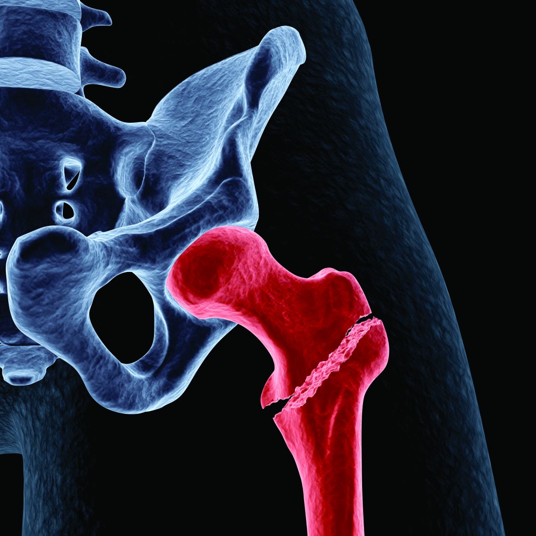
Marathon running does not increase arthritis risk: Survey
Runners who had undergone knee or hip surgery or had a previous hip or knee injury that prevented running were most likely to have arthritis, researchers found. Family history of arthritis, higher body mass index (BMI), and older age were also associated with increased risk of the condition.
The study was presented at the American Academy of Orthopaedic Surgeons 2023 Annual Meeting.
It has generally been thought that running may increase risk of osteoarthritis because it puts more load on joints than walking or standing, noted Grace Hsiao-Wei Lo, MD, an assistant professor of immunology, allergy, and rheumatology at the Baylor College of Medicine, Houston, who was not involved with the work. Research in this area has yielded mixed results: A 2017 analysis of multiple studies found that competitive runners did have higher rates of arthritis than recreational runners, while another study conducted by Dr. Lo found that runners did not have an increased risk of knee osteoarthritis, compared with nonrunners. A 2018 study showed that marathon runners had lower instances of arthritis, compared with the general population.
In this new study, researchers surveyed 3,804 runners who participated in the 2019 or 2021 Chicago Marathon about their running history, average mileage per week, and average running pace. The survey also asked about known risk factors for osteoarthritis, including BMI, family history of arthritis, and past knee and hip injuries that prevented running.
Runners, on average, were about 44 years old and ran 27.9 miles per week. The largest proportion of respondents had completed 2-5 marathons (37.3%), around 21% of respondents had finished 6-10 marathons, and 17% were running their first marathon. Study participants had an average of 15 years of running experience, 1,892 reported a previous hip or knee injury, and 413 had undergone knee or hip surgery. Overall, 36.4% reported experiencing hip or knee pain in the past year, and 7.3% had been diagnosed with arthritis.
Researchers found that there was no association between the risk of osteoarthritis and weekly mileage, years spent running, number of marathons completed, or running pace. Respondents who had undergone knee or hip surgery had the highest risk of osteoarthritis (odds ratio, 5.85; P < .0001), followed by those with a history of knee or hip injuries that prevented running (OR, 5.04; P < .0001). Other identified risk factors were family history of arthritis (OR, 3.47; P < .0001), BMI (OR, 1.10; P < .0001), and older age (OR, 1.08; P < .0001).
The news should be encouraging for runners, said Matthew Hartwell, MD, an orthopedic surgeon at the University of California, San Francisco, who led the research. If someone does not have injuries or surgeries that keep them from running, “you can still continue to run,” he said. “There may not necessarily be this dose-response relationship where the more you run, the more you break down your knee or your hip.”
Still, 24.2% of runners reported that their physician had advised them to reduce their mileage or stop running altogether. Most runners (94.2%) said they planned to run another marathon.
“The results of this study are consistent with the experiences of many lifelong runners and observations of sports medicine professionals that osteoarthritis is not an inevitable consequence of distance running,” said Brett Toresdahl, MD, a sports medicine physician at the Hospital for Special Surgery in New York, who was not involved with the study.
Still, he emphasized that more research is necessary to understand whether running contributes to the risk of developing osteoarthritis. The participants in the study were current marathoners, he noted, so it is likely they have healthy joints that can tolerate running longer distances. “If there is a subset of people who have joints that are negatively affected by running, they wouldn’t likely be registering for a marathon,” he said in an email interview.
Dr. Lo added that comparing these marathoners to a group who did not run would help assess whether running can be harmful to joints. “To be fair, this is a challenging subject to study,” she said. “Osteoarthritis has a long natural history, and so it is difficult to evaluate this kind of question over many years of running and many years of evaluation of arthritis.”
While the research does not answer the question of whether running can lead to osteoarthritis, it helps show the need for long-term research on how running affects joints over time as well as one’s general health, Dr. Toresdahl noted. “I would not be surprised if future longitudinal research will come to the same conclusion that running for the majority of patients is a net benefit for overall health and at least net neutral for joint health when done in moderation,” he said.
Dr. Hartwell, Dr. Lo, and Dr. Toresdahl report no relevant financial relationships.
A version of this article originally appeared on Medscape.com.
FROM AAOS 2023
Guidelines: Don’t delay total joint arthroplasty for additional nonoperative therapies
Patients with moderate to severe osteoarthritis (OA) or osteonecrosis (ON) eligible for total joint arthroplasty (TJA) who have failed one or more nonoperative therapies should proceed directly to surgery, according to new guidelines from the American College of Rheumatology and the American Association of Hip and Knee Surgeons.
“One of the reasons for creating this guideline was that many patients have been subjected to delays for surgery after completing nonoperative therapy, despite persistent moderate to severe pain, loss of function, and moderate to severe radiographic OA or ON,” said coauthors Susan M. Goodman, MD, a rheumatologist at Hospital for Special Surgery in New York, and Charles Hannon, MD, an orthopedic surgeon at Washington University in St. Louis, in an email interview with this news organization. “This guideline supports surgery being performed in an expeditious fashion after the decision has been made to proceed with surgery by both the physician and patient through a shared decision-making process,” they said.
The guidelines also state that obesity by itself should not be a reason to delay TJA. “We could not find a rationale for a strict cut off for weight/body mass index (BMI). Our literature review revealed that though many adverse events were, in fact, increased in patients with morbid obesity, there is also an increase in adverse events for those who had bariatric surgery prior to their arthroplasty,” they added, noting that patients need to be made aware of the increased risk for adverse events in patients with obesity. Though the guidelines do not pose any BMI cutoffs, they state that weight loss should be “strongly encouraged.” These new recommendations are conditional, and all had a “low” to “very low” certainty of evidence; however, there was high consensus on the recommendations from the expert panel.
The guidelines also recommended:
- Delaying TJA to achieve smoking and nicotine cessation or reduction.
- Delaying TJA to improve glycemic control in patients with diabetes, although the group did not recommend any specific measure or threshold.
- Not delaying TJA in patients with a severe deformity, bone loss, or a neuropathic joint.
The new guidelines formalize what many surgeons have already been doing for the past few years, said Arjun Saxena, MD, MBA, an orthopedic surgeon in Philadelphia who was not involved with the guidelines. “A lot of total joint programs have really focused on patient optimization, including smoking cessation, glycemic control, and weight loss prior to surgery,” he said.
Most importantly, the guidelines put an emphasis on how the decision to proceed with TJA should be a shared decision between a physician and patient, he added. Some insurance companies with prior authorization policies may require a patient to try additional nonoperative therapies before approving surgery, creating barriers to care, he said. “Hopefully [these new recommendations] will help third parties understand that joint replacement is a big decision – most doctors aren’t going to recommend that unless it’s necessary or something that is going to help patients,” he said. “I understand that there is a certain need for preauthorization, but just having strict guidelines isn’t appropriate. You really need to look at the whole picture,” he added.
The full manuscript has been submitted for review and is expected to be jointly published in American College of Rheumatology and the American Association of Hip and Knee Surgeons journals later this year.
Dr. Saxena consults for the orthopedic implant company Corin.
A version of this article originally appeared on Medscape.com.
Running does not cause lasting cartilage damage
Running does not appear to cause sustained wear and tear of healthy knee cartilage, with research suggesting that the small, short-term changes to cartilage after a run reverse within hours.
A systematic review and meta-analysis published in the most recent issue of Osteoarthritis and Cartilage presents the findings involving 396 adults, which compared the “before” and “after” state of healthy knee cartilage in runners.
Running is often thought to be detrimental to joint health, wrote Sally Coburn, PhD candidate at the La Trobe Sport & Exercise Medicine Research Centre at La Trobe University in Melbourne and coauthors, but this perception is not supported by evidence.
For the analysis, the researchers included studies that looked at either knee or hip cartilage using MRI to assess its size, shape, structure, and/or composition both in the 48 hours before a single bout of running and in the 48 hours after. The analysis aimed to include adults with or at risk of osteoarthritis, but only 57 of the 446 knees in the analysis fit these criteria.
In studies where participants underwent MRI within 20 minutes of running, there was an immediate postrun decrease in the volume of cartilage, ranging from –3.3% for weight-bearing femoral cartilage to –4.1% for tibial cartilage volume. This also revealed a decrease in T1 and T2 relaxation times, which are specialized MRI measures that reflect the composition of cartilage and which can indicate a breakdown of cartilage structure in the case of diseases such as arthritis.
Reversal of short-term cartilage changes
However, within 48 hours of the run, data from studies that repeated the MRIs more than once after the initial prerun scan suggested these changes reversed back to prerun levels.

“We were able to pool delayed T2 relaxation time measures from studies that repeated scans of the same participants 60 minutes and 91 minutes post-run and found no effect of running on tibiofemoral joint cartilage composition,” the authors write.
For example, one study in marathon runners found no difference in cartilage thickness in the tibiofemoral joint between baseline and at 2-10 hours and 12 hours after the marathon. Another showed the immediate post-run decrease in patellofemoral joint cartilage thickness had reverted back to prerun levels when the scan was repeated 24 hours after the run.
“The changes are very minimal and not inconsistent with what’s expected for your cartilage which is functioning normally,” Ms. Coburn told this news organization.
Sparse data in people with osteoarthritis
The authors said there were not enough data from individuals with osteoarthritis to be able to pool and quantify their cartilage changes. However, one study in the analysis found that cartilage lesions in people considered at risk of osteoarthritis because of prior anterior cruciate ligament reconstruction were unchanged after running.
Another suggested that the decrease in femoral cartilage volume recorded at 15 minutes persisted at 45 minutes, while a separate study found significantly increased T2 relaxation times at 45 minutes after a run in those with knee osteoarthritis but not in those without osteoarthritis.
Senior author Adam Culvenor, PhD, senior research fellow at the La Trobe Centre, said their analysis suggested running was healthy, with small changes in cartilage that resolve quickly, but “we really don’t know yet if running is safe for people with osteoarthritis,” he said. “We need much more work in that space.”
Overall, the study evidence was rated as being of low certainty, which Dr. Coburn said was related to the small numbers in each study, which in turn relates to the cost and logistical challenges of the specialized MRI scan used.
“Study of a repeated exposure over a long duration of time on a disease that has a long natural history, like osteoarthritis, is challenging in that most funding agencies will not fund studies longer than 5 years,” Grace Hsiao-Wei Lo, MD, of the department of immunology, allergy, and rheumatology at the Baylor College of Medicine in Houston, said in an email.
Dr. Lo, who was not involved with this review and meta-analysis, said there are still concerns about the effect of running on knee osteoarthritis among those with the disease, although there are some data to suggest that among those who self-select to run, there are no negative outcomes for the knee.
An accompanying editorial noted that research into the effect of running on those with osteoarthritis was still in its infancy. “This would help to guide clinical practice on how to support people with osteoarthritis, with regard to accessing the health benefits of running participation,” write Jean-Francois Esculier, PT, PhD, from the University of British Columbia, Vancouver, and Christian Barton, PhD, with the La Trobe Centre, pointing out there were a lack of evidence-based clinical recommendations for people with osteoarthritis who want to start or continue running.
It’s a question that PhD candidate Michaela Khan, MSc, is trying to answer at the University of British Columbia. “Our lab did a pilot study for my current study now, and they found that osteoarthritic cartilage took a little bit longer to recover than their healthy counterparts,” Ms. Khan said. Her research is suggesting that people with osteoarthritis not only can run, but even those with severe disease, who might be candidates for knee replacement, can run long distances.
Commenting on the analysis, Ms. Khan said the main take-home message was that healthy cartilage seems to recover after running, and that there is not an ongoing effect of ‘wear and tear.’
“That’s changing the narrative that if you keep running, it will wear away your cartilage, it’ll hurt your knees,” she said. “Now, we have a good synthesis of scientific evidence to prove maybe otherwise.”
Ms. Coburn and Dr. Culvenor report grant support from the National Health & Medical Research Council of Australia, and another author reports grant support from the U.S. National Institute of Arthritis and Musculoskeletal and Skin Diseases. The authors, as well as Dr. Lo and Ms. Khan, report relevant financial relationships.
Running does not appear to cause sustained wear and tear of healthy knee cartilage, with research suggesting that the small, short-term changes to cartilage after a run reverse within hours.
A systematic review and meta-analysis published in the most recent issue of Osteoarthritis and Cartilage presents the findings involving 396 adults, which compared the “before” and “after” state of healthy knee cartilage in runners.
Running is often thought to be detrimental to joint health, wrote Sally Coburn, PhD candidate at the La Trobe Sport & Exercise Medicine Research Centre at La Trobe University in Melbourne and coauthors, but this perception is not supported by evidence.
For the analysis, the researchers included studies that looked at either knee or hip cartilage using MRI to assess its size, shape, structure, and/or composition both in the 48 hours before a single bout of running and in the 48 hours after. The analysis aimed to include adults with or at risk of osteoarthritis, but only 57 of the 446 knees in the analysis fit these criteria.
In studies where participants underwent MRI within 20 minutes of running, there was an immediate postrun decrease in the volume of cartilage, ranging from –3.3% for weight-bearing femoral cartilage to –4.1% for tibial cartilage volume. This also revealed a decrease in T1 and T2 relaxation times, which are specialized MRI measures that reflect the composition of cartilage and which can indicate a breakdown of cartilage structure in the case of diseases such as arthritis.
Reversal of short-term cartilage changes
However, within 48 hours of the run, data from studies that repeated the MRIs more than once after the initial prerun scan suggested these changes reversed back to prerun levels.
“We were able to pool delayed T2 relaxation time measures from studies that repeated scans of the same participants 60 minutes and 91 minutes post-run and found no effect of running on tibiofemoral joint cartilage composition,” the authors write.
For example, one study in marathon runners found no difference in cartilage thickness in the tibiofemoral joint between baseline and at 2-10 hours and 12 hours after the marathon. Another showed the immediate post-run decrease in patellofemoral joint cartilage thickness had reverted back to prerun levels when the scan was repeated 24 hours after the run.
“The changes are very minimal and not inconsistent with what’s expected for your cartilage which is functioning normally,” Ms. Coburn told this news organization.
Sparse data in people with osteoarthritis
The authors said there were not enough data from individuals with osteoarthritis to be able to pool and quantify their cartilage changes. However, one study in the analysis found that cartilage lesions in people considered at risk of osteoarthritis because of prior anterior cruciate ligament reconstruction were unchanged after running.
Another suggested that the decrease in femoral cartilage volume recorded at 15 minutes persisted at 45 minutes, while a separate study found significantly increased T2 relaxation times at 45 minutes after a run in those with knee osteoarthritis but not in those without osteoarthritis.
Senior author Adam Culvenor, PhD, senior research fellow at the La Trobe Centre, said their analysis suggested running was healthy, with small changes in cartilage that resolve quickly, but “we really don’t know yet if running is safe for people with osteoarthritis,” he said. “We need much more work in that space.”
Overall, the study evidence was rated as being of low certainty, which Dr. Coburn said was related to the small numbers in each study, which in turn relates to the cost and logistical challenges of the specialized MRI scan used.
“Study of a repeated exposure over a long duration of time on a disease that has a long natural history, like osteoarthritis, is challenging in that most funding agencies will not fund studies longer than 5 years,” Grace Hsiao-Wei Lo, MD, of the department of immunology, allergy, and rheumatology at the Baylor College of Medicine in Houston, said in an email.
Dr. Lo, who was not involved with this review and meta-analysis, said there are still concerns about the effect of running on knee osteoarthritis among those with the disease, although there are some data to suggest that among those who self-select to run, there are no negative outcomes for the knee.
An accompanying editorial noted that research into the effect of running on those with osteoarthritis was still in its infancy. “This would help to guide clinical practice on how to support people with osteoarthritis, with regard to accessing the health benefits of running participation,” write Jean-Francois Esculier, PT, PhD, from the University of British Columbia, Vancouver, and Christian Barton, PhD, with the La Trobe Centre, pointing out there were a lack of evidence-based clinical recommendations for people with osteoarthritis who want to start or continue running.
It’s a question that PhD candidate Michaela Khan, MSc, is trying to answer at the University of British Columbia. “Our lab did a pilot study for my current study now, and they found that osteoarthritic cartilage took a little bit longer to recover than their healthy counterparts,” Ms. Khan said. Her research is suggesting that people with osteoarthritis not only can run, but even those with severe disease, who might be candidates for knee replacement, can run long distances.
Commenting on the analysis, Ms. Khan said the main take-home message was that healthy cartilage seems to recover after running, and that there is not an ongoing effect of ‘wear and tear.’
“That’s changing the narrative that if you keep running, it will wear away your cartilage, it’ll hurt your knees,” she said. “Now, we have a good synthesis of scientific evidence to prove maybe otherwise.”
Ms. Coburn and Dr. Culvenor report grant support from the National Health & Medical Research Council of Australia, and another author reports grant support from the U.S. National Institute of Arthritis and Musculoskeletal and Skin Diseases. The authors, as well as Dr. Lo and Ms. Khan, report relevant financial relationships.
High HDL-C levels linked to increased fracture risk
High levels of high-density lipoprotein cholesterol (HDL-C) in older adults are associated with a higher risk of sustaining a fracture than lower HDL-C levels, a new study suggests.
“Two animal studies showing that HDL-C reduces bone mineral density by reducing osteoblast number and function provide a plausible explanation for why high HDL-C may increase the risk of fractures,” Monira Hussain, MBBS, MPH, PhD, of Monash University in Melbourne, told this news organization. “So, it was not surprising when our analyses provided evidence that amongst those in the highest quintile of HDL-C (> 74 mg/dL), there was a [33%] increased risk of fractures.”
After adjustment, one standard deviation increment in HDL-C level was associated with a 14% higher risk of fracture during a 4-year follow-up.
Based on this and other studies, Dr. Hussain said, “I believe that the finding of a very high HDL-C [should] alert clinicians to a higher risk of mortality, fractures, and possibly other threats to their patient’s health.”
The study was published online in JAMA Cardiology.
Independent risk factor
For this report, the researchers conducted a post hoc analysis of data from the Aspirin in Reducing Events in the Elderly (ASPREE) clinical trial and the ASPREE-Fracture substudy.
ASPREE was a double-blind, randomized, placebo-controlled primary prevention trial of aspirin. Participants were 16,703 community-dwelling Australians and 2,411 individuals from the United States with a mean age of 75 and without evident cardiovascular disease, dementia, physical disability, or life-limiting chronic illness.
The ASPREE-Fracture substudy collected data on fractures reported post randomization from the Australian participants. Fractures were confirmed by imaging and adjudicated by an expert panel and included both traumatic and minimal trauma fractures.
Of the 16,262 participants who had a plasma HDL-C measurement at baseline (55% women), 1,659 (10.2%) experienced at least one fracture over a median of 4 years. This included 711 minimal trauma fractures (for example, falls from standing height) and 948 other trauma fractures, mainly falls on stairs, ladders, or stools.
Higher rates of fractures occurred in the highest quintile of HDL-C level where the mean level was 89 mg/dL. At baseline, participants in that quintile had a lower BMI, a high prevalence of current/former smoking and current alcohol use, 12 years or longer of school, more physical activity, and higher use of antiosteoporosis medication. They also had less chronic kidney disease, diabetes, prefrailty/frailty, or treatment with lipid-lowering drugs.
In a fully adjusted model, each standard deviation increment in HDL-C level was associated with a 14% higher risk of fractures (hazard ratio, 1.14). When analyzed in quintiles, compared with participants in Q1, those in Q5 had a 33% higher risk for fracture (HR, 1.33).
Prevalence rates were similar between the sexes. The increase in fracture risk appeared to be independent of traditional risk factors for fractures, including age, sex, physical activity, alcohol use, frailty, BMI, smoking status, diabetes, chronic kidney disease, use of lipid-lowering or antiosteoporosis drugs, and education, the authors note.
The results persisted in sensitivity analyses in restricted subgroups of interest and in stratified analyses – including, for example, only minimal fractures; participants not taking antiosteoporosis drugs or statins; never smokers; nondrinkers; and those engaging in minimal physical activity (walking less than 30 minutes per day).
No association was observed between non–HDL-C levels and fractures.
The authors conclude that the study “provides robust evidence that higher levels of HDL-C are associated with incident fractures in both male and female individuals, independent of conventional risk factors.”
Clinically useful?
Commenting on the study for this news organization, Marilyn Tan, MD, clinic chief of the Endocrine Clinic and clinical associate professor of medicine at Stanford (Calif.) University, said, “I certainly would not recommend anyone do anything to actively lower their HDL levels. HDL levels are largely determined by genetics, diet, and lifestyle, with some effects from certain medications/supplements. Studies have demonstrated that moderately higher HDL levels may be protective for atherosclerosis.”
In the current study, she said, “Causation has not been proven, and importantly there is no evidence that reducing HDL levels reduces fracture risk. Also, this association between raised HDL levels and fracture risk has not been demonstrated consistently in other studies.”
Furthermore, she noted, the preclinical trials on which the authors based their hypothesis – that is, an association between HDL and a reduction in the number and function of osteoblasts – “has not been demonstrated widely in human subjects.”
“We have a large armamentarium of FDA-approved treatments for osteoporosis that have been clinically proven to reduce fracture risk very significantly, and these are the tools [in addition to lifestyle changes] we should use to reduce fracture risk,” Dr. Tan concluded.
John Wilkins, MD, of Northwestern University, Chicago, and Anand Rohatgi, MD, MSCS, of UT Southwestern Medical Center, Dallas, also point out some limitations of the study in a related editorial.
They note the inclusion of predominantly healthy adults with a mean age of 75, a population that could yield different findings from middle-aged cohorts with chronic illnesses, as well as a lack of clarity regarding the possible role of alcohol intake among the study participants.
Furthermore, the editorialists write, although significant associations were shown in this study, “models were not adjusted for detailed measures of exercise/activity, triglycerides, or any other lipids, including other HDL compositional measures such as HDL-P or ApoA-I levels. There was no assessment of whether HDL-C improved discrimination, reclassification, or any other validated measures of risk prediction performance.
“Taken together,” they conclude, “this study alone leaves several unanswered questions as to whether high HDL-C could be a useful biomarker to detect fracture risk.”
No commercial funding was disclosed. The authors report no relevant financial relationships.
A version of this article originally appeared on Medscape.com.
‘Very doable’ low-dose workout enough to treat knee OA
Exercise helps patients with knee osteoarthritis, but more isn’t necessarily better, new research shows.
A low-dose exercise regimen helped patients with knee OA about as much as a more intense workout plan, according to trial results published online in Annals of Internal Medicine.
Both high and low doses of exercise reduced pain and improved function and quality of life.
The improvements with the lower-dose plan and its 98% adherence rate are encouraging, said Nick Trasolini, MD, assistant professor of orthopedic surgery at Atrium Health Wake Forest Baptist Medical Center, Winston-Salem, N.C.
“This is a very doable amount of medical exercise therapy for patients with knee osteoarthritis, and one that makes a big difference in patient-reported symptoms,” Dr. Trasolini, who was not involved in the study, said in an interview.
What’s the right dose?
Exercise is a go-to treatment for knee OA, but the precise dose to recommend has been unclear. To study this question, Tom Arild Torstensen, MSc, RPT, with Karolinska Institutet, Huddinge, Sweden, and Holten Institute, Stockholm, and colleagues conducted a trial at four centers in Sweden and Norway.
The study included 189 men and women with knee OA. Participants were randomly assigned to low- or high-dose exercise plans, which they performed three times per week for 12 weeks under the supervision of a physiotherapist.
Participants in the high-dose group performed 11 exercises during each session, which lasted 70-90 minutes.
The low-dose regimen consisted of five exercises – cycling, squats, step-ups, step-downs, and knee extensions – performed for 20–30 minutes.
The researchers measured outcomes using the Knee Injury and Osteoarthritis Outcome Score, which assesses pain, other symptoms, function in daily living, function in sports and recreation, and knee-related quality of life.
“Patients in both groups improved significantly over time, but high-dose exercise was not superior to low-dose exercise in most comparisons,” the study investigators reported
High-dose exercise was associated with better function in sports and recreational activity and knee-related quality of life at 6 months. Those differences did not persist at 1 year, however. The researchers reported no safety concerns with either intervention.
Adherence was “nearly perfect” in the low-dose group. It was slightly lower in the high-dose group, the researchers said.
“Interestingly, it seems that high-dose treatment could be preferable to low-dose treatment in the long run for people who lead active lives,” they wrote. “This should be the subject of future studies.”
All clinical practice guidelines for knee OA recommend exercise, but “we do not know the optimal dose,” Kim Bennell, PhD, a research physiotherapist at the University of Melbourne, said in an interview.
Dose has components, including number of times per week, number of exercises, sets and repetitions, intensity, and duration of exercise sessions, Dr. Bennell said.
“These results suggest that an exercise program that involves less time and fewer exercises can still offer benefits and may be easier for patients to undertake and stick at than one that involves greater time and effort,” she said.
The study was supported by the Swedish Rheumatic Fund. Dr. Trasolini and Dr. Bennell have disclosed no relevant financial relationships.
A version of this article originally appeared on Medscape.com.
FROM ANNALS OF INTERNAL MEDICINE

Corticosteroid injections may worsen knee OA progression
Injecting hyaluronic acid (HA) instead, or managing the condition without injections, may better preserve knee structure and cartilage, according to results of two related studies presented at the annual meeting of the Radiological Society of North America.
The findings come nonrandomized, observational cohort studies, leading knee OA experts to call for further study in randomized trial settings. In the meantime, shared decision-making between patients and clinicians is advised on the use of these injections.
For knee OA, most patients seek a noninvasive treatment for symptomatic relief. “At least 10% of these patients undergo local treatment with injectable corticosteroids or hyaluronic acid,” the lead author of one of the studies, Upasana Upadhyay Bharadwaj, MD, research fellow in musculoskeletal radiology at the University of California, San Francisco, said in a video press release.
Researchers in both studies used data and images from the Osteoarthritis Initiative (OAI), a multicenter, longitudinal, observational study of 4,796 U.S. patients aged 45-79 years with knee OA. Participants were enrolled from February 2004 to May 2006.
The OAI maintains a natural history database of information regarding participants’ clinical evaluation data, x-rays, MRI scans, and a biospecimen repository. Data are available to researchers worldwide.
Two studies draw similar conclusions
In one study, Dr. Bharadwaj and colleagues found that HA injections appeared to show decreased knee OA progression in bone marrow lesions.
They investigated 8 patients who received one CS injection, 12 who received one HA injection, and 40 control persons who received neither treatment. Participants were propensity-score matched by age, sex, body mass index (BMI), Kellgren-Lawrence (KL) grade, Western Ontario and McMaster Universities Osteoarthritis Index (WOMAC), and Physical Activity Scale for the Elderly (PASE).
The researchers semiquantitatively graded three Tesla MRI scans that had been obtained at baseline, 2 years before the injection, and 2 years after the injection, using whole-organ MRI score (WORMS) for the meniscus, bone marrow lesions, cartilage, joint effusion, and ligaments.
They quantified OA progression using the difference in WORMS between baseline and 2-year follow-up, and they used linear regression models, adjusted for age, sex, BMI, KL grade, WOMAC, and PASE, to identify the link between type of injection and progression of WORMS.
At 2 years, the authors found a significant association between CS injection and postinjection progression of WORMS over 2 years for the knee overall, the lateral meniscus, lateral cartilage, and medial cartilage. There was no significant link between HA injection and postinjection progression of WORMS or between either injection type and progression of pain, as quantified by WOMAC. There was also no significant difference in progression of WORMS over the 2 years prior to injection for CS and HA injections.
“Corticosteroid injections must be administered with caution with respect to long-term effects on osteoarthritis,” Dr. Bharadwaj advised. “Hyaluronic acid injections, on the other hand, may slow down progression of knee osteoarthritis and alleviate long-term effects while offering similar symptomatic relief to corticosteroid injections. Overall, they are perhaps a safer alternative when looking at medium- and long-term disease course of knee osteoarthritis.”
In the second study, lead author Azad Darbandi, MS, a fourth-year medical student at Chicago Medical School, North Chicago, and colleagues found that patients who received CS injections experienced significantly more medial joint space narrowing.
They identified 210 knees with imaging at baseline and at 48 months that received CS injections, and 59 that received HA injections; 6,827 knees served as controls. The investigators matched 50 patients per group on the basis of confounding factors, which included age, sex, BMI, comorbidities, surgery, and semiquantitative imaging outcomes at baseline. They performed ANCOVA testing using 48-month semiquantitative imaging outcomes as dependent variables and confounding variables as covariates.
The researchers analyzed joint space narrowing, KL grade, and tibia/femur medial/lateral compartment osteophyte formation and sclerosis.
At 4 years, the average KL grade in the CS group was 2.79, it was 2.11 in the HA group,;and it was 2.37 in the control group. Intergroup comparisons showed significant differences in KL grade between CS and HA groups and between CS and control groups. Medial compartment joint space narrowing was 1.56 in the CS group, 1.11 in the HA group, and 1.18 in controls. There was a significant difference between the CS and control groups. Other dependent variables were not significant.
“These preliminary results suggest that corticosteroid injections accelerated the radiographic progression of osteoarthritis, specifically medial joint space narrowing and Kellgren-Lawrence grading, whereas hyaluronic acid injections did not,” Mr. Darbandi said in an interview.
“OA radiographic progression does not always correlate with clinical progression, and further research is needed,” he added.
Proper matching of patients at baseline for confounding factors is a strength of the study, Mr. Darbandi said, while the retrospective study design is a weakness.
Experts share their perspectives on the preliminary results
Michael M. Kheir, MD, assistant professor of orthopedic surgery at the University of Michigan Health System, who was not involved in the studies, said he would like to see further related research.
“Perhaps steroid injections are not as benign as they once seemed,” he added. “They should be reserved for patients who already have significant arthritis and are seeking temporary relief prior to surgical reconstruction with a joint replacement, or for patients with recalcitrant pain after having already tried HA injections.”
William A. Jiranek, MD, professor and orthopedic surgeon at Duke Health in Morrisville, N.C., who also was not involved in the studies, was not surprised by the findings.
“It is important to do these studies to learn that steroid injections do not come with zero cost,” he said.
“I am pretty sure that a percentage of these patients had no cartilage loss at all,” he added. “We need to understand which OA phenotypes are not at risk of progressive cartilage loss from steroid injections.”
Annunziato (Ned) Amendola, MD, professor and sports medicine orthopedic surgeon at Duke Health in Durham, N.C., who was also not involved in the studies, said he would like to know how injection effectiveness and activity level are related.
“If the injections were effective at relieving pain, and the patients were more active, that may have predisposed to more joint wear,” he said. “It’s like tires that last longer if you don’t abuse them.”
Shared decision-making and further research recommended
Amanda E. Nelson, MD, associate professor of medicine in the division of rheumatology, allergy, and immunology at the University of North Carolina at Chapel Hill, said: “The lack of randomization introduces potential biases around why certain therapies (CS injection, HA injection, or neither) were selected over others (such as disease severity, preference, comorbid conditions, other contraindications, etc), thus making interpretation of the findings challenging.
“The causal relationship remains in question, and questions around the efficacy of intra-articular HA in particular, and the ideal settings for intra-articular therapy in general, persist,” noted Dr. Nelson, who was also not involved in the studies. “Thus, shared decision-making between patients and their providers is essential when considering these options.”
C. Kent Kwoh, MD, professor of medicine and medical imaging at the University of Arizona and director of the University of Arizona Arthritis Center, both in Tucson, said in an interview that these types of studies are important because CS injections are common treatments for knee OA, they are recommended in treatment guidelines, and other good options are lacking.
But he pointed out that the results of these two studies need to be interpreted with caution and should not be used to decide the course of treatment.
“These data are hypothesis generating. They suggest association, but they do not show causation,” said Dr. Kwoh, who was also not involved in the studies. “Both studies are secondary analyses of data collected from the OAI, which was not specifically designed to answer the questions these studies are posing.
“The OAI was not a treatment study, and participants were seen only once a year or so. They may have had joint injections anytime from only days to around 1 year before their visit, and their levels of activity or pain just prior to or just after their joint injections were not reported,” Dr. Kwoh explained.
The reasons why patients did or did not receive a specific joint injection – including their socioeconomic status, race, access to insurance, and other confounding factors – were not assessed and may have affected the results, he added.
The fact that both studies used the same data and came to the same conclusions gives the conclusions some strength, he said, but “the gold standard to understanding causation would be a randomized, controlled trial.”
Mr. Darbandi’s research received grant support from Boeing, His c-authors, as well as all experts not involved in the studies, reported no relevant financial relationshiips. Dr. Bharadwaj did not provide conflict-of-interest and funding details. Dr. Kwoh reported membership on panels that have developed guidelines for the management of knee OA.
A version of this article first appeared on Medscape.com.
FROM RSNA 2022
Roy Altman: Physician, father, teacher, editor
Roy Altman, MD, was a father, a husband, a teacher, a physician, an editor, and a mentor. He died November 30 as a result of complications related to Parkinson’s disease.
Altman was an editorial advisory board member for MDedge Rheumatology/Rheumatology News since the publications’ start in 2002. He also treated patients and taught students at the University of Miami and the University of California, Los Angeles. A father of four adult children and a grandfather to nine grandchildren, Altman was also a husband to Linda, “his lifelong partner.”
“[He] had this tremendous editorial expertise to be able to manage different journals at the same time without conflict,” said Marc Hochberg, MD, MPH, a professor of medicine, epidemiology, and public health and head of the division of rheumatology and clinical immunology at the University of Maryland School of Medicine, Baltimore. “This was done in the days before electronic publishing, in the days before everything was being done on the computer.”
Publications where Altman served as editor include Osteoarthritis and Cartilage and Seminars in Arthritis and Rheumatism.
In addition to editing articles, Altman also had an active patient panel, while lecturing and traveling internationally, Hochberg said. Altman accomplished this “while maintaining a very active family life,” remembers Hochberg, who learned from Altman about ways to achieve work-life balance.
“That’s important when you’re a junior faculty member – and you’re trying to develop your academic career – that you prioritize what you need to do to be successful in the world of academic medicine, especially rheumatology,” said Hochberg, who described Altman as a “very close friend.”
Teacher, physician, and mentor
John FitzGerald, MD, PhD, MBA, chief of clinical rheumatology at the University of California, Los Angeles, looks back on the more than 15 years that Altman drove 1 hour each way to teach fellows about topics including osteoarthritis. “He really valued teaching. ... that was a lot of his focus,” said FitzGerald, who adds that Altman also enjoyed his relationships with patients.
Altman joined the University of California, Los Angeles, faculty as a professor of medicine in the division of rheumatology and immunology in 2003. Before arriving at UCLA, he was chief of rheumatology and immunology at the University of Miami for many years.
“His patients loved him because he was providing treatments and services that they really had trouble finding elsewhere,” FitzGerald said. Altman entrusted FitzGerald with giving him injections between patients or during lunch for the arthritis that increasingly bothered him.
“He was very appreciative of having the care provided to him. ... he was also sort of playful and teasing,” FitzGerald said.
Altman also took on the role of teacher at home, according to his daughter, Evie. “My dad loved to diagnose; he enjoyed piecing together the disparate physical and radiologic clues to determine the cause of disease or pain. When I was much younger, he taught me how to spot someone with Paget’s Disease from the other end of the room.”
“He loved teaching, imparting his knowledge to others,” she added, remembering the slide sessions Altman presented to his students at their family home. “We loved [the slide sessions] because we could get pizza for dinner, and we got really good at spotting the biological markers of rheumatoid arthritis.”
Aaron Altman said his father was “impassioned by bringing people alleviation of their pain and suffering. It drove him to his very core.”
Researcher and family man
Evie Altman said in an interview that many people don’t know her father was one of the first practitioners of arthroscopy. “He would bring the then-rigid scopes into our elementary school class for ‘show and tell.’ Also, he traveled to the Bolivian jungles in the late 1970s to gather specific red ants whose venom locals had reported as an arthritic treatment,” she said.
Ultimately, Altman isolated the venom’s active ingredient, said Evie, who remembers that her father studied the effect of the treatment for many years. “This eventually led to his research with capsaicin peppers, which was developed into a cream used widely today.”
“Growing up, our garage freezer always had serum and patient urine in one section, away from the ice cream,” Evie added.
In 2011, Sally Koch Kubetin reported for Rheumatology News on Altman’s habit of donning his wife’s handpainted ties. His motivation? It was “born of the sensible desire to be recognizable in a busy world,” she wrote.
Evie said that her mother started painting ties for her father early in his career. Two of his favorite ties, she said, are the “Lady and the Tramp” tie featuring the two dogs eating spaghetti from the same bowl and the Winnie the Pooh tie. “Mostly, he loved the ties because my mom made them,” she said.
Student and military doctor
Altman received his medical degree from the University of Miami School of Medicine after earning an undergraduate degree from Michigan State University, East Lansing, Kubetin reported. The University of Miami School of Medicine was also where he did his internship, residency, and fellowship in rheumatology.
Kubetin reported that Altman’s military service interrupted his training. During his service as a lieutenant in the U.S. Navy active duty reserve, he was a general medical officer and cared for sailors and officers at Naval Air Station Point Mugu in Oxnard, Calif.
Altman had a second academic appointment as a professor of orthopedics when he was in Miami, according to the Rheumatology News profile. In addition, he was the clinical director of the geriatric research, education, and clinical center, and chief of the arthritis section in the division of medicine at the Miami Veterans Affairs Medical Center.
“Persistence, stubbornness, and all around stick-to-it-iveness defined him in our eyes,” said his daughter, Sarah. His daughter, Ruth, credits her father’s kindness and patience with informing how she practices veterinary medicine. “When I went to rounds with dad on Saturdays, he went to a nearby flower place downtown to get mom flowers almost every week if he could,” she said.
Altman announced at the family’s annual summer reunion in 2021 that he would stop seeing patients, said his daughter, Evie. He continued to present slide sessions and grand rounds to medical students at UCLA until early this year. Altman continued to edit journals and review articles until he died from complications of Parkinson’s disease.
He published articles in medical journals that number in the thousands, according to a website established by Altman’s family that honors his life. Altman was born in Astoria, N.Y., on May 16, 1937, and moved to Miami Beach with his parents when he was young.
Ask knee OA patients about stair climbing difficulty
Asking knee osteoarthritis patients a simple question – do you have difficulty climbing stairs? – may predict the risk of future functional limitation, according to research presented at the annual meeting of the American College of Rheumatology. Finding out that the patient has difficulty also opens avenues for further evaluation and intervention, said Jason Jakiela, a PhD candidate at the University of Delaware, Newark, who led the study. “We like to view it as a kind of yellow flag,” Mr. Jakiela said in an interview.
Another expert agreed. “I think this is useful for clinical rheumatologists,” said C. Kent Kwoh, MD, professor of medicine and medical imaging at the University of Arizona, Tucson, and director of the University of Arizona Arthritis Center. He commented on the study findings but was not involved in the study. Another common question asked of OA patients, about pain, may not be as useful as asking about difficulty climbing stairs, he said. “Their pain level can go up and down and can be quite varied.”

Osteoarthritis affects more than 32.5 million adults, according to the CDC, and the knee is a common site.
Study details, results
Mr. Jakiela and his team, including Daniel White, PT, ScD, MSC, associate professor of physical therapy at the University of Delaware, Newark, used data from the Osteoarthritis Initiative (OAI). They assessed stair climbing difficulty at baseline with the question: Does your health now limit you in climbing several flights of stairs? Respondents could answer that they were limited a lot, a little, or not at all.

The researchers evaluated functional limitation using two measures: Walking speed and Western Ontario and McMaster Universities Osteoarthritis Index physical function (WOMAC-PF) scores. A walking speed of < 1.22 m/s over 20 meters, the speed needed to safely cross a timed intersection, represented poor function. A WOMAC-PF score of 28/68 or more was also used to define low functioning.
The analyses included only people free of functional limitations at baseline. Each measure was conducted at the start and then at 12, 24, 36, 48, 72, and 96 months’ follow-up visits.
While 2,952 participants (mean age 60.1, 54% female, mean body mass index 27.9) were in the walking speed sample, 3,983 participants (mean age 61.2, 57% female, mean BMI 28.2) were in the WOMAC-PF sample.

When compared with people who had no limitations, those limited a little had a 47% greater risk of gait speed functional limitation and those limited a lot had a 61% greater risk at follow-up. There was a 70% greater risk for functional limitation defined by WOMAC-PF score at follow-up among people who were limited a little in stair climbing when compared with those not limited at all, and people with a lot of limitations had 161% greater risk. Slow gait speed has been linked with mortality.
Over the 8-year follow-up, 973 in the walking speed sample and 578 in the WOMAC-PF sample developed functional limitation.
Starting the conversation
The question about stair climbing difficulty is a good “jumping-off point,” Mr. Jakiela said. “It opens up a line of questioning.” With knee OA, stair climbing difficulty is often the first reported limitation. That difficulty could capture a variety of issues, he said. Patients could be struggling with strength issues, cardiovascular problems, or balance deficits, for instance.
It signals there may be a trajectory of slow decline coming in this patient, Mr. Jakiela said.
“It’s a signal that something is not right,” Dr. White said in an interview. “We don’t know what is wrong.” While questions about stairs have routinely been asked of OA patients, the study findings suggest the answer to the question about having difficulty could help predict a patient’s future course, he said.
After patients reported a little or a lot of difficulty with stair climbing, the average time to reach functional limitation status was about 3 years, Mr. Jakiela said. That gives health care providers time to ask more questions about the patient’s condition and potentially intervene, depending on the details of the difficulty. If it’s a balance issue, physical therapy might help, for example.
While gait speed is a tried-and-true indication, collecting answers about stair climbing difficulty is easier and quicker for clinicians than assessing gait speed, which requires more time as well as office space, Mr. Jakiela said. It’s also intuitive for the patients to recall, the researchers said.
More practical takeaways
Finding out whether functional limitation is likely, based on the stair question, can help health care providers consider nonpharmacologic interventions, Dr. Kwoh agreed, such as physical therapy or braces. “It doesn’t have to be drugs. We have limited drugs for OA at the moment. We don’t have a so-called DMARD drug [for OA].”

NSAIDs have side effects, and people are very familiar with the issues of opioids, he said. It’s important, he added, for the health care provider, if referring to a physical therapist, to find the right one. To help those dealing with knee OA, a PT in sports medicine might be a good choice, he said.
Mr. Jakiela has no disclosures. Dr. Kwoh and Dr. White have no relevant disclosures.
Asking knee osteoarthritis patients a simple question – do you have difficulty climbing stairs? – may predict the risk of future functional limitation, according to research presented at the annual meeting of the American College of Rheumatology. Finding out that the patient has difficulty also opens avenues for further evaluation and intervention, said Jason Jakiela, a PhD candidate at the University of Delaware, Newark, who led the study. “We like to view it as a kind of yellow flag,” Mr. Jakiela said in an interview.
Another expert agreed. “I think this is useful for clinical rheumatologists,” said C. Kent Kwoh, MD, professor of medicine and medical imaging at the University of Arizona, Tucson, and director of the University of Arizona Arthritis Center. He commented on the study findings but was not involved in the study. Another common question asked of OA patients, about pain, may not be as useful as asking about difficulty climbing stairs, he said. “Their pain level can go up and down and can be quite varied.”
Osteoarthritis affects more than 32.5 million adults, according to the CDC, and the knee is a common site.
Study details, results
Mr. Jakiela and his team, including Daniel White, PT, ScD, MSC, associate professor of physical therapy at the University of Delaware, Newark, used data from the Osteoarthritis Initiative (OAI). They assessed stair climbing difficulty at baseline with the question: Does your health now limit you in climbing several flights of stairs? Respondents could answer that they were limited a lot, a little, or not at all.
The researchers evaluated functional limitation using two measures: Walking speed and Western Ontario and McMaster Universities Osteoarthritis Index physical function (WOMAC-PF) scores. A walking speed of < 1.22 m/s over 20 meters, the speed needed to safely cross a timed intersection, represented poor function. A WOMAC-PF score of 28/68 or more was also used to define low functioning.
The analyses included only people free of functional limitations at baseline. Each measure was conducted at the start and then at 12, 24, 36, 48, 72, and 96 months’ follow-up visits.
While 2,952 participants (mean age 60.1, 54% female, mean body mass index 27.9) were in the walking speed sample, 3,983 participants (mean age 61.2, 57% female, mean BMI 28.2) were in the WOMAC-PF sample.
When compared with people who had no limitations, those limited a little had a 47% greater risk of gait speed functional limitation and those limited a lot had a 61% greater risk at follow-up. There was a 70% greater risk for functional limitation defined by WOMAC-PF score at follow-up among people who were limited a little in stair climbing when compared with those not limited at all, and people with a lot of limitations had 161% greater risk. Slow gait speed has been linked with mortality.
Over the 8-year follow-up, 973 in the walking speed sample and 578 in the WOMAC-PF sample developed functional limitation.
Starting the conversation
The question about stair climbing difficulty is a good “jumping-off point,” Mr. Jakiela said. “It opens up a line of questioning.” With knee OA, stair climbing difficulty is often the first reported limitation. That difficulty could capture a variety of issues, he said. Patients could be struggling with strength issues, cardiovascular problems, or balance deficits, for instance.
It signals there may be a trajectory of slow decline coming in this patient, Mr. Jakiela said.
“It’s a signal that something is not right,” Dr. White said in an interview. “We don’t know what is wrong.” While questions about stairs have routinely been asked of OA patients, the study findings suggest the answer to the question about having difficulty could help predict a patient’s future course, he said.
After patients reported a little or a lot of difficulty with stair climbing, the average time to reach functional limitation status was about 3 years, Mr. Jakiela said. That gives health care providers time to ask more questions about the patient’s condition and potentially intervene, depending on the details of the difficulty. If it’s a balance issue, physical therapy might help, for example.
While gait speed is a tried-and-true indication, collecting answers about stair climbing difficulty is easier and quicker for clinicians than assessing gait speed, which requires more time as well as office space, Mr. Jakiela said. It’s also intuitive for the patients to recall, the researchers said.
More practical takeaways
Finding out whether functional limitation is likely, based on the stair question, can help health care providers consider nonpharmacologic interventions, Dr. Kwoh agreed, such as physical therapy or braces. “It doesn’t have to be drugs. We have limited drugs for OA at the moment. We don’t have a so-called DMARD drug [for OA].”
NSAIDs have side effects, and people are very familiar with the issues of opioids, he said. It’s important, he added, for the health care provider, if referring to a physical therapist, to find the right one. To help those dealing with knee OA, a PT in sports medicine might be a good choice, he said.
Mr. Jakiela has no disclosures. Dr. Kwoh and Dr. White have no relevant disclosures.
FROM ACR 2022
Joint replacements: Should there be BMI cutoffs?
For patients with severe arthritis, joint replacement is considered when more conservative treatments have failed. Because patients with obesity have a higher risk of complications during and after surgery, some surgeons, hospitals, and insurance companies have adopted body mass index cutoffs as a basis for deciding whether to offer patients these elective surgeries. But some experts argue that these cutoffs are arbitrary, exclude patients who can still benefit from the surgery, and can increase disparities in care.
“By enforcing cutoffs in general, you’re losing the ability for each surgeon to determine who they want to operate on,” said Daniel Wiznia, MD, assistant professor of orthopedic surgery at Yale University, New Haven, Conn. He is on the leadership committee of the Movement Is Life Caucus, a nonprofit group focused on eliminating disparities in musculoskeletal health. “For every surgeon, it’s up to them to decide if they feel comfortable doing the surgery,” he noted in an interview. “My guidance for that would be, don’t just say no because of the number – look at the patient’s entire medical profile.”
According to the Centers for Disease Control and Prevention, nearly 42% of adults in the United States have a BMI over 30, and 9.2% of adults have a BMI over 40. This excess weight puts additional stress on joints: When a person is walking, experts estimate that the force on the knees can be two to three times someone’s body weight. Over time, this pressure can wear down the cartilage on joints.
As a result, people who are overweight or obese are more likely to develop osteoarthritis and to need joint replacements. According to a Canadian study, patients with a BMI of 30-35 are 3.4 times as likely to require a hip replacement and are 8.5 times as likely to require a knee replacement compared to individuals with a BMI in the “healthy weight” range. With a BMI above 40, individuals were 8.5 times more likely to need a hip replacement and were 32.7 times as likely to need a knee replacement.
More complications, greater expense
While there are no universally recommended BMI cutoffs for joint replacement surgery, it is not uncommon for institutions to require that patients have a BMI below a certain value (usually 35-40) to proceed with surgery. A 2013 survey of physicians from the American Association of Hip and Knee Surgeons found that 52% of surgeons required a BMI below 40 to qualify for surgery.
One of the main reasons for these cutoffs is the elevated risk of complications during and after surgery. Research suggests that obesity is associated with higher rates of wound dehiscence, prosthetic joint infection (PJI), and revision total joint arthroplasty. One 2016 study suggests that patients with a BMI of 35-39.9 are twice as likely to experience PJI compared to patients with a BMI below 35. For patients with a BMI of 40 or higher, PJI is four times as likely.
Another study found that patients whose BMI is 35-40 and who undergo total joint arthroplasty have a 6.4-fold greater risk of deep incision infection. For those with a BMI over 40, that rises to a 12.9-fold increased risk compared to patients with a BMI of 18.5-25. Patients with obesity tend to have other comorbidities that can increase the risk of complications during surgery, such as type 2 diabetes, coronary artery disease, and chronic kidney disease.
Because of the increased risk of complications, health care costs tend to be higher for patients with obesity. The growing popularity of bundled health payments can discourage operating on patients who are more likely to experience complications, such as patients with high BMIs, noted Dr. Wiznia.
Research suggests that minorities and people with lower socioeconomic status are disproportionately affected by these cutoffs. According to the CDC, among non-Hispanic Black Americans and Hispanic Americans, rates of obesity are higher than among their White counterparts, and these patients are less likely to undergo joint replacement. Strictly enforcing this eligibility criterion can worsen those disparities. A study involving 21,294 adults over age 50 from the National Health and Nutrition Examination Survey (NHANES) found that requiring a BMI of under 35 for total joint arthroplasty resulted in Black patients being 39% less likely to be eligible for surgery than White patients. And individuals with an annual household income under $45,000 were 19% less likely to qualify for surgery than those with a household income above $45,000.
BMI no better than other risk factors
Although high BMI is independently associated with a higher risk of complications, the increased risk of complications conferred by a BMI at or above 40 is similar to or lower than those of other comorbidities that surgeons generally accept, said Nicholas Giori, MD, PhD, professor of orthopedic surgery at Stanford (Calif.) University, and chief of orthopedic surgery at the VA Palo Alto Health Care System. These other comorbidities include age older than 75, hypertension that requires medication, and insulin-controlled diabetes. “The independent risk of just having the diagnosis of insulin-dependent diabetes is actually comparable to the independent risk of having obesity by itself,” he told this news organization, “and all of us operate on [patients with] diabetes.”

Also, there is no BMI at which the risk of complications suddenly increases, according to the American Academy of Orthopaedic Surgeons. “It’s a rising complication rate as you go into higher BMIs,” Dr. Giori said. “If you operate on someone with a BMI of 39 vs. 41, you’re not going to find that much of a difference [in risk].” But if a medical system enforced a hard BMI cutoff of 40, one patient would qualify for surgery while the other would be barred.
Weight not as “modifiable” as previously thought
Weight is often considered a “modifiable factor” for a person considering undergoing total joint arthroplasty, but research suggests that the issue is more complicated. “Obesity is tricky, because some people are successful [in weight loss],” said Dr. Giori. Those tend to be the more memorable stories. “But a large majority have a really hard time losing substantial weight – enough to make a difference in risk,” he continued.
A study conducted in North Carolina found that restricting patients with a BMI over 40 from having elective total joint arthroplasty procedures until their weight was optimized did not result in successful weight loss. Only 20% of patients who originally presented with a BMI above this limit eventually underwent surgery after 2 years, and fewer than half of these patients had achieved a BMI of less than 40 at the time of their surgery. A third of all patients in the study did not return to the orthopedic office after their first visit.
“To hold a hard cutoff when it’s very, very hard to modify ... is essentially telling people that they are not going to ever have surgery,” Dr. Giori said; “I think that can be unfair to some patients.”
Bariatric surgery is often suggested for patients with obesity who have not experienced successful weight loss with diet and lifestyle changes alone, but bariatric surgery comes with its own complications. Research on outcomes from total joint arthroplasty among patients with who have lost weight with bariatric surgery has yielded mixed results. “I rarely push anyone hard to go that route but present it as an option for certain patients,” said Benjamin M. Stronach, MD, an orthopedic surgeon at the University of Arkansas for Medical Sciences, in Little Rock. He usually brings up bariatric surgery with patients with a BMI in the high 40s or higher to gauge their interest. If patients are already considering weight loss surgery, his office provides referrals.
But even bariatric surgery does not result in successful long-term weight loss for every patient, Dr. Stronach said. He’s seeing more and more patients who come for consultations after having undergone bariatric surgery 10 to 15 years ago. These patients lost a significant amount of weight, but then gained the weight back. He noted that bariatric surgery can be very successful for some patients who adhere to their postbariatric regimen. “We typically see fairly impressive results in the short term,” he said.
Patients with obesity benefit from joint replacement
Although patients with obesity are at higher risk for complications from joint replacement surgery, research suggests that these patients can still benefit greatly from these surgeries and that these surgeries remain cost-effective. Some studies have found that patients with obesity tend to have worse outcomes after surgery than patients who are not obese, but often, patients with high BMIs are starting from a lower point, with greater joint pain and limited mobility, Dr. Giori said. But the improvements – that is, net change in measured outcomes – can be greater for obese patients.
“Several studies have shown equal or greater improvements in validated outcome scores, function, and satisfaction compared with nonobese patients after surgery,” authors wrote in a recent review article in which they discuss how to optimize joint replacement surgery for patients with obesity. The article, published in the November 2022 issue of the Journal of the American Academy of Orthopaedic Surgeon (JAAOS), is part of a collection of review articles by the Movement Is Life Caucus.
Encourage weight loss, but look beyond the number
Rather than adhering to strict BMI cutoffs, some experts urge surgeons to consider the patient as a whole and to evaluate each individual’s overall health and potential risk. Dr. Giori generally considers high BMI as just another comorbidity when assessing a patient’s overall risk. “For a person who only has a high BMI but is otherwise healthy, I see no reason not to go ahead and schedule that person for surgery, because reducing the patient’s BMI will not substantially reduce the patient’s complication risk, and a delay in surgery may adversely affect the patient’s quality of life and ability to earn a living,” he said.
“If someone is between a BMI of 40 and 45, we are definitely going to have a discussion about weight,” Dr. Stronach said. He generally counsels against surgery for any patient with a BMI at 45 or above. He wants patients to have a BMI below 40 before surgery but considers individual cases for exceptions. “We will still move forward at times with someone with a BMI of 41, as an example, who is otherwise healthy,” he said. Similarly, if a patient has lost a significant amount of weight (e.g., the patient’s BMI was reduced from 50 to 41), the patient is actively engaged in improving their health, and surgeons believe the patient has significantly reduced their risk, “a lot of time, we’re not going to draw a line in the sand right at [a BMI of] 40,” he said.
While using a BMI of under 35 or 40 as a guideline when starting to work with patients is reasonable, working toward a weight loss of 5%-10% of total body weight is another goal to consider, authors advise in the JAAOS obesity review article. Research suggests that even a 5% reduction in overall body weight can reduce surgical complications and can improve a patient’s glucose and lipid levels and cardiac profile. Referrals to dietitians and weight loss programs, as well as behavioral counseling, can also be useful in initiating weight loss and keeping patients engaged in the process, the authors wrote.
Consider a patient’s comorbidities
Many patients with obesity have comorbidities, such as type 2 diabetes and hypertension, that can also be optimized for surgery so as to lower a patient’s overall risk profile. For patients with diabetes, achieving an A1c of 8% or lower can be a reasonable goal and can reduce risk. “We’ve found that an HbA1c level of 8% or less is something that virtually all diabetics (though not everybody) can reach, and it’s something that can be reached in a reasonable amount of time,” Dr. Giori said. Preoperative use of beta-blockers, continued use of ACE inhibitors or angiotensin receptor blockers, and behavioral modifications can improve a patient’s cardiac health before surgery.
Malnutrition can be a correctable problem for patients, regardless of BMI. In the Movement Is Life collection of optimization articles, experts recommend that orthopedists screen for malnutrition with blood tests for albumin, vitamin D, transferrin, and total lymphocyte count. Patients with malnutrition should be screened for food insecurity, experts advise, and surgical candidates with deficiencies can be given supplements of omega-3 fatty acids, arginine, and protein shakes.
Surgeon comfort and shared decision-making
Dr. Wiznia emphasized that the patient and surgeon need to discuss the risks of surgery, concerns about potential complications, and how a complication could affect the patient’s life moving forward. “Ultimately, the surgeon needs to make the decision [of whether or not to proceed [with surgery] with the patient,” he said, “but not every surgeon is going to feel comfortable operating on these patients, and not every medical institution is going to have the equipment and the investments to support surgeons doing it.”
Dr. Giori agreed that surgeons should proceed only with surgical cases they feel comfortable with. Certain surgeons may decide not to operate on individuals with higher BMIs because of the potential complications and can refer these patients to more specialized care centers. Operating on larger patients is more difficult and requires surgical skills and expertise that the surgeon may not have, he noted. “What I do object to is a system-wide BMI cutoff – for example, if an insurance company won’t pay for you to have a joint replacement, regardless of where you go or who your surgeon is,” Dr. Giori added. “I think that’s wrong, because it’s not patient centered and it’s basically excluding people from having a life-altering operation.”
Dr. Giori and Dr. Wiznia report no relevant financial relationships. Dr. Stronach is a consultant for DJ Orthopaedics, Johnson & Johnson, and MiCare Path.
A version of this article first appeared on Medscape.com.
For patients with severe arthritis, joint replacement is considered when more conservative treatments have failed. Because patients with obesity have a higher risk of complications during and after surgery, some surgeons, hospitals, and insurance companies have adopted body mass index cutoffs as a basis for deciding whether to offer patients these elective surgeries. But some experts argue that these cutoffs are arbitrary, exclude patients who can still benefit from the surgery, and can increase disparities in care.
“By enforcing cutoffs in general, you’re losing the ability for each surgeon to determine who they want to operate on,” said Daniel Wiznia, MD, assistant professor of orthopedic surgery at Yale University, New Haven, Conn. He is on the leadership committee of the Movement Is Life Caucus, a nonprofit group focused on eliminating disparities in musculoskeletal health. “For every surgeon, it’s up to them to decide if they feel comfortable doing the surgery,” he noted in an interview. “My guidance for that would be, don’t just say no because of the number – look at the patient’s entire medical profile.”
According to the Centers for Disease Control and Prevention, nearly 42% of adults in the United States have a BMI over 30, and 9.2% of adults have a BMI over 40. This excess weight puts additional stress on joints: When a person is walking, experts estimate that the force on the knees can be two to three times someone’s body weight. Over time, this pressure can wear down the cartilage on joints.
As a result, people who are overweight or obese are more likely to develop osteoarthritis and to need joint replacements. According to a Canadian study, patients with a BMI of 30-35 are 3.4 times as likely to require a hip replacement and are 8.5 times as likely to require a knee replacement compared to individuals with a BMI in the “healthy weight” range. With a BMI above 40, individuals were 8.5 times more likely to need a hip replacement and were 32.7 times as likely to need a knee replacement.
More complications, greater expense
While there are no universally recommended BMI cutoffs for joint replacement surgery, it is not uncommon for institutions to require that patients have a BMI below a certain value (usually 35-40) to proceed with surgery. A 2013 survey of physicians from the American Association of Hip and Knee Surgeons found that 52% of surgeons required a BMI below 40 to qualify for surgery.
One of the main reasons for these cutoffs is the elevated risk of complications during and after surgery. Research suggests that obesity is associated with higher rates of wound dehiscence, prosthetic joint infection (PJI), and revision total joint arthroplasty. One 2016 study suggests that patients with a BMI of 35-39.9 are twice as likely to experience PJI compared to patients with a BMI below 35. For patients with a BMI of 40 or higher, PJI is four times as likely.
Another study found that patients whose BMI is 35-40 and who undergo total joint arthroplasty have a 6.4-fold greater risk of deep incision infection. For those with a BMI over 40, that rises to a 12.9-fold increased risk compared to patients with a BMI of 18.5-25. Patients with obesity tend to have other comorbidities that can increase the risk of complications during surgery, such as type 2 diabetes, coronary artery disease, and chronic kidney disease.
Because of the increased risk of complications, health care costs tend to be higher for patients with obesity. The growing popularity of bundled health payments can discourage operating on patients who are more likely to experience complications, such as patients with high BMIs, noted Dr. Wiznia.
Research suggests that minorities and people with lower socioeconomic status are disproportionately affected by these cutoffs. According to the CDC, among non-Hispanic Black Americans and Hispanic Americans, rates of obesity are higher than among their White counterparts, and these patients are less likely to undergo joint replacement. Strictly enforcing this eligibility criterion can worsen those disparities. A study involving 21,294 adults over age 50 from the National Health and Nutrition Examination Survey (NHANES) found that requiring a BMI of under 35 for total joint arthroplasty resulted in Black patients being 39% less likely to be eligible for surgery than White patients. And individuals with an annual household income under $45,000 were 19% less likely to qualify for surgery than those with a household income above $45,000.
BMI no better than other risk factors
Although high BMI is independently associated with a higher risk of complications, the increased risk of complications conferred by a BMI at or above 40 is similar to or lower than those of other comorbidities that surgeons generally accept, said Nicholas Giori, MD, PhD, professor of orthopedic surgery at Stanford (Calif.) University, and chief of orthopedic surgery at the VA Palo Alto Health Care System. These other comorbidities include age older than 75, hypertension that requires medication, and insulin-controlled diabetes. “The independent risk of just having the diagnosis of insulin-dependent diabetes is actually comparable to the independent risk of having obesity by itself,” he told this news organization, “and all of us operate on [patients with] diabetes.”
Also, there is no BMI at which the risk of complications suddenly increases, according to the American Academy of Orthopaedic Surgeons. “It’s a rising complication rate as you go into higher BMIs,” Dr. Giori said. “If you operate on someone with a BMI of 39 vs. 41, you’re not going to find that much of a difference [in risk].” But if a medical system enforced a hard BMI cutoff of 40, one patient would qualify for surgery while the other would be barred.
Weight not as “modifiable” as previously thought
Weight is often considered a “modifiable factor” for a person considering undergoing total joint arthroplasty, but research suggests that the issue is more complicated. “Obesity is tricky, because some people are successful [in weight loss],” said Dr. Giori. Those tend to be the more memorable stories. “But a large majority have a really hard time losing substantial weight – enough to make a difference in risk,” he continued.
A study conducted in North Carolina found that restricting patients with a BMI over 40 from having elective total joint arthroplasty procedures until their weight was optimized did not result in successful weight loss. Only 20% of patients who originally presented with a BMI above this limit eventually underwent surgery after 2 years, and fewer than half of these patients had achieved a BMI of less than 40 at the time of their surgery. A third of all patients in the study did not return to the orthopedic office after their first visit.
“To hold a hard cutoff when it’s very, very hard to modify ... is essentially telling people that they are not going to ever have surgery,” Dr. Giori said; “I think that can be unfair to some patients.”
Bariatric surgery is often suggested for patients with obesity who have not experienced successful weight loss with diet and lifestyle changes alone, but bariatric surgery comes with its own complications. Research on outcomes from total joint arthroplasty among patients with who have lost weight with bariatric surgery has yielded mixed results. “I rarely push anyone hard to go that route but present it as an option for certain patients,” said Benjamin M. Stronach, MD, an orthopedic surgeon at the University of Arkansas for Medical Sciences, in Little Rock. He usually brings up bariatric surgery with patients with a BMI in the high 40s or higher to gauge their interest. If patients are already considering weight loss surgery, his office provides referrals.
But even bariatric surgery does not result in successful long-term weight loss for every patient, Dr. Stronach said. He’s seeing more and more patients who come for consultations after having undergone bariatric surgery 10 to 15 years ago. These patients lost a significant amount of weight, but then gained the weight back. He noted that bariatric surgery can be very successful for some patients who adhere to their postbariatric regimen. “We typically see fairly impressive results in the short term,” he said.
Patients with obesity benefit from joint replacement
Although patients with obesity are at higher risk for complications from joint replacement surgery, research suggests that these patients can still benefit greatly from these surgeries and that these surgeries remain cost-effective. Some studies have found that patients with obesity tend to have worse outcomes after surgery than patients who are not obese, but often, patients with high BMIs are starting from a lower point, with greater joint pain and limited mobility, Dr. Giori said. But the improvements – that is, net change in measured outcomes – can be greater for obese patients.
“Several studies have shown equal or greater improvements in validated outcome scores, function, and satisfaction compared with nonobese patients after surgery,” authors wrote in a recent review article in which they discuss how to optimize joint replacement surgery for patients with obesity. The article, published in the November 2022 issue of the Journal of the American Academy of Orthopaedic Surgeon (JAAOS), is part of a collection of review articles by the Movement Is Life Caucus.
Encourage weight loss, but look beyond the number
Rather than adhering to strict BMI cutoffs, some experts urge surgeons to consider the patient as a whole and to evaluate each individual’s overall health and potential risk. Dr. Giori generally considers high BMI as just another comorbidity when assessing a patient’s overall risk. “For a person who only has a high BMI but is otherwise healthy, I see no reason not to go ahead and schedule that person for surgery, because reducing the patient’s BMI will not substantially reduce the patient’s complication risk, and a delay in surgery may adversely affect the patient’s quality of life and ability to earn a living,” he said.
“If someone is between a BMI of 40 and 45, we are definitely going to have a discussion about weight,” Dr. Stronach said. He generally counsels against surgery for any patient with a BMI at 45 or above. He wants patients to have a BMI below 40 before surgery but considers individual cases for exceptions. “We will still move forward at times with someone with a BMI of 41, as an example, who is otherwise healthy,” he said. Similarly, if a patient has lost a significant amount of weight (e.g., the patient’s BMI was reduced from 50 to 41), the patient is actively engaged in improving their health, and surgeons believe the patient has significantly reduced their risk, “a lot of time, we’re not going to draw a line in the sand right at [a BMI of] 40,” he said.
While using a BMI of under 35 or 40 as a guideline when starting to work with patients is reasonable, working toward a weight loss of 5%-10% of total body weight is another goal to consider, authors advise in the JAAOS obesity review article. Research suggests that even a 5% reduction in overall body weight can reduce surgical complications and can improve a patient’s glucose and lipid levels and cardiac profile. Referrals to dietitians and weight loss programs, as well as behavioral counseling, can also be useful in initiating weight loss and keeping patients engaged in the process, the authors wrote.
Consider a patient’s comorbidities
Many patients with obesity have comorbidities, such as type 2 diabetes and hypertension, that can also be optimized for surgery so as to lower a patient’s overall risk profile. For patients with diabetes, achieving an A1c of 8% or lower can be a reasonable goal and can reduce risk. “We’ve found that an HbA1c level of 8% or less is something that virtually all diabetics (though not everybody) can reach, and it’s something that can be reached in a reasonable amount of time,” Dr. Giori said. Preoperative use of beta-blockers, continued use of ACE inhibitors or angiotensin receptor blockers, and behavioral modifications can improve a patient’s cardiac health before surgery.
Malnutrition can be a correctable problem for patients, regardless of BMI. In the Movement Is Life collection of optimization articles, experts recommend that orthopedists screen for malnutrition with blood tests for albumin, vitamin D, transferrin, and total lymphocyte count. Patients with malnutrition should be screened for food insecurity, experts advise, and surgical candidates with deficiencies can be given supplements of omega-3 fatty acids, arginine, and protein shakes.
Surgeon comfort and shared decision-making
Dr. Wiznia emphasized that the patient and surgeon need to discuss the risks of surgery, concerns about potential complications, and how a complication could affect the patient’s life moving forward. “Ultimately, the surgeon needs to make the decision [of whether or not to proceed [with surgery] with the patient,” he said, “but not every surgeon is going to feel comfortable operating on these patients, and not every medical institution is going to have the equipment and the investments to support surgeons doing it.”
Dr. Giori agreed that surgeons should proceed only with surgical cases they feel comfortable with. Certain surgeons may decide not to operate on individuals with higher BMIs because of the potential complications and can refer these patients to more specialized care centers. Operating on larger patients is more difficult and requires surgical skills and expertise that the surgeon may not have, he noted. “What I do object to is a system-wide BMI cutoff – for example, if an insurance company won’t pay for you to have a joint replacement, regardless of where you go or who your surgeon is,” Dr. Giori added. “I think that’s wrong, because it’s not patient centered and it’s basically excluding people from having a life-altering operation.”
Dr. Giori and Dr. Wiznia report no relevant financial relationships. Dr. Stronach is a consultant for DJ Orthopaedics, Johnson & Johnson, and MiCare Path.
A version of this article first appeared on Medscape.com.
NSAIDs for knee osteoarthritis may worsen pain over time
CHICAGO – Taking NSAIDs for knee osteoarthritis may worsen inflammation and pain over time, suggest new data revealed at the annual meeting of the Radiological Society of North America.
Johanna Luitjens, MD, a postdoctoral scholar in the department of radiology and biomedical Imaging at the University of California, San Francisco, told this news organization that NSAIDs are frequently used to treat OA pain because inflammation is one of the main drivers of OA, but whether they actually help outcomes has been unclear. Her study suggests that they don’t help – and may actually worsen – outcomes.
In particular, this study looked at the impact of NSAIDs on synovitis – the inflammation of the membrane lining the knee joint – by using MRI-based structural biomarkers.
OA, the most common form of arthritis, affects more than 32 million adults in the United States and more than 500 million people worldwide.
No approved therapy to reduce OA progression
Little is known of the long-term effects of NSAIDs on OA progression. Currently, there’s no approved therapy to cure OA or to reduce its advance.
Dr. Luitjens noted, however, that the synovial membrane mediates development and progression of OA and may be a good therapeutic target.
Researchers studied participants from the Osteoarthritis Initiative (OAI) cohort with moderate to severe OA who used NSAIDs regularly for at least 1 year between baseline and 4-year follow-up. All participants had high-quality 3T MRI of the knee at baseline and after 4 years. Images were scored for biomarkers of inflammation, including cartilage thickness and composition.
Dr. Luitjens and associates studied 721 participants who matched the inclusion criteria (129 with and 592 participants without regular NSAID use). The available data did not further specify amounts of NSAIDs used.
At baseline, significantly higher signal intensity in the infrapatellar fat pad (IFP) was seen in patients who used NSAID, compared with controls (adjusted difference in score, 0.26; 95% confidence interval, –0.5 to –0.129; P = .039).
In addition, at the end of the study period, there was a significantly greater increase in signal intensity of IFP (adjusted difference in score, 0.46; 95% CI, 0.2-0.72; P < .001) and higher increase in effusion synovitis (adjusted difference in score, 0.27; 95% CI, 0.06-0.47; P = .01) in NSAID users, compared with controls.
IFP size and synovial proliferation score did not different significantly between groups at the start of the study and showed no significant change over time.
The results showed no long-term benefit of NSAID use. Joint inflammation and cartilage quality were worse at baseline in the participants taking NSAIDs, compared with the control group, and worsened at 4-year follow-up.
Design limits strength
Amanda E. Nelson, MD, associate professor of medicine, division of rheumatology, allergy, and immunology at the University of North Carolina at Chapel Hill, cautioned against assuming causality, pointing out that the OAI is an observational cohort study. (Dr. Nelson was not involved in the OAI or Dr. Luitjens’ analysis.)

“[The OAI is] large and well known, but it wasn’t designed to compare these groups, and this was a small subset,” she said in an interview. Without randomization, it’s hard to judge the results.
“It may be that people on NSAIDs for the duration of the study had more pain and had more disease to begin with, or had more symptoms or had failed other treatments,” she said, adding that the effect sizes were small.
Measures such as the IFP are ranked 0-3, so “the clinical difference of a 0.26 difference on a 0-3 scale is a bit uncertain,” she said.
Dr. Luitjens said that the researchers tried to adjust for potential confounders but agreed that randomized controlled trials are needed to better advise physicians and patients on the benefits or harms of using NSAIDs for OA.
Weighing the risks in older adults
Una Makris, MD, associate professor of internal medicine in the division of rheumatic diseases at the University of Texas Southwestern Medical Center, Dallas, noted that NSAIDs are “not always the safest option.”

“We are still in desperate need of disease-modifying drugs in OA with rigorous randomized trials to show efficacy for outcomes that are most meaningful to patients,” Dr. Makris, who was not involved in the study, told this news organization.
“OA is most common in older adults, those often with multiple comorbidities, so we must always weigh the risks – including known adverse effects which can be amplified in older adults – and benefits with the goal of improved function and less pain,” Dr. Makris said.
NSAID use also should be considered in the context of body mass index, cardiovascular risk, prior trauma or injury, other medication use, and behavioral factors, including physical activity, she said.
Dr. Luitjens, Dr. Nelson, and Dr. Makris reported no relevant financial relationships.
A version of this article first appeared on Medscape.com.
CHICAGO – Taking NSAIDs for knee osteoarthritis may worsen inflammation and pain over time, suggest new data revealed at the annual meeting of the Radiological Society of North America.
Johanna Luitjens, MD, a postdoctoral scholar in the department of radiology and biomedical Imaging at the University of California, San Francisco, told this news organization that NSAIDs are frequently used to treat OA pain because inflammation is one of the main drivers of OA, but whether they actually help outcomes has been unclear. Her study suggests that they don’t help – and may actually worsen – outcomes.
In particular, this study looked at the impact of NSAIDs on synovitis – the inflammation of the membrane lining the knee joint – by using MRI-based structural biomarkers.
OA, the most common form of arthritis, affects more than 32 million adults in the United States and more than 500 million people worldwide.
No approved therapy to reduce OA progression
Little is known of the long-term effects of NSAIDs on OA progression. Currently, there’s no approved therapy to cure OA or to reduce its advance.
Dr. Luitjens noted, however, that the synovial membrane mediates development and progression of OA and may be a good therapeutic target.
Researchers studied participants from the Osteoarthritis Initiative (OAI) cohort with moderate to severe OA who used NSAIDs regularly for at least 1 year between baseline and 4-year follow-up. All participants had high-quality 3T MRI of the knee at baseline and after 4 years. Images were scored for biomarkers of inflammation, including cartilage thickness and composition.
Dr. Luitjens and associates studied 721 participants who matched the inclusion criteria (129 with and 592 participants without regular NSAID use). The available data did not further specify amounts of NSAIDs used.
At baseline, significantly higher signal intensity in the infrapatellar fat pad (IFP) was seen in patients who used NSAID, compared with controls (adjusted difference in score, 0.26; 95% confidence interval, –0.5 to –0.129; P = .039).
In addition, at the end of the study period, there was a significantly greater increase in signal intensity of IFP (adjusted difference in score, 0.46; 95% CI, 0.2-0.72; P < .001) and higher increase in effusion synovitis (adjusted difference in score, 0.27; 95% CI, 0.06-0.47; P = .01) in NSAID users, compared with controls.
IFP size and synovial proliferation score did not different significantly between groups at the start of the study and showed no significant change over time.
The results showed no long-term benefit of NSAID use. Joint inflammation and cartilage quality were worse at baseline in the participants taking NSAIDs, compared with the control group, and worsened at 4-year follow-up.
Design limits strength
Amanda E. Nelson, MD, associate professor of medicine, division of rheumatology, allergy, and immunology at the University of North Carolina at Chapel Hill, cautioned against assuming causality, pointing out that the OAI is an observational cohort study. (Dr. Nelson was not involved in the OAI or Dr. Luitjens’ analysis.)
“[The OAI is] large and well known, but it wasn’t designed to compare these groups, and this was a small subset,” she said in an interview. Without randomization, it’s hard to judge the results.
“It may be that people on NSAIDs for the duration of the study had more pain and had more disease to begin with, or had more symptoms or had failed other treatments,” she said, adding that the effect sizes were small.
Measures such as the IFP are ranked 0-3, so “the clinical difference of a 0.26 difference on a 0-3 scale is a bit uncertain,” she said.
Dr. Luitjens said that the researchers tried to adjust for potential confounders but agreed that randomized controlled trials are needed to better advise physicians and patients on the benefits or harms of using NSAIDs for OA.
Weighing the risks in older adults
Una Makris, MD, associate professor of internal medicine in the division of rheumatic diseases at the University of Texas Southwestern Medical Center, Dallas, noted that NSAIDs are “not always the safest option.”
“We are still in desperate need of disease-modifying drugs in OA with rigorous randomized trials to show efficacy for outcomes that are most meaningful to patients,” Dr. Makris, who was not involved in the study, told this news organization.
“OA is most common in older adults, those often with multiple comorbidities, so we must always weigh the risks – including known adverse effects which can be amplified in older adults – and benefits with the goal of improved function and less pain,” Dr. Makris said.
NSAID use also should be considered in the context of body mass index, cardiovascular risk, prior trauma or injury, other medication use, and behavioral factors, including physical activity, she said.
Dr. Luitjens, Dr. Nelson, and Dr. Makris reported no relevant financial relationships.
A version of this article first appeared on Medscape.com.
AT RSNA 2022