User login
Dermabrasion Versus Tretinoin: The Stretch Mark Showdown

Striae cutis distensae, colloquially known as stretch marks, are extremely common and are difficult to treat. Hexsel et al (Dermatol Surg. 2014;40:537-544) published a randomized pilot study to evaluate the efficacy of superficial dermabrasion versus topical tretinoin in the treatment of striae cutis distensae. Thirty-two women were enrolled and all had early (<6 months) striae rubra measuring up to a maximum of 5 mm in length. Exclusion criteria included any striae alba in the area. Two treatment groups were randomized: one received 16 weekly sessions of localized superficial dermabrasion, and the other group applied tretinoin cream 0.05% daily for 16 weeks. All participants were assessed at 4, 8, 12, and 16 weeks. A 3-mm diamond tip rotating at 10,000 revolutions per minute was used for the dermabrasion. The clinical end point of the superficial dermabrasion was no pain or bleeding. Tretinoin was applied once daily at night. The 5-point global aesthetic improvement scale was used to assess patient improvement and biopsies were done in willing participants.
Both groups experienced significant reduction in the width and length of the striae cutis distensae from baseline (P<.05). No differences were seen between the 2 groups, and the global aesthetic improvement scale score did not differ between the 2 groups. Fourteen participants had adverse events including pruritus, erythema, burning, scaling, crusts, swelling, and papules. There was no statistically significant difference in adverse events between treatments; however, the tretinoin group did experience more scaling, pruritus, and erythema. Eight biopsies were done in the dermabrasion group and 1 was done in the tretinoin group. The dermabrasion group showed histologic evidence of epidermal and dermal improvement with a decrease in elastolysis, collagen fragmentation, and epidermal atrophy, as well as an increase in neocollagenesis.
What’s the issue?
Striae cutis distensae is a common and often distressing skin condition. It is well known that treating early striae rubra may be more efficacious than treating later striae alba. Many methods have been employed to treat striae rubra including lasers and creams. This study places superficial dermabrasion against tretinoin in a prospective manner. Although both treatment groups saw improvement, the tretinoin group was noted to experience more scaling, pruritus, and erythema, which can affect treatment adherence and compliance. Dermabrasion was shown to have beneficial effects via histologic examination, not just on the appearance of the striae but also on the skin structures. Dermabrasion can be a suitable option for patients who cannot pursue more costly laser treatments or expensive tretinoin prescriptions. When a patient presents with striae, what is your go-to treatment?
We want to know your views! Tell us what you think.
Reader Comment
How do you rationalize that dermabrasion is less expensive than tretinoin? Patients can purchase it online if their insurance won't cover it.
--Deborah Ohlhausen, MD
Author Comment
This article points out that dermabrasion can be a suitable treatment option. Tretinoin can be expensive if not covered by insurance. Online purchase is not recommended due to the question of safety and efficacy. Therefore, dermabrasion may be a suitable alternative as pointed out by this study.
--Anthony M. Rossi, MD

Mastering the Masseter Muscle: Tailored Treatment of Masseter Hypertrophy

In the article “Classification of Masseter Hypertrophy for Tailored Botulinum Toxin Type A Treatment” (Plast Reconstr Surg. 2014;134:209e-218e), Xie et al systemically classified and compared degrees of masseter hypertrophy and then prospectively used this system to tailor treatment with botulinum toxin type A. The authors identified 5 different bulging types of a contracted masseter: minimal, mono, double, triple, and excessive. Ultrasound studies and cadaver dissections were used to identify the bulging type and showed that the masseter consists of 3 different muscle layers that exhibit different directions of muscle contraction. These muscle layers are innervated by separate nerve branches that originate from the nervus massetericus, the most prominent part of the masseter bulge corresponding to the distribution region of the nervus massetericus in the central lower one-third region. The authors concluded that an injection close to the nerve endings at this site of insertion would allow for a reduced injection dosage and limited dispersion. Therefore, they chose the most prominent point of the bulging masseter while clenching as the ideal initial injection point.
What’s the issue?
The off-label use of botulinum toxin type A for the treatment of masseter hypertrophy is becoming more popular. It has been used for aesthetic volume reduction of the masseter and lower-face recontouring. Masseter hypertrophy can cause a prominent mandibular angle resulting in a wide counter of the lower face, which can be a cause of aesthetic concern in women and men as well as individuals of many ethnicities, such as those of Asian descent. The reported doses of neurotoxin as well as injection points and techniques vary, making it difficult to translate into clinical practice. Xie et al have suggested a tailored approach to the treatment of masseter hypertrophy rather than injecting in a prototypical approach. The authors had the patient clench and palpated the masseter to classify it into 1 of 5 types. Then the main injection point was into the belly of the most prominent bulge. Further injection points (range, 1–3) were used depending on the type of masseter hypertrophy. The minimal dose of botulinum toxin type A used was 20 U with the highest amount being 40 U. The greatest effect in reduction of masseter hypertrophy occurred at 3 months. Adverse effects encountered were on par with prior reports and included unnatural smile, concavity below the zygomatic arch, and even paradoxical bulging. The overall complication rate was 9.1% (20/220), but it was 60% among patients who received higher doses. With this classification system and injection pattern described, the authors noted a reduction in injection dose and therefore complication rates as well as significantly reduced masseter volume and improved lower face contour (P<.01). When it comes to masseter injections and lower-face shaping, what is your method?
Nonsurgical Alternatives for Skin Cancer Treatment: Report From the AAD Meeting
An important topic at the 2014 Summer AAD Meeting in Chicago, Illinois, was the nonsurgical treatment of skin cancers, including adjuvant therapy, topical creams, photodynamic therapy, and radiation for melanoma and nonmelanoma skin cancers. Dr. Anthony M. Rossi discusses the benefits of some of these nonsurgical treatment options for skin cancers and describes how he uses them in his practice. He also discusses how to determine which treatment option is best for each patient and emphasizes the importance of patient compliance and close follow-up.
An important topic at the 2014 Summer AAD Meeting in Chicago, Illinois, was the nonsurgical treatment of skin cancers, including adjuvant therapy, topical creams, photodynamic therapy, and radiation for melanoma and nonmelanoma skin cancers. Dr. Anthony M. Rossi discusses the benefits of some of these nonsurgical treatment options for skin cancers and describes how he uses them in his practice. He also discusses how to determine which treatment option is best for each patient and emphasizes the importance of patient compliance and close follow-up.
An important topic at the 2014 Summer AAD Meeting in Chicago, Illinois, was the nonsurgical treatment of skin cancers, including adjuvant therapy, topical creams, photodynamic therapy, and radiation for melanoma and nonmelanoma skin cancers. Dr. Anthony M. Rossi discusses the benefits of some of these nonsurgical treatment options for skin cancers and describes how he uses them in his practice. He also discusses how to determine which treatment option is best for each patient and emphasizes the importance of patient compliance and close follow-up.

The Cosmeceutical Effect

In a JAMA Facial Plastic Surgery online article, Bhattacharyya et al published an article that investigated the antiaging effects of 4 different commercial topical agents. Hairless mice were used as subjects and skin samples were collected from them. The cohorts included nonirradiated mice (control population), mice irradiated with UVB for 8 weeks, mice irradiated with UVB and then exposed to a topical cosmeceutical applied for 5 weeks, and mice who were exposed to UVB but not exposed to cosmeceuticals. The 4 cosmeceuticals were as follows: antioxidant mixture consisting of ferulic acid (CE Ferulic with L-ascorbic acid, alpha-tocopherol, and ferulic acid; SkinCeuticals); peptide cream (Replenix Peptide Cream with acetyl hexapeptide-8, acetyl dipeptide-1, palmitoyl tripeptide-3, and Macrocystis pyrifera extract; Topix Pharmaceuticals, Inc); estrogen cream (Estriol-M 0.3% facial serum; Madison Pharmacy Associates, Inc.); and retinoic acid (Renova with tretinoin 0.05%; Ortho Dermatological).
The exposure to UVB (80% UVB radiation in the range of 280–340 nm) was shown to induce wrinkle formation after 13 weeks. Epidermal thickness, sebocyte counts, and proliferating cell nuclear antigen were measured as outcomes. The authors concluded that the peptide cream, antioxidant mixture, estrogen cream, and retinoic acid cosmeceuticals attenuated this radiation-induced wrinkle formation. There was a statistical trend of reversal of irradiation-induced epidermal thickness with the peptide cream and antioxidant mixture. The retinoic acid augmented epidermal width and sebocyte counts, and the estrogen cream was effective in restoring surface features but enhanced thickness of epidermis in irradiated specimens. All of the groups had higher proliferating cell nuclear antigen scores, except the peptide group, which brought it down to control level.
What’s the issue?
Photoaging is a common cosmetic concern among many patients who seek to find a topical treatment. The peptide cream, antioxidant mixture, and estrogen cream reduced wrinkle formation in 5 weeks. The estrogen cream and retinoic acid treatment actually augmented the epidermal thickness to a level higher than after irradiation. The authors concluded that of the 4 cosmeceuticals tested, the peptide cream and the antioxidant mixture were the most effective overall in reversing photoaging effects, while the retinoic acid was deemed least effective. This study provides a good in vivo look at the histologic effects of common cosmeceutical preparations. With this evidence, what would you prescribe for photoaging?
Bruises Be Gone! Treatment of Bruising

An article in Dermatologic Surgery, “Comparative Study on Bruise Reduction Treatments After Bruise Induction Using the Pulsed Dye Laser,” (2013;39:1459-1464) compared the of effectiveness different modalities in reducing time for bruise resolution. The investigators compared cold compresses; hydrogen peroxide; over-the-counter bruise serum containing primrose oil, vitamin E oil, and glycerin; and pulsed dye laser (PDL). Seventeen patients (Fitzpatrick skin types I–IV) were enrolled and had bruise induction with a PDL to produce five 2×2-cm zones of bruising on the lower abdomen. Excluding the control, bruises were randomly treated using a cold compress, bruise serum, 3% hydrogen peroxide–soaked gauze, or PDL. Subjects and 2 blinded physician evaluators graded the bruise severity using a visual scale on days 0, 3, and 7. The investigators found that treatment did not result in statistically significantly shorter bruise resolution time than in controls. They reported that PDL-treated bruises took a longer time to resolve than controls.
What’s the issue?
Although this study found that the PDL-treated bruises took a longer time to resolve, there have been other studies that have shown PDL to hasten time to bruise resolution, which could be due to the fact that the initial bruises in this particular study were actually induced by PDL. In another study, DeFatta et al (Arch Facial Plast Surg. 2009;11:99-103) found maximal efficacy when PDL was performed on or after postoperative day 5. It was reasoned that treatment before then was less effective because of the depth of red blood cell extravasation and overlying inflammation and edema. Karen and Hale (Dermatol Surg. 2010;36:1328-1331) reported that the optimal time for PDL treatment was between 48 and 72 hours, suggesting that hemoglobin predominates during this period instead of the bilirubin predominance seen in the later stages of ecchymosis. Their parameters were 7.5 J/cm2, 10-mm spot, 6-millisecond pulse duration, and cryogen 30 milliseconds with a 20-millisecond delay for a single pass. What do you use for posttreatment bruising?
Pulmonary Embolism During Temporal Filling: The Middle Temporal Vein

In an article published online on February 27, 2014, in JAMA Facial Plastic Surgery, Jiang et al reported on a potentially fatal complication during the use of autologous fat transfer for facial augmentation. They described 3 patients with a nonthrombotic pulmonary embolism during autologous fat injection to the temple area. Two of 3 patients were under local anesthesia and 1 patient was under general anesthesia. The 2 patients under local anesthesia complained of sudden diaphoresis, dyspnea, and tachypnea. The other patient who was under general anesthesia had sudden cardiac and respiratory arrest and subsequently died. Autopsy confirmed a pulmonary embolism. The authors identified the middle temporal vein (MTV) as the culprit vessel that was cannulized. In this report, 10 cadaveric dissections were done to identify and characterize the MTV.
What’s the issue?
Facial augmentation with autologous fat as well as other filler materials has become increasingly popular. Therefore, knowledge of anatomy and vasculature are of utmost importance to help mitigate potential complications. The anatomic levels of the temporal region from superficial to deep include the epidermis, dermis, subcutaneous adipose, superficial musculoaponeurotic system, superficial temporal fascia, superficial layer of the deep temporal fascia, superficial temporal fat-pad, deep layer of the deep temporal fascia, and temporalis muscle. The MTV arises from 2 to 4 tributaries at the area of the lateral orbital angle. It is lifted by the superficial temporal fat-pad, and because it lies between the superficial and deep layers of the deep temporal fascia, the vein walls are kept patent and do not collapse during injection. The sentinel vein is one of the MTV’s tributaries and has the same characteristics. It lies lateral to the lateral orbital rim and perforates through the superficial layer of the deep temporal fascia. Another reason why the MTV is a risk factor for cannulization is its large caliber. The stem can be as wide as 3.15 +/- 0.13 mm. This study found a mean (standard deviation) of 2.06 (0.17) mm from the point of origin and 3.02 (0.23) mm at the palpebral fissure plane. The authors made the recommendation to use blunt-tipped needles during fat augmentation in this area as well as multiple injection sites with slow injection. This technique also can be applied to filling with other materials. Pulling back on the syringe before injection also can help as well as injecting small amounts in a steady retrograde fashion. Before your next filler patient, is it time for an anatomy review?
Diagnostic and Prognostic Biomarkers and Therapeutic Targets in Melanoma
Quality-of-Life Improvement With the Use of Semipermanent Fillers for HIV Lipoatrophy

In the January 2014 issue of Aesthetic Surgery Journal (2014;34:118-132), van Rozelaar et al studied the quality-of-life (QOL) effects and magnetic resonance imaging (MRI) changes seen in patients who are human immunodeficiency virus (HIV) positive and are treated with semipermanent fillers, poly-L-lactic acid (PLLA) and calcium hydroxylapatite (CaHA). They followed an 82-patient cohort for 1 year; all patients had facial lipoatrophy (FLA) of grades 2 to 4. Forty-one patients had PLLA injected (mean volume, 58.2 mL; range, 12–105 mL) and 41 patients had CaHA injected (mean volume, 9.1 mL; range, 3–23 mL) done in multiple sessions. The MRI examinations were performed prior to treatment and again 12 months after. The severity of FLA as well as QOL was measured using self-reported questionnaires based on the 36-Item Short Form Health Survey, Medical Outcomes Study HIV Health Survey, and Center for Epidemiologic Studies Depression Scale formats.
Of the patients enrolled, 49 patients completed the 1-year follow-up posttreatment MRI: 26 treated with PLLA and 23 treated with CaHA. Eleven CaHA patients (47.8%) were treated in the buccal region only, while 6 patients (23.1%) in the PLLA group were injected in the buccal region only. No significant change in total subcutaneous thickness (TST) was observed at the level of the mandibular head. Injection of PLLA and CaHA showed an increase in TST buccally (P=.69) and temporally (P=.26). Temporal TST increase was more pronounced in PLLA patients compared with CaHA-treated patients. Of note, collagen formation also was observed in 25 PLLA patients (96.2%) and 19 CaHA patients (82.6%) and was defined as well-demarcated hypointense subcutaneous tissue on MRI. Adipose tissue also was shown to increase significantly in both groups (19 PLLA patients [73.1%] and 15 CaHA patients [65.2%]). The MRI examinations revealed only 1 nodule in a PLLA patient.
Quality of life improved significantly on all subscales tested (P<.01), including role functioning (physical and emotional), social functioning, and mental health. Depressive symptoms also were reported to decrease significantly over time (P<.001). Interestingly, the percentage of patients receiving sickness benefits because of lipoatrophy decreased significantly over time (P<.001). Patients treated with CaHA had more favorable scores than patients treated with PLLA regarding self-rated severity of lipoatrophy.
What’s the issue?
It is well known that FLA is a cutaneous side effect of both HIV itself and medication usage, which has notable QOL effects. This prospective study showed that the use of semipermanent fillers had a remarkable impact on self-reported QOL parameters. The authors also reported that it was the first study using MRI as a measurement tool. Although this cohort study did have a notable number of patients who did not complete the MRI evaluation, it did show that there were lasting results 1 year after treatment. The treatment of FLA with PLLA and CaHA injections increased TST in both buccal and temporal regions, which was associated with QOL improvement in all subscales after start of treatment. The CaHA cohort obtained higher scores in the QOL assessment, which was postulated to be the result of CaHA’s immediate results. With the wide use of fillers, it is important to understand the QOL effects for all populations. Do you think all populations have similar increases in QOL?
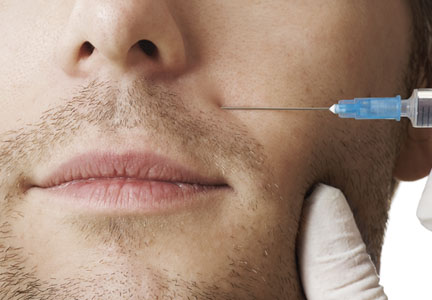
Battle of the Sexes: “Brotox”

In the October 2013 issue of Dermatologic Surgery (2013;39:1434-1443), Keaney and Alster reviewed the use of botulinum toxin in men. Although botulinum toxin injection was the most common cosmetic procedure performed in 2011 in both sexes, men represented only 6% of participants. The authors reviewed the sexual differences in the anatomy of treatment areas. As described, men were noted to have more prominent supraorbital ridges, which contributed to a flatter eyebrow contour that was positioned lower along the orbital rim compared to women. Also the medial supraorbital ridge blended into the glabellar complex on men, giving a greater forward projection. Additionally, forehead height and width were greater in men and there was a greater backward slope compared to women. The female forehead had a greater contour and the female orbit was proportionally larger in relation to skull size. With regard to facial musculature, men were thought to have greater skeletal muscle mass. Men were reported to have greater upward vertical movement for facial expressions, which was thought to contribute to the differences in rhytide severity and distribution.
The authors also reviewed 2 published studies that accounted for sex in study design or subgroup analysis. In a study performed by Brandt et al (Dermatol Surg. 2009;35:1893-1901) of abobotulinumtoxinA for the treatment of glabellar lines, 158 participants (23 men [15%]) were randomized (2:1 ratio) to receive a single 50-U injection of abobotulinumtoxinA or placebo. This study found that women in the abobotulinumtoxinA group were more likely to respond than men, and therefore Brandt et al concluded that treatment of the male glabella required an abobotulinumtoxinA dose of more than 50 U.
What’s the issue?
The number of men seeking botulinum toxin injections has been increasing and therefore it is important to understand the inherent anatomic differences. There have been numerous clinical articles and trials using botulinum toxin but not all include men. It is important to consider the differences in muscle mass and anatomic differences when injecting botulinum toxin in men. Although clinical trials and studies provide guidelines on dosing, it is important to assess each patient individually according to muscle strength and mass. The standardized doses used in clinical trials may not be appropriate for all patients, especially men, because this patient population is not equally represented in these trials. Some male patients may state that botulinum toxin did not work, which may be due to inherent underdosing for the muscle size or strength. It is important to understand and appreciate these anatomic and structural differences in men when utilizing botulinum toxin. With a growing proportion of men seeking cosmetic treatments, are you equipped to assess the nuances?
Looking Forward: Juvéderm Voluma for Midface Volume

Callan et al (Clin Cosmet Investig Dermatol. 2013;6:81-89) reported on the efficacy and safety profile of Juvéderm Voluma (Allergan, Inc) for midface volume deficiency in 103 participants over 24 months. Study participants received treatment with the hyaluronic acid filler to the malar area in 1 or 2 sessions over a 4-week period. An additional treatment was administered at week 78 of the study. At least a 1-point improvement on the midface volume deficit scale (MFVDS) and on the global aesthetic improvement scale (GAIS) was defined as clinically meaningful improvement. Of the 103 participants enrolled, 84% had moderate or significant volume deficiency at baseline. At week 8, 96% were reported to be responders on the MFVDS and 98% deemed themselves responders on the GAIS.
Seventy-two participants completed 24 months of treatment; 45 of these participants did not receive supplementary filler at week 78. Forty-three of the 45 (95.6%) participants were deemed responders on the MFVDS. At end of the study (n=72), 66 participants were either satisfied or very satisfied with the product, with 70 participants indicating that they would recommend the product to others. Adverse events included bruising, swelling, pain/tenderness, erythema, eyelid edema, and vasovagal syncope. Injection-site bruising and swelling were the most commonly reported adverse events. There was a single case of swelling in the left tear trough area, which occurred approximately 17 weeks after the week 4 treatment with the study product and 2 months after bilateral administration of Juvéderm Ultra (Allergan, Inc) to the tear troughs (done outside the study), which was speculated to have led to bilateral hardening of the Juvéderm Voluma implant. Oral prednisolone 5 mg daily was administered over 5 days and Hyalase (sanofi-aventis Australia)(100–150 U) was injected 3 times over a 3-week period. The swelling completely resolved approximately 1 month after the third Hyalase session.
What’s the issue?
Juvéderm Voluma currently is not available in the United States. It differs from other hyaluronic acid fillers in that it has a lower cohesivity but higher gel hardness that results in a more viscous solution. These properties are purported to be more suited for deeper injection in the skin at the deep dermal/subcutaneous level and submuscular/supraperiosteal level, which would allow for higher lift of tissue and volume restoration, while still being injected via a small-diameter needle. Although this filler has been used outside the United States, it will be interesting to see the adaptation and adoption in the US filler market. Is it the future of fillers or another one to add to the toolbox?