Article
Moving Beyond Traditional Methods for Treatment of Acne Keloidalis Nuchae
Acne keloidalis nuchae (AKN) is a chronic inflammatory condition commonly affecting the occipital scalp and posterior...
Article

Moving Beyond Traditional Methods for Treatment of Acne Keloidalis Nuchae
The Comparison
A A 25-year-old man of Hispanic ethnicity with pink papules, pustules, and large keloidal tumors on the occipital...
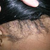
Article
Act Fast With Traction Alopecia to Avoid Permanent Hair Loss
Often caused or worsened by hairstyling practices, traction alopecia can be reversible with proper treatment in its early stages but may result in...
Article
Act Fast With Traction Alopecia to Avoid Permanent Hair Loss
Traction alopecia is a common type of alopecia that ultimately can result in permanent hair loss.
Article
Plantar Hyperpigmentation
Plantar hyperpigmentation is a benign finding in many individuals and is especially prevalent in those with darker skin...
Article
Plantar Hyperpigmentation
Though it is a benign finding in many individuals, especially those with darker skin tones, it is important to differentiate plantar...
Article
Dx Across the Skin Color Spectrum: Longitudinal Melanonychia
A brown, black, or gray pigmented linear band that spans the length of the nail plate due to excess melanin, longitudinal melanonychia may be...
Article

Papulosquamous Dermatophytid Reaction in a Child With Tinea Capitis
High clinical suspicion for id reaction in patients of the appropriate age group and demographic—pediatric patients of African descent—is...
Article

Longitudinal Melanonychia
A patient presented with variable shades of brown covering more than 50% of the nail width. What's the condition and what are the key clinical...
Article

Allergic contact dermatitis
Allergic contact dermatitis (ACD) is an inflammatory condition of the skin caused by an immunologic response to 1 or more identifiable allergens...
Article

Allergic Contact Dermatitis
A Black woman with a rash on the neck that developed after new products were applied to the hair. What’s the condition and what...
Article

Squamous Cell Carcinoma
An African woman presents with a growth on the lower lip decades after a large facial burn. What's the condition and what are the key clinical...
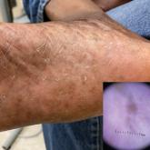
Article
 of the keratoacanthoma type on the arm.")
Squamous cell carcinoma
Cutaneous squamous cell carcinoma (SCC) develops from a malignant tumor of the keratinocytes, eccrine glands, or pilosebaceous units that invades...
Article

Systemic lupus erythematosus
Systemic lupus erythematosus (SLE) is a chronic autoimmune condition that affects the kidneys, lungs, brain, and heart, although it is not limited...
Article

Systemic Lupus Erythematosus
This Black woman has malar erythema and hyperpigmentation with sparing of the nasolabial folds. What's the condition and what are the key clinical...