User login
There Was a Farmer Had a Rash ...
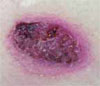
A61-year-old farmer from Iowa with a medical history significant for non-Hodgkin’s lymphoma in remission presented for evaluation and treatment of two ulcerating lesions, located on his left forearm and left thigh of two months’ duration. He denied a history of pulmonary symptoms, fever, or unintentional weight loss. Physical examination was negative for lymphadenopathy or splenomegaly. Two large, beefy hyperkeratotic plaques with an underlying border with pustules were noted. The lesion shown is on the left lateral leg and is 5.3 x 4.0 cm.
What is your diagnosis:
- Cutaneous sarcoidosis;
- Recurrent lymphoma with skin metastasis;
- Blastomycosis;
- Pyoderma gangrenosum; or
- Anthrax.
Discussion
The answer is C: blastomycosis. A pathology specimen from a left arm skin punch biopsy was read as pseudoepitheliomatous hyperplasia, dermal abscess, and broad-based yeast organisms suggestive of blastomycosis. Fungal cultures grew a white-like colony of hyphae suggestive of blastomycosis. DNA probe was positive for blastomycosis dermatitidis. Interestingly, his fungal serologies were negative. The patient was started on itraconazole 200 mg twice daily.
Blastomycosis typically presents in one of two ways:
- Pulmonary infection and/or
- Cutaneous infection.
Typically, the spores of Blastomyces dermatitidis are inhaled from the soil, decomposed vegetation, or rotting wood, and the respiratory system is the first site of infection. Occupations with frequent outdoor exposure in highly endemic areas (including the southeastern states of the United States) connote increased risk. Case series have documented inoculation arising after outdoor activities in the woods near water sources.
Although pulmonary infection is subclinical in 50% of inhalational cases, one study found that pulmonary manifestations were present in 154 of 170 cases (91%) with cough (90%), fever (75%), night sweats (68%), weight loss (66%), chest pain (63%), dyspnea (54%), and aches (50%). Therefore, respiratory symptoms may signal the disease.
According to another review of 100 cases in an endemic area, pulmonary blastomycosis should be considered for any pulmonary infiltrate, especially in the upper lobes. For this patient, because there were no pulmonary symptoms or cutaneous trauma, the most likely etiology is via hematogenous spread. However, whereas the chest radiograph did not show classic signs of blastomycosis (one or more densely consolidated areas of pneumonia or nodular infiltrates), it did show slight fibrosis and pleural thickenings in the apices that is occasionally associated with blastomycosis infection.
As in this case, a presenting cutaneous lesion may be the first sign of disease. The cutaneous findings are usually the result of hematogenous spread; however, uncommon primary cutaneous blastomycosis can occur after direct inoculation from trauma to the skin. Classically, they are described as well-demarcated papulopustules and verrucous plaques with central scarring and black crusting. But the skin lesions can present in many forms and often confound the differential diagnosis. Other cutaneous infectious etiologies include verrucae, nocardiosis, cutaneous tuberculosis, and other dimorphic fungi. However, other dimorphic fungi are less likely to infect the skin. Inflammatory conditions, such as pyoderma gangrenosum and sarcoidosis, must be considered. Ulcerating squamous cell carcinoma is also a consideration.
Blastomycosis is recognized histologically by its broad-based budding and thick, double-contoured walls of the yeast forms found at body temperature (37º C) while it grows as tan or white mold at room temperature. Cultures can be drawn from sputum, pus, or urine. Severe disease often requires systemic antifungal treatment, whereas more moderate to mild disease can be treated topically. TH
References
- Baumgardner DJ, Halsmer SE, Eagan G. Symptoms of pulmonary blastomycosis: northern Wisconsin, United States. Wilderness Environ Med. 2004;15:250-256.
- Patel RG, Patel B, Petrini MF, et al. Clinical presentation, radiographic findings, and diagnostic methods of pulmonary blastomycosis: a review of 100 consecutive cases. South Med J. 1999;92:289-295.
- Bolognia J, ed. Dermatology. Barcelona, Spain: Mosby; 2003.
- Kasper DL, ed. Harrison’s Online Principles of Internal Medicine. 16th ed. New York: McGraw-Hill; 2005.