User login
Rivaroxaban vs dalteparin for VTE in cancer

Rivaroxaban is a feasible alternative to low-molecular-weight heparin (LMWH) for the treatment of venous thromboembolism (VTE) in cancer patients, according to researchers.
In the SELECT-D trial, cancer patients with VTE received 6 months of treatment with rivaroxaban or the LMWH dalteparin.
Patients who received rivaroxaban had a lower rate of VTE recurrence but higher rates of bleeding than those who received dalteparin.
These results were published in the Journal of Clinical Oncology. The research was supported by Bayer AG.
The trial enrolled 406 patients who had cancer and VTE. Sixty-nine percent of patients were receiving cancer treatment (83% chemotherapy) at baseline, and 58% had metastases.
Patients were randomized to receive dalteparin or rivaroxaban for 6 months. Dalteparin was given at 200 IU/kg daily for the first month, then at 150 IU/kg daily for months 2 to 6. Rivaroxaban was given at 15 mg twice daily for 3 weeks, then at 20 mg once daily for the rest of the treatment period.
The 6-month cumulative VTE recurrence rate was 11% in the dalteparin arm and 4% in the rivaroxaban arm (hazard ratio [HR]=0.43; 95% CI, 0.19 to 0.99).
The 6-month cumulative rate of major bleeding was 4% in the dalteparin arm and 6% in the rivaroxaban arm (HR=1.83; 95% CI, 0.68 to 4.96).
And the 6-month cumulative rate of clinically relevant non-major bleeding was 4% in the dalteparin arm and 13% in the rivaroxaban arm (HR=3.76; 95% CI, 1.63 to 8.69).
The researchers said these results suggest rivaroxaban can be an alternative to LMWH in cancer patients.
“Clinicians were already adopting [rivaroxaban] into practice for non-cancer patients, and now they have data from this study to indicate that this form of treatment is an alternative option for many cancer patients who have a clot,” said study author Annie Young, SRN, PhD, of Warwick Medical School at University of Warwick in Coventry, UK.
“We now need to be sitting down with each one of our cancer patients with VTE, discussing their preference alongside looking at all their clinical details—including whether the cancer lesion is still there, what other medications are being taken, and what other conditions the patient has—so that we can choose the optimal VTE treatment for each patient.”
Group calls on WHO to help fight HTLV-1

A group of scientists and activists are calling on the World Health Organization (WHO) to fight the spread of human T-cell leukemia virus subtype 1 (HTLV-1).
The group has written a letter to the WHO highlighting the global prevalence of HTLV-1 infection and recommending strategies to prevent the transmission of HTLV-1.
An abbreviated version of the letter was published in The Lancet. The full letter is available on the Global Virus Network (GVN)* website.
“Since my colleagues and I discovered HTLV-1 . . ., we have learned that this destructive and lethal virus is causing much devastation in communities with high prevalence,” said Robert C. Gallo, MD, co-founder and director of GVN and a professor at the University of Maryland School of Medicine in Baltimore.
“During the GVN meeting last September, I was astounded to learn of the hyper-endemic numbers in the Aboriginal population of Australia. HTLV-1 is endemic in other regions, including several islands of the Caribbean, and in countries such as Brazil, Iran, Japan, and Peru. We hope that the WHO will agree with us and begin to take action in promoting prevention strategies against HTLV-1.”
HTLV-1 prevalence
In their letter, Dr Gallo and his colleagues cite statistics of HTLV-1 prevalence around the world.
The authors note that, in a hospital-based cohort study conducted in Central Australia, 33.6% of Indigenous people tested HTLV-1 positive, with the incidence rate reaching 48.5% in older men.
Research has suggested that, in Brazil, the HTLV-1 prevalence rate is 1.8% in the general population, 1.3% in blood donors in certain regions, and 1.05% in pregnant women.
It is estimated that nearly 1 million people are HTLV-1-positive in Japan, and 850,000 to 1.7 million people in Nigeria are infected with the virus.
In Gabon, the HTLV-1 prevalence in adults is believed to be 5% to 10%. And in Central African Republic, 7% of older, female Pygmies in the southern region were found to be infected with HTLV-1.
Studies have indicated that 6.1% of the general population in Jamaica is positive for HTLV-1, and other Caribbean islands have similar prevalence rates.
The authors note that HTLV-1 and -2 have also been detected in non-endemic areas due to migration and sexual transmission.
It is estimated that 20,000 to 30,000 people are infected with HTLV-1 in the UK, and 10,000 to 25,000 people are infected in metropolitan France.
Other estimates suggest that 266,000 people are infected with HTLV-1 or -2 in the US, and 3600 people with HTLV-1 associated myelopathy/tropical spastic paraparesis (HAM/TSP) remain undiagnosed.
“The general neglect, globally, of the importance of HTLV-1 as a sexually transmitted infection that causes a range of debilitating inflammatory diseases does our patients, who request a sexual health screen, a disservice,” said Graham P. Taylor, MDMB, DSc, a professor at Imperial College London in the UK.
“It is also important to recognize the importance of mother-to-child transmission of HTLV-1 in the development of adult T-cell leukemia/lymphoma (ATLL) decades later. Despite the availability of highly sensitive and specific diagnostic tests for infection and a proven intervention, except for Japan, there are no antenatal screening programs. Evaluating the cost-effectiveness of such programs should now be a priority.”
“To prevent mother-to-child infection, the Japanese government has been offering HTLV-1 screening for all pregnant women without cost,” noted Yoshi Yamano, MD, PhD, a professor at St. Marianna University School of Medicine in Kawasaki, Japan.
“Taking a leadership role to promote research, it also provides grants for clinical trials and patient registries focused on ATLL and HAM/TSP.”
Preventing transmission
The letter outlines 5 strategies to prevent or reduce transmission of HTLV-1. The authors recommend:
- Protecting the sexually active population with routine HTLV-1 testing in sexual health clinics and through promotion of CMPC—Counsel & Monitor HTLV-1-positive patients, notify Partners, and promote Condom usage
- Protecting blood and organ donors and recipients by testing for HTLV, avoiding use of products that may be infected, and promoting CMPC
- Protecting mothers, babies, and fathers via routine antenatal care testing, advising HTLV-1-positive mothers against breastfeeding (when feasible), and promoting CMPC
- Protecting injectable drug users by promoting HTLV-1 testing, providing safe needles through needle exchange program, and promoting CMPC
- Supporting the general population and healthcare providers by providing access to an up-to-date WHO HTLV-1 Fact Sheet that can help healthcare providers diagnose HTLV-1 and related diseases and help patients protect themselves from HTLV-1.
“This virus has been underestimated since the time of its discovery perhaps because it is restricted to certain regions or because it is not terribly infectious,” said William Hall, MD, PhD, co-founder of GVN and a professor at the University College Dublin in Ireland.
“However, for decades, it has been known that HTLV-1 is highly carcinogenic and causes severe paralytic neurologic disease and immune disorders that can lead to bacterial infections. It is time that the WHO publicize prevention strategies against this devastating virus.”
*GVN is an international coalition of medical virologists dedicated to identifying, researching, fighting, and preventing current and emerging pandemic viruses that pose a threat to public health.
MSC product can treat refractory GVHD
MONTRÉAL—Results from a phase 3 trial suggest a mesenchymal stem cell (MSC) product can treat steroid-refractory, acute graft-versus-host disease (GVHD) in children.
The product, remestemcel-L (MSC-100-IV), produced an overall response rate of 69% at day 28, with complete resolution of GVHD in 29% of patients.
Adverse events (AEs) in this trial were consistent with the known safety profile of remestemcel-L.
Joanne Kurtzberg, MD, of Duke University Medical Center in Durham, North Carolina, presented these results at ISCT 2018.
The trial was sponsored by Mesoblast International Sàrl, the company developing remestemcel-L.
Remestemcel-L consists of human MSCs derived from donor bone marrow and expanded in culture.
Patients
The trial enrolled 55 patients who had acute GVHD and had failed to respond to steroid treatment. This was defined as progression within 3 days or no improvement within 7 days of consecutive treatment with at least 2 mg/kg/day of methylprednisolone or an equivalent product.
The patients had a median age of 7.6 years (range, 0.6 years to 17.9 years) at baseline, and 64% were male. Underlying diseases include acute myeloid leukemia (32.7%), acute lymphoblastic leukemia (21.8%), anemia (9.1%), chronic myeloid leukemia (7.3%), sickle cell disease (5.5%), juvenile myelomonocytic leukemia (3.6%), myelodysplastic syndromes (3.6%), and “other” disease (16.4%).
Most patients (87%) had received myeloablative conditioning, most (76%) had an unrelated donor, and roughly half (51%) received an HLA-mismatched transplant.
Fifty-five percent of patients received a bone marrow transplant, 25% received peripheral blood stem cells, and 20% received cord blood.
Forty-seven percent of patients had grade D GVHD at baseline, 42% had grade C, and 11% had grade B. Thirty-six percent of patients had multi-organ involvement (all with lower gastrointestinal), 38% had lower gastrointestinal involvement only, and 26% had skin involvement only.
Results
Fifty-four patients were treated with remestemcel-L. They received 8 injections over 4 weeks (twice weekly), consisting of 2 million cells per kg per injection.
The overall response rate at day 28 was 69%. Twenty-nine percent of patients achieved a complete response, defined as resolution of acute GVHD in all involved organs.
Forty percent of patients achieved a partial response, defined as organ-level improvement of at least one stage without worsening of any other organ.
All patients reported at least one treatment-emergent AE, and 61% had serious treatment-emergent AEs. The most common of these were infection (33%) and respiratory events (20%).
Four patients withdrew from the trial before day 100. One patient couldn’t receive treatment, 1 withdrew due to an AE (somnolence), 1 had parental consent withdrawn, and 1 was taken off study by the principal investigator.
There were 11 on-study deaths, but none were considered related to remestemcel-L. Eight deaths were due to infection, 1 due to GVHD progression, and 2 due to primary cancer relapse.
The day-100 survival analysis is pending.
Galinpepimut-S receives orphan designation for MM
The US Food and Drug Administration (FDA) has granted orphan drug designation to galinpepimut-S (GPS) as a treatment for multiple myeloma (MM).
GPS is an immunotherapeutic that targets malignancies characterized by overexpression of the Wilms tumor 1 (WT1) antigen.
GPS consists of 4 peptide chains, 2 of which are modified chains that induce an innate immune response (CD4+/CD8+) against the WT1 antigen and access a range of HLA types.
When GPS is administered to a patient, the induced immune response has the potential to recognize and destroy cancer cells and provide ongoing support to the immune system so it can continue to target and destroy residual cancer cells.
GPS also has orphan designation from the FDA for the treatment of acute myeloid leukemia and malignant plural mesothelioma.
Phase 2 trial
GPS has been investigated in a phase 2 trial of MM patients. Results from this trial were recently presented at the 44th Annual Meeting of the EBMT.
Researchers evaluated GPS in combination with lenalidomide as maintenance therapy in MM patients who received an autologous stem cell transplant (ASCT).
The study enrolled 19 patients who began receiving GPS within 22 days of ASCT. They received 6 doses every 2 weeks. (Injection sites were pre-stimulated with granulocyte-macrophage colony-stimulating factor.)
Patients received 6 additional monthly doses of GPS as well as lenalidomide maintenance (10 mg daily) starting on day 100 post-ASCT.
Twelve patients received all 12 doses of GPS. Eleven patients achieved a complete response or very good partial response. All of these patients had CD4 immune responses, and 9 of them had CD8 immune responses.
The progression-free survival was 81% at 12 months and 62% at 18 months. The median progression-free survival was 23.6 months (range, 15.2 to not reached).
The overall survival was 88% at 18 months, and the median overall survival was not reached.
About orphan designation
The FDA grants orphan designation to products intended to treat, diagnose, or prevent diseases/disorders that affect fewer than 200,000 people in the US.
The designation provides incentives for sponsors to develop products for rare diseases. This may include tax credits toward the cost of clinical trials, prescription drug user fee waivers, and 7 years of market exclusivity if the product is approved.
Team identifies new prognostic factor for MF

New research suggests DNA sequencing can reveal which patients with early stage mycosis fungoides (MF) will develop aggressive disease.
By sequencing the T-cell receptor beta gene (TCRB) in skin biopsies from MF patients, investigators were able to measure the tumor clone frequency (TCF), or the percentage of all T cells that represent the MF clone.
The researchers found the TCF could predict progression-free survival (PFS) and overall survival (OS).
In fact, for patients with early stage MF, TCF was a stronger predictor of PFS than other established prognostic factors.
“While more work needs to be done, we think this approach has the potential to prospectively identify a subgroup of patients who are destined to develop aggressive, life-threatening disease and treat them in a more aggressive fashion with the intent to better manage and, ideally, cure their cancer,” said Thomas Kupper, MD, of Brigham and Women’s Hospital in Boston, Massachusetts.
“At the same time, we believe we can provide reassurance to patients with a low-risk (low TCF) profile that they are likely to survive indefinitely with conventional conservative therapies. As a physician who has treated this disease for decades, I am excited to be involved with work that so directly and profoundly affects the care and management of these patients.”
Dr Kupper and his colleagues described this work in Science Translational Medicine.
The researchers had previously published a study showing that high-throughput sequencing of the TCRB gene was an accurate way to diagnose cutaneous T-cell lymphoma (CTCL), including MF.
With the current study, the investigators set out to determine if the method could be used to predict progression and survival in patients with CTCL.
The team sequenced TCRB in lesional skin biopsies from 309 CTCL patients, most of whom had MF. The discovery cohort had 208 patients (177 with MF), and the validation cohort had 101 patients (87 with MF).
The sequencing produced a snapshot of the TCRB genes from a large number of cells at the site of the lesion. And the researchers could use this to measure TCF.
The team tested the association of TCF with prognosis in all CTCL patients in the discovery cohort and found a TCF greater than 25% in the skin was significantly associated with reduced PFS (P<0.001) and OS (P<0.001). Results were similar in the validation cohort (P<0.001 for PFS and OS).
The investigators also found that TCF was significantly associated with PFS (P<0.001) and OS (P<0.001) in MF patients but not in patients with Sézary syndrome. The team noted that they did not assess the predictive value of TCF in the blood of Sézary patients.
In a multivariable analysis, TCF was still significantly associated with PFS (P<0.001) and OS (P<0.001) in the MF patients.
The researchers also assessed potential prognostic variables in the early stage MF patients and found a TCF greater than 25% was a stronger predictor of PFS than any other established prognostic factor. This includes disease stage, presence of plaques, elevated lactate dehydrogenase, age, large-cell transformation, and CLIPI score.
“In reviewing the results, 2 different patients could have identical-looking skin lesions, but one might have a TCF of 8%, and one would have a TCF of 40%,” Dr Kupper noted. “The latter patient was highly likely to progress—something we would never have been able to predict before this discovery.”
“The TCF was independent of how thick or thin the skin lesions were. Most importantly, compared to all other currently used means of trying to predict which patients would progress, TCF was by far the most sensitive and specific.”
The high-throughput sequencing in this study was performed using ImmunoSEQ, an assay developed by Adaptive Biotechnologies. The company did not sponsor the study, but company employees were involved in the research.
Producing compatible RBCs for transfusion

Researchers say they have found a way to generate customized red blood cells (RBCs) that might one day be used to avoid transfusion incompatibilities.
The team used genome editing to modify an erythroblast cell line, making it deficient in antigens responsible for common transfusion incompatibilities.
The cells in this line could then be differentiated to generate RBCs with broad transfusion compatibility.
The researchers stressed that many obstacles must be overcome before this approach can be translated to a clinical product. However, this work does demonstrate proof of principle.
Ashley Mark Toye, PhD, of the University of Bristol in Bristol, UK, and his colleagues described this work in EMBO Molecular Medicine.
With previous work, Dr Toye and his colleagues generated an immortalized human erythroblast cell line known as the Bristol Erythroid Line Adult (BEL-A). The team showed that this cell line can be induced to generate RBCs in the lab.
With the current study, the researchers engineered BEL-A to express fewer antigens and therefore be less immunogenic.
The team first conducted a 15-month survey in England to investigate which antigens most commonly caused challenges in identifying a matched donated unit for transfusion recipients. The survey revealed 56 patients who had rare blood types with alloantibodies against RBC antigens.
Twenty-two of the patients had alloantibodies to antigens located on glycophorin B (GPB), U, S, or s. Nineteen patients had alloantibodies to antigens within the Rh blood group system.
Ten patients had alloantibodies to the Duffy blood group, and 2 previously untransfused patients had Fy (a-b-). Ten patients had Kell antigen alloantibodies. Eight patients had the Bombay or para-Bombay phenotype. And 3 patients each had alloantibodies to Lu and Kidd antigens.
To create compatible RBCs, the researchers first removed these blood groups from BEL-A using CRISPR-Cas9 genome editing.
The team created several new cell lines that lacked individual blood groups before creating a single cell line in which the 5 most common blood groups were removed—ABO (Bombay phenotype), Rh (Rhnull), Kell (K0), Duffy (Duffynull), and GPB (S-s-U-).
RBCs derived from this modified cell line could, theoretically, serve most of the challenging cases the researchers identified.
“Blood made using genetically edited cells could one day provide compatible transfusions for a group of patients for whom blood matching is difficult or impossible to achieve within the donor population,” Dr Toye said. “However, much more work will still be needed to produce blood cells suitable for patient use.”
Cooperation can drive T-ALL, study shows
Overexpression of HOXA9 and activated JAK/STAT signaling cooperate to drive the development of T-cell acute lymphoblastic leukemia (ALL), according to researchers.
The team found that JAK3 mutations are significantly associated with elevated HOXA9 expression in T-ALL, and co-expression of HOXA9 and JAK3 mutations prompt “rapid” leukemia development in mice.
In addition, STAT5 and HOXA9 occupy similar genetic loci, which results in increased JAK-STAT signaling in leukemia cells.
These discoveries, and results of subsequent experiments, suggested that PIM1 and JAK1 are potential therapeutic targets for T-ALL.
Jan Cools, PhD, of VIB-KU Leuven Center for Cancer Biology in Leuven, Belgium, and his colleagues described this research in Cancer Discovery.
“JAK3/STAT5 mutations are important in ALL since they stimulate the growth of the cells,” Dr Cools said. “[W]e found that JAK3/STAT5 mutations frequently occur together with HOXA9 mutations.”
In analyzing data from 2 cohorts of T-ALL patients, the researchers found that IL7R/JAK/STAT5 mutations were more frequent in HOXA+ cases, and HOXA9 was “the most important upregulated gene of the HOXA cluster.” (HOXA9 expression levels were significantly higher than HOXA10 or HOXA11 levels.)
“We examined the cooperation between JAK3/STAT5 mutation and HOXA9, [and] we observed that HOXA9 boosts the effects of other genes, leading to tumor development,” said study author Charles de Bock, PhD, of VIB-KU Leuven.
“As a result, when JAK3/STAT5 mutations and HOXA9 are both present, leukemia develops more rapidly and aggressively.”
The researchers found that co-expression of HOXA9 and the JAK3 M511I mutation led to rapid leukemia development in mice. Animals with co-expression of HOXA9 and JAK3 (M511I) developed leukemia that was characterized by an increase in peripheral white blood cell counts that exceeded 10,000 cells/mm3 within 30 days.
In addition, these mice had a significant decrease in disease-free survival compared to mice with JAK3 (M511I) alone. The median disease-free survival was 25 days and 126.5 days, respectively (P<0.0001).
Further analysis revealed co-localization of HOXA9 and STAT5. The researchers also found that HOXA9 enhances transcriptional activity of STAT5 in leukemia cells.
The team said this reconfirms STAT5 as “a major central player” in T-ALL, and it suggests that STAT5 target genes such as PIM1 could be therapeutic targets for T-ALL.
To test this theory, the researchers inhibited both PIM1 and JAK1 in JAK/STAT mutant T-ALL. The PIM1 inhibitor AZD1208 and the JAK1/2 inhibitor ruxolitinib demonstrated synergy and significantly reduced leukemia burden in vivo.
Daratumumab approved for newly diagnosed MM
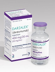
The US Food and Drug Administration (FDA) has granted another approval for the CD38-directed antibody daratumumab (Darzalex®).
Daratumumab is now approved for use in combination with bortezomib, melphalan, and prednisone (VMP) to treat patients with newly diagnosed multiple myeloma (MM) who are ineligible for autologous stem cell transplant.
Daratumumab was first approved by the FDA in 2015 as a monotherapy for MM patients who have received at least 3 prior lines of therapy, including a proteasome inhibitor (PI) and an immunomodulatory agent, or who are double-refractory to a PI and an immunomodulatory agent.
In 2016, daratumumab was approved for use in combination with lenalidomide and dexamethasone, or bortezomib and dexamethasone, to treat MM patients who have received at least 1 prior therapy.
In 2017, daratumumab was approved for use in combination with pomalidomide and dexamethasone to treat MM patients who have received at least 2 prior therapies, including lenalidomide and a PI.
For full prescribing information, visit www.darzalex.com.
Phase 3 trial
The FDA’s approval of daratumumab in combination with VMP is supported by data from the phase 3 ALCYONE (MMY3007) study. Results from this study were presented at the 2017 ASH Annual Meeting and simultaneously published in NEJM.
ALCYONE enrolled 706 patients with newly diagnosed MM who were not eligible for high-dose chemotherapy with autologous stem cell transplant. Patients were randomized to receive VMP or daratumumab plus VMP (D-VMP).
The overall response rates were 91% in the D-VMP arm and 74% in the VMP arm (P<0.0001). Rates of complete response were 43% and 24%, respectively. And rates of minimal residual disease negativity were 22% and 6%, respectively.
The median progression-free survival (PFS) was not reached in the D-VMP arm and was 18.1 months in the VMP arm. The 12-month PFS was 87% and 76%, respectively. The 18-month PFS was 72% and 50%, respectively.
The most common treatment-emergent adverse events (TEAEs; in the D-VMP and VMP arms, respectively) were neutropenia (50% and 53%), thrombocytopenia (49% and 54%), anemia (28% and 38%), peripheral sensory neuropathy (28% and 34%), upper respiratory tract infection (26% and 14%), diarrhea (24% and 25%), pyrexia (23% and 21%), and nausea (21% and 22%).
Infusion-related reactions occurred in 28% of patients in the D-VMP arm and 0% of those in the VMP arm.
The rate of grade 3/4 infections was higher in the D-VMP arm than the VMP arm—23% and 15%, respectively. In both arms, most infections resolved.
The most common grade 3/4 TEAEs (in the D-VMP and VMP arms, respectively) were neutropenia (40% and 39%), thrombocytopenia (34% and 38%), and anemia (16% and 20%).
The rate of discontinuation due to AEs was 5% in the D-VMP arm and 9% in the VMP arm.
‘Long-term benefits’ with nivolumab in cHL
Nivolumab can provide “long-term benefits” in patients with relapsed or refractory classical Hodgkin lymphoma (cHL), according to researchers.
Extended follow-up of the CheckMate-205 study showed that nearly 70% of patients responded to nivolumab.
The median duration of response was 17 months, and the median progression-free survival (PFS) was 15 months.
The most common drug-related AEs were fatigue, diarrhea, infusion-related reactions, rash, nausea, and pruritus.
Philippe Armand, MD, of the Dana-Farber Cancer Institute in Boston, Massachusetts, and his colleagues reported these results in the Journal of Clinical Oncology.
The study was sponsored by Bristol-Myers Squibb Company.
Patients
This phase 2 trial enrolled 243 adults with relapsed or refractory cHL who had undergone autologous hematopoietic stem cell transplant (auto-HSCT).
Their median age was 34 (range, 26-46), and 58% were male. Fifty-seven percent had stage IV disease, 20% had stage III, 21% had stage II, and 2% had stage I.
Patients had received a median of 4 prior therapies (range, 3-5). The median time from diagnosis to first nivolumab dose was 4.5 years (range, 2.4-7.6), and the median time from most recent auto-HSCT to first nivolumab dose was 2.0 years (range, 0.9-4.9).
The researchers divided patients into 3 cohorts according to exposure to brentuximab vedotin (BV):
- Cohort A was BV-naïve (n=63)
- Cohort B received BV only after auto-HSCT (n=80)
- Cohort C received BV before and/or after auto-HSCT (n=100).
Baseline characteristics were generally similar across the cohorts. However, cohort A had fewer prior treatments (median of 2 vs 4 in cohorts B and C).
Cohort B had the longest interval between diagnosis and first nivolumab dose (6.2 years vs 3.1 in cohort A and 3.5 in C) and between most recent auto-HSCT and first nivolumab dose (3.4 years vs 1 in cohort A and 1.7 in C).
Treatment
All patients received nivolumab at 3 mg/kg once every 2 weeks until disease progression or unacceptable toxicity.
In cohort C, patients who were in complete response (CR) for 1 year had to discontinue nivolumab, but they could resume treatment if they relapsed within 2 years.
A protocol amendment allowed patients to continue treatment despite progression if they had stable performance status and were deriving “perceived clinical benefit.”
At a median follow-up of 18 months, 40% percent of all patients were still on treatment, including 48% of patients in cohort A, 40% in cohort B, and 35% in cohort C.
Safety
The most common drug-related AEs were fatigue (23%), diarrhea (15%), infusion-related reactions (14%), rash (12%), nausea (10%), and pruritus (10%).
The most common grade 3/4 drug-related AEs were lipase increase (5%), neutropenia (3%), ALT increase (2%), AST increase (2%), and amylase increase (2%).
The most common serious drug-related AEs were infusion-related reactions (2%), pneumonitis (1%), pneumonia (1%), pleural effusion (1%), and pyrexia (1%).
Seven percent of patients discontinued treatment due to AEs. The most common of these were pneumonitis (2%) and autoimmune hepatitis (1%).
There were no deaths due to drug-related AEs.
Efficacy
The objective response rate was 69% overall, 65% in cohort A, 68% in cohort B, and 73% in cohort C.
CR was the best response for 16% of all patients, 29% of cohort A, 13% of cohort B, and 12% of cohort C.
The median duration of response was 17 months overall, 20 months for cohort A, 16 months for cohort B, 15 months for cohort C, and 20 months for patients who achieved a CR.
The researchers said responses were similar irrespective of BV treatment sequence.
The median PFS was 15 months for all patients, 18 months for cohort A, 15 months for cohort B, 12 months for cohort C, and 22 months for patients who achieved a CR.
The median overall survival was not reached in any of the cohorts.
The 12-month overall survival was 92% overall, 93% in cohort A, 95% in cohort B, 90% in cohort C, and 100% in patients who achieved a CR.
Subsequent HSCT
Forty-four patients proceeded to allogeneic HSCT after nivolumab, and the median post-HSCT follow-up was 5.5 months (range, 2.9-11.8).
At 6 months, the rate of transplant-related mortality was 13%, and the rate of disease progression was 7%.
The rate of grade 2-4 acute graft-vs-host disease (GVHD) was 30%, the rate of grade 3-4 acute GVHD was 20%, and the rate of chronic GVHD was 15%.
CAR T-cell therapy bridges to HSCT in AML patient

A case report suggests an investigational chimeric antigen receptor (CAR) T-cell therapy can provide a bridge to transplant in relapsed/refractory acute myeloid leukemia (AML).
The therapy, known as CYAD-01, prompted a morphologic leukemia-free state in an AML patient.
This patient went on to receive an allogeneic hematopoietic stem cell transplant (allo-HSCT) and achieve a complete molecular remission, which was ongoing 6 months after transplant.
Investigators reported no adverse events related to CYAD-01.
This report was published in haematologica.
The patient is enrolled in the THINK trial (NCT03018405), which is sponsored by Celyad SA, the company developing CYAD-01.
According to Celyad, CYAD-01 consists of autologous T cells expressing a CAR based on the natural killer group 2 member D receptor (NKG2D), a transmembrane receptor expressed by natural killer cells and some T-cell subsets.
The AML patient who received CYAD-01 was 52 years old at trial enrollment. He had +8/del(7)(q22q36), FLT3/NPM1 wild-type AML.
The patient’s disease was primary refractory to 7+3 induction, so he went on to receive salvage chemotherapy with cladribine, cytarabine, G-CSF, and mitoxantrone. He achieved a complete response to this treatment and received 2 cycles of consolidation with cladribine and cytarabine.
The patient’s subsequent allo-HSCT was delayed to allow for pulmonary function test recovery. In the meantime, he relapsed.
At this point, the patient enrolled in the THINK trial. He underwent apheresis and received CYAD-01 infusions at the initial dose level of 3 x 10⁸ cells every 2 weeks for 3 administrations.
The patient achieved a morphologic leukemia-free state at 3 months, which enabled him to undergo allo-HSCT.
The patient achieved a complete molecular remission after transplant. He remained in remission at last follow-up—9 months after his first CYAD-01 infusion and 6 months after allo-HSCT.
The investigators said CYAD-01 was well tolerated in this patient. He did not develop cytokine release syndrome or experience neurotoxic effects. The patient had only non-related grade 1 adverse events.
“The THINK study case report provides the first clinical validity of CYAD-01 as a tumor-specific antigen-receptor and AML as a disease sensitive to gene-engineered cell therapies,” said study investigator David Sallman, MD, of Moffitt Cancer Center in Tampa, Florida.
“As antigen targeting offers significant challenges in AML, this outcome brings hope for the further use of gene-engineered T cells for patients with AML [who] have run out of therapeutic options. It’s all the more striking that this outcome was observed without any prior lymphodepletion, highlighting the potential of using a physiologic antigen-receptor.”