User login
"Testing" Your Diagnostic Skills

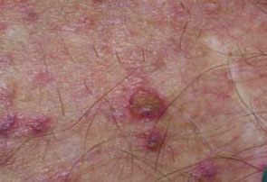
A72-year-old male with a history of renal transplantation underwent laparoscopic cholecystectomy for cholelithiasis. On post-operative day one, he developed worsening abdominal pain. An urgent exploratory laparotomy revealed no abnormalities. Over the next few days, he experienced worsening confusion and agitation. He developed respiratory failure, as well as a cutaneous eruption over the face, chest, abdomen, back, and upper extremities.
What is the most sensitive test for diagnosis of this eruption?
- Tzanck smear;
- Darkfield microscopy;
- Direct fluorescent antibody testing;
- Polymerase chain reaction; or
- Viral culture.
Discussion
The answer is D: polymerase chain reaction. Varicella zoster virus (VZV) was detected by rapid polymerase chain reaction from a skin biopsy specimen and from a sample of cerebrospinal fluid. It was also detected by viral culture from tracheal secretions. The patient was diagnosed with disseminated herpes zoster with VZV encephalitis and pneumonitis. He was treated with intravenous acyclovir, 1 g every eight hours for 14 days. His respiratory function and mental status gradually improved.
Herpes zoster is a painful vesicular eruption confined to the distribution of a sensory dermatome. It is caused by the reactivation of latent VZV located in the dorsal root ganglion of the affected dermatome. Triggers for reactivation include stress, trauma, fever, radiation therapy, and immunosuppression. The reactivation of VZV can also present in a disseminated fashion. Herpes zoster is classified as disseminated when more than 20 vesicular lesions occur outside of the primary and adjacent dermatomes.1 Disseminated herpes zoster can occur in immunocompetent patients, but it usually occurs in the setting of immunosuppression.2 It is seen most often in the settings of HIV, malignancy, and immunosuppressive therapy.1
The cutaneous lesions of herpes zoster begin as erythematous macules and papules that progress to vesicles. The vesicular lesions can evolve into pustules. The vesicles become crusts after seven to 10 days. Intense pain often precedes the cutaneous eruption, which can lead to misdiagnosis as myocardial infarction, pleurisy, acute surgical abdomen, or herniated intervertebral disk.2
Disseminated herpes zoster can also involve the central nervous system, lungs, liver, heart, and gastrointestinal tract. Pulmonary involvement is the most common of the possible visceral manifestations. Visceral zoster occurs in 10% of immunocompromised patients with cutaneous zoster.1
The differential diagnosis of disseminated herpes zoster includes bullous impetigo, insect bites, erythema multiforme, papular urticaria, drug eruption, contact dermatitis, and other viral exanthems such as coxsackie virus, rickettsial pox, and smallpox. The best initial test is a Tzanck smear of a scraping from the base of a lesion. The presence of multinucleated giant cells and epithelial cells with acidophilic intranuclear inclusions provides a rapid diagnosis. The most sensitive test is polymerase chain reaction of vesicular fluid, but this test is not universally available.3 The diagnosis can also be made by viral culture, but this can be difficult because VZV is a labile virus.2,3 Other diagnostic tests include direct fluorescent antibody testing, serology, and immunohistochemical stains on a skin biopsy specimen.
Treatment with intravenous acyclovir is indicated in immunocompromised patients with disseminated VZV.1 Treatment should be started within 72 hours of the onset of the vesicular eruption. Acyclovir-resistant VZV should be treated with foscarnet.2 There are also reports of physicians combining acyclovir with plasma exchange, a treatment option that may be beneficial in decreasing the viral load.4 Varicella zoster virus is transmissible to susceptible individuals by direct contact or by respiratory transmission from a patient with pulmonary involvement. Airborne and contact precautions are recommended until all of the patient’s vesicles have crusted. TH
References
- Stratman E. Visceral zoster as the presenting feature of disseminated herpes zoster. J Am Acad Dermatol. 2002;46(5):771-774.
- McCrary ML, Severson J, Tyring SK. Varicella zoster virus. J Am Acad Dermatol. 1999;41(1):1-14.
- Sauerbrei A, Eichhorn U, Schacke M, et al. Laboratory diagnosis of herpes zoster. J Clin Virol. 1999;14(1):31-36.
- Lee C, Koike M, Oshimi K, et al. Acyclovir combined with plasma exchange for disseminated varicella-zoster virus infection after bone marrow transplantation [in Japanese]. Rinsho Ketsueki. 2006;47(3):210-213.
The Case of the Nonhealing Wound
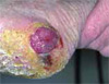
An 85-year-old female developed a sore on the left foot (see image above) during the past six months. Throughout that time she underwent periodic debridement and local wound care with gentamicin ointment followed by the use of silver sulfadiazine cream dressings, an Unna Boot, and a surgical shoe with heel relief. Despite treatment her wound increased in size, bleeds easily, but it is not painful.
WHAT IS YOUR DIAGNOSIS?
- Pyogenic granuloma;
- Squamous cell carcinoma;
- Amelanotic melanoma;
- erkel cell carcinoma; or
- Hypertrophic granulation tissue?
Discussion
The correct answer is C: amelanotic melanoma. The patient’s skin biopsy revealed a nodular malignant melanoma with ulceration, Clark’s level V, Breslow thickness at least 5.8 mm. She underwent wide local excision with sentinel lymph node biopsy, which was negative for tumor. The defect was repaired with a split-thickness skin graft and temporary wound vacuum. She is being closely monitored for local recurrence and in-transit metastasis.
Melanoma classically presents as an asymmetric, irregularly hyperpigmented lesion with ill-defined borders; however, some melanomas have little to no pigment and can be easily confused with other benign or malignant entities. Amelanotic melanomas comprise about 2% to 8% of all melanomas.1-2 A seemingly amelanotic lesion may have an area of subtle pigmentation peripherally that can be a clue to the diagnosis.2-3 The prognosis of amelanotic melanomas is the same as that of pigmented melanomas and is contingent upon depth of invasion, location, and patient age and gender. Unfortunately, the diagnosis of an amelanotic melanoma is often delayed, leading to more advanced tumors. Treatment is analogous to pigmented melanomas.2
A rapidly proliferating amelanotic melanoma can be clinically confused with a pyogenic granuloma, a benign vascular hyperplasia. Pyogenic granulomas present as solitary, discrete, erythematous papules or pedunculated growths on cutaneous and mucosal surfaces. They are often friable and may ulcerate. Pyogenic granulomas are more common in children and young adults, but they can occur at any age. If a pyogenic granuloma is not surgically excised, its growth will eventually stabilize, leading to involution, necrosis, or shrinkage to a fibrotic papule.4
Hypertrophic granulation tissue is another benign entity that can resemble an amelanotic melanoma. The production of granulation tissue is a normal response in the early proliferative stage of wound healing. Granulation tissue has abundant vascular structures, which give it an erythematous, edematous, and friable appearance. As wound healing progresses, granulation tissue is replaced with new epidermis through re-epithelialization.5 Failure of a wound to show signs of progressive healing should prompt a biopsy to distinguish normal granulation tissue from malignancy. Amelanotic melanoma has been reported in cases of nonhealing diabetic foot ulcers.6
Amelanotic melanoma can also be difficult to clinically distinguish from other malignant growths, such as squamous cell carcinoma. More common in elderly patients, squamous cell carcinoma commonly presents as a pink to erythematous, scaly papule, or plaque on a sun-exposed surface. Treatment of superficial squamous cell carcinoma, such as Bowen’s disease, with cryotherapy or cautery is highly effective; however, if an amelanotic melanoma is mistakenly treated as Bowen’s disease, then the delay in eventual histological diagnosis may result in an advanced stage amelanotic melanoma.7
Merkel cell carcinoma is a highly aggressive tumor that typically presents as an erythematous to violaceous, painless, solitary nodule or plaque that grows rapidly. It usually affects older patients and commonly occurs on the head. It has a high likelihood of local recurrence, metastasis, and poor prognosis.8 Merkel cell carcinomas are rare, and they elicit the same differential diagnoses as amelanotic melanomas. Histological differentiation from amelanotic melanoma is necessary. TH
References
- Adler M, White C. Amelanotic malignant melanoma. Semin Cutan Med Surg. 1997;16:122-130.
- Koch SE, Lange JR. Amelanotic melanoma: the great masquerader. J Am Acad Dermatol. 2000 May;42(5 Pt 1):731-734.
- Bono A, Maurichi A, Moglia D, et al. Clinical and dermatoscopic diagnosis of early amelanotic melanoma. Melanoma Res. 2001;11:491-494.
- Lin RL, Janniger CK. Pyogenic granuloma. Cutis. 2004 Oct;74(4):229-33.
- Freedburg IM, Eisen AZ, Klaus W, et al. Fitzpatrick’s Dermatology in General Medicine, 6th ed. New York: McGraw-Hill 2003;243.
- Gregson CL, Allain TJ. Amelanotic malignant melanoma disguised as a diabetic foot ulcer. Diabet Med. 2004 Aug;21(8):924-927.
- Holder JE, Colloby PS, Fletcher A, et al. Amelanotic superficial spreading malignant melanoma mimicking Bowen’s disease. Br J Dermatol. 1996 Mar;134(3):519-521.
- Agelli M, Clegg LX. Epidemiology of primary Merkel cell carcinoma in the United States. J Am Acad Dermatol. 2003 Nov;49(5):832-841.