User login
Fishing for a Diagnosis

A63-year-old female with a history of hypertension and nicotine dependence presented with acute substernal chest pain. The patient reported that the pain was a dull pressure, 5/10 in severity, which radiated into her neck and left arm and was associated with dyspnea.
The patient was on atenolol for hypertension. She reported a 30-year smoking history. A physical exam revealed an anxious, hypertensive (180/95), tachycardic (123 bpm) female. Findings on cardiovascular exam were otherwise unremarkable. Electrocardiogram revealed a 2-3 mm ST-segment elevation in leads V2-V6. Laboratory tests revealed an elevated troponin T level (0.32 ng/mL [nl < 0.03 ng/mL]) and an elevated creatinine kinase-MB fraction (8.2 ng/mL [nl < 6.2 ng/mL]).
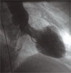
Using the above information, the diagnosis of an ST-segment elevation MI was made. The patient went in for urgent cardiac catheterization, which revealed normal coronary anatomy. A left ventriculogram demonstrated moderate hypokinesis of the apical segment and a left ventricular ejection fraction of 34%. TH
What is the most likely cause of the patient’s ECG changes, elevated cardiac biomarkers, and reduced left ventricular function?
- Left ventricular aneurysm
- ST-segment elevation myocardial infarction
- Left ventricular apical ballooning syndrome
- Myocarditis
- Amyloidosis
Discussion
The answer is C: left ventricular apical ballooning syndrome. Transient left ventricular apical ballooning syndrome is a recently described cardiac condition that mimics the clinical presentation of atherosclerotic acute coronary syndrome. Also known as Takotsubo cardiomyopathy, after a round-bottomed, narrow-necked Japanese fishing pot used for trapping octopus, transient left ventricular apical ballooning syndrome was first described in Japan by Dote and colleagues more than a decade ago.1
Typical findings include patients with ischemia-like chest pain and dyspnea, ST-segment elevation and evolutionary T-wave inversion noted on ECG, mildly elevated levels of cardiac biomarkers, and transient apical wall motion abnormalities. These findings occur in the absence of obstructive coronary atherosclerosis. The condition is predominantly seen in postmenopausal women, and most episodes occur after an event causing physical or emotional stress.
The etiology of this condition is widely debated. Many feel that an exaggerated sympathetic response is the critical mechanism of this syndrome. One study has shown that patients with this syndrome had supraphysiologic levels of plasma catecholamines and stress-related neuropeptides.
Treatment is mainly supportive once ST-segment elevation myocardial infarction has been ruled out with a coronary angiogram. Beta-blocker therapy may be appropriate due to presumed catecholamine surge. Short-term cardiac monitoring is also prudent to evaluate for dysrhythmia. Finally, anticoagulation may be considered to prevent mural thrombosis formation.
The prognosis for patients with transient left ventricular apical ballooning syndrome is favorable, with most patients regaining normal systolic ventricular function within several months, and recurrence is rare. Follow-up echocardiographic evaluation is commonly conducted to ensure adequate resolution of systolic left ventricular dysfunction.
Universal diagnostic criteria for transient left ventricular apical ballooning syndrome have not been established. One diagnostic algorithm recently published includes four criteria: 1) transient regional wall motion abnormalities of the left ventricular apical and midventricular segments; 2) absence of obstructive coronary disease or plaque rupture; 3) new ST-segment elevation and/or T-wave inversion; and 4) absence of an obvious alternative cause (e.g., recent head trauma, extensive intracranial bleeding, myocarditis, pheochromocytoma, hypertrophic cardiomyopathy). To make the diagnosis of transient left ventricular apical ballooning syndrome, all four criteria must be met.
As awareness of transient left ventricular apical ballooning syndrome increases, and as evaluation of left ventricular function becomes standard, this diagnosis is becoming more common. In patients presenting with ST-segment elevation and suspected acute coronary syndromes, one must keep apical ballooning syndrome in the differential. Prompt recognition and aggressive supportive treatment are indicated, and recovery of systolic function can be expected. TH
References
- Dote K, Sato H, Tateishi H, et al. Myocardial stunning due to simultaneous multivessel coronary spasms: a review of 5 cases [in Japanese]. J Cardiol. 1991;21(2):203-214.
A63-year-old female with a history of hypertension and nicotine dependence presented with acute substernal chest pain. The patient reported that the pain was a dull pressure, 5/10 in severity, which radiated into her neck and left arm and was associated with dyspnea.
The patient was on atenolol for hypertension. She reported a 30-year smoking history. A physical exam revealed an anxious, hypertensive (180/95), tachycardic (123 bpm) female. Findings on cardiovascular exam were otherwise unremarkable. Electrocardiogram revealed a 2-3 mm ST-segment elevation in leads V2-V6. Laboratory tests revealed an elevated troponin T level (0.32 ng/mL [nl < 0.03 ng/mL]) and an elevated creatinine kinase-MB fraction (8.2 ng/mL [nl < 6.2 ng/mL]).
Using the above information, the diagnosis of an ST-segment elevation MI was made. The patient went in for urgent cardiac catheterization, which revealed normal coronary anatomy. A left ventriculogram demonstrated moderate hypokinesis of the apical segment and a left ventricular ejection fraction of 34%. TH
What is the most likely cause of the patient’s ECG changes, elevated cardiac biomarkers, and reduced left ventricular function?
- Left ventricular aneurysm
- ST-segment elevation myocardial infarction
- Left ventricular apical ballooning syndrome
- Myocarditis
- Amyloidosis
Discussion
The answer is C: left ventricular apical ballooning syndrome. Transient left ventricular apical ballooning syndrome is a recently described cardiac condition that mimics the clinical presentation of atherosclerotic acute coronary syndrome. Also known as Takotsubo cardiomyopathy, after a round-bottomed, narrow-necked Japanese fishing pot used for trapping octopus, transient left ventricular apical ballooning syndrome was first described in Japan by Dote and colleagues more than a decade ago.1
Typical findings include patients with ischemia-like chest pain and dyspnea, ST-segment elevation and evolutionary T-wave inversion noted on ECG, mildly elevated levels of cardiac biomarkers, and transient apical wall motion abnormalities. These findings occur in the absence of obstructive coronary atherosclerosis. The condition is predominantly seen in postmenopausal women, and most episodes occur after an event causing physical or emotional stress.
The etiology of this condition is widely debated. Many feel that an exaggerated sympathetic response is the critical mechanism of this syndrome. One study has shown that patients with this syndrome had supraphysiologic levels of plasma catecholamines and stress-related neuropeptides.
Treatment is mainly supportive once ST-segment elevation myocardial infarction has been ruled out with a coronary angiogram. Beta-blocker therapy may be appropriate due to presumed catecholamine surge. Short-term cardiac monitoring is also prudent to evaluate for dysrhythmia. Finally, anticoagulation may be considered to prevent mural thrombosis formation.
The prognosis for patients with transient left ventricular apical ballooning syndrome is favorable, with most patients regaining normal systolic ventricular function within several months, and recurrence is rare. Follow-up echocardiographic evaluation is commonly conducted to ensure adequate resolution of systolic left ventricular dysfunction.
Universal diagnostic criteria for transient left ventricular apical ballooning syndrome have not been established. One diagnostic algorithm recently published includes four criteria: 1) transient regional wall motion abnormalities of the left ventricular apical and midventricular segments; 2) absence of obstructive coronary disease or plaque rupture; 3) new ST-segment elevation and/or T-wave inversion; and 4) absence of an obvious alternative cause (e.g., recent head trauma, extensive intracranial bleeding, myocarditis, pheochromocytoma, hypertrophic cardiomyopathy). To make the diagnosis of transient left ventricular apical ballooning syndrome, all four criteria must be met.
As awareness of transient left ventricular apical ballooning syndrome increases, and as evaluation of left ventricular function becomes standard, this diagnosis is becoming more common. In patients presenting with ST-segment elevation and suspected acute coronary syndromes, one must keep apical ballooning syndrome in the differential. Prompt recognition and aggressive supportive treatment are indicated, and recovery of systolic function can be expected. TH
References
- Dote K, Sato H, Tateishi H, et al. Myocardial stunning due to simultaneous multivessel coronary spasms: a review of 5 cases [in Japanese]. J Cardiol. 1991;21(2):203-214.