Article
Purpura Fulminans Induced by Vibrio vulnificus
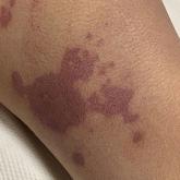
Purpura fulminans is an acute, life-threatening condition characterized by intravascular thrombosis and hemorrhagic necrosis of the skin.
Purpura fulminans is an acute, life-threatening condition characterized by intravascular thrombosis and hemorrhagic necrosis of the skin.