Editor’s Note: This commentary, written by members of the Cleveland Clinic Breast Cancer Screening Task Force, was not independently peer-reviewed.
In November 2009, the US Preventive Services Task Force (USPSTF) announced its new guidelines for breast cancer screening—and created an instant controversy by suggesting that fewer screening tests be done.1
The November 2009 update recommended that most women wait until age 50 to get their first screening mammogram instead of getting it at age 40, that they get a mammogram every other year instead of every year, and that physicians not teach their patients breast self-examination anymore. However, on December 4, 2009, the USPSTF members voted to modify the recommendation for women under age 50, stating that the decision to start screening mammography every 2 years should be individualized, taking into account the patient’s preferences after being apprised of the possible benefits and harms.2
Various professional and advocacy groups have reacted differently to the new guidelines, and as a result, women are unsure about the optimal screening for breast cancer.
NEW GUIDELINES ARE BASED ON TWO STUDIES
The USPSTF commissioned two studies, which it used to formulate the new recommendations.3,4 Its goal was to evaluate the current evidence for the efficacy of several screening tests and schedules in reducing breast cancer mortality rates.
An updated systematic review
Nelson et al3 performed a systematic review of studies of the benefit and harm of screening with mammography, clinical breast examination, and breast self-examination.
Screening mammography continued to demonstrate a reduction in deaths due to breast cancer. The risk reduction ranged from 14% to 32% in women age 50 to 69. Similarly, it was calculated to reduce the incidence of deaths due to breast cancer by 15% in women age 39 to 49. However, this younger age group has a relatively low incidence of breast cancer, and therefore, according to this analysis, 556 women need to undergo one round of screening to detect one case of invasive breast cancer, and 1,904 women need to be offered screening (over several rounds, which varied by trial) to prevent one breast cancer death.3
Most of the harm of screening in the 39-to-49-year age category was due to false-positive results, which were more common in this group than in older women. The authors calculated that after every round of screening mammography, about 84 of every 1,000 women in the younger age category need additional imaging and about 9 need a biopsy. The issue of overdiagnosis (detection of cancers that would have never been a problem in one’s lifetime) was not specifically addressed for this age category, and in different studies, estimates of overdiagnosis rates for all age groups varied widely, from less than 1% to 30%.
Beyond age 70, the authors reported the data insufficient for evaluating the benefit and harm of screening mammography.
Breast self-examination was found to offer no benefit, based largely on two randomized studies, one in St. Petersburg, Russia,5 and the other in Shanghai, China,6 both places where screening mammography was not routinely offered. These studies and one observational study in the United States7 failed to show a reduction in breast cancer mortality rates with breast self-examination.
Clinical breast examination (ie, by a health care provider) lacked sufficient data to draw conclusions.
A study based on statistical models of mammography
Mandelblatt et al4 used statistical modeling to estimate the effect of mammographic screening at various ages and at different intervals.
The authors used six statistical models previously shown to give similar qualitative estimates of the contribution of screening in reducing breast cancer mortality rates. They estimated the number of mammograms required relative to the number of cancers detected, the number of breast cancer deaths prevented, and the harms (false-positive mammograms, unnecessary biopsies, and overdiagnosis) incurred with 20 different screening strategies, ie, screening with different starting and stopping ages and at intervals of either 1 or 2 years.
They estimated that screening every other year would achieve most of the benefit of screening every year, with less harm. Looking at the different strategies and models, on average, biennial screening would, by their calculations, achieve about 81% of the mortality reduction achieved with annual screening. Compared with screening women ages 50 to 69 only, extending screening to women age 40 to 49 would reduce the cancer mortality rate by 3% more, while extending it up to age 79 would reduce it by another 7% to 8%.
In terms of harm, the models predicted more false-positive studies if screening were started before age 50 and if it were done annually rather than biennially. They also predicted that more unnecessary biopsies would be done with annual screening than with screening every 2 years. The models suggested that the risk for overdiagnosis was higher in older age groups because of higher rates of death from causes other than breast cancer, and that the overdiagnosis rate was also somewhat higher with annual than with 2-year screening.
WHAT WOULD LESS SCREENING MEAN?
Our practice has been to initiate annual screening with mammography at age 40 and to continue as long as the patient’s life expectancy is at least 10 years.
According to the models used by Mandelblatt et al,4 screening 1,000 women every year, starting at age 40 and continuing until age 84, would result in 177 to 227 life-years gained compared with no screening. In contrast, screening only women age 50 to 74 and only every other year (as advocated in the new guidelines) would entail about one-third the number of mammograms but would result in fewer life-years gained per 1,000 women screened: between 96 and 128. If we take the mean of the estimates from the six models, adherence to the new screening guidelines would be estimated to result in about 79 fewer life-years gained for every 1,000 women screened. On the other hand, each woman screened would need to undergo about 25 fewer screening mammograms in her lifetime.4
KEY POINTS ABOUT BREAST CANCER SCREENING
Together, these studies demonstrate several points about breast cancer screening.
Importantly, randomized controlled trials and model analyses continue to show that screening mammography reduces the breast cancer mortality rate.
The studies and models also reinforce the concept that those at greatest risk get the most benefit from screening. Because the incidence of breast cancer rises with age, the probability of a true-positive result is higher in women over age 50 than it is in younger women, and, therefore, the screening test performs better.
On the other hand, women at high risk of dying of other causes, such as those over age 75, achieve less benefit from screening, as some of the cancers detected in this manner may not contribute to their death even if they are not detected early.
Screening is therefore best targeted at people who are healthy but who are at sufficient risk for the disease in question to justify the screening.
CLEVELAND CLINIC’S POSITION
In December 2009, the Cleveland Clinic Breast Cancer Screening Task Force, a multidisciplinary panel of breast cancer experts, breast radiologists, and primary care providers, convened to review the literature and set forth institutional recommendations for breast cancer screening for healthy women. The authors of this paper are members of this task force. Our consensus recommendations:
We continue to recommend annual mammography for most healthy women over age 40.
Screening every other year is an option for older postmenopausal women, as they are likely to achieve most of the benefit of annual screening with this schedule.
We agree with the USPSTF finding that there are insufficient data to provide evidenced-based recommendations regarding the benefits and harms of clinical breast examination. However, breast examination was done as part of the screening in many of the randomized trials of mammography and cannot easily be separated from mammography. Therefore, we believe that careful examination of the breasts remains an important consideration in the general physical examination.
The USPSTF recommendation not to teach breast self-examination was based on studies that probably do not apply to the US population. Therefore, we continue to recommend that women be familiar with their breasts and report any changes to their physicians.
How we reached these conclusions
The task force discussions focused heavily on at what age mammography should be started and how often it should be done. In addition to an in-depth review of the studies on which the USPSTF recommendations were based, we considered a review posted on the Society of Breast Imaging (SBI) Web site.8
A key point from the SBI’s review is that although breast cancer occurs less often in women under age 50, approximately 1 in 69 women are diagnosed with invasive cancer when in their 40s. Some—probably a minority— have a family history of breast cancer and thus warrant earlier screening on that basis.
Breast cancer is, therefore, an important public health concern for women ages 40 to 49. While mammography is an imperfect test, it has a demonstrated ability to find cancers at an earlier stage in this age group. The SBI statement also summarized data suggesting that the 40-to-49-year age group would experience significantly fewer lives saved by screening if the mammography interval were increased from once a year to every other year (ie, by approximately one-half—from 36% of deaths prevented with annual screening to 18% deaths prevented with screening every other year).
Screening every other year is also expected to result in fewer lives saved in women ages 50 to 69 (39% of deaths prevented by biennial screening instead of 44% to 46% with annual screening). However, this proportion of deaths prevented with more frequent (ie, annual) screening is smaller than in the younger age group. Breast cancers that arise before menopause are considered biologically more aggressive, so the longer the interval between screening tests, the lower the likelihood of detecting some of these potentially more lethal cancers.
We believe, for several reasons, that the randomized trials may have underestimated the benefit of mammography. The trials included in the USPSTF studies did not use modern mammographic techniques such as digital mammography. Some of the trials used single-view mammography, which may be less sensitive. Also, the rate of compliance with screening in these randomized trials was only about 70%, which would lead to an underestimation of the number of lives saved with mammography screening. Yet in spite of these limitations, the data continue to show a reduction in breast cancer deaths in all age categories studied.
Other issues the task force considered
Harms of screening are acceptable. We agree that the need for additional imaging or possibly breast biopsy is an acceptable consequence of screening for most women, especially when weighed against the potential benefit of improving survival. Nelson et al3 briefly discussed the risk of inducing other cancers through radiation exposure, and any such risk appears to be low enough that it is overshadowed by the reduction in the breast cancer mortality rate achieved from screening.
The USPSTF studies did not address the issue of cost, which is another potential harm of screening. However, screening mammography is relatively inexpensive compared with other potentially life-saving screening tests.
Our position differs from that of the American College of Physicians (ACP), which has endorsed the USPSTF recommendation for reduced breast cancer screening. The USPSTF has been a leading group in providing practice recommendations based on high-level evidence predominantly from randomized controlled clinical trials, and its recommendations have been consistently followed by the ACP and many of its members, including Cleveland Clinic physicians. It is, therefore, not without considerable discussion that we have come to our consensus.
Evidence for less screening was not compelling. One of our concerns about the new USPSTF recommendations is that the changes are based largely on a model analysis of the efficiency of different screening strategies rather than on randomized controlled trials comparing different strategies. We did not find this level of present evidence to be sufficiently compelling to make a change in our practice that may result in loss of lives from breast cancer.
Screening guidelines will continue to change over time as technology improves and new data are introduced. In the future, risk-assessment strategies such as incorporating genetic profiles may allow us to use factors more predictive than age to target our screening population.
While we continue to strive for better means of early detection and cancer prevention, the Cleveland Clinic task force is currently recommending yearly screening with mammography and breast examination for most women, starting at age 40.
References
US Preventive Services Task Force. Screening for breast cancer: U.S. Preventive Services Task Force recommendation statement. Ann Intern Med2009; 151:716–726.
US Preventive Services Task Force (USPSTF). Recommendation statement from USPSTF: screening for breast cancer. Medscape. http://www.medscape.com/viewarticle/714016. Accessed 12/28/2009.
Nelson HD, Tyne K, Naik A, et al. Screening for breast cancer: an update for the U.S. Preventive Services Task Force. Ann Intern Med2009; 151:727–737.
Mandelblatt JS, Cronin KA, Bailey S, et al. Effects of mammography screening under different screening schedules: model estimates of potential benefits and harms. Ann Intern Med2009; 151:738–747.
Semiglazov VF, Manikhas AG, Moiseenko VM, et al. Results of a prospective randomized investigation [Russia (St. Petersburg)/WHO] to evaluate the significance of self-examination for the early detection of breast cancer [in Russian]. Vopr Onkol2003; 49:434–441. Cited by Nelson et al (see reference 3, above).
Thomas DB, Gao DL, Ray RM, et al. Randomized trial of breast self-examination in Shanghai: final results. J Natl Cancer Inst2002; 94:1445–1457. Cited by Nelson et al (see reference 3, above).
Tu SP, Reisch LM, Taplin SH, Kreuter W, Elmore JG. Breast self-examination: self-reported frequency, quality, and associated outcomes. J Cancer Educ2006; 21:175–181. Cited by Nelson et al (see reference 3, above).
Halle C.F. Moore, MD Taussig Cancer Institute, Cleveland Clinic; Member, Cleveland Clinic Breast Cancer Screening Task Force
G. Thomas Budd, MD Taussig Cancer Institute, Cleveland Clinic; Member, Cleveland Clinic Breast Cancer Screening Task Force
Andrea Sikon, MD Department of Internal Medicine, Center for Specialized Women’s Health, Cleveland Clinic; Member, Cleveland Clinic Breast Cancer Screening Task Force
Alice Rim, MD Vice Chair, Imaging Institute; Section Head, Breast Imaging, Department of Diagnostic Radiology, Cleveland Clinic; Member, Cleveland Clinic Breast Cancer Screening Task Force
Melanie Chellman-Jeffers, MD Section of Breast Imaging, Department of Diagnostic Radiology, Imaging Institute, Cleveland Clinic; Member, Cleveland Clinic Breast Cancer Screening Task Force
Joseph Crowe, MD Chairman, Breast Services, Department of General Surgery, Taussig Cancer Institute, Cleveland Clinic; Member, Cleveland Clinic Breast Cancer Screening Task Force
Address: Halle C.F. Moore, MD, Taussig Cancer Institute, R35, Cleveland Clinic, 9500 Euclid Avenue, Cleveland, OH 44195; mooreh1@ccf.org
Author and Disclosure Information
Halle C.F. Moore, MD Taussig Cancer Institute, Cleveland Clinic; Member, Cleveland Clinic Breast Cancer Screening Task Force
G. Thomas Budd, MD Taussig Cancer Institute, Cleveland Clinic; Member, Cleveland Clinic Breast Cancer Screening Task Force
Andrea Sikon, MD Department of Internal Medicine, Center for Specialized Women’s Health, Cleveland Clinic; Member, Cleveland Clinic Breast Cancer Screening Task Force
Alice Rim, MD Vice Chair, Imaging Institute; Section Head, Breast Imaging, Department of Diagnostic Radiology, Cleveland Clinic; Member, Cleveland Clinic Breast Cancer Screening Task Force
Melanie Chellman-Jeffers, MD Section of Breast Imaging, Department of Diagnostic Radiology, Imaging Institute, Cleveland Clinic; Member, Cleveland Clinic Breast Cancer Screening Task Force
Joseph Crowe, MD Chairman, Breast Services, Department of General Surgery, Taussig Cancer Institute, Cleveland Clinic; Member, Cleveland Clinic Breast Cancer Screening Task Force
Address: Halle C.F. Moore, MD, Taussig Cancer Institute, R35, Cleveland Clinic, 9500 Euclid Avenue, Cleveland, OH 44195; mooreh1@ccf.org
Editor’s Note: This commentary, written by members of the Cleveland Clinic Breast Cancer Screening Task Force, was not independently peer-reviewed.
In November 2009, the US Preventive Services Task Force (USPSTF) announced its new guidelines for breast cancer screening—and created an instant controversy by suggesting that fewer screening tests be done.1
The November 2009 update recommended that most women wait until age 50 to get their first screening mammogram instead of getting it at age 40, that they get a mammogram every other year instead of every year, and that physicians not teach their patients breast self-examination anymore. However, on December 4, 2009, the USPSTF members voted to modify the recommendation for women under age 50, stating that the decision to start screening mammography every 2 years should be individualized, taking into account the patient’s preferences after being apprised of the possible benefits and harms.2
Various professional and advocacy groups have reacted differently to the new guidelines, and as a result, women are unsure about the optimal screening for breast cancer.
NEW GUIDELINES ARE BASED ON TWO STUDIES
The USPSTF commissioned two studies, which it used to formulate the new recommendations.3,4 Its goal was to evaluate the current evidence for the efficacy of several screening tests and schedules in reducing breast cancer mortality rates.
An updated systematic review
Nelson et al3 performed a systematic review of studies of the benefit and harm of screening with mammography, clinical breast examination, and breast self-examination.
Screening mammography continued to demonstrate a reduction in deaths due to breast cancer. The risk reduction ranged from 14% to 32% in women age 50 to 69. Similarly, it was calculated to reduce the incidence of deaths due to breast cancer by 15% in women age 39 to 49. However, this younger age group has a relatively low incidence of breast cancer, and therefore, according to this analysis, 556 women need to undergo one round of screening to detect one case of invasive breast cancer, and 1,904 women need to be offered screening (over several rounds, which varied by trial) to prevent one breast cancer death.3
Most of the harm of screening in the 39-to-49-year age category was due to false-positive results, which were more common in this group than in older women. The authors calculated that after every round of screening mammography, about 84 of every 1,000 women in the younger age category need additional imaging and about 9 need a biopsy. The issue of overdiagnosis (detection of cancers that would have never been a problem in one’s lifetime) was not specifically addressed for this age category, and in different studies, estimates of overdiagnosis rates for all age groups varied widely, from less than 1% to 30%.
Beyond age 70, the authors reported the data insufficient for evaluating the benefit and harm of screening mammography.
Breast self-examination was found to offer no benefit, based largely on two randomized studies, one in St. Petersburg, Russia,5 and the other in Shanghai, China,6 both places where screening mammography was not routinely offered. These studies and one observational study in the United States7 failed to show a reduction in breast cancer mortality rates with breast self-examination.
Clinical breast examination (ie, by a health care provider) lacked sufficient data to draw conclusions.
A study based on statistical models of mammography
Mandelblatt et al4 used statistical modeling to estimate the effect of mammographic screening at various ages and at different intervals.
The authors used six statistical models previously shown to give similar qualitative estimates of the contribution of screening in reducing breast cancer mortality rates. They estimated the number of mammograms required relative to the number of cancers detected, the number of breast cancer deaths prevented, and the harms (false-positive mammograms, unnecessary biopsies, and overdiagnosis) incurred with 20 different screening strategies, ie, screening with different starting and stopping ages and at intervals of either 1 or 2 years.
They estimated that screening every other year would achieve most of the benefit of screening every year, with less harm. Looking at the different strategies and models, on average, biennial screening would, by their calculations, achieve about 81% of the mortality reduction achieved with annual screening. Compared with screening women ages 50 to 69 only, extending screening to women age 40 to 49 would reduce the cancer mortality rate by 3% more, while extending it up to age 79 would reduce it by another 7% to 8%.
In terms of harm, the models predicted more false-positive studies if screening were started before age 50 and if it were done annually rather than biennially. They also predicted that more unnecessary biopsies would be done with annual screening than with screening every 2 years. The models suggested that the risk for overdiagnosis was higher in older age groups because of higher rates of death from causes other than breast cancer, and that the overdiagnosis rate was also somewhat higher with annual than with 2-year screening.
WHAT WOULD LESS SCREENING MEAN?
Our practice has been to initiate annual screening with mammography at age 40 and to continue as long as the patient’s life expectancy is at least 10 years.
According to the models used by Mandelblatt et al,4 screening 1,000 women every year, starting at age 40 and continuing until age 84, would result in 177 to 227 life-years gained compared with no screening. In contrast, screening only women age 50 to 74 and only every other year (as advocated in the new guidelines) would entail about one-third the number of mammograms but would result in fewer life-years gained per 1,000 women screened: between 96 and 128. If we take the mean of the estimates from the six models, adherence to the new screening guidelines would be estimated to result in about 79 fewer life-years gained for every 1,000 women screened. On the other hand, each woman screened would need to undergo about 25 fewer screening mammograms in her lifetime.4
KEY POINTS ABOUT BREAST CANCER SCREENING
Together, these studies demonstrate several points about breast cancer screening.
Importantly, randomized controlled trials and model analyses continue to show that screening mammography reduces the breast cancer mortality rate.
The studies and models also reinforce the concept that those at greatest risk get the most benefit from screening. Because the incidence of breast cancer rises with age, the probability of a true-positive result is higher in women over age 50 than it is in younger women, and, therefore, the screening test performs better.
On the other hand, women at high risk of dying of other causes, such as those over age 75, achieve less benefit from screening, as some of the cancers detected in this manner may not contribute to their death even if they are not detected early.
Screening is therefore best targeted at people who are healthy but who are at sufficient risk for the disease in question to justify the screening.
CLEVELAND CLINIC’S POSITION
In December 2009, the Cleveland Clinic Breast Cancer Screening Task Force, a multidisciplinary panel of breast cancer experts, breast radiologists, and primary care providers, convened to review the literature and set forth institutional recommendations for breast cancer screening for healthy women. The authors of this paper are members of this task force. Our consensus recommendations:
We continue to recommend annual mammography for most healthy women over age 40.
Screening every other year is an option for older postmenopausal women, as they are likely to achieve most of the benefit of annual screening with this schedule.
We agree with the USPSTF finding that there are insufficient data to provide evidenced-based recommendations regarding the benefits and harms of clinical breast examination. However, breast examination was done as part of the screening in many of the randomized trials of mammography and cannot easily be separated from mammography. Therefore, we believe that careful examination of the breasts remains an important consideration in the general physical examination.
The USPSTF recommendation not to teach breast self-examination was based on studies that probably do not apply to the US population. Therefore, we continue to recommend that women be familiar with their breasts and report any changes to their physicians.
How we reached these conclusions
The task force discussions focused heavily on at what age mammography should be started and how often it should be done. In addition to an in-depth review of the studies on which the USPSTF recommendations were based, we considered a review posted on the Society of Breast Imaging (SBI) Web site.8
A key point from the SBI’s review is that although breast cancer occurs less often in women under age 50, approximately 1 in 69 women are diagnosed with invasive cancer when in their 40s. Some—probably a minority— have a family history of breast cancer and thus warrant earlier screening on that basis.
Breast cancer is, therefore, an important public health concern for women ages 40 to 49. While mammography is an imperfect test, it has a demonstrated ability to find cancers at an earlier stage in this age group. The SBI statement also summarized data suggesting that the 40-to-49-year age group would experience significantly fewer lives saved by screening if the mammography interval were increased from once a year to every other year (ie, by approximately one-half—from 36% of deaths prevented with annual screening to 18% deaths prevented with screening every other year).
Screening every other year is also expected to result in fewer lives saved in women ages 50 to 69 (39% of deaths prevented by biennial screening instead of 44% to 46% with annual screening). However, this proportion of deaths prevented with more frequent (ie, annual) screening is smaller than in the younger age group. Breast cancers that arise before menopause are considered biologically more aggressive, so the longer the interval between screening tests, the lower the likelihood of detecting some of these potentially more lethal cancers.
We believe, for several reasons, that the randomized trials may have underestimated the benefit of mammography. The trials included in the USPSTF studies did not use modern mammographic techniques such as digital mammography. Some of the trials used single-view mammography, which may be less sensitive. Also, the rate of compliance with screening in these randomized trials was only about 70%, which would lead to an underestimation of the number of lives saved with mammography screening. Yet in spite of these limitations, the data continue to show a reduction in breast cancer deaths in all age categories studied.
Other issues the task force considered
Harms of screening are acceptable. We agree that the need for additional imaging or possibly breast biopsy is an acceptable consequence of screening for most women, especially when weighed against the potential benefit of improving survival. Nelson et al3 briefly discussed the risk of inducing other cancers through radiation exposure, and any such risk appears to be low enough that it is overshadowed by the reduction in the breast cancer mortality rate achieved from screening.
The USPSTF studies did not address the issue of cost, which is another potential harm of screening. However, screening mammography is relatively inexpensive compared with other potentially life-saving screening tests.
Our position differs from that of the American College of Physicians (ACP), which has endorsed the USPSTF recommendation for reduced breast cancer screening. The USPSTF has been a leading group in providing practice recommendations based on high-level evidence predominantly from randomized controlled clinical trials, and its recommendations have been consistently followed by the ACP and many of its members, including Cleveland Clinic physicians. It is, therefore, not without considerable discussion that we have come to our consensus.
Evidence for less screening was not compelling. One of our concerns about the new USPSTF recommendations is that the changes are based largely on a model analysis of the efficiency of different screening strategies rather than on randomized controlled trials comparing different strategies. We did not find this level of present evidence to be sufficiently compelling to make a change in our practice that may result in loss of lives from breast cancer.
Screening guidelines will continue to change over time as technology improves and new data are introduced. In the future, risk-assessment strategies such as incorporating genetic profiles may allow us to use factors more predictive than age to target our screening population.
While we continue to strive for better means of early detection and cancer prevention, the Cleveland Clinic task force is currently recommending yearly screening with mammography and breast examination for most women, starting at age 40.
References
US Preventive Services Task Force. Screening for breast cancer: U.S. Preventive Services Task Force recommendation statement. Ann Intern Med2009; 151:716–726.
US Preventive Services Task Force (USPSTF). Recommendation statement from USPSTF: screening for breast cancer. Medscape. http://www.medscape.com/viewarticle/714016. Accessed 12/28/2009.
Nelson HD, Tyne K, Naik A, et al. Screening for breast cancer: an update for the U.S. Preventive Services Task Force. Ann Intern Med2009; 151:727–737.
Mandelblatt JS, Cronin KA, Bailey S, et al. Effects of mammography screening under different screening schedules: model estimates of potential benefits and harms. Ann Intern Med2009; 151:738–747.
Semiglazov VF, Manikhas AG, Moiseenko VM, et al. Results of a prospective randomized investigation [Russia (St. Petersburg)/WHO] to evaluate the significance of self-examination for the early detection of breast cancer [in Russian]. Vopr Onkol2003; 49:434–441. Cited by Nelson et al (see reference 3, above).
Thomas DB, Gao DL, Ray RM, et al. Randomized trial of breast self-examination in Shanghai: final results. J Natl Cancer Inst2002; 94:1445–1457. Cited by Nelson et al (see reference 3, above).
Tu SP, Reisch LM, Taplin SH, Kreuter W, Elmore JG. Breast self-examination: self-reported frequency, quality, and associated outcomes. J Cancer Educ2006; 21:175–181. Cited by Nelson et al (see reference 3, above).
The authors thank Dr. Keller for his readership. (On a personal note, Dr. Chellman-Jeffers spent her childhood in the Los Angeles area near his practice.) Dr. Keller brings up several interesting points regarding breast MRI, a subject that fills entire subspecialty textbooks.
On the subject of a palpable abnormality, a breast MRI’s field of view encompasses the entire breast, and although breast MRI is quite sensitive, it is known to have a lower specificity than other modalities.1 This means that more findings—which may or may not be related to the actual palpable abnormality—will lead to more studies and more biopsies, with proportionately fewer cancers found.
As for regions of tissue coverage with mammography, the axillary tail is actually more consistently imaged with mammography and ultrasonography than with MRI because of the cardiac pulsation artifact in the plane of the heart, as well as the breastcoil image centering on the breast. MRI-guided biopsy in the axilla is also generally not possible. These limitations are typical for breast MRI equipment. The expense of breast MRI is indeed considerable, but cost is not the main reason for the preference of other modalities.
In contrast, targeted ultrasonography is exquisitely suited to specifically image a palpable abnormality. With its small field of view (4 cm and smaller), a very high percentage of palpable masses can be seen. It is also more personal and comfortable and can be patient-directed. You can ask the patient to physically show you what is being felt and then scan it in real time. Needle biopsy can then be performed, often during the same visit (at many facilities), using ultrasonography as a real-time guidance tool in any location within the breast, including the axilla.
In the algorithm implied by your question, the patient feels a lump and has a negative diagnostic mammogram (including specific, problem-directed views) and targeted ultrasonography, which, again, is more focused than MRI and more capable of imaging the axilla or areas out of the breast coil for this purpose. Then, based on clinical suspicion or patient anxiety, these two very good tests are disregarded or not believed. At this point, the patient should be seen by a specialist, usually a surgeon, for evaluation for palpation-guided biopsy. It is true that some palpable masses are not identified by mammography and ultrasonography. But it is also true that MRI does not find every cancer, and it can find many more lesions that are not cancerous and that have a dubious relation to the original area of concern. This can easily turn into the proverbial wild-goose chase. No matter the outcome of the MRI, the patient still needs to be seen by a surgeon.
Our two major indications for breast MRI are currently in the preoperative extent-of-disease workup for known breast cancer and as an additional screening examination for high-risk patients (lifetime risk greater than 20%–25% by BRCAPRO, Gail, or other model method per the 2007 American Cancer Society guidelines2). We always require a comparative review of mammography in the completed interpretation of breast MRI and, as such, do not consider MRI a viable (or statistically proven) substitute for screening mammography for patients with sensitive breasts. Breast MRI is in fact more physically challenging for most patients than mammography, because the patient needs to remain motionless in a prone position in an enclosed space for an extended period of time (our protocol is 17 minutes). Gadolinium contrast must also be given, which requires renal function laboratory tests and intravenous access. The study must also be scheduled in all premenopausal patients in the postmenstrual phase of her cycle (around days 7–14) to avoid diffuse hormonally related enhancement and to minimize false-positive results.
References
Orel S. Who should have breast magnetic resonance imaging evaluation?J Clin Oncol2008; 26:703–711.
Saslow D, Boetes C, Burke W, et al; American Cancer Society Breast Cancer Advisory Group. American Cancer Society guidelines for breast cancer screening with MRI as an adjunct to mammography, CA Cancer J Clin2007; 57:75–89. Erratum in: CA Cancer J Clin 2007; 57:185.
The authors thank Dr. Keller for his readership. (On a personal note, Dr. Chellman-Jeffers spent her childhood in the Los Angeles area near his practice.) Dr. Keller brings up several interesting points regarding breast MRI, a subject that fills entire subspecialty textbooks.
On the subject of a palpable abnormality, a breast MRI’s field of view encompasses the entire breast, and although breast MRI is quite sensitive, it is known to have a lower specificity than other modalities.1 This means that more findings—which may or may not be related to the actual palpable abnormality—will lead to more studies and more biopsies, with proportionately fewer cancers found.
As for regions of tissue coverage with mammography, the axillary tail is actually more consistently imaged with mammography and ultrasonography than with MRI because of the cardiac pulsation artifact in the plane of the heart, as well as the breastcoil image centering on the breast. MRI-guided biopsy in the axilla is also generally not possible. These limitations are typical for breast MRI equipment. The expense of breast MRI is indeed considerable, but cost is not the main reason for the preference of other modalities.
In contrast, targeted ultrasonography is exquisitely suited to specifically image a palpable abnormality. With its small field of view (4 cm and smaller), a very high percentage of palpable masses can be seen. It is also more personal and comfortable and can be patient-directed. You can ask the patient to physically show you what is being felt and then scan it in real time. Needle biopsy can then be performed, often during the same visit (at many facilities), using ultrasonography as a real-time guidance tool in any location within the breast, including the axilla.
In the algorithm implied by your question, the patient feels a lump and has a negative diagnostic mammogram (including specific, problem-directed views) and targeted ultrasonography, which, again, is more focused than MRI and more capable of imaging the axilla or areas out of the breast coil for this purpose. Then, based on clinical suspicion or patient anxiety, these two very good tests are disregarded or not believed. At this point, the patient should be seen by a specialist, usually a surgeon, for evaluation for palpation-guided biopsy. It is true that some palpable masses are not identified by mammography and ultrasonography. But it is also true that MRI does not find every cancer, and it can find many more lesions that are not cancerous and that have a dubious relation to the original area of concern. This can easily turn into the proverbial wild-goose chase. No matter the outcome of the MRI, the patient still needs to be seen by a surgeon.
Our two major indications for breast MRI are currently in the preoperative extent-of-disease workup for known breast cancer and as an additional screening examination for high-risk patients (lifetime risk greater than 20%–25% by BRCAPRO, Gail, or other model method per the 2007 American Cancer Society guidelines2). We always require a comparative review of mammography in the completed interpretation of breast MRI and, as such, do not consider MRI a viable (or statistically proven) substitute for screening mammography for patients with sensitive breasts. Breast MRI is in fact more physically challenging for most patients than mammography, because the patient needs to remain motionless in a prone position in an enclosed space for an extended period of time (our protocol is 17 minutes). Gadolinium contrast must also be given, which requires renal function laboratory tests and intravenous access. The study must also be scheduled in all premenopausal patients in the postmenstrual phase of her cycle (around days 7–14) to avoid diffuse hormonally related enhancement and to minimize false-positive results.
References
Orel S. Who should have breast magnetic resonance imaging evaluation?J Clin Oncol2008; 26:703–711.
Saslow D, Boetes C, Burke W, et al; American Cancer Society Breast Cancer Advisory Group. American Cancer Society guidelines for breast cancer screening with MRI as an adjunct to mammography, CA Cancer J Clin2007; 57:75–89. Erratum in: CA Cancer J Clin 2007; 57:185.
A 28-year-old woman comes in for her annual checkup. Her physician notices a palpable, painless, 1-cm, well-demarcated mass in the left breast at the 3 o’clock position 2 cm from the nipple, with no associated skin changes, nipple retraction, or discharge. The patient has no personal or family history of breast cancer.
Given the patient’s age, physical findings, and medical history, the clinician believes it unlikely that the patient has cancer. How should she proceed with the workup of this patient?
PHYSICAL FINDINGS OF A BREAST MASS ARE NOT EXCLUSIVE
Figure 1. A simple cyst in the left breast. All three mammographic views—craniocaudal (A), mediolateral oblique (B), and spot-compression (C)—show a round, well-circumscribed mass in the mid-breast. Ultrasonography (D) shows a round, well-circumscribed anechoic lesion with a sharply defined posterior wall and posterior acoustic enhancement.Breast cancer is the most common female malignancy and the second-leading cause of cancer deaths in the United States.1 The incidence is low in young women and increases with advancing age. Benign breast disease is common in young women and less common in postmenopausal women.2,3 However, the discovery of a breast mass, whether by the woman herself or by a clinician, is a common occurrence and distressing for any woman.
Benign lesions tend to have discrete, well-defined margins and are typically mobile. Malignant lesions may be firm, may have indistinct borders, and are often immobile.2 Although most breast masses found by palpation are benign, imaging is the critical next step in the workup to help determine if the mass is benign or malignant.
Benign palpable masses include:
Figure 2. Fibroadenoma. On mammography, the craniocaudal (A) and mediolateral oblique (B) views with a bright metallic marker (arrows) show a round, well-circumscribed mass in the upper outer quadrant of the left breast. Ultrasonography (C) shows an oval, well-circumscribed, mildly heterogeneous, hypoechoic mass that is wider than tall, indicating a benign mass.Cysts (Figure 1)
Fibroadenomas (Figure 2)
Prominent fat lobules
Lymph nodes
Oil cysts
Lipomas
Hamartomas (Figure 3)
Hematomas
Fat necrosis
Galactoceles.
Malignant palpable masses include:
Figure 3. Hamartoma. Craniocaudal (A) and mediolateral oblique (B) mammographic views of the left breast show an apparently encapsulated, heterogeneous mass that contains fat mixed with fibroglandular tissue.Invasive ductal and lobular carcinoma (Figure 4)
Ductal carcinoma in situ (which rarely presents as a palpable mass.)
HISTORY AND PHYSICAL EXAMINATION
To ensure that imaging provides the most useful information about a palpable breast lump, it is important to first do a careful history and physical examination. Important aspects of the history include family history, personal history of breast cancer, and any previous breast biopsies. The onset and duration of the palpable mass, changes in its size, the relationship of these changes to the menstrual cycle, and the presence or lack of tenderness are additional important elements of the history.
Figure 4. Infiltrating ductal carcinoma. Craniocaudal (A) and mediolateral oblique (B) mammographic views of the right breast show an irregular, mildly spiculated, high-density lesion in the posterior, medial breast. Ultrasonography (C) shows an irregularly shaped hypoechoic mass which is taller than wide (a profile tending to indicate malignancy) and has mild posterior acoustic shadowing.On examination, it is important to note the clock-face location, size, texture, tenderness, and mobility of the lump. Accompanying nipple discharge and skin erythema or retraction are also important to report. In addition to conveying the location of the mass to the radiologist, it is equally important that the patient know what features the physician feels. This way, if the clinical information from the ordering physician is not available at the time of the radiologic evaluation, the patient will be able to guide the radiologist to the region of concern.
IMAGING TECHNIQUES
Mammography and ultrasonography are the primary imaging studies for evaluating palpable breast masses. Typically, in women under age 30, ultrasonography is the first or the only test ordered to evaluate the abnormality.4 In women age 30 or older, diagnostic mammography is typically the first test ordered. If mammography indicates that the palpable mass is not benign, then ultrasonography is the next study to be done.3 Although a powerful tool, magnetic resonance imaging of the breast does not currently have a role in the workup of a palpable abnormality and should not be used as a decision-delaying tactic or in place of biopsy.
Screening or diagnostic mammography?
Mammography is used in both screening and diagnosis. Screening mammography consists of two standard views of each breast—craniocaudal and mediolateral oblique—and is appropriate for asymptomatic women.
Women age 30 or older who present with a palpable breast mass require diagnostic mammography, in which standard mammographic views are obtained, as well as additional views (eg, tangential or spot-compression views) to better define the area of clinical concern. In a tangential view, a metallic skin marker is placed on the skin overlying the site of the palpable abnormality.
On mammography, a suspicious palpable mass has an irregular shape with spiculated margins. A benign mass typically has a round shape with well-circumscribed margins. If the palpable abnormality is not mammographically benign (eg, if it does not look like a lymph node, lipoma, or degenerating fibroadenoma), then ultrasonography is performed.
Mammography is less sensitive in younger women (ie, under age 30) because their breast tissue tends to be dense and glandular, whereas the tissue becomes more “fat-replaced” with age.3
Ultrasonography plays a complementary role
Ultrasonography complements diagnostic mammography and can be used as a first imaging study to evaluate a palpable breast mass in a young woman (ie, under age 30) with dense breast tissue. Ultrasonography is helpful in distinguishing cystic lesions from solid masses. It helps the radiologist delineate the shape, borders, and acoustic properties of the mass. It is also performed when a palpable mass is mammographically occult. When a mass appears suspicious on either mammography or ultrasonography, ultrasonography can be used to guide biopsy.
A suspicious mass on ultrasonography classically appears “taller than wide” and has posterior acoustic shadowing. Microlobulations and a spiculated margin also raise concern for malignancy. A benign sonographic appearance of a palpable mass includes a “wider than tall” (ellipsoid) shape, with homogeneous echogenicity, and four or fewer gentle lobulations. A thin, echogenic capsule also suggests the mass is benign.
Core-needle biopsy with ultrasonographic guidance
Core-needle biopsy is performed with a large-diameter (14-gauge to 18-gauge) needle to obtain tissue cores for histologic analysis. It has gained popularity over fine-needle aspiration because it includes surrounding tissue architecture, thus providing a more definitive histologic diagnosis.
Pathologic information obtained from core-needle biopsy allows the radiologist and surgeon to counsel the patient and determine the best surgical management or follow-up imaging study. If a clinician performs fine-needle biopsy in the office, it should be preceded by an imaging workup of the palpable finding.
WHAT IS APPROPRIATE FOR OUR 28-YEAR-OLD PATIENT?
Because she is under age 30, ultrasonography is the initial study of choice to evaluate the mass. If a simple cyst is detected, she can be reassured that the lesion is benign, and no subsequent follow-up is required. If the lesion is a solid mass with benign features, mammography may be considered, the patient may be followed with short-interval imaging (every 6 months) depending on patient-specific factors such as family history, or the mass can be biopsied. If the lesion is a solid mass with suspicious or malignant features, mammography with spot-compression views should be performed, and the patient should undergo core-needle biopsy with ultrasonographic guidance.
In a patient age 30 or older, diagnostic mammography is the imaging study of first choice.3 If the mass is clearly benign on mammography, no additional imaging would be necessary. If mammography fails to image the mass or shows it to have benign features such as fat, then the patient can undergo ultrasonography for further evaluation and confirmation of the clinical and mammographic findings. If the mass appears suspicious or malignant on mammography, ultrasonography is the next step, as it can help characterize the lesion and be used to guide core-needle biopsy.
IF A PREGNANT WOMAN HAS A PALPABLE BREAST MASS
Most publications on breast cancer in pregnancy report a prevalence of 3 per 10,000 pregnancies, accounting for 3% of all breast cancers diagnosed.5 Therefore, imaging evaluation of a palpable mass should not be postponed.
Hormonal changes throughout pregnancy may increase the nodularity of breast tissue, raising the concern of palpable masses. Additionally, there is a higher prevalence of galactoceles and lactating adenomas in these patients. Because contrasting fatty breast tissue is lost during pregnancy and because of the need to minimize radiation exposure, ultrasonography is often the imaging test of first choice. If mammography is required, the radiation dose is very low and the patient’s abdomen and pelvis can be shielded.6 In this situation, the patient can be reassured that the imaging test is not jeopardizing her fetus.
WHAT WORKUP IS REQUIRED IN MEN?
Breast cancer in men is rare, accounting for less than 0.5% of all cases.7 Most often, a palpable breast mass in a man presents as unilateral gynecomastia. Gynecomastia occurs in a bimodal age distribution (in the 2nd and 7th decades) and has a variety of hormonal and drug-related causes. Despite the low prevalence of breast cancer in men, the combination of mammography and ultrasonography is recommended for evaluation at all ages.
References
Klein S. Evaluation of palpable breast masses. Am Fam Physician2005; 71:1731–1738.
Pruthi S. Detection and evaluation of a palpable breast mass. Mayo Clin Proc2001; 76:641–648.
Harvey JA. Sonography of palpable breast masses. Semin Ultrasound CT MR2006; 27:284–297.
Mehta TS. Current uses of ultrasound in the evaluation of the breast. Radiol Clin North Am2003; 41:841–856.
Gallenberg MM, Lopines CL. Breast cancer and pregnancy. Semin Oncol1989; 16:369–376.
Barnavon Y, Wallack MK. Management of the pregnant patient with carcinoma of the breast. Surg Gynecol Obstet1990; 171:347–352.
Cardenosa G. The Core Curriculum: Breast Imaging. Philadelphia: Lippincott Williams and Wilkins, 2003;304.
Lauren Stein, MD Imaging Institute, Cleveland Clinic
Melanie Chellman-Jeffers, MD Center for Specialized Women’s Health and Section of Breast Imaging, Department of Diagnostic Radiology, Imaging Institute, Cleveland Clinic
Address: Melanie Chellman-Jeffers, MD, Imaging Institute, Section of Breast Imaging, A10, Cleveland Clinic, 9500 Euclid Avenue, Cleveland, OH 44195; e-mail chellmm@ccf.org
Author and Disclosure Information
Lauren Stein, MD Imaging Institute, Cleveland Clinic
Melanie Chellman-Jeffers, MD Center for Specialized Women’s Health and Section of Breast Imaging, Department of Diagnostic Radiology, Imaging Institute, Cleveland Clinic
Address: Melanie Chellman-Jeffers, MD, Imaging Institute, Section of Breast Imaging, A10, Cleveland Clinic, 9500 Euclid Avenue, Cleveland, OH 44195; e-mail chellmm@ccf.org
A 28-year-old woman comes in for her annual checkup. Her physician notices a palpable, painless, 1-cm, well-demarcated mass in the left breast at the 3 o’clock position 2 cm from the nipple, with no associated skin changes, nipple retraction, or discharge. The patient has no personal or family history of breast cancer.
Given the patient’s age, physical findings, and medical history, the clinician believes it unlikely that the patient has cancer. How should she proceed with the workup of this patient?
PHYSICAL FINDINGS OF A BREAST MASS ARE NOT EXCLUSIVE
Figure 1. A simple cyst in the left breast. All three mammographic views—craniocaudal (A), mediolateral oblique (B), and spot-compression (C)—show a round, well-circumscribed mass in the mid-breast. Ultrasonography (D) shows a round, well-circumscribed anechoic lesion with a sharply defined posterior wall and posterior acoustic enhancement.Breast cancer is the most common female malignancy and the second-leading cause of cancer deaths in the United States.1 The incidence is low in young women and increases with advancing age. Benign breast disease is common in young women and less common in postmenopausal women.2,3 However, the discovery of a breast mass, whether by the woman herself or by a clinician, is a common occurrence and distressing for any woman.
Benign lesions tend to have discrete, well-defined margins and are typically mobile. Malignant lesions may be firm, may have indistinct borders, and are often immobile.2 Although most breast masses found by palpation are benign, imaging is the critical next step in the workup to help determine if the mass is benign or malignant.
Benign palpable masses include:
Figure 2. Fibroadenoma. On mammography, the craniocaudal (A) and mediolateral oblique (B) views with a bright metallic marker (arrows) show a round, well-circumscribed mass in the upper outer quadrant of the left breast. Ultrasonography (C) shows an oval, well-circumscribed, mildly heterogeneous, hypoechoic mass that is wider than tall, indicating a benign mass.Cysts (Figure 1)
Fibroadenomas (Figure 2)
Prominent fat lobules
Lymph nodes
Oil cysts
Lipomas
Hamartomas (Figure 3)
Hematomas
Fat necrosis
Galactoceles.
Malignant palpable masses include:
Figure 3. Hamartoma. Craniocaudal (A) and mediolateral oblique (B) mammographic views of the left breast show an apparently encapsulated, heterogeneous mass that contains fat mixed with fibroglandular tissue.Invasive ductal and lobular carcinoma (Figure 4)
Ductal carcinoma in situ (which rarely presents as a palpable mass.)
HISTORY AND PHYSICAL EXAMINATION
To ensure that imaging provides the most useful information about a palpable breast lump, it is important to first do a careful history and physical examination. Important aspects of the history include family history, personal history of breast cancer, and any previous breast biopsies. The onset and duration of the palpable mass, changes in its size, the relationship of these changes to the menstrual cycle, and the presence or lack of tenderness are additional important elements of the history.
Figure 4. Infiltrating ductal carcinoma. Craniocaudal (A) and mediolateral oblique (B) mammographic views of the right breast show an irregular, mildly spiculated, high-density lesion in the posterior, medial breast. Ultrasonography (C) shows an irregularly shaped hypoechoic mass which is taller than wide (a profile tending to indicate malignancy) and has mild posterior acoustic shadowing.On examination, it is important to note the clock-face location, size, texture, tenderness, and mobility of the lump. Accompanying nipple discharge and skin erythema or retraction are also important to report. In addition to conveying the location of the mass to the radiologist, it is equally important that the patient know what features the physician feels. This way, if the clinical information from the ordering physician is not available at the time of the radiologic evaluation, the patient will be able to guide the radiologist to the region of concern.
IMAGING TECHNIQUES
Mammography and ultrasonography are the primary imaging studies for evaluating palpable breast masses. Typically, in women under age 30, ultrasonography is the first or the only test ordered to evaluate the abnormality.4 In women age 30 or older, diagnostic mammography is typically the first test ordered. If mammography indicates that the palpable mass is not benign, then ultrasonography is the next study to be done.3 Although a powerful tool, magnetic resonance imaging of the breast does not currently have a role in the workup of a palpable abnormality and should not be used as a decision-delaying tactic or in place of biopsy.
Screening or diagnostic mammography?
Mammography is used in both screening and diagnosis. Screening mammography consists of two standard views of each breast—craniocaudal and mediolateral oblique—and is appropriate for asymptomatic women.
Women age 30 or older who present with a palpable breast mass require diagnostic mammography, in which standard mammographic views are obtained, as well as additional views (eg, tangential or spot-compression views) to better define the area of clinical concern. In a tangential view, a metallic skin marker is placed on the skin overlying the site of the palpable abnormality.
On mammography, a suspicious palpable mass has an irregular shape with spiculated margins. A benign mass typically has a round shape with well-circumscribed margins. If the palpable abnormality is not mammographically benign (eg, if it does not look like a lymph node, lipoma, or degenerating fibroadenoma), then ultrasonography is performed.
Mammography is less sensitive in younger women (ie, under age 30) because their breast tissue tends to be dense and glandular, whereas the tissue becomes more “fat-replaced” with age.3
Ultrasonography plays a complementary role
Ultrasonography complements diagnostic mammography and can be used as a first imaging study to evaluate a palpable breast mass in a young woman (ie, under age 30) with dense breast tissue. Ultrasonography is helpful in distinguishing cystic lesions from solid masses. It helps the radiologist delineate the shape, borders, and acoustic properties of the mass. It is also performed when a palpable mass is mammographically occult. When a mass appears suspicious on either mammography or ultrasonography, ultrasonography can be used to guide biopsy.
A suspicious mass on ultrasonography classically appears “taller than wide” and has posterior acoustic shadowing. Microlobulations and a spiculated margin also raise concern for malignancy. A benign sonographic appearance of a palpable mass includes a “wider than tall” (ellipsoid) shape, with homogeneous echogenicity, and four or fewer gentle lobulations. A thin, echogenic capsule also suggests the mass is benign.
Core-needle biopsy with ultrasonographic guidance
Core-needle biopsy is performed with a large-diameter (14-gauge to 18-gauge) needle to obtain tissue cores for histologic analysis. It has gained popularity over fine-needle aspiration because it includes surrounding tissue architecture, thus providing a more definitive histologic diagnosis.
Pathologic information obtained from core-needle biopsy allows the radiologist and surgeon to counsel the patient and determine the best surgical management or follow-up imaging study. If a clinician performs fine-needle biopsy in the office, it should be preceded by an imaging workup of the palpable finding.
WHAT IS APPROPRIATE FOR OUR 28-YEAR-OLD PATIENT?
Because she is under age 30, ultrasonography is the initial study of choice to evaluate the mass. If a simple cyst is detected, she can be reassured that the lesion is benign, and no subsequent follow-up is required. If the lesion is a solid mass with benign features, mammography may be considered, the patient may be followed with short-interval imaging (every 6 months) depending on patient-specific factors such as family history, or the mass can be biopsied. If the lesion is a solid mass with suspicious or malignant features, mammography with spot-compression views should be performed, and the patient should undergo core-needle biopsy with ultrasonographic guidance.
In a patient age 30 or older, diagnostic mammography is the imaging study of first choice.3 If the mass is clearly benign on mammography, no additional imaging would be necessary. If mammography fails to image the mass or shows it to have benign features such as fat, then the patient can undergo ultrasonography for further evaluation and confirmation of the clinical and mammographic findings. If the mass appears suspicious or malignant on mammography, ultrasonography is the next step, as it can help characterize the lesion and be used to guide core-needle biopsy.
IF A PREGNANT WOMAN HAS A PALPABLE BREAST MASS
Most publications on breast cancer in pregnancy report a prevalence of 3 per 10,000 pregnancies, accounting for 3% of all breast cancers diagnosed.5 Therefore, imaging evaluation of a palpable mass should not be postponed.
Hormonal changes throughout pregnancy may increase the nodularity of breast tissue, raising the concern of palpable masses. Additionally, there is a higher prevalence of galactoceles and lactating adenomas in these patients. Because contrasting fatty breast tissue is lost during pregnancy and because of the need to minimize radiation exposure, ultrasonography is often the imaging test of first choice. If mammography is required, the radiation dose is very low and the patient’s abdomen and pelvis can be shielded.6 In this situation, the patient can be reassured that the imaging test is not jeopardizing her fetus.
WHAT WORKUP IS REQUIRED IN MEN?
Breast cancer in men is rare, accounting for less than 0.5% of all cases.7 Most often, a palpable breast mass in a man presents as unilateral gynecomastia. Gynecomastia occurs in a bimodal age distribution (in the 2nd and 7th decades) and has a variety of hormonal and drug-related causes. Despite the low prevalence of breast cancer in men, the combination of mammography and ultrasonography is recommended for evaluation at all ages.
References
Klein S. Evaluation of palpable breast masses. Am Fam Physician2005; 71:1731–1738.
Pruthi S. Detection and evaluation of a palpable breast mass. Mayo Clin Proc2001; 76:641–648.
Harvey JA. Sonography of palpable breast masses. Semin Ultrasound CT MR2006; 27:284–297.
Mehta TS. Current uses of ultrasound in the evaluation of the breast. Radiol Clin North Am2003; 41:841–856.
Gallenberg MM, Lopines CL. Breast cancer and pregnancy. Semin Oncol1989; 16:369–376.
Barnavon Y, Wallack MK. Management of the pregnant patient with carcinoma of the breast. Surg Gynecol Obstet1990; 171:347–352.
Cardenosa G. The Core Curriculum: Breast Imaging. Philadelphia: Lippincott Williams and Wilkins, 2003;304.
Typically, in women under age 30, ultrasonography is the first or the only test ordered to evaluate the abnormality. In women age 30 or older, diagnostic mammography is typically the first test ordered.
On mammography, a suspicious palpable mass has an irregular shape with spiculated margins. A benign mass typically has a round shape with well-circumscribed margins.
When mammography is required during pregnancy, the patient can be reassured that it will not jeopardize her fetus because the radiation dose is very low and the abdomen and pelvis can be shielded.
Early detection of breast cancer is vital to reducing the morbidity and mortality associated with this disease. After a brief overview of breast cancer epidemiology and risk assessment, this article describes screening and diagnostic imaging techniques as they are currently practiced to promote early breast cancer detection. We conclude with a review of image-guided needle biopsy techniques and a recommended approach to breast cancer screening in the general population.
EPIDEMIOLOGY OF BREAST CANCER: DAUNTING BUT SLOWLY IMPROVING
After nonmelanoma skin cancers, breast cancer is the most common form of cancer in women today, accounting for more than 1 in 4 cancers diagnosed in US women.1 If the current incidence of breast cancer remains constant, US females born today have an average risk of 12.7% of being diagnosed with breast cancer during their lifetime (ie, 1-in-8 lifetime risk), based on National Cancer Institute statistics.2,3 The American Cancer Society estimated that 178,480 new cases of invasive breast cancer and 62,030 new cases of in situ breast cancer would be diagnosed in the United States in 2007, and that 40,460 US women would die from breast cancer that year.1 Only lung cancer accounts for more cancer deaths in women.
The role of race and ethnicity
Breast cancer risk varies by race and ethnicity in the United States. After age 40 years, white women have a higher incidence of breast cancer compared with African American women; conversely, before age 40, African American women have a higher incidence compared with white women. African American women are more likely than their white counterparts to die from their breast cancer at any age. Incidence and death rates from breast cancer are lower among Asian American, American Indian, and Hispanic women compared with both white and African American women.1
Recent hopeful trends
Despite the daunting incidence numbers reviewed above, recent years have seen encouraging trends in US breast cancer epidemiology.
For invasive breast cancer, the growth in incidence rates slowed during the 1990s, and rates actually declined by 3.5% per year during the period 2001–2004.1 These changes are likely attributable to multiple factors, including variations in rates of mammography screening and decreased use of hormone replacement therapy after the 2002 publication of results from the Women’s Health Initiative trial. Still, the trend is encouraging.
Incidence rates of in situ breast cancer rose rapidly during the 1980s and 1990s, largely due to increased diagnosis by mammography, but have plateaued since 2000 among women aged 50 years or older while continuing to rise modestly in younger women.1
Meanwhile, the overall death rate from breast cancer in women declined by 2.2% annually from 1990 to 2004.1
RISK FACTORS AND RISK MODELING
Risk factors for breast cancer have been well described and include the following:
Age ( ≥ 65 years vs < 65 years, although risk increases across all ages up to 80 years)
Family history of breast cancer
Late age at first full-term pregnancy (> 30 years)
Never having a full-term pregnancy
Early menarche and/or late menopause
Certain genetic mutations for breast cancer (eg, in the BRCA1, BRCA2, ATM, and CHEK2 genes)
Certain breast disorders, such as atypical hyperplasia or lobular carcinoma in situ
High breast tissue density
High bone density (postmenopausal)
High-dose radiation to the chest.
The above risk factors are, in general, fixed. More elusive risk factors, in that they are variable and modifiable, include obesity, use of exogenous hormones (recent and long-term hormone replacement therapy; recent oral contraceptive use), alcohol use, tobacco use, diet, and a low level of physical activity. Breast implants are not a risk factor for breast cancer, though their presence does obscure breast tissue on imaging, limiting the detectability of a tumor when it does develop (see “Screening the Surgically Altered Breast” below).
Women with a genetic predisposition to breast cancer merit special consideration. Hereditary breast cancers account for about 5% to 10% of breast cancer cases, and the BRCA1 and BRCA2 mutations are responsible for 80% to 90% of these cases, while other gene mutations (noted above) or genetic syndromes account for the rest. Clinical options for managing women with a genetic predisposition include surveillance, chemoprevention, and prophylactic surgery.4 Detailed discussion of the management of these women is beyond the scope of this article, but readers are referred to www.nccn.org/professionals/physician_gls/PDF/ genetics_screening.pdf for practice guidelines from the National Comprehensive Cancer Network.5
Tools for risk assessment
Several tools are available to predict a woman’s risk of developing breast cancer. Probably the most widely used is the Gail model,6 which was published in 1989 and forms the statistical basis for the National Cancer Institute’s Breast Cancer Risk Assessment Tool, which is available for downloading at www.cancer.gov/bcrisktool.7 The model uses a woman’s personal medical and reproductive histories and her family history of breast cancer to predict her 5-year and lifetime risk of developing invasive breast cancer. Factors included in the risk calculation are age, race, number of first-degree relatives with a history of breast cancer, age at first live birth (or nulliparity), age at menarche, number of breast biopsies, and presence or absence of a history of atypical hyperplasia. The relative risk for each of these factors is multiplied to generate a composite risk. The Gail model has been validated for white women but has been shown to underestimate breast cancer risk in African American women; it remains to be validated for Hispanic women, Asian women, and other subgroups of women.7
The commonly taught “triple test” for palpable breast lesions is another risk model that incorporates clinical findings. It consists of a physical examination, mammography, and fine-needle aspiration8 (in the “modified triple test,” ultrasonography replaces mammography9). When all three elements of the test are concordant (either all benign or all malignant), the triple test has been reported to have 100% diagnostic accuracy.8,9
A WORD ABOUT BREAST EXAMINATION
Breast self-examination
The role of breast self-examination is controversial in the literature. There are currently no data to support the contention that it increases detection of breast cancer. As a result, the American Cancer Society no longer recommends that all women perform monthly breast self-exams, although it advises that all women be told about the potential benefits and limitations of breast self-examination (Table 1).10 Research suggests that structured breast self-examination is less important than self-awareness. Women who detect breast tumors themselves typically find them outside of a structured examination, such as when bathing or getting dressed.1
Clinical breast examination
As noted in Table 1, regular clinical breast examinations are recommended by the American Cancer Society for asymptomatic women at average risk for breast cancer, with the recommended frequency depending on the woman’s age.10 The US Preventive Services Task Force takes the stance that there is insufficient evidence to recommend for or against breast cancer screening with clinical breast examination alone.11 While it is unclear precisely what contribution clinical breast exams make to the detection of breast cancer, they certainly provide clinicians an opportunity to raise awareness about breast cancer and educate patients about breast symptoms, risk factors, and new detection technologies.10
SCREENING MAMMOGRAPHY
Screening mammography is the single most effective method of early breast cancer detection,1 and the American Cancer Society recommends that women at average risk for breast cancer have annual screening mammograms beginning at age 40 years (Table 1).10
The evidence base
The primary evidence supporting the recommendation for screening mammography comes from eight randomized trials that studied the effectiveness of screening mammography for cancer detection in Sweden,12,13 the United States,14 Canada,15,16 and the United Kingdom.17 Overall, breast cancers detected by screening mammography are smaller and have a more favorable history and tumor biology than those detected clinically without the use of imaging. A pooled analysis of the most recent data from all randomized trials of screening mammography in women aged 39 to 74 years showed a 24% reduction in mortality (95% CI, 18% to 30%) in women undergoing screening mammography, although not all individual trials showed a statistically significant mortality reduction.10
The screening procedure at a glance
A screening mammogram, as distinguished from a diagnostic mammogram (Table 2), consists of two standard radiographic views of each breast (mediolateral oblique and craniocaudal).18 The woman being screened is advised to wear no powders or deodorants and should be asymptomatic. Women with symptoms (eg, breast lump, focal tenderness, nipple discharge) should be scheduled for a diagnostic mammogram (Table 2), not a screening mammogram.
The mammography technologist obtains the standard radiographs of each breast, and computer-assisted detection software can be applied to the mammogram films to aid in the identification of abnormalities as a computer-generated second opinion. Although computer-assisted detection is not currently standard of care, it is available at most institutions. The films are read later by a radiologist who will interpret them according to the American College of Radiology’s standard system of describing mammogram findings, called the Breast Imaging Reporting and Data System (BI-RADS). In this system, results are assigned a category rating on a scale from 0 to 6 (Table 3). This standardization allows physicians to use consistent language, ensures better follow-up of suspicious findings, and reduces interobserver variability.
Analog vs digital
Figure 1. Normal dense digital mammogram images showing right and left mediolateral oblique views (panels A and B, respectively) and right and left craniocaudal views (panels C and D, respectively).Breast radiographs can be obtained by the traditional film-screen (analog) method or obtained digitally (Figure 1).
Digital mammograms are radiographs that are acquired digitally and allow digital enhancement to aid in interpretation. When receiving a digital mammogram, the woman being screened still undergoes compression and positioning as for a conventional film-screen mammogram, and the images are still produced with x-rays. However, digitization allows manipulation of the images as they are being interpreted, enabling the radiologist to focus on areas of interest or to “window” and “level” the image, similar to adjusting the tint and contrast on a television set.
Research trials comparing digital and film mammography, such as the Digital Mammographic Imaging Screening Trial (DMIST),19 have found digital mammography to be especially helpful in women with extremely dense breasts, who have an elevated risk for breast cancer. For women with fatty breasts the differences between the types of mammogram are less significant.
The type of mammogram a woman receives generally depends on the equipment available at the site she visits. Digital mammography units currently cost approximately 3 times as much as corresponding film-screen units, yet digital mammograms command reimbursement rates only about 1.6 times higher than those for film mammograms (Table 4). A hard copy of the digitized image can be printed, although the hope is that eventually fewer mammogram images will be printed and space-saving electronic storage will supplant storage of printed films.
For further detail on digital mammography, readers are referred to the recent review by D’Orsi and Newell.20
SCREENING THE SURGICALLY ALTERED BREAST
Following surgical cancer treatment or reconstructive surgery, screening of remaining breast tissue for cancer is still performed and is just as essential to patient care as presurgery screening. The first line of defense for any patient with a surgically altered breast is mammography.
When a patient has had breast reconstruction following mastectomy, it is presumed that very little breast tissue remains. There is no standard of care for screening the nonbreast tissue introduced by the reconstructive procedure. Nonetheless, at our institution we perform a single mediolateral oblique projection on any flap-reconstructed breast in light of rare anecdotal accounts of cancer found in and around the reconstructed breast. When problem-solving is needed to evaluate a new palpable abnormality, special angled views (tangential) and directed ultrasonography can be used. We do not routinely perform screening mammography on mastectomy patients who have had reconstruction with implants, but we can investigate areas of clinical concern (eg, due to palpable masses) with directed ultrasonography.21
The cosmetically altered breast presents its own issues in cancer detection. Both silicone-gel and saline implants obscure breast tissue that could contain cancer. For this reason, special implant-displaced views are performed that allow visualization of a larger portion of breast tissue beyond that allowed by standard mammograms. Therefore, an asymptomatic patient with implants who presents for screening mammography will have eight mammography views obtained instead of the routine four views.22
Patients who have had breast reduction, excisional biopsy, or prior breast conservation surgery (lumpectomy and radiation) are screened in a routine manner with mammography.23 Patients who have had prior surgical procedures often have architectural distortion at the surgical site, which is generally stable over time. Any prior surgical procedure can predispose the patient to the development of fat necrosis, which is a benign entity but can mimic cancer in its early phases through the development of calcifications and, occasionally, a new palpable lump. We most commonly confront this issue in the period 2 to 4 years after the operation.24 Occasionally the findings are such that a biopsy is needed to determine whether fat necrosis is the cause. In this population, magnetic resonance imaging (MRI) can also be used as an adjunctive tool, and can sometimes clarify the presence of fat necrosis and other postoperative findings, such as seroma, hematoma, or inflammation. In other instances, only a biopsy can determine what a particular finding represents.
DIAGNOSTIC MAMMOGRAPHY
Any mammography performed for a problem-solving purpose is considered diagnostic mammography (Table 2); the exam is tailored to the patient’s individual issue.25 Diagnostic mammography requires the presence of a qualified radiologist at the time of imaging. The goal is to come to a final conclusion about the mammographic or clinical finding at the time of the patient’s visit. Special views are usually performed that include, but are not limited to, spot-compression or spot-magnification views, depending on the finding.26 The patient is then given a same-day written account of the results at the conclusion of the study.
Examples of problems that may prompt diagnostic mammography include patient-reported palpable findings, screening mammography findings that are recalled for further investigation, or physician-detected findings. Often, ultrasonography is also used at the same visit and its results are integrated with the mammography findings to arrive at the final impression.
BREAST ULTRASONOGRAPHY AND BREAST MRI
Ultrasonography and MRI are two very useful adjunctive tools for breast lesion detection and analysis. At this time, however, neither is a replacement for screening mammography as a primary screening modality; rather, each is used in a complementary fashion for lesion analysis and biopsy guidance.10,27
Ultrasonography: Best for further study of areas of interest
Ultrasonography uses high-frequency sound waves to create a picture using a probe directed to an area of interest in the breast. The optimal probe for breast imaging is one typically operating in a frequency of 12 to 18 MHz and 4 cm in scanning width.
Because ultrasonography provides views of only a small area of breast tissue at a time, it is operator and patient dependent. It is best used when a known area of interest needs further evaluation, such as when a patient reports a palpable abnormality or when a mass is detected on mammography.
Ultrasonography uses no ionizing radiation, so it is especially helpful in young or pregnant women who present with a palpable abnormality. It is also useful for patients who have recently undergone a surgical procedure. As ultrasonography is currently used, no compression is needed and it can be performed easily in patients with limited mobility. Needle biopsies are most easily performed using ultrasonographic guidance.
MRI: An emerging adjunct under study in high-risk patients
Breast MRI is an emerging modality under active research that shows promise for adjunctive breast imaging. It is commonly being used as a tool for local staging in women with newly diagnosed breast cancer.28,29 Current research is focused on its suitability as a screening modality, in conjunction with mammography, in high-risk populations based on family history and other factors addressed in the Gail model6 and similar risk models.
The limitations of breast MRI include its high cost, unsuitability for some patients (eg, the obese [due to table weight constraints], patients with pacemakers, patients with renal failure), the potential for unnecessary biopsies due to decreased specificity, lack of portability, and the length of time required for imaging.
Figure 2. Contrast-enhanced breast MRI in the axial projection demonstrating multiple malignant masses in the left breast.Breast MRI is a four-dimensional study, with time as the fourth dimension (in addition to length, width, and depth). The patient receives an intravenous line and is given gadolinium for contrast enhancement. Imaging time depends on the protocol used and is specific to the imaging center, but it typically involves approximately 20 minutes of motionless scan time for the patient.30 Lesions are detectable by their level of vascularity, and diagnostic images are dependent on adequate contrast enhancement (Figure 2). Several software packages are commercially available that perform post-processing of breast MRI data. Although cancer on MRI has a characteristic enhancement curve, there is much overlap with benign entities; as a result, morphologic characteristics of the lesion—such as size, shape, and borders—are paramount.31
When a lesion is initially detected with MRI, an attempt is usually made to identify it with ultrasonography as well, owing to the ease of ultrasonography-guided biopsy.32 It is important, however, for an imaging center that performs breast MRI to be able to perform biopsies using MRI guidance since not all lesions are identifiable by other modalities.33 Breast MRI studies are not easily portable between imaging facilities since a typical study contains a thousand or more images that are best viewed on a site-specific workstation monitor.
HISTOLOGIC CONFIRMATION
Once an abnormality is detected on imaging, a confirmatory histologic diagnosis is needed before embarking on medical or surgical treatments. Image-guided biopsy plays a critical role in this regard. In our breast imaging section, we perform ultrasonography-guided core needle biopsy and aspiration, stereotactic needle biopsy, and MRI-guided needle biopsy, as well as wire localizations on the day of surgery. All procedures performed are considered minimally invasive and are suitable for a vast majority of patients for whom they are recommended.34
Ultrasonography-guided procedures
Figure 3. “Pre-fire” (top) and “post-fire” (bottom) ultrasonographic views of an 18-gauge percutaneous needle core biopsy of a suspicious breast mass.Ultrasonography-guided core needle biopsy is the modality of choice for most patients when a suspicious abnormality is visible on ultrasonography.35 Generally, the patient is placed in an angled supine position, with her arm elevated for optimal lesion accessibility. Following administration of a local anesthetic, a small nick is made in the skin and a specialized 14- or 18gauge spring-loaded core biopsy needle is inserted during real-time imaging with the ultrasonographic probe (Figure 3). Several samples are obtained, and the pathologic diagnosis is generally available within a few working days. Breast core biopsy needles are also commercially available as handheld vacuum-assisted devices, which can sample larger amounts of tissue in a short time but are more expensive and often accompanied by a noisy vacuum device.
Ultrasonography-guided fine-needle aspiration is an additional option for patients when core biopsy cannot be performed because the lesion is located adjacent to sensitive structures, such as implants or the pectoralis muscle. Fine-needle aspiration is also used to evaluate complicated breast cysts and, occasionally, lymph nodes. Drawbacks of fine-needle aspiration (relative to larger core needle biopsy) are that it is limited to cytologic, not histologic, examination and that it yields a higher false-negative rate.
Stereotactically guided procedures
Stereotactic core biopsy is performed when lesions—usually calcifications, but sometimes masses—are visible only on mammography.36,37 “Stereotactic” refers to the means by which the target is localized, ie, with a “stereo pair” of digital mammogram pictures with a small field of view. The patient is placed in a prone position with the breast of interest placed through a hole at the undersurface of the table in a light compression. The biopsy unit is attached to a dedicated computer that calculates coordinates. The needle is then brought to the coordinate position for sampling to take place.
The biopsy needle used for this procedure is vacuum-assisted, which means the needle is placed only one time, and samples in the vicinity of the target are vacuumed into a reservoir for retrieval. If the target is calcifications, a specimen radiograph is routinely performed to verify adequate sample acquisition before the patient leaves the biopsy table.38 When the original target is no longer visible, a titanium marker clip is often placed. This facilitates localization of the biopsied area should surgery be needed.
Stereotactic biopsy has several limitations that ultrasonography-guided biopsy does not. The patient must be cooperative and mobile enough to get on the table and hold a prone position for the duration of the procedure (about 45 minutes). If the patient is taking warfarin or has a bleeding diathesis, preprocedure steps such as clinical evaluation to check the international normalized ratio and prothrombin time, or even stopping the warfarin temporarily, may be needed to minimize bleeding during the procedure, as a 9- or 12-gauge needle is used. Stereotactic biopsy is also limited by lesion position. A far posterior lesion may not be accessible if it does not reach through the hole in the table. Also, there is a limit to the compressed thinness of breast tissue that can be biopsied. Finally, most tables used for stereotactic biopsy have a functioning weight limit of 300 pounds.
Open surgical biopsy
A final option is open surgical biopsy, which is used when the more minimally invasive techniques are equivocal, discordant, or impossible due to the limitations noted above, or when atypical cells are found.
HOW SHOULD WE SCREEN OUR PATIENTS?
The various screening options for breast cancer are listed in Table 4, along with their market approval status and Medicare reimbursement levels.
For women at average risk for breast cancer, the American Cancer Society recommends an annual mammogram and clinical breast examination by a physician beginning at age 40 (Table 1).10
For women at high risk for developing breast cancer (> 20% to 25% lifetime risk, based on the Gail model6 or similar risk models), breast MRI should be considered as an adjunct to annual screening mammography (Table 5).39 Evidence is currently insufficient, however, to support the adjunctive use of breast MRI for women with other risk factors (Table 5), although studies are ongoing.39
In conclusion, the process of finding breast cancer includes regular screening with mammography and clinical breast examination (plus MRI in high-risk women) and the diagnostic modalities of ultrasonography, MRI, and diagnostic mammography. Our ultimate goal is to find cancer at the earliest time possible by all means necessary for the individual patient.
Gail MH, Brinton LA, Byar DP, et al. Projecting individualized probabilities of developing breast cancer for white females who are being examined annually. J Natl Cancer Inst 1989; 81:1879–1886.
Breast cancer risk assessment tool. An interactive tool for measuring the risk of invasive breast cancer. National Cancer Institute Web site. http://www.cancer.gov/bcrisktool/. Accessed January 21, 2008.
Vetto J, Pommier R, Schmidt W, et al. Use of the “triple test” for palpable breast lesions yields high diagnostic accuracy and cost savings. Am J Surg 1995; 169:519–522.
Vetto JT, Pommier RF, Schmidt WA, Eppich H, Alexander PW. Diagnosis of palpable breast lesions in younger women by the modified triple test is accurate and cost-effective. Arch Surg 1996; 131:967–974.
Smith RA, Saslow D, Sawyer KA, et al. American Cancer Society guidelines for breast cancer screening: update 2003. CA Cancer J Clin 2003; 53:141–169.
U.S. Preventive Services Task Force. Screening for breast cancer: recommendations and rationale. Ann Intern Med 2002; 137:344–346.
Nyström L, Andersson I, Bjurstam N, et al. Long-term effects of mammography screening: updated overview of the Swedish randomized trials. Lancet 2002; 359:909–919.
Tabar L, Fagerberg G, Chen HH, et al. Efficacy of breast cancer screening by age: new results from the Swedish Two-County Trial. Cancer 1995; 75:2507–2517.
Shapiro S, Venet W, Strax P, Venet L. Periodic Screening for Breast Cancer: The Health Insurance Plan Project and Its Sequelae, 1963-1986. Baltimore, MD: Johns Hopkins University Press; 1988.
Miller AB, To T, Baines CJ, Wall C. Canadian National Breast Screening Study-2: 13-year results of a randomized trial in women aged 50-59 years. J Natl Cancer Inst 2000; 92:1490–1499.
Miller AB, To T, Baines CJ, Wall C. The Canadian National Breast Screening Study-1: breast cancer mortality after 11 to 16 years of follow-up: a randomized screening trial of mammography in women age 40 to 49 years. Ann Intern Med 2002; 137:305–312.
Alexander FE, Anderson TJ, Brown HK, et al. 14 years of follow-up from the Edinburgh randomized trial of breast-cancer screening. Lancet 1999; 353:1903–1908.
Eklund GW, Cardenosa G. The art of mammographic positioning. Radiol Clin North Am 1992; 30:21–53.
Pisano E, Gatsonis C, Hendrick E, et al. Diagnostic performance of digital versus film mammography for breast cancer screening. N Engl J Med 2005; 353:1773–1783.
D’Orsi CJ, Newell MS. Digital mammography: clinical implementation and clinical trials. Semin Roentgenol 2007; 42:236–242.
Fajardo LL, Roberts CC, Hunt KR. Mammographic surveillance of breast cancer patients: should the masectomy site be imaged? AJR Am J Roentgenol 1993; 161:953–955.
Eklund GW, Busby RC, Miller SH, Job JS. Improved imaging of the augmented breast. AJR Am J Roentgenol 1988; 151:469–473.
Mendelson EB. Evaluation of the postoperative breast. Radiol Clin North Am 1992; 30:107–138.
Philpotts LE, Lee CH, Haffty BG, et al. Mammographic findings of recurrent breast cancer after l
Sickles EA. Practical solutions to common mammographic problems: tailoring the examination. AJR Am J Roentgenol 1988; 151:31–39.
Jackson VP. The role of US in breast imaging. Radiology 1990; 177:305–311.
Lehman CD, Gatsonis C, Kuhl CK, et al. MRI evaluation of the contralateral breast in women with recently diagnosed breast cancer. N Engl J Med 2007; 356:1295–1303.
Liberman L. Breast MR imaging in assessing extent of disease. Magn Reson Imaging Clin N Am 2006; 14:339–349.
Kuhl C. The current status of breast MR imaging. Part I. Choice of technique, image interpretation, diagnostic accuracy, and transfer to clinical practice. Radiology 2007; 244:356–378.
Flickinger FW, Allison JD, Sherry RM, Wright JC. Differentiation of benign from malignant breast masses by time-intensity evaluation of contrast-enhanced MRI. Magn Reson Imaging 1993; 11:617–620.
Chellman-Jeffers MR, Listinsky J, Dinunzio A, Lieber M, Rim A. Utility of second look ultrasound as an adjunct to contrast-enhanced MRI of the breast. Paper presented at: American Roentgen Ray Society Meeting; May 4, 2006; Vancouver, BC. Abstract 269.
Orel SG, Schnall MD, Newman RW, Powell CM, Torosian MH, Rosato EF. MR imaging-guided localization and biopsy of breast lesions: initial experience. Radiology 1994; 193:97–102.
Liberman L. Percutaneous imaging-guided core breast biopsy: state of the art at the millennium. AJR Am J Roentgenol 2000; 174:1191–1199.
Fornage BD, Coan JD, David CL. Ultrasound-guided needle biopsy of the breast and other interventional procedures. Radiol Clin North Am 1992; 30:167–185.
Parker SH, Lovin JD, Jobe WE, et al. Nonpalpable breast lesions: stereotactic automated large-core biopsies. Radiology 1991; 180:403–407.
Parker SH, Burbank F, Jackman RJ, et al. Percutaneous large-core breast biopsy: a multi-institutional study. Radiology 1994; 193:3 59–364.
Liberman L, Evans WP III, Dershaw DD, et al. Radiography of microcalcifications in stereotaxic mammary core biopsy specimens. Radiology 1994; 190:223–225.
Saslow D, Boetes C, Burke W, et al. American Cancer Society guidelines for breast screening with MRI as an adjunct to mammography. CA Cancer J Clin 2007; 57:75–89.
Early detection of breast cancer is vital to reducing the morbidity and mortality associated with this disease. After a brief overview of breast cancer epidemiology and risk assessment, this article describes screening and diagnostic imaging techniques as they are currently practiced to promote early breast cancer detection. We conclude with a review of image-guided needle biopsy techniques and a recommended approach to breast cancer screening in the general population.
EPIDEMIOLOGY OF BREAST CANCER: DAUNTING BUT SLOWLY IMPROVING
After nonmelanoma skin cancers, breast cancer is the most common form of cancer in women today, accounting for more than 1 in 4 cancers diagnosed in US women.1 If the current incidence of breast cancer remains constant, US females born today have an average risk of 12.7% of being diagnosed with breast cancer during their lifetime (ie, 1-in-8 lifetime risk), based on National Cancer Institute statistics.2,3 The American Cancer Society estimated that 178,480 new cases of invasive breast cancer and 62,030 new cases of in situ breast cancer would be diagnosed in the United States in 2007, and that 40,460 US women would die from breast cancer that year.1 Only lung cancer accounts for more cancer deaths in women.
The role of race and ethnicity
Breast cancer risk varies by race and ethnicity in the United States. After age 40 years, white women have a higher incidence of breast cancer compared with African American women; conversely, before age 40, African American women have a higher incidence compared with white women. African American women are more likely than their white counterparts to die from their breast cancer at any age. Incidence and death rates from breast cancer are lower among Asian American, American Indian, and Hispanic women compared with both white and African American women.1
Recent hopeful trends
Despite the daunting incidence numbers reviewed above, recent years have seen encouraging trends in US breast cancer epidemiology.
For invasive breast cancer, the growth in incidence rates slowed during the 1990s, and rates actually declined by 3.5% per year during the period 2001–2004.1 These changes are likely attributable to multiple factors, including variations in rates of mammography screening and decreased use of hormone replacement therapy after the 2002 publication of results from the Women’s Health Initiative trial. Still, the trend is encouraging.
Incidence rates of in situ breast cancer rose rapidly during the 1980s and 1990s, largely due to increased diagnosis by mammography, but have plateaued since 2000 among women aged 50 years or older while continuing to rise modestly in younger women.1
Meanwhile, the overall death rate from breast cancer in women declined by 2.2% annually from 1990 to 2004.1
RISK FACTORS AND RISK MODELING
Risk factors for breast cancer have been well described and include the following:
Age ( ≥ 65 years vs < 65 years, although risk increases across all ages up to 80 years)
Family history of breast cancer
Late age at first full-term pregnancy (> 30 years)
Never having a full-term pregnancy
Early menarche and/or late menopause
Certain genetic mutations for breast cancer (eg, in the BRCA1, BRCA2, ATM, and CHEK2 genes)
Certain breast disorders, such as atypical hyperplasia or lobular carcinoma in situ
High breast tissue density
High bone density (postmenopausal)
High-dose radiation to the chest.
The above risk factors are, in general, fixed. More elusive risk factors, in that they are variable and modifiable, include obesity, use of exogenous hormones (recent and long-term hormone replacement therapy; recent oral contraceptive use), alcohol use, tobacco use, diet, and a low level of physical activity. Breast implants are not a risk factor for breast cancer, though their presence does obscure breast tissue on imaging, limiting the detectability of a tumor when it does develop (see “Screening the Surgically Altered Breast” below).
Women with a genetic predisposition to breast cancer merit special consideration. Hereditary breast cancers account for about 5% to 10% of breast cancer cases, and the BRCA1 and BRCA2 mutations are responsible for 80% to 90% of these cases, while other gene mutations (noted above) or genetic syndromes account for the rest. Clinical options for managing women with a genetic predisposition include surveillance, chemoprevention, and prophylactic surgery.4 Detailed discussion of the management of these women is beyond the scope of this article, but readers are referred to www.nccn.org/professionals/physician_gls/PDF/ genetics_screening.pdf for practice guidelines from the National Comprehensive Cancer Network.5
Tools for risk assessment
Several tools are available to predict a woman’s risk of developing breast cancer. Probably the most widely used is the Gail model,6 which was published in 1989 and forms the statistical basis for the National Cancer Institute’s Breast Cancer Risk Assessment Tool, which is available for downloading at www.cancer.gov/bcrisktool.7 The model uses a woman’s personal medical and reproductive histories and her family history of breast cancer to predict her 5-year and lifetime risk of developing invasive breast cancer. Factors included in the risk calculation are age, race, number of first-degree relatives with a history of breast cancer, age at first live birth (or nulliparity), age at menarche, number of breast biopsies, and presence or absence of a history of atypical hyperplasia. The relative risk for each of these factors is multiplied to generate a composite risk. The Gail model has been validated for white women but has been shown to underestimate breast cancer risk in African American women; it remains to be validated for Hispanic women, Asian women, and other subgroups of women.7
The commonly taught “triple test” for palpable breast lesions is another risk model that incorporates clinical findings. It consists of a physical examination, mammography, and fine-needle aspiration8 (in the “modified triple test,” ultrasonography replaces mammography9). When all three elements of the test are concordant (either all benign or all malignant), the triple test has been reported to have 100% diagnostic accuracy.8,9
A WORD ABOUT BREAST EXAMINATION
Breast self-examination
The role of breast self-examination is controversial in the literature. There are currently no data to support the contention that it increases detection of breast cancer. As a result, the American Cancer Society no longer recommends that all women perform monthly breast self-exams, although it advises that all women be told about the potential benefits and limitations of breast self-examination (Table 1).10 Research suggests that structured breast self-examination is less important than self-awareness. Women who detect breast tumors themselves typically find them outside of a structured examination, such as when bathing or getting dressed.1
Clinical breast examination
As noted in Table 1, regular clinical breast examinations are recommended by the American Cancer Society for asymptomatic women at average risk for breast cancer, with the recommended frequency depending on the woman’s age.10 The US Preventive Services Task Force takes the stance that there is insufficient evidence to recommend for or against breast cancer screening with clinical breast examination alone.11 While it is unclear precisely what contribution clinical breast exams make to the detection of breast cancer, they certainly provide clinicians an opportunity to raise awareness about breast cancer and educate patients about breast symptoms, risk factors, and new detection technologies.10
SCREENING MAMMOGRAPHY
Screening mammography is the single most effective method of early breast cancer detection,1 and the American Cancer Society recommends that women at average risk for breast cancer have annual screening mammograms beginning at age 40 years (Table 1).10
The evidence base
The primary evidence supporting the recommendation for screening mammography comes from eight randomized trials that studied the effectiveness of screening mammography for cancer detection in Sweden,12,13 the United States,14 Canada,15,16 and the United Kingdom.17 Overall, breast cancers detected by screening mammography are smaller and have a more favorable history and tumor biology than those detected clinically without the use of imaging. A pooled analysis of the most recent data from all randomized trials of screening mammography in women aged 39 to 74 years showed a 24% reduction in mortality (95% CI, 18% to 30%) in women undergoing screening mammography, although not all individual trials showed a statistically significant mortality reduction.10
The screening procedure at a glance
A screening mammogram, as distinguished from a diagnostic mammogram (Table 2), consists of two standard radiographic views of each breast (mediolateral oblique and craniocaudal).18 The woman being screened is advised to wear no powders or deodorants and should be asymptomatic. Women with symptoms (eg, breast lump, focal tenderness, nipple discharge) should be scheduled for a diagnostic mammogram (Table 2), not a screening mammogram.
The mammography technologist obtains the standard radiographs of each breast, and computer-assisted detection software can be applied to the mammogram films to aid in the identification of abnormalities as a computer-generated second opinion. Although computer-assisted detection is not currently standard of care, it is available at most institutions. The films are read later by a radiologist who will interpret them according to the American College of Radiology’s standard system of describing mammogram findings, called the Breast Imaging Reporting and Data System (BI-RADS). In this system, results are assigned a category rating on a scale from 0 to 6 (Table 3). This standardization allows physicians to use consistent language, ensures better follow-up of suspicious findings, and reduces interobserver variability.
Analog vs digital
Figure 1. Normal dense digital mammogram images showing right and left mediolateral oblique views (panels A and B, respectively) and right and left craniocaudal views (panels C and D, respectively).Breast radiographs can be obtained by the traditional film-screen (analog) method or obtained digitally (Figure 1).
Digital mammograms are radiographs that are acquired digitally and allow digital enhancement to aid in interpretation. When receiving a digital mammogram, the woman being screened still undergoes compression and positioning as for a conventional film-screen mammogram, and the images are still produced with x-rays. However, digitization allows manipulation of the images as they are being interpreted, enabling the radiologist to focus on areas of interest or to “window” and “level” the image, similar to adjusting the tint and contrast on a television set.
Research trials comparing digital and film mammography, such as the Digital Mammographic Imaging Screening Trial (DMIST),19 have found digital mammography to be especially helpful in women with extremely dense breasts, who have an elevated risk for breast cancer. For women with fatty breasts the differences between the types of mammogram are less significant.
The type of mammogram a woman receives generally depends on the equipment available at the site she visits. Digital mammography units currently cost approximately 3 times as much as corresponding film-screen units, yet digital mammograms command reimbursement rates only about 1.6 times higher than those for film mammograms (Table 4). A hard copy of the digitized image can be printed, although the hope is that eventually fewer mammogram images will be printed and space-saving electronic storage will supplant storage of printed films.
For further detail on digital mammography, readers are referred to the recent review by D’Orsi and Newell.20
SCREENING THE SURGICALLY ALTERED BREAST
Following surgical cancer treatment or reconstructive surgery, screening of remaining breast tissue for cancer is still performed and is just as essential to patient care as presurgery screening. The first line of defense for any patient with a surgically altered breast is mammography.
When a patient has had breast reconstruction following mastectomy, it is presumed that very little breast tissue remains. There is no standard of care for screening the nonbreast tissue introduced by the reconstructive procedure. Nonetheless, at our institution we perform a single mediolateral oblique projection on any flap-reconstructed breast in light of rare anecdotal accounts of cancer found in and around the reconstructed breast. When problem-solving is needed to evaluate a new palpable abnormality, special angled views (tangential) and directed ultrasonography can be used. We do not routinely perform screening mammography on mastectomy patients who have had reconstruction with implants, but we can investigate areas of clinical concern (eg, due to palpable masses) with directed ultrasonography.21
The cosmetically altered breast presents its own issues in cancer detection. Both silicone-gel and saline implants obscure breast tissue that could contain cancer. For this reason, special implant-displaced views are performed that allow visualization of a larger portion of breast tissue beyond that allowed by standard mammograms. Therefore, an asymptomatic patient with implants who presents for screening mammography will have eight mammography views obtained instead of the routine four views.22
Patients who have had breast reduction, excisional biopsy, or prior breast conservation surgery (lumpectomy and radiation) are screened in a routine manner with mammography.23 Patients who have had prior surgical procedures often have architectural distortion at the surgical site, which is generally stable over time. Any prior surgical procedure can predispose the patient to the development of fat necrosis, which is a benign entity but can mimic cancer in its early phases through the development of calcifications and, occasionally, a new palpable lump. We most commonly confront this issue in the period 2 to 4 years after the operation.24 Occasionally the findings are such that a biopsy is needed to determine whether fat necrosis is the cause. In this population, magnetic resonance imaging (MRI) can also be used as an adjunctive tool, and can sometimes clarify the presence of fat necrosis and other postoperative findings, such as seroma, hematoma, or inflammation. In other instances, only a biopsy can determine what a particular finding represents.
DIAGNOSTIC MAMMOGRAPHY
Any mammography performed for a problem-solving purpose is considered diagnostic mammography (Table 2); the exam is tailored to the patient’s individual issue.25 Diagnostic mammography requires the presence of a qualified radiologist at the time of imaging. The goal is to come to a final conclusion about the mammographic or clinical finding at the time of the patient’s visit. Special views are usually performed that include, but are not limited to, spot-compression or spot-magnification views, depending on the finding.26 The patient is then given a same-day written account of the results at the conclusion of the study.
Examples of problems that may prompt diagnostic mammography include patient-reported palpable findings, screening mammography findings that are recalled for further investigation, or physician-detected findings. Often, ultrasonography is also used at the same visit and its results are integrated with the mammography findings to arrive at the final impression.
BREAST ULTRASONOGRAPHY AND BREAST MRI
Ultrasonography and MRI are two very useful adjunctive tools for breast lesion detection and analysis. At this time, however, neither is a replacement for screening mammography as a primary screening modality; rather, each is used in a complementary fashion for lesion analysis and biopsy guidance.10,27
Ultrasonography: Best for further study of areas of interest
Ultrasonography uses high-frequency sound waves to create a picture using a probe directed to an area of interest in the breast. The optimal probe for breast imaging is one typically operating in a frequency of 12 to 18 MHz and 4 cm in scanning width.
Because ultrasonography provides views of only a small area of breast tissue at a time, it is operator and patient dependent. It is best used when a known area of interest needs further evaluation, such as when a patient reports a palpable abnormality or when a mass is detected on mammography.
Ultrasonography uses no ionizing radiation, so it is especially helpful in young or pregnant women who present with a palpable abnormality. It is also useful for patients who have recently undergone a surgical procedure. As ultrasonography is currently used, no compression is needed and it can be performed easily in patients with limited mobility. Needle biopsies are most easily performed using ultrasonographic guidance.
MRI: An emerging adjunct under study in high-risk patients
Breast MRI is an emerging modality under active research that shows promise for adjunctive breast imaging. It is commonly being used as a tool for local staging in women with newly diagnosed breast cancer.28,29 Current research is focused on its suitability as a screening modality, in conjunction with mammography, in high-risk populations based on family history and other factors addressed in the Gail model6 and similar risk models.
The limitations of breast MRI include its high cost, unsuitability for some patients (eg, the obese [due to table weight constraints], patients with pacemakers, patients with renal failure), the potential for unnecessary biopsies due to decreased specificity, lack of portability, and the length of time required for imaging.
Figure 2. Contrast-enhanced breast MRI in the axial projection demonstrating multiple malignant masses in the left breast.Breast MRI is a four-dimensional study, with time as the fourth dimension (in addition to length, width, and depth). The patient receives an intravenous line and is given gadolinium for contrast enhancement. Imaging time depends on the protocol used and is specific to the imaging center, but it typically involves approximately 20 minutes of motionless scan time for the patient.30 Lesions are detectable by their level of vascularity, and diagnostic images are dependent on adequate contrast enhancement (Figure 2). Several software packages are commercially available that perform post-processing of breast MRI data. Although cancer on MRI has a characteristic enhancement curve, there is much overlap with benign entities; as a result, morphologic characteristics of the lesion—such as size, shape, and borders—are paramount.31
When a lesion is initially detected with MRI, an attempt is usually made to identify it with ultrasonography as well, owing to the ease of ultrasonography-guided biopsy.32 It is important, however, for an imaging center that performs breast MRI to be able to perform biopsies using MRI guidance since not all lesions are identifiable by other modalities.33 Breast MRI studies are not easily portable between imaging facilities since a typical study contains a thousand or more images that are best viewed on a site-specific workstation monitor.
HISTOLOGIC CONFIRMATION
Once an abnormality is detected on imaging, a confirmatory histologic diagnosis is needed before embarking on medical or surgical treatments. Image-guided biopsy plays a critical role in this regard. In our breast imaging section, we perform ultrasonography-guided core needle biopsy and aspiration, stereotactic needle biopsy, and MRI-guided needle biopsy, as well as wire localizations on the day of surgery. All procedures performed are considered minimally invasive and are suitable for a vast majority of patients for whom they are recommended.34
Ultrasonography-guided procedures
Figure 3. “Pre-fire” (top) and “post-fire” (bottom) ultrasonographic views of an 18-gauge percutaneous needle core biopsy of a suspicious breast mass.Ultrasonography-guided core needle biopsy is the modality of choice for most patients when a suspicious abnormality is visible on ultrasonography.35 Generally, the patient is placed in an angled supine position, with her arm elevated for optimal lesion accessibility. Following administration of a local anesthetic, a small nick is made in the skin and a specialized 14- or 18gauge spring-loaded core biopsy needle is inserted during real-time imaging with the ultrasonographic probe (Figure 3). Several samples are obtained, and the pathologic diagnosis is generally available within a few working days. Breast core biopsy needles are also commercially available as handheld vacuum-assisted devices, which can sample larger amounts of tissue in a short time but are more expensive and often accompanied by a noisy vacuum device.
Ultrasonography-guided fine-needle aspiration is an additional option for patients when core biopsy cannot be performed because the lesion is located adjacent to sensitive structures, such as implants or the pectoralis muscle. Fine-needle aspiration is also used to evaluate complicated breast cysts and, occasionally, lymph nodes. Drawbacks of fine-needle aspiration (relative to larger core needle biopsy) are that it is limited to cytologic, not histologic, examination and that it yields a higher false-negative rate.
Stereotactically guided procedures
Stereotactic core biopsy is performed when lesions—usually calcifications, but sometimes masses—are visible only on mammography.36,37 “Stereotactic” refers to the means by which the target is localized, ie, with a “stereo pair” of digital mammogram pictures with a small field of view. The patient is placed in a prone position with the breast of interest placed through a hole at the undersurface of the table in a light compression. The biopsy unit is attached to a dedicated computer that calculates coordinates. The needle is then brought to the coordinate position for sampling to take place.
The biopsy needle used for this procedure is vacuum-assisted, which means the needle is placed only one time, and samples in the vicinity of the target are vacuumed into a reservoir for retrieval. If the target is calcifications, a specimen radiograph is routinely performed to verify adequate sample acquisition before the patient leaves the biopsy table.38 When the original target is no longer visible, a titanium marker clip is often placed. This facilitates localization of the biopsied area should surgery be needed.
Stereotactic biopsy has several limitations that ultrasonography-guided biopsy does not. The patient must be cooperative and mobile enough to get on the table and hold a prone position for the duration of the procedure (about 45 minutes). If the patient is taking warfarin or has a bleeding diathesis, preprocedure steps such as clinical evaluation to check the international normalized ratio and prothrombin time, or even stopping the warfarin temporarily, may be needed to minimize bleeding during the procedure, as a 9- or 12-gauge needle is used. Stereotactic biopsy is also limited by lesion position. A far posterior lesion may not be accessible if it does not reach through the hole in the table. Also, there is a limit to the compressed thinness of breast tissue that can be biopsied. Finally, most tables used for stereotactic biopsy have a functioning weight limit of 300 pounds.
Open surgical biopsy
A final option is open surgical biopsy, which is used when the more minimally invasive techniques are equivocal, discordant, or impossible due to the limitations noted above, or when atypical cells are found.
HOW SHOULD WE SCREEN OUR PATIENTS?
The various screening options for breast cancer are listed in Table 4, along with their market approval status and Medicare reimbursement levels.
For women at average risk for breast cancer, the American Cancer Society recommends an annual mammogram and clinical breast examination by a physician beginning at age 40 (Table 1).10
For women at high risk for developing breast cancer (> 20% to 25% lifetime risk, based on the Gail model6 or similar risk models), breast MRI should be considered as an adjunct to annual screening mammography (Table 5).39 Evidence is currently insufficient, however, to support the adjunctive use of breast MRI for women with other risk factors (Table 5), although studies are ongoing.39
In conclusion, the process of finding breast cancer includes regular screening with mammography and clinical breast examination (plus MRI in high-risk women) and the diagnostic modalities of ultrasonography, MRI, and diagnostic mammography. Our ultimate goal is to find cancer at the earliest time possible by all means necessary for the individual patient.
Gail MH, Brinton LA, Byar DP, et al. Projecting individualized probabilities of developing breast cancer for white females who are being examined annually. J Natl Cancer Inst 1989; 81:1879–1886.
Breast cancer risk assessment tool. An interactive tool for measuring the risk of invasive breast cancer. National Cancer Institute Web site. http://www.cancer.gov/bcrisktool/. Accessed January 21, 2008.
Vetto J, Pommier R, Schmidt W, et al. Use of the “triple test” for palpable breast lesions yields high diagnostic accuracy and cost savings. Am J Surg 1995; 169:519–522.
Vetto JT, Pommier RF, Schmidt WA, Eppich H, Alexander PW. Diagnosis of palpable breast lesions in younger women by the modified triple test is accurate and cost-effective. Arch Surg 1996; 131:967–974.
Smith RA, Saslow D, Sawyer KA, et al. American Cancer Society guidelines for breast cancer screening: update 2003. CA Cancer J Clin 2003; 53:141–169.
U.S. Preventive Services Task Force. Screening for breast cancer: recommendations and rationale. Ann Intern Med 2002; 137:344–346.
Nyström L, Andersson I, Bjurstam N, et al. Long-term effects of mammography screening: updated overview of the Swedish randomized trials. Lancet 2002; 359:909–919.
Tabar L, Fagerberg G, Chen HH, et al. Efficacy of breast cancer screening by age: new results from the Swedish Two-County Trial. Cancer 1995; 75:2507–2517.
Shapiro S, Venet W, Strax P, Venet L. Periodic Screening for Breast Cancer: The Health Insurance Plan Project and Its Sequelae, 1963-1986. Baltimore, MD: Johns Hopkins University Press; 1988.
Miller AB, To T, Baines CJ, Wall C. Canadian National Breast Screening Study-2: 13-year results of a randomized trial in women aged 50-59 years. J Natl Cancer Inst 2000; 92:1490–1499.
Miller AB, To T, Baines CJ, Wall C. The Canadian National Breast Screening Study-1: breast cancer mortality after 11 to 16 years of follow-up: a randomized screening trial of mammography in women age 40 to 49 years. Ann Intern Med 2002; 137:305–312.
Alexander FE, Anderson TJ, Brown HK, et al. 14 years of follow-up from the Edinburgh randomized trial of breast-cancer screening. Lancet 1999; 353:1903–1908.
Eklund GW, Cardenosa G. The art of mammographic positioning. Radiol Clin North Am 1992; 30:21–53.
Pisano E, Gatsonis C, Hendrick E, et al. Diagnostic performance of digital versus film mammography for breast cancer screening. N Engl J Med 2005; 353:1773–1783.
D’Orsi CJ, Newell MS. Digital mammography: clinical implementation and clinical trials. Semin Roentgenol 2007; 42:236–242.
Fajardo LL, Roberts CC, Hunt KR. Mammographic surveillance of breast cancer patients: should the masectomy site be imaged? AJR Am J Roentgenol 1993; 161:953–955.
Eklund GW, Busby RC, Miller SH, Job JS. Improved imaging of the augmented breast. AJR Am J Roentgenol 1988; 151:469–473.
Mendelson EB. Evaluation of the postoperative breast. Radiol Clin North Am 1992; 30:107–138.
Philpotts LE, Lee CH, Haffty BG, et al. Mammographic findings of recurrent breast cancer after l
Sickles EA. Practical solutions to common mammographic problems: tailoring the examination. AJR Am J Roentgenol 1988; 151:31–39.
Jackson VP. The role of US in breast imaging. Radiology 1990; 177:305–311.
Lehman CD, Gatsonis C, Kuhl CK, et al. MRI evaluation of the contralateral breast in women with recently diagnosed breast cancer. N Engl J Med 2007; 356:1295–1303.
Liberman L. Breast MR imaging in assessing extent of disease. Magn Reson Imaging Clin N Am 2006; 14:339–349.
Kuhl C. The current status of breast MR imaging. Part I. Choice of technique, image interpretation, diagnostic accuracy, and transfer to clinical practice. Radiology 2007; 244:356–378.
Flickinger FW, Allison JD, Sherry RM, Wright JC. Differentiation of benign from malignant breast masses by time-intensity evaluation of contrast-enhanced MRI. Magn Reson Imaging 1993; 11:617–620.
Chellman-Jeffers MR, Listinsky J, Dinunzio A, Lieber M, Rim A. Utility of second look ultrasound as an adjunct to contrast-enhanced MRI of the breast. Paper presented at: American Roentgen Ray Society Meeting; May 4, 2006; Vancouver, BC. Abstract 269.
Orel SG, Schnall MD, Newman RW, Powell CM, Torosian MH, Rosato EF. MR imaging-guided localization and biopsy of breast lesions: initial experience. Radiology 1994; 193:97–102.
Liberman L. Percutaneous imaging-guided core breast biopsy: state of the art at the millennium. AJR Am J Roentgenol 2000; 174:1191–1199.
Fornage BD, Coan JD, David CL. Ultrasound-guided needle biopsy of the breast and other interventional procedures. Radiol Clin North Am 1992; 30:167–185.
Parker SH, Lovin JD, Jobe WE, et al. Nonpalpable breast lesions: stereotactic automated large-core biopsies. Radiology 1991; 180:403–407.
Parker SH, Burbank F, Jackman RJ, et al. Percutaneous large-core breast biopsy: a multi-institutional study. Radiology 1994; 193:3 59–364.
Liberman L, Evans WP III, Dershaw DD, et al. Radiography of microcalcifications in stereotaxic mammary core biopsy specimens. Radiology 1994; 190:223–225.
Saslow D, Boetes C, Burke W, et al. American Cancer Society guidelines for breast screening with MRI as an adjunct to mammography. CA Cancer J Clin 2007; 57:75–89.
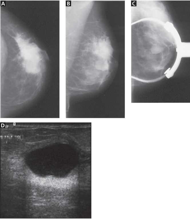
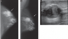
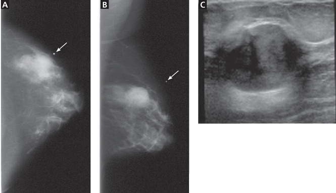

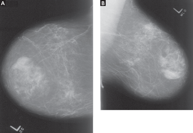
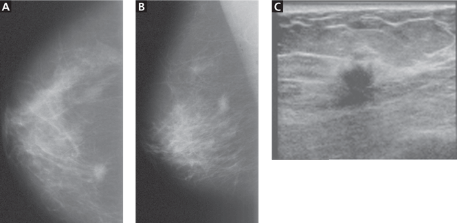
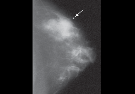


 and “post-fire” (bottom) ultrasonographic views of an 18-gauge percutaneous needle core biopsy of a suspicious breast mass.")
