User login
Large study links brown fat with lower rates of cardiometabolic disease
People who have brown fat detected on imaging seem to be at reduced risk of cardiac and metabolic conditions, ranging from type 2 diabetes to hypertension and coronary artery disease, with a notably strong effect in people with obesity, according to a new study of more than 52,000 individuals who had PET/CT scans as part of cancer evaluation.
Although this has been studied for decades in newborns and animals, only in the past decade have scientists appreciated that some adults have brown fat, typically around the neck and shoulders.
The new study, by far the largest of its kind in humans, appears to confirm the health benefits of brown fat suggested by previous studies, Tobias Becher, MD, and colleagues from The Rockefeller University, New York, wrote in their article published online Jan. 4 in Nature Medicine.
“Our study indicates an important contribution of brown adipose tissue to cardiometabolic health and suggests ... [it] has therapeutic potential in humans,” they stated.
But Caroline M. Apovian, MD, Center for Weight Management and Wellness, Brigham and Women’s Hospital, Boston, is more cautious in her interpretation of the findings.
“It’s nice to see that what we believe about this is correct, and it’s great to see that with obesity and more brown fat there is reduced diabetes and hypertension, but it’s only an association,” she said in an interview.
“This is a good study, but I don’t think we have an understanding of exactly why some people have more brown fat than others, how white fat becomes brown fat, the role of therapeutics, or if it’s important to try to create more brown fat.
“We don’t know if it’s a matter of exercise or something like living in a colder environment, so we need to find out whether or not brown fat is, for instance, a genetic issue, and if it is, if there is a way to increase it in humans,” she added.
And the fact that the study included patients with or being screened for cancer is one of the most important limitations of the study, Dr. Apovian noted.
Brown fat detected in 10% of participants
Contrary to white fat, which stores energy, brown fat is thermogenic, activated by cold conditions, and instead burns energy. And although animal studies have shown a link between brown fat and improvements in glucose and lipid homeostasis, the effects of brown fat in humans are not well understood.
Dr. Becher and colleagues explained that large-scale studies of brown fat have been practically impossible because the tissue only shows up on medical imaging and it would be unethical to expose people to radiation just to study brown fat.
But they realized that, across the street from their lab, many thousands of people visit Memorial Sloan Kettering Cancer Center each year to undergo PET/CT scans for cancer evaluation.
Because radiologists routinely take note when brown adipose tissue is detected to prevent its misinterpretation as a tumor, the information was readily available with the scan data.
“We realized this could be a valuable resource to get us started with looking at brown fat at a population scale,” Dr. Becher said in a press statement from The Rockefeller University.
So they reviewed 134,529 PET/CT scans from 52,487 individuals attending Memorial Sloan Kettering between June 2009 and March 2018 for indications ranging from cancer diagnosis to treatment or surveillance.
Participants were classified by the presence or absence of brown adipose tissue and researchers were able to use electronic health records to comprehensively examine associations between brown fat and rates of disease.
Overall, brown adipose tissue was identified in 5,070 (9.7%) of patients, with higher rates of brown fat among women than men (13.8% vs. 4.9%; P < .0001) and reduced rates with advancing age (P < .0001), as has been observed in previous studies.
The researchers noted, however, that this rate of around 10% of people having brown fat is likely an underestimate because the patients had been instructed to avoid cold exposure, exercise, and caffeine – all of which are thought to increase brown adipose tissue – prior to having their scans.
Does brown fat mitigate some harms of obesity?
Among those with brown fat, the rate of type 2 diabetes was 4.6% compared with 9.5% in those with no detected brown fat (P < .0001), and in a multivariate analysis, the odds ratio (OR) for type 2 diabetes in the presence of brown fat was 0.44.
The occurrence of coronary artery disease was significantly lower in those with brown fat (OR, 0.68; P = .0002), as was cerebrovascular disease (OR, 0.77; P = .0317), heart failure (OR, 0.62; P = .0043), and hypertension (OR, 0.85; P = .0014).
Brown fat also was associated with notable improvements in glucose, triglycerides, and HDL-C levels (all P < .0001), while no differences were seen in measures of LDL-Cs or total cholesterol.
Leukocyte and platelet counts were significantly decreased in individuals with brown fat (both P < .0001).
The findings “suggest potential roles for brown adipose beyond regulation of lipid and glucose metabolism,” the authors wrote.
Most notably, the effects were more pronounced in people with obesity. For example, the prevalence of type 2 diabetes in those with obesity and brown fat was less than half the rate in those with obesity without brown fat (7.5% vs. 20.3%; P < .0001).
This could indicate that brown adipose tissue “might play a role in mitigating the deleterious effects of obesity,” the researchers stated.
“Future research should aim to improve our understanding of brown adipose tissue regulation in humans and to develop mechanisms to safely modulate [its activity],” they concluded.
The study received funding from the American Diabetes Association, the Sinsheimer Foundation, and the National Center for Advancing Translational Sciences of the U.S. Department of Health & Human Services. The authors and Dr. Apovian have reported no relevant financial relationships.
A version of this article first appeared on Medscape.com.
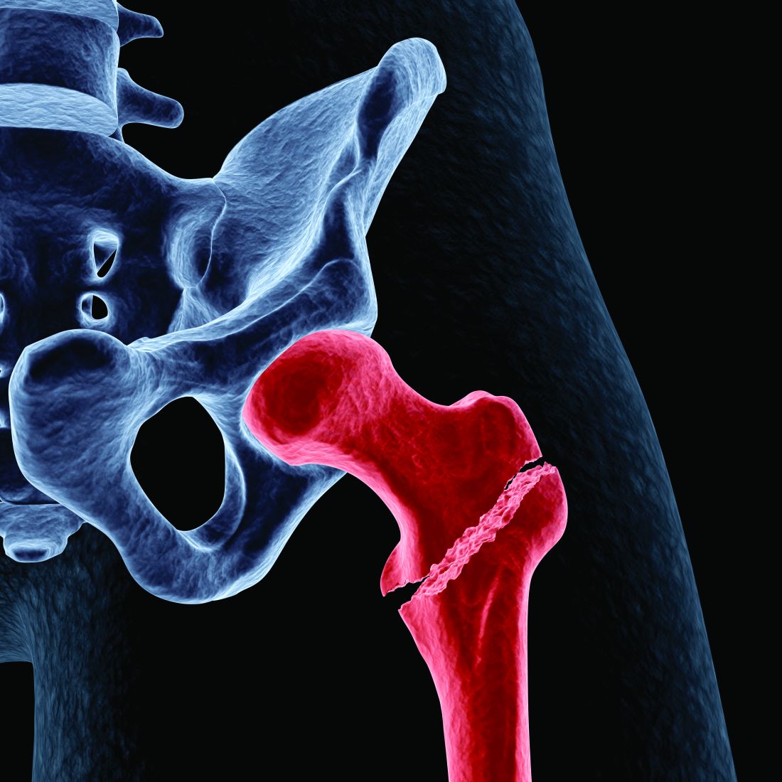
Temper enthusiasm for long-term treatment with bisphosphonates?
Women treated with oral bisphosphonate drugs for osteoporosis for 5 years get no additional benefit – in terms of hip fracture risk – if the treatment is extended for another 5 years, new research shows.
“We found that hip fracture risk in women did not differ if women stopped bisphosphonate use after 5 years or stayed on the medication for 10 years,” coauthor Joan C. Lo, MD, Kaiser Permanente Northern California, Oakland, said in an interview.
The new study, published Dec. 7 in JAMA Network Open, did show a small benefit in continuing the treatment through 7 years vs. 5 years, but it wasn’t clear if this was significant.
“Whether there is a benefit to staying on the drug for 7 years needs to be further studied in randomized trials,” Dr. Lo stressed.
It is well established that oral bisphosphonates are effective in reducing the risk for fracture within the first 3-5 years of treatment; however, evidence on the effects of treatment beyond 5 years is lacking.
The most recent guidance from the American Society of Bone and Mineral Research (ASBMR) on the issue, which were released in 2015, recommends continuation of bisphosphonates beyond 5 years for high-risk patients, but it recommends a “drug holiday” for low-risk patients.
Study adds important new evidence
However, that guidance acknowledges that data are limited regarding long-term use. This large new study adds important new evidence to the discussion, Robert A. Adler, MD, who was a member of the ASBMR Task Force for the recent guidance, said in an interview.
“[With the lack of recent research,] this new study from Kaiser Permanente is of great interest,” said Dr. Adler, chief of endocrinology and metabolism at Central Virginia Veterans Affairs Health Care System and professor of internal medicine and of epidemiology at Virginia Commonwealth University, Richmond.
“It is new data and suggests we might temper our enthusiasm for long-term treatment with bisphosphonates,” he said.
“Importantly, it is the first large observational trial and is closer to a real-world setting than a randomized controlled trial,” he said.
But, Dr. Adler emphasized: “The take-home message is that while this suggests that patients can probably be given a drug holiday for a couple of years ... they should be retested, and if they appear to be at an increased risk of fracture, they probably should restart again.
“Osteoporosis is a chronic disorder,” he emphasized. “It isn’t cured by any of our treatments, and as people get older, they are at a higher fracture risk.
“So we really need to follow our patients for a lifetime and reassess their fracture risk every couple of years – whether they are still on therapy or on a drug holiday.”
Possible that 7 years is better than 5 but remains to be proven
The new study involved data from Kaiser Permanente Northern and Southern California on 29,685 women who had completed 5 years of treatment with oral bisphosphonates, including alendronate, risedronate, or ibandronate, between 2002 and 2014.
Among the women, 11,105 (37%) continued taking the drugs beyond 5 years to 7 years, and 2,725 (9.2%) completed a total of 10 years of treatment.
Their median age was 71. Among those for whom bone mineral density data were available, 37% had osteoporosis after the first 5 years of treatment.
During these 5 years of treatment, 507 hip fractures occurred.
The cumulative incidence of hip fracture among for those who discontinued study therapy at entry, i.e., those who underwent treatment for 5 years, was 23.0 per 1,000 individuals.
After 7 years of treatment, the rate was 20.8 per 1000. For those who continued therapy for 10 years, the rate was 26.8 per 1000 individuals.
The rate in the 7-year treatment group was based on patients taking a 6-month drug holiday after the initial 5 years, but the results are hard to interpret, Dr. Lo said.
“It’s possible that 7 years is better than 5, but this is not a randomized trial, and some of the data analyses done in the study suggest more research should be done to look at a benefit after 7 years.
“At the end of the day, doctors and women need to decide at 5 years what an individual woman’s risk fracture risk is and determine if she should stay on the drug longer,” Dr. Lo emphasized.
Limitations: Subgroups not identified, adherence hard to assess
The uncertainty of any benefit of treatment with bisphosphonates beyond 5 years is further reflected in U.S. recommendations – the Food and Drug Administration has concluded on the basis of pooled data from the extension phase of major clinical trials that any advantages of treatment beyond 3-5 years are unclear.
Key limitations of the current study include the fact that the incidence of hip fracture was not evaluated in low-risk vs. high-risk subgroups; therefore, “these findings may not be applicable to older women at higher risk of osteoporotic fracture,” the authors wrote.
Furthermore, the study did not assess outcomes of fractures other than hip fractures, such as vertebral fractures, they noted.
Dr. Adler pointed out that another limitation is that adherence in the trial was defined as taking 60% of prescribed pills.
“I think this is the biggest weakness with the study,” he said. “Particularly with medications like oral bisphosphonates that don’t really make patients feel any different, it’s a real challenge to make sure patients continue to take these drugs properly.”
The findings should give some reassurance for patients who take a break from the drugs after 5 years. However, reassessment of their risk is critical, Dr. Adler reiterated.
The study was supported by a grant from the National Institute on Aging and the National Institute of Arthritis, Musculoskeletal, and Skin Diseases of the National Institutes of Health. The authors and Adler have disclosed no relevant financial relationships.
A version of this article first appeared on Medscape.com.
To D or not to D? Vitamin D doesn’t reduce falls in older adults
Higher doses of vitamin D supplementation not only show no benefit in the prevention of falls in older adults at increased risk of falling, compared with the lowest doses, but they appear to increase the risk, new research shows.

Based on the findings, supplemental vitamin D above the minimum dose of 200 IU/day likely has little benefit, lead author Lawrence J. Appel, MD, MPH, told this news organization.
“In the absence of any benefit of 1,000 IU/day versus 2,000 IU/day [of vitamin D supplementation] on falls, along with the potential for harm from doses above 1,000 IU/day, it is hard to recommend a dose above 200 IU/day in older-aged persons, unless there is a compelling reason,” asserted Dr. Appel, director of the Welch Center for Prevention, Epidemiology, and Clinical Research at Johns Hopkins Bloomberg School of Public Health in Baltimore.
“More is not always better – and it may even be worse,” when it comes to vitamin D’s role in the prevention of falls, he said.
The research, published in Annals of Internal Medicine, adds important evidence in the ongoing struggle to prevent falls, says Bruce R. Troen, MD, in an accompanying editorial.
“Falls and their deleterious consequences remain a substantial risk for older adults and a huge challenge for health care teams,” writes Dr. Troen, a physician-investigator with the Veterans Affairs Western New York Healthcare System.
However, commenting in an interview, Dr. Troen cautions: “There are many epidemiological studies that are correlative, not causative, that do show a likelihood for benefit [with vitamin D supplementation]. … Therefore, there’s no reason for clinicians to discontinue vitamin D in individuals because of this study.”
“If you’re monitoring an older adult who is frail and has multiple comorbidities, you want to know what their vitamin D level is [and] provide them an appropriate supplement if needed,” he emphasized.
Some guidelines already reflect the lack of evidence of any role of vitamin D supplementation in the prevention of falls, including those of the 2018 U.S. Preventive Services Task Force, which, in a reversal of its 2012 recommendation, now does not recommend vitamin D supplementation for fall prevention in older persons without osteoporosis or vitamin D deficiency, Dr. Appel and colleagues note.
No prevention of falls regardless of baseline vitamin D
As part of STURDY (Study to understand fall reduction and vitamin D in you), Dr. Appel and colleagues enrolled 688 community-dwelling participants who had an elevated risk of falling, defined as a serum 25-hydroxyvitamin D [25(OH)D] level of 25 to 72.5 nmol/L (10-29 ng/dL).
Participants were a mean age of 77.2 years and had a mean total 25(OH)D level of 55.3 nmol/L at enrollment.
They were randomized to one of four doses of vitamin D3, including 200 IU/day (the control group), or 1,000, 2,000, or 4,000 IU/day.
The highest doses were found to be associated with worse – not better – outcomes including a shorter time to hospitalization or death, compared with the 1,000-IU/day group. The higher-dose groups were therefore switched to a dose of 1,000 IU/day or lower, and all participants were followed for up to 2 years.
Overall, 63% experienced falls over the course of the study, which, though high, was consistent with the study’s criteria of participants having an elevated fall risk.
Of the 667 participants who completed the trial, no benefit in prevention of falling was seen across any of the doses, compared with the control group dose of 200 IU/day, regardless of participants’ baseline vitamin D levels.
Safety analyses showed that even in the 1,000-IU/day group, a higher risk of first serious fall and first fall with hospitalization was seen compared with the 200-IU/day group.
A limitation is that the study did not have a placebo group, however, “200 IU/day is a very small dose, probably homeopathic,” Dr. Appel said. “It was likely close to a placebo,” he said.
Caveats: comorbidities, subgroups
In his editorial, Dr. Troen notes other studies, including VITAL (Vitamin D and Omega-3 Trial) also found no reduction in falls with higher vitamin D doses; however, that study did not show any significant risks with the higher doses.
He adds that the current study lacks information on subsets of participants.
“We don’t have enough information about the existing comorbidities and medications that these people are on to be able to pull back the layers. Maybe there is a subgroup that should not be getting 4,000 IU, whereas another subgroup may not be harmed and you may decide that patient can benefit,” he said.
Furthermore, the trial doesn’t address groups such as nursing home residents.
“I have, for instance, 85-year-olds with vitamin D levels of maybe 20 nmol/L with multiple medical issues, but levels that low were not included in the study, so this is a tricky business, but the bottom line is first, do no harm,” he said.
“We really need trials that factor in the multiple different aspects so we can come up, hopefully, with a holistic and interdisciplinary approach, which is usually the best way to optimize care for frail older adults,” he concluded.
The study received funding from the National Institute of Aging.
A version of this article originally appeared on Medscape.com.
Higher doses of vitamin D supplementation not only show no benefit in the prevention of falls in older adults at increased risk of falling, compared with the lowest doses, but they appear to increase the risk, new research shows.
Based on the findings, supplemental vitamin D above the minimum dose of 200 IU/day likely has little benefit, lead author Lawrence J. Appel, MD, MPH, told this news organization.
“In the absence of any benefit of 1,000 IU/day versus 2,000 IU/day [of vitamin D supplementation] on falls, along with the potential for harm from doses above 1,000 IU/day, it is hard to recommend a dose above 200 IU/day in older-aged persons, unless there is a compelling reason,” asserted Dr. Appel, director of the Welch Center for Prevention, Epidemiology, and Clinical Research at Johns Hopkins Bloomberg School of Public Health in Baltimore.
“More is not always better – and it may even be worse,” when it comes to vitamin D’s role in the prevention of falls, he said.
The research, published in Annals of Internal Medicine, adds important evidence in the ongoing struggle to prevent falls, says Bruce R. Troen, MD, in an accompanying editorial.
“Falls and their deleterious consequences remain a substantial risk for older adults and a huge challenge for health care teams,” writes Dr. Troen, a physician-investigator with the Veterans Affairs Western New York Healthcare System.
However, commenting in an interview, Dr. Troen cautions: “There are many epidemiological studies that are correlative, not causative, that do show a likelihood for benefit [with vitamin D supplementation]. … Therefore, there’s no reason for clinicians to discontinue vitamin D in individuals because of this study.”
“If you’re monitoring an older adult who is frail and has multiple comorbidities, you want to know what their vitamin D level is [and] provide them an appropriate supplement if needed,” he emphasized.
Some guidelines already reflect the lack of evidence of any role of vitamin D supplementation in the prevention of falls, including those of the 2018 U.S. Preventive Services Task Force, which, in a reversal of its 2012 recommendation, now does not recommend vitamin D supplementation for fall prevention in older persons without osteoporosis or vitamin D deficiency, Dr. Appel and colleagues note.
No prevention of falls regardless of baseline vitamin D
As part of STURDY (Study to understand fall reduction and vitamin D in you), Dr. Appel and colleagues enrolled 688 community-dwelling participants who had an elevated risk of falling, defined as a serum 25-hydroxyvitamin D [25(OH)D] level of 25 to 72.5 nmol/L (10-29 ng/dL).
Participants were a mean age of 77.2 years and had a mean total 25(OH)D level of 55.3 nmol/L at enrollment.
They were randomized to one of four doses of vitamin D3, including 200 IU/day (the control group), or 1,000, 2,000, or 4,000 IU/day.
The highest doses were found to be associated with worse – not better – outcomes including a shorter time to hospitalization or death, compared with the 1,000-IU/day group. The higher-dose groups were therefore switched to a dose of 1,000 IU/day or lower, and all participants were followed for up to 2 years.
Overall, 63% experienced falls over the course of the study, which, though high, was consistent with the study’s criteria of participants having an elevated fall risk.
Of the 667 participants who completed the trial, no benefit in prevention of falling was seen across any of the doses, compared with the control group dose of 200 IU/day, regardless of participants’ baseline vitamin D levels.
Safety analyses showed that even in the 1,000-IU/day group, a higher risk of first serious fall and first fall with hospitalization was seen compared with the 200-IU/day group.
A limitation is that the study did not have a placebo group, however, “200 IU/day is a very small dose, probably homeopathic,” Dr. Appel said. “It was likely close to a placebo,” he said.
Caveats: comorbidities, subgroups
In his editorial, Dr. Troen notes other studies, including VITAL (Vitamin D and Omega-3 Trial) also found no reduction in falls with higher vitamin D doses; however, that study did not show any significant risks with the higher doses.
He adds that the current study lacks information on subsets of participants.
“We don’t have enough information about the existing comorbidities and medications that these people are on to be able to pull back the layers. Maybe there is a subgroup that should not be getting 4,000 IU, whereas another subgroup may not be harmed and you may decide that patient can benefit,” he said.
Furthermore, the trial doesn’t address groups such as nursing home residents.
“I have, for instance, 85-year-olds with vitamin D levels of maybe 20 nmol/L with multiple medical issues, but levels that low were not included in the study, so this is a tricky business, but the bottom line is first, do no harm,” he said.
“We really need trials that factor in the multiple different aspects so we can come up, hopefully, with a holistic and interdisciplinary approach, which is usually the best way to optimize care for frail older adults,” he concluded.
The study received funding from the National Institute of Aging.
A version of this article originally appeared on Medscape.com.
Lung cancer CT scan is chance for ‘opportunistic’ osteoporosis check
Low-dose chest CT for lung cancer screening provides the opportunity to simultaneously screen patients for osteoporosis, detecting notably higher rates of osteoporosis in men than the traditional tool of DXA, research published in the Journal of Bone and Mineral Research shows.
“Our large-scale, multicenter study of bone density measured from routine low-dose CT scans demonstrated the great potential of using low-dose CT for the opportunistic screening of osteoporosis as an alternative to standard DXA scans,” said senior author Wei Tian, MD, of the Chinese Academy of Engineering and Peking University, in a press statement from the journal.
“Our study revealed the unexpectedly high prevalence of osteoporosis in men, which may impact on the management strategy of men in the future,” Dr. Tian added.
Josephine Therkildsen, MD, of Herning Hospital, Denmark, who has conducted similar research using cardiac CT scans, said the findings add important new insights into the issue of opportunistic screening.
“The results are highly interesting, as they show that low-dose CT-based opportunistic screening could identify a substantial number of patients with low lumbar bone mineral density (BMD) with the future potential to diagnose osteoporosis and initiate relevant treatment before a fracture occurs,” she told this news organization.
Perry J. Pickhardt, MD, chief of gastrointestinal imaging at the University of Wisconsin School of Medicine and Public Health in Madison, agrees. He said in an interview that CT scans of the chest and abdomen, commonly performed for a variety of clinical indications and widespread in most developed countries, can in fact be essential for the detection of a multitude of other concerns – yet are underused for those other purposes.
Use of CT in this way “would likely be very cost effective and clinically efficacious,” he said, adding: “We are seeing greatly increased interest in leveraging this extra information that is contained within every CT scan.” And, “Importantly, artificial intelligence advances now allow for automated approaches, which should allow for expanded use.”
Lung cancer CT scans shed light on osteoporosis prevalence
In the study, led by Xiaoguang Cheng, MD, PhD, of the department of radiology, Beijing Jishuitan Hospital, China, researchers examined lung cancer CT screening data from the prospective China Biobank Project to determine the prevalence of osteoporosis in China.
This included the thoracic low-dose CT scans of 69,095 adults, including 40,733 men and 28,362 women, taken between 2018 and 2019.
To screen for osteoporosis, they used quantitative CT software to evaluate lumbar spine (L1-L2) trabecular volume BMD (vBMD) and diagnostic criteria from the American College of Radiology. Using the vBMD measures from the CT imaging, they found the prevalence of osteoporosis among those over 50 years of age in the Chinese population to be 29% for women (49 million) and 13.5% for men (22.8 million).
Interestingly, the osteoporosis prevalence rate among women was comparable to estimates in the population derived from DXA (29.1%); however, the rate in men was twice that estimated from DXA scans (6.5%).
Decreases in trabecular vBMD with age were observed in both genders. However, declines were steeper among women, who had higher peak trabecular vBMD (185.4 mg/cm3), compared with men (176.6 mg/cm3) at age 30-34 years, but significantly lower measures (62.4 mg/cm3) than men (92.1 mg/cm3) at age 80 years.
The prevalence of osteoporosis in women increased from 2.8% at age 50-54 years to 79.8% at age 85 or older, while in men, the prevalence was 3.2% at age 50-54 years and 44.1% at age 85 or older.
“This is the first study to establish Chinese reference data for vBMD using opportunistic screening from low-dose chest CT in a large population cohort,” the authors write.
“The opportunistic screening of osteoporosis using low-dose CT is clinically feasible and requires no additional exposure to ionizing radiation.”
In addition, no additional equipment or patient time was required, suggesting that “this approach has potential for opportunistic screening for osteoporosis.”
They note, however, that further cohort studies are needed to assess clinical utility of this method.
CT ‘likely a more accurate measure’ of volumetric BMD
Dr. Pickhardt said the differences in osteoporosis prevalence observed between DXA and CT-derived measures in men likely reflect the greater accuracy of CT.
“DXA is a planar technique with a number of drawbacks,” he said in an interview. “CT provides a more direct volumetric measure and is likely a more accurate method for BMD assessment.”
He speculated that the greater differences between DXA versus CT seen in men than women “may relate to sex differences in cortical bone of vertebral bodies, which cannot be separated from the underlying trabecular bone with DXA (whereas CT directly measures the inner trabecular bone).”
The authors note that, although areal BMD (aBMD) derived from DXA is required for osteoporosis diagnosis according to World Health Organization criteria, “trabecular vBMD derived from CT can be also used for diagnosis based on thresholds published by the American College of Radiology of 120 mg/cm3 and 80 mg/cm3 to define osteopenia and osteoporosis, respectively, thresholds that were subsequently confirmed for the Chinese population.”
Furthermore, vBMD has been shown in some studies to be more strongly related to fracture risk, compared with DXA aBMD measures.
Importantly, in another recent study involving 9,223 adults, Dr. Pickhardt and colleagues reported that bone and muscle biomarkers derived from CT were comparable to the Fracture Risk Assessment Tool score for the presymptomatic prediction of future osteoporotic fractures.
Dr. Pickhardt is an advisor to Bracco Imaging and Zebra Medical Vision. Dr. Therkildsen has reported no relevant financial relationships.
This article first appeared on Medscape.com.
New study pinpoints how Mediterranean diet reduces diabetes risk
The known reduction in the risk of type 2 diabetes associated with adoption of the Mediterranean diet appears specifically attributed to its beneficial effects on some key factors, a new study published online in JAMA Network Open reveals.
While a reduction in body mass index may be somewhat obvious, other mechanisms include beneficial effects on insulin resistance, lipoprotein metabolism, and inflammation.
However, the diet’s antidiabetes effect does not appear to extend to people whose weight is considered healthy (BMI under 25 kg/m2), according to the findings.
“It is striking to see in these U.S. women how strong the long-term antidiabetic properties of a Mediterranean-type dietary pattern are,” senior author Samia Mora, MD, of the Center for Lipid Metabolomics, Brigham and Women’s Hospital, Harvard Medical School, Boston, said in an interview.
“While it was known that the Mediterranean diet has many health benefits in particular on metabolism and inflammation, it was not previously known which of these biological pathways may be contributing to the lower risk of diabetes and to what magnitude.
“Our findings support the idea that by improving their diet, people can improve their future risk of type 2 diabetes, particularly if they are overweight or have obesity,” she added.
“And it’s important to note that many of these changes don’t happen right away. While metabolism can change over a short period of time, our study indicates that there are longer term changes happening that may provide protection over decades.”
Mediterranean diet reduced diabetes risk in those with BMI ≥ 25 kg/m2
The Mediterranean diet, with an emphasis on healthy olive oil as the predominant source of oil, favors fruits, vegetables, legumes, nuts, seeds, fish, and dairy products, while limiting intake of red and processed meats as well as sweets.
The diet has been linked to as much as a 25%-30% reduction in the risk of diabetes in previous observational studies.
To investigate the precise mechanisms that underlie the prevention of diabetes, lead author Shafqat Ahmad, PhD, also of Harvard, and colleagues examined data from 25,317 healthy women participating in the Women’s Health Study who had baseline assessments between September 1992 and May 1995. They were a mean age of 52.9 years at baseline.
Over the course of the study, 2,307 participants developed type 2 diabetes.
With a mean follow-up of 19.8 years, those who had the highest self-reported adherence to the Mediterranean diet (a score ≥ 6 on a scale of 0-6) at baseline, had as much as a 30% lower risk of developing type 2 diabetes after multivariate adjustments, compared to those with a lower Mediterranean diet score (a score ≤ 3; hazard ratio, 0.70).
The diabetes-related biomarkers that contributed the most to the reduced risk were insulin resistance, accounting for 65% of the reduction, followed by BMI (55.5%), high-density lipoprotein measures (53%), and inflammation (52.5%).
Other factors, though to a lesser degree, included branched-chain amino acids (34.5%), very low-density lipoprotein measures (32.0%), low-density lipoprotein measures (31.0%), blood pressure (29.0%), and apolipoproteins (23.5%).
Differences in hemoglobin A1c levels had a limited effect on the risk (2%).
Notably, a subgroup analysis looking at effects of the diet according to baseline BMI showed the reductions in type 2 diabetes associated with higher intake of the Mediterranean diet extended only to those with an above normal weight (BMI ≥ 25 kg/m2).
Dr. Mora noted that, as this was not a prespecified analysis, these findings should be viewed as hypothesis-generating, but are consistent with the well-known increase in diabetes risk seen with a higher BMI.
“[The finding] fits with the biology and pathogenesis of type 2 diabetes that is driven in large part by excess weight, in particular for visceral adiposity and its resulting metabolic dysregulation and inflammation,” she said.
“We know from other studies, such as the Nurses’ Health Study, that the risk for type 2 diabetes in women increases even at BMI levels below 25 kg/m2, but the risk goes up exponentially at around a BMI of 25 and higher.”
Strong role of insulin resistance a surprise
The strong role of insulin resistance was a surprise, Dr. Mora added.
“We were surprised that insulin resistance, measured by a simple blood biomarker, would have the strongest mediating effect – even stronger than BMI – for the Mediterranean diet on risk of diabetes,” she noted.
“This could represent an opportunity to intervene earlier and more intensively on improving insulin resistance through dietary approaches such as the Mediterranean diet, especially [because] insulin resistance can precede by years and decades the overt hyperglycemia and clinical diagnosis of diabetes.”
Yet another surprise was that A1c had no substantial mediating effect on the reduction of diabetes risk with the Mediterranean diet.
“This could suggest that the cat is out of the bag by the time the A1c rises,” Dr. Mora observed.
A study limitation is that the Women’s Health Study consisted of well-educated U.S. women who were health professionals and predominantly White, so the results may not be generalizable to men or individuals of other races or ethnicities.
In addition, BMI was self-reported and participants were not uniformly screened for diabetes, therefore surveillance bias could be possible.
However, the findings suggest that “even a small increase in adherence to the Mediterranean diet has substantial benefits over many years in preventing diabetes, among many other health benefits such as lowering insulin resistance and inflammation, improving lipid metabolism, and lowering blood pressure,” Mora said.
“And of course, the more the adherence, the more the benefit.”
The study received support through grants from the National Institutes of Health, the National Heart, Lung, and Blood Institute, the National Institute of Diabetes and Digestive and Kidney Diseases, the American Heart Association, and the Molino Family Trust. A coauthor is listed as a coinventor on patents held by Brigham and Women’s Hospital related to the use of inflammatory biomarkers in cardiovascular disease (licensed to AstraZeneca and Siemens).
A version of this article originally appeared on Medscape.com.
Statins beneficial in elderly, guidelines should be strengthened
Contrary to historical evidence, two new studies show.
“By contrast with previous historical studies, our data show that LDL cholesterol is an important risk factor for myocardial infarction and atherosclerotic cardiovascular disease in a contemporary primary prevention cohort of individuals aged 70 to 100 years,” Borge Nordestgaard, MD, of the University of Copenhagen, and colleagues noted in the first of the two studies, published this week in the Lancet.
“By lowering LDL cholesterol in healthy individuals aged 70-100 years, the potential for preventing myocardial infarctions and atherosclerotic cardiovascular disease is huge, and at a substantially lower number needed to treat when compared with those aged 20-69 years,” they added.
“These findings support the concept of the cumulative burden of LDL cholesterol over one’s lifetime and the progressive increase in risk for atherosclerotic cardiovascular disease, including myocardial infarction, with age,” added Frederick J. Raal, PhD, and Farzahna Mohamed, MB BCh, of the University of the Witwatersrand, Johannesburg, South Africa, in an editorial published with both new studies in the Lancet (2020 Nov 10. doi: 10.1016/S0140-6736[20]32333-3).
The studies underscore the need for clinicians to consider continued risks associated with elevated LDL cholesterol in older age, they stressed, adding that statins are also beneficial for younger persons at risk to prevent conditions from worsening.
“The average age of patients in all the trials analyzed was older than 60 years, an age when atherosclerotic cardiovascular disease is already well established,” the editorialists wrote.
“Lipid-lowering therapy should be initiated at a younger age, preferably before age 40 years, in those at risk to delay the onset of atherosclerosis, rather than try to manage the condition once fully established or advanced,” they stressed.
No RCTs have included patients older than 70
For persons aged 40-75 years, elevated LDL cholesterol levels are a known risk factor for MI and atherosclerotic cardiovascular disease, and there is consensus in guidelines regarding treatment with statins.
However, the risk for people older than 70 is controversial. Some studies show little or no association between elevated LDL cholesterol levels and an increased risk for MI.
Contributing to the uncertainty is that few of the randomized, controlled trials that have investigated the question have included patients aged older than 70 years.
As a consequence, many practice guidelines have noted that the level of evidence in older patients is low, and some organizations have lowered the strength of recommendations regarding the treatment for older patients in comparison with younger patients.
Primary prevention: CV events increase with elevated LDL cholesterol in older age
Dr. Nordestgaard and colleagues studied data on 91,131 people living in Copenhagen who did not have atherosclerotic cardiovascular disease or diabetes at baseline and were not taking statins.
Of the participants, 10,592 were aged 70-79 years, and 3,188 participants were aged 80-100 years.
Over an average follow-up period of 7.7 years, 1,515 participants had a first MI, and 3,389 developed atherosclerotic cardiovascular disease.
In the primary-prevention cohort, after multivariate adjustment, the risk of having a heart attack per 1.0 mmol/L increase in LDL cholesterol was increased in the group overall (hazard ratio, 1.34). The increased risk was observed for all age groups, including those aged 80-100 years (HR, 1.28), 70-79 (HR, 1.25), 60-69 (HR, 1.29), 50-59 (HR, 1.28), and 20-49 (HR, 1.68).
Risk for atherosclerotic cardiovascular disease was also raised per 1.0 mmol/L increase in LDL cholesterol overall (HR, 1.16) and in all age groups, particularly those aged 70-100 years.
Greater elevations in LDL cholesterol (5.0 mmol/L or higher, indicative of possible familial hypercholesterolemia) were associated with a notably higher risk for heart attack after multivariate adjustment in people aged 80-100 (HR, 2.99). Risk was also higher among those aged 70-79 (HR, 1.82).
The highest incidence was in those older than 70. The rate was 8.5 heart attacks per 1,000 people per year among those aged 80-100 and 5.2 heart attacks per 1,000 in those aged 70-79. The rates were 2.5 per 1,000 among those 60-69, 1.8 for those aged 50-59, and 0.8 for those aged 20-49.
“The absolute risk [of cardiovascular events] is of course much higher in the elderly than those under the age of 75, but what was a surprise was how clear our results were on a relative risk scale, that the risk associated with elevated LDL [cholesterol] was as high in people aged 80-100 as the younger patients,” Dr. Nordestgaard said in an interview.
With regard to the benefits of cholesterol-lowering drugs, the study showed that the number needed to prevent one heart attack over 5 years was 80 among those aged 80-100; the number was 439 for people aged 50-59.
With regard to stronger statins, when moderate-intensity statins were used, the number needed to treat to prevent one cardiovascular disease event of any type dropped to 42 for patients aged 80-100. It was 88 for those aged 70-79, 164 for those aged 60-69, 345 for those aged 50-59, and 769 for those aged 20-49.
“The clinical significance of this is that it appears those in older age groups indeed benefit from cholesterol-lowering therapy,” Dr. Nordestgaard said. “I think many people have this idea that LDL [cholesterol] is not important over the age of about 70-75, but that’s not the case.”
“These robust findings are novel,” he and his colleagues stressed.
Despite these observational findings, the South African editorialists noted that “whether lipid-lowering therapy should be initiated for primary prevention in people aged 75 years or older is unclear,” owing to the host of risks and benefits that need to be balanced.
The findings of an ongoing randomized, placebo-controlled trial (STAREE) may answer this question, they wrote. It is investigating primary prevention in 18,000 older patients (≥70 years) who are being randomly assigned to receive atorvastatin 40 mg/d or placebo. The study is seeking to determine whether statin treatment extends the length of a disability-free life, which will be assessed on the basis of survival outside permanent residential care. Results are expected in 2022-2023.
Unequivocal reductions in events in elderly, comparable with younger patients
In the second study (Lancet. 2020 Nov 10. doi: 10.1016/S0140-6736[20]32332-1), Baris Gencer, MD, of Brigham and Women’s Hospital, Boston, =and colleagues evaluated the effects of statins and other cholesterol-lowering drugs, including ezetimibe and proprotein convertase subtilisin/kexin type 9 inhibitors, in older versus younger patients.
The systematic review and meta-analysis of 29 randomized controlled trials, also published in the Lancet, were presented virtually as a poster as part of the 2020 American Heart Association scientific session. It included data on 244,090 patients, including 21,492 aged 75 years and older.
The meta-analysis included studies of cardiovascular outcomes of a guideline-recommended LDL cholesterol–lowering drug, with a median follow-up of at least 2 years and inclusion of data on patients aged 75 years and older.
The results showed that over a median follow-up of 2.2 to 6 years, statin use by older patients was associated with a relative risk reduction of major vascular events of 26% per 1 mmol/L reduction in LDL cholesterol (P = .0019), which was comparable with a risk reduction of 15% per 1 mmol/L reduction in LDL cholesterol for patients younger than 75 years (P = .37, compared with older patients).
Treatment of older patients with LDL cholesterol–lowering drugs was also associated with significantly improved outcomes in cardiovascular death (risk ratio, 0.85), MI (RR, 0.80), stroke (RR, 0.73), and coronary revascularization (RR, 0.80).
“We found an unequivocal reduction in the risk of major vascular events with both statin and nonstatin LDL cholesterol-lowering treatments, which was similar to that seen in younger patients,” the authors wrote.
“Cholesterol-lowering medications are affordable drugs that have reduced risk of heart disease for millions of people worldwide, but until now, their benefits for older people have remained less certain,” said lead author Marc Sabatine, MD, also of Brigham and Women’s Hospital, in a Lancet press release.
“Our analysis indicates that these therapies are as effective in reducing cardiovascular events and deaths in people aged 75 years and over as they are in younger people. We found no offsetting safety concerns, and together, these results should strengthen guideline recommendations for the use of cholesterol-lowering medications, including statin and nonstatin therapy, in elderly people.”
The editorialists agreed: “More than 80% of fatal cardiovascular events occur in individuals older than 65 years, and the incidence of cardiovascular events is increasing in those older than 80 years; therefore, the findings of Gencer and colleagues’ study should encourage the use of lipid-lowering therapy in older patients.”
The authors of the two studies have disclosed no relevant financial relationships. Dr. Raal has received research grants, honoraria, or consulting fees for advisory board membership, professional input, and lectures on lipid-lowering drug therapy from Amgen, Regeneron, Sanofi, Novartis, and the Medicines Company.
A version of this article originally appeared on Medscape.com.
First-of-its kind guideline on lipid monitoring in endocrine diseases
Endocrine diseases of any type – not just diabetes – can represent a cardiovascular risk and patients with those disorders should be screened for high cholesterol, according to a new clinical practice guideline from the Endocrine Society.
“The simple recommendation to check a lipid panel in patients with endocrine diseases and calculate cardiovascular risk may be practice changing because that is not done routinely,” Connie Newman, MD, chair of the Endocrine Society committee that developed the guideline, said in an interview.
“Usually the focus is on assessment and treatment of the endocrine disease, rather than on assessment and treatment of atherosclerotic cardiovascular disease risk,” said Newman, an adjunct professor of medicine in the department of medicine, division of endocrinology, diabetes & metabolism, at New York University.
Whereas diabetes, well-known for its increased cardiovascular risk profile, is commonly addressed in other cardiovascular and cholesterol practice management guidelines, the array of other endocrine diseases are not typically included.
“This guideline is the first of its kind,” Dr. Newman said. “The Endocrine Society has not previously issued a guideline on lipid management in endocrine disorders [and] other organizations have not written guidelines on this topic.
“Rather, guidelines have been written on cholesterol management, but these do not describe cholesterol management in patients with endocrine diseases such as thyroid disease [hypothyroidism and hyperthyroidism], Cushing’s syndrome, acromegaly, growth hormone deficiency, menopause, male hypogonadism, and obesity,” she noted.
But these conditions carry a host of cardiovascular risk factors that may require careful monitoring and management.
“Although endocrine hormones, such as thyroid hormone, cortisol, estrogen, testosterone, growth hormone, and insulin, affect pathways for lipid metabolism, physicians lack guidance on lipid abnormalities, cardiovascular risk, and treatment to reduce lipids and cardiovascular risk in patients with endocrine diseases,” she explained.
Vinaya Simha, MD, an internal medicine specialist at the Mayo Clinic in Rochester, Minn., agrees that the guideline is notable in addressing an unmet need.
Recommendations that stand out to Dr. Simha include the suggestion of adding eicosapentaenoic acid (EPA) ethyl ester to reduce the risk of cardiovascular disease in adults with diabetes or atherosclerotic cardiovascular disease who have elevated triglyceride levels despite statin treatment.
James L. Rosenzweig, MD, an endocrinologist at Hebrew SeniorLife in Boston, agreed that this is an important addition to an area that needs more guidance.
“Many of these clinical situations can exacerbate dyslipidemia and some also increase the cardiovascular risk to a greater extent in combination with elevated cholesterol and/or triglycerides,” he said in an interview.
“In many cases, treatment of the underlying disorder appropriately can have an important impact in resolving the lipid disorder. In others, more aggressive pharmacological treatment is indicated,” he said.
“I think that this will be a valuable resource, especially for endocrinologists, but it can be used as well by providers in other disciplines.”
Key recommendations for different endocrine conditions
The guideline, published in the Journal of Clinical Endocrinology & Metabolism, details those risks and provides evidence-based recommendations on their management and treatment.
Key recommendations include:
- Obtain a lipid panel and evaluate cardiovascular risk factors in all adults with endocrine disorders.
- In patients with and risk factors for cardiovascular disease, start statin therapy in addition to lifestyle modification to reduce cardiovascular risk. “This could mean earlier treatment because other guidelines recommend consideration of therapy at age 40,” Dr. Newman said.
- Statin therapy is also recommended for adults over 40 with with a duration of diabetes of more than 20 years and/or microvascular complications, regardless of their cardiovascular risk score. “This means earlier treatment of patients with type 1 diabetes with statins in order to reduce cardiovascular disease risk,” Dr. Newman noted.
- In patients with hyperlipidemia, rule out as the cause before treating with lipid-lowering medications. And among patients who are found to have hypothyroidism, reevaluate the lipid profile when the patient has thyroid hormone levels in the normal range.
- Adults with persistent endogenous Cushing’s syndrome should have their lipid profile monitored. Statin therapy should be considered in addition to lifestyle modifications, irrespective of the cardiovascular risk score.
- In postmenopausal women, high cholesterol or triglycerides should be treated with statins rather than hormone therapy.
- Evaluate and treat lipids and other cardiovascular risk factors in women who enter menopause early (before the age of 40-45 years).
Nice summary of ‘risk-enhancing’ endocrine disorders
Dr. Simha said in an interview that the new guideline is “probably the first comprehensive statement addressing lipid treatment in patients with a broad range of endocrine disorders besides diabetes.”
“Most of the treatment recommendations are congruent with other current guidelines such as the American College of Cardiology/American Heart Association [guidelines], but there is specific mention of which endocrine disorders represent enhanced cardiovascular risk,” she explained.
The new recommendations are notable for including “a nice summary of how different endocrine disorders affect lipid values, and also which endocrine disorders need to be considered as ‘risk-enhancing factors,’ ” Dr. Simha noted.
“The use of EPA in patients with hypertriglyceridemia is novel, compared to the ACC/AHA recommendation. This reflects new data which is now available,” she added.
The American Association of Clinical Endocrinologists also just issued a new algorithm on lipid management and prevention of cardiovascular disease in which treatment of hypertriglyceridemia is emphasized.
In addition, the new Endocrine Society guideline “also mentions an LDL [cholesterol] treatment threshold of 70 mg/dL, and 55 mg/dL in some patient categories, which previous guidelines have not,” Dr. Simha noted.
Overall, Dr. Newman added that the goal of the guideline is to increase awareness of key issues with endocrine diseases that may not necessarily be on clinicians’ radars.
“We hope that it will make a lipid panel and cardiovascular risk evaluation routine in adults with endocrine diseases and cause a greater focus on therapies to reduce heart disease and stroke,” she said.
Dr. Newman, Dr. Simha, and Dr. Rosenzweig reported no relevant financial relationships.
A version of this article originally appeared on Medscape.com.
‘Landmark’ study pushed detection of covert consciousness in TBI
Compelling advances in the ability to detect signs of consciousness in unconscious patients who have experienced traumatic brain injury (TBI) are leading to unprecedented changes in the field. There is now hope of improving outcomes and even sparing lives of patients who may otherwise have been mistakenly assessed as having no chance of recovery.

That research, published in the New England Journal of Medicine in June 2019, linked the promising signals of consciousness in comatose patients, detected only on imaging, with remarkable outcomes a year later.
“This was a landmark study,” said Brian L. Edlow, MD, in a presentation on the issue of covert consciousness at the virtual annual meeting of the American Neurological Association.
“Importantly, it is the first compelling evidence that early detection of covert consciousness also predicts 1-year outcomes in the Glasgow Outcome Scale Extended (GOSE), showing that covert consciousness in the ICU appears to be relevant for predicting long-term outcomes,” said Dr. Edlow, who is associate director of the Center for Neurotechnology and Neurorecovery, Massachusetts General Hospital, in Boston.
The researchers showed that 15% of unconscious patients with acute brain injury in the study exhibited significant brain activity on EEG in response to stimuli that included verbal commands such as envisioning that they are playing tennis.
Although other studies have shown similar effects with task-based stimuli, the New England Journal of Medicine study further showed that a year later, the patients who had shown signs of covert consciousness, also called “cognitive motor dissociation” (CMD), were significantly more likely to have a good functional outcome, said the study’s senior author, Jan Claassen, MD, director of critical care neurology at Columbia University, New York, who also presented at the ANA session.
“Importantly, a year later after injury, we found that 44% of patients with CMD and only 14% of non-CMD patients had a good functional outcome, defined as a GOSE score indicating a state where they can at least take care of themselves for 8 hours in a day,” he said.
“[Whether] these patients in a CMD state represent a parallel state or a transitory state on the road to recovery remains to be shown,” he said.
Jennifer Frontera, MD, a professor in the department of neurology at NYU Langone Health in New York and comoderator of the session, agreed that the research is “remarkable.”
“Also,” she said, “it is practical, since many could potentially apply and validate his algorithms, since EEG technology is portable and widely available.”
Research has ushered in a ‘sea change’ in neurocritical care
The research has helped push forward recommendations on the treatment of unconscious patients, Dr. Edlow said. “This has led to a sea change in our field just over the last 2 years, with multiple guidelines published suggesting that it may be time for us to consider incorporating task-based fMRI and EEG techniques into our clinical assessment of patients with disorders of consciousness,” Dr. Edlow said.
Among those updating their recommendations was the American Academy of Neurology, which revised guidelines on practice parameters for patients in a persistent vegetative state. Those guidelines had not been updated since 1995.
Although concluding that “no diagnostic assessment procedure had moderate or strong evidence for use,” the guidelines acknowledge that “it is possible that a positive electromyographic (EMG) response to command, EEG reactivity to sensory stimuli, laser-evoked potentials, and the Perturbational Complexity Index can distinguish a minimally conscious state from vegetative state/unresponsive wakefulness syndrome (VS/UWS).”
Earlier this year, the European Academy of Neurology followed suit with updated guidelines of its own. In the EAN guideline, the academy’s Panel on Coma, Disorders of Consciousness recommends that task-based fMRI, EEG, and other advanced assessments be performed as part of a composite assessment of consciousness and that a patient’s best performance or highest level of consciousness on any of those tests should be a reflection of their diagnosis, Dr. Edlow explained.
“What this means is that our field is moving toward a multimodal assessment of consciousness in the ICU as well as beyond, in the subacute to chronic setting, whereby the behavioral exam, advanced DG, and advanced MRI methods all also contribute to the diagnosis of consciousness,” he said.
The standard for assessment of disorders of consciousness is the Coma Recovery Scale–Revised, with a 25-item scale for diagnosis, prediction of outcome, and assessment of potential treatment efficacy.
But much uncertainty can remain despite the assessment, Dr. Claassen said. “Behavioral assessments of patients with acute brain injury are challenging because examinations fluctuate, and there’s variability between assessors,” he said. “Nevertheless, patients and their families demand guidance from us.”
Dr. Edlow pointed out that the largest study to date of the causes of death among patients with TBI in the ICU underscores the need for better assessments.
The study of more than 600 patients at six level l trauma centers in Canada showed that 70% of patients who died in the ICU from TBI did so as the result of the withdrawal of life-sustaining therapy. However, only about a half (57%) had an unreactive pupil, and only about a quarter (23.7%) had evidence of herniation on CT, findings that are commonly associated with a poor prognosis.
“What emerges from this is that the manner in which the clinicians communicated the prognosis to families was a primary determinant of decisions to withdraw life-sustaining therapy,” Dr. Edlow said.
Negative response not necessarily conclusive
Dr. Edlow added a word of caution that the science is still far from perfect. He noted that, for 25% of healthy patients who are given a motor imagery task, neuroimaging might not show a response, implying that the lack of a signal may not be conclusive.
He described the case of a patient who was comatose at the time she was scanned on day 3 after injury and who showed no responses to language, music, or motor imagery during the MRI, yet a year later, she was functionally independent, back in the workforce, and had very few residual symptoms from her trauma.
“So if a patient does not show a response, that does not prove the patient is not conscious, and it does not prove that the patient is likely to have a poor outcome,” Dr. Edlow said. Such cases underscore the need for more advances in understanding the inner workings of brain injury.
Dr. Edlow and his colleagues are embarking on a trial of the effects of intravenous methylphenidate in targeting the stimulation of dopaminergic circuits within the subcortical ascending arousal network in patients with severe brain injuries.
“The scientific premise of the trial is that personalized brain network mapping in the ICU can identify patients whose connectomes are amenable to neuromodulation,” Dr. Edlow and his colleague report in an article in Neurocritical Care.
The trial, called STIMPACT (Stimulant Therapy Targeted to Individualized Connectivity Maps to Promote ReACTivation of Consciousness), is part of the newly launched Connectome-based Clinical Trial Platform, which the authors describe as “a new paradigm for developing and testing targeted therapies that promote early recovery of consciousness in the ICU.”
Such efforts are essential, given the high stakes of TBI outcomes, Dr. Edlow said.
“Let’s be clear about the stakes of an incorrect prognosis,” he said. “If we’re overly pessimistic, then a patient who could have potential for meaningful recovery will likely die in our ICU. On the other hand, if we are overly optimistic, then a patient could end up in a vegetative or minimally conscious state that he or she may never have found to be acceptable,” he said.
Access to technologies a ‘civil right?’
Some ethicists in the field are recommending that patients be given access to the advanced techniques as a civil right, similar to the rights described in the Convention on the Rights of Persons With Disabilities, which was adopted by the United Nations in 2008, Dr. Edlow noted.
“So the question that we as clinicians are going to face moving forward from an ethical standpoint is, if we have access to these techniques, is it an ethical obligation to offer them now?” he said.
Dr. Edlow underscored the need to consider the reality that “there are profound issues relating to resource allocation and access to these advanced techniques, but we’re going to have to consider this together as we move forward.”
Dr. Edlow has received funding from the National Institutes of Health. Dr. Claassen is a minority shareholder with ICE Neurosystems. Dr. Frontera has disclosed no relevant financial relationships.
A version of this article originally appeared on Medscape.com.
Compelling advances in the ability to detect signs of consciousness in unconscious patients who have experienced traumatic brain injury (TBI) are leading to unprecedented changes in the field. There is now hope of improving outcomes and even sparing lives of patients who may otherwise have been mistakenly assessed as having no chance of recovery.
That research, published in the New England Journal of Medicine in June 2019, linked the promising signals of consciousness in comatose patients, detected only on imaging, with remarkable outcomes a year later.
“This was a landmark study,” said Brian L. Edlow, MD, in a presentation on the issue of covert consciousness at the virtual annual meeting of the American Neurological Association.
“Importantly, it is the first compelling evidence that early detection of covert consciousness also predicts 1-year outcomes in the Glasgow Outcome Scale Extended (GOSE), showing that covert consciousness in the ICU appears to be relevant for predicting long-term outcomes,” said Dr. Edlow, who is associate director of the Center for Neurotechnology and Neurorecovery, Massachusetts General Hospital, in Boston.
The researchers showed that 15% of unconscious patients with acute brain injury in the study exhibited significant brain activity on EEG in response to stimuli that included verbal commands such as envisioning that they are playing tennis.
Although other studies have shown similar effects with task-based stimuli, the New England Journal of Medicine study further showed that a year later, the patients who had shown signs of covert consciousness, also called “cognitive motor dissociation” (CMD), were significantly more likely to have a good functional outcome, said the study’s senior author, Jan Claassen, MD, director of critical care neurology at Columbia University, New York, who also presented at the ANA session.
“Importantly, a year later after injury, we found that 44% of patients with CMD and only 14% of non-CMD patients had a good functional outcome, defined as a GOSE score indicating a state where they can at least take care of themselves for 8 hours in a day,” he said.
“[Whether] these patients in a CMD state represent a parallel state or a transitory state on the road to recovery remains to be shown,” he said.
Jennifer Frontera, MD, a professor in the department of neurology at NYU Langone Health in New York and comoderator of the session, agreed that the research is “remarkable.”
“Also,” she said, “it is practical, since many could potentially apply and validate his algorithms, since EEG technology is portable and widely available.”
Research has ushered in a ‘sea change’ in neurocritical care
The research has helped push forward recommendations on the treatment of unconscious patients, Dr. Edlow said. “This has led to a sea change in our field just over the last 2 years, with multiple guidelines published suggesting that it may be time for us to consider incorporating task-based fMRI and EEG techniques into our clinical assessment of patients with disorders of consciousness,” Dr. Edlow said.
Among those updating their recommendations was the American Academy of Neurology, which revised guidelines on practice parameters for patients in a persistent vegetative state. Those guidelines had not been updated since 1995.
Although concluding that “no diagnostic assessment procedure had moderate or strong evidence for use,” the guidelines acknowledge that “it is possible that a positive electromyographic (EMG) response to command, EEG reactivity to sensory stimuli, laser-evoked potentials, and the Perturbational Complexity Index can distinguish a minimally conscious state from vegetative state/unresponsive wakefulness syndrome (VS/UWS).”
Earlier this year, the European Academy of Neurology followed suit with updated guidelines of its own. In the EAN guideline, the academy’s Panel on Coma, Disorders of Consciousness recommends that task-based fMRI, EEG, and other advanced assessments be performed as part of a composite assessment of consciousness and that a patient’s best performance or highest level of consciousness on any of those tests should be a reflection of their diagnosis, Dr. Edlow explained.
“What this means is that our field is moving toward a multimodal assessment of consciousness in the ICU as well as beyond, in the subacute to chronic setting, whereby the behavioral exam, advanced DG, and advanced MRI methods all also contribute to the diagnosis of consciousness,” he said.
The standard for assessment of disorders of consciousness is the Coma Recovery Scale–Revised, with a 25-item scale for diagnosis, prediction of outcome, and assessment of potential treatment efficacy.
But much uncertainty can remain despite the assessment, Dr. Claassen said. “Behavioral assessments of patients with acute brain injury are challenging because examinations fluctuate, and there’s variability between assessors,” he said. “Nevertheless, patients and their families demand guidance from us.”
Dr. Edlow pointed out that the largest study to date of the causes of death among patients with TBI in the ICU underscores the need for better assessments.
The study of more than 600 patients at six level l trauma centers in Canada showed that 70% of patients who died in the ICU from TBI did so as the result of the withdrawal of life-sustaining therapy. However, only about a half (57%) had an unreactive pupil, and only about a quarter (23.7%) had evidence of herniation on CT, findings that are commonly associated with a poor prognosis.
“What emerges from this is that the manner in which the clinicians communicated the prognosis to families was a primary determinant of decisions to withdraw life-sustaining therapy,” Dr. Edlow said.
Negative response not necessarily conclusive
Dr. Edlow added a word of caution that the science is still far from perfect. He noted that, for 25% of healthy patients who are given a motor imagery task, neuroimaging might not show a response, implying that the lack of a signal may not be conclusive.
He described the case of a patient who was comatose at the time she was scanned on day 3 after injury and who showed no responses to language, music, or motor imagery during the MRI, yet a year later, she was functionally independent, back in the workforce, and had very few residual symptoms from her trauma.
“So if a patient does not show a response, that does not prove the patient is not conscious, and it does not prove that the patient is likely to have a poor outcome,” Dr. Edlow said. Such cases underscore the need for more advances in understanding the inner workings of brain injury.
Dr. Edlow and his colleagues are embarking on a trial of the effects of intravenous methylphenidate in targeting the stimulation of dopaminergic circuits within the subcortical ascending arousal network in patients with severe brain injuries.
“The scientific premise of the trial is that personalized brain network mapping in the ICU can identify patients whose connectomes are amenable to neuromodulation,” Dr. Edlow and his colleague report in an article in Neurocritical Care.
The trial, called STIMPACT (Stimulant Therapy Targeted to Individualized Connectivity Maps to Promote ReACTivation of Consciousness), is part of the newly launched Connectome-based Clinical Trial Platform, which the authors describe as “a new paradigm for developing and testing targeted therapies that promote early recovery of consciousness in the ICU.”
Such efforts are essential, given the high stakes of TBI outcomes, Dr. Edlow said.
“Let’s be clear about the stakes of an incorrect prognosis,” he said. “If we’re overly pessimistic, then a patient who could have potential for meaningful recovery will likely die in our ICU. On the other hand, if we are overly optimistic, then a patient could end up in a vegetative or minimally conscious state that he or she may never have found to be acceptable,” he said.
Access to technologies a ‘civil right?’
Some ethicists in the field are recommending that patients be given access to the advanced techniques as a civil right, similar to the rights described in the Convention on the Rights of Persons With Disabilities, which was adopted by the United Nations in 2008, Dr. Edlow noted.
“So the question that we as clinicians are going to face moving forward from an ethical standpoint is, if we have access to these techniques, is it an ethical obligation to offer them now?” he said.
Dr. Edlow underscored the need to consider the reality that “there are profound issues relating to resource allocation and access to these advanced techniques, but we’re going to have to consider this together as we move forward.”
Dr. Edlow has received funding from the National Institutes of Health. Dr. Claassen is a minority shareholder with ICE Neurosystems. Dr. Frontera has disclosed no relevant financial relationships.
A version of this article originally appeared on Medscape.com.
FROM ANA 2020
Time-restricted eating shows no weight-loss benefit in RCT
The popular new weight-loss approach of eating within a restricted window of time during the day, allowing for an extended period of fasting – also known as intermittent fasting – does not result in greater weight loss, compared with nonrestricted meal timing, results from a randomized clinical trial show.
“I was very surprised by all of [the results],” senior author Ethan J. Weiss, MD, said in an interview.
“Part of the reason we did the study was because I had been doing time-restricted eating myself for years and even recommending it to friends and patients as an effective weight-loss tool,” said Dr. Weiss, of the Cardiovascular Research Institute, University of California, San Francisco.
“But no matter how you slice it, prescription of time-restricted eating – at least this version –is not a very effective weight-loss strategy,” Dr. Weiss said.
The study, published online in JAMA Internal Medicine by Dylan A. Lowe, PhD, also of the University of California, San Francisco, involved 116 participants who were randomized to a 12-week regimen of either three structured meals per day or time-restricted eating, with instructions to eat only between 12:00 p.m. and 8:00 p.m. and to completely abstain from eating at other times.
The participants were not given any specific instructions regarding caloric or macronutrient intake “so as to offer a simple, real-world recommendation to free-living individuals,” the authors wrote.
Although some prior research has shown improvements in measures such as glucose tolerance with time-restricted eating, studies showing weight loss with the approach, including one recently reported by Medscape Medical News, have been small and lacked control groups.
“To my knowledge this is the first randomized, controlled trial and definitely the biggest,” Dr. Weiss. “I think it is the most comprehensive dataset available in people, at least for this intervention.”
Participants used app to log details
At baseline, participants had a mean weight of 99.2 kg (approximately 219 lb). Their mean age was 46.5 years and 60.3% were men. They were drawn from anywhere in the United States and received study surveys through a custom mobile study application on the Eureka Research Platform. They were given a Bluetooth weight scale to use daily, which was connected with the app, and randomized to one of the two interventions. A subset of 50 participants living near San Francisco underwent in-person testing.
At the end of the 12 weeks, those in the time-restricted eating group (n = 59) did have a significant decrease in weight, compared with baseline (−0.94 kg; P = .01), while weight loss in the consistent-meal group (n = 57) was not significant (−0.68 kg; P = .07).
But importantly, the difference in weight loss between the groups was not significant (−0.26 kg; P = .63).
There were no significant differences in secondary outcomes of fasting insulin, glucose, hemoglobin A1c, or blood lipids within or between the time-restricted eating and consistent-meal group either. Nor were there any significant differences in resting metabolic rate.
Although participants did not self-report their caloric intake, the authors estimated that the differences were not significant using mathematical modeling developed at the National Institutes of Health.
Rates of adherence to the diets were 92.1% in the consistent-meal group versus 83.5% in the time-restricted group.
Not all diets are equal: Time-restricted eating group lost more lean mass
In a subset analysis, loss of lean mass was significantly greater in the time-restricted eating group, compared with the consistent-meals group, in terms of both appendicular lean mass (P = .009) and the appendicular lean mass index (P = .005).
In fact, as much as 65% of the weight lost (1.10 kg of the average 1.70 kg) in the time-restricted eating group consisted of lean mass, while much less was fat mass (0.51 kg).
“The proportion of lean mass loss in this study (approximately 65%) far exceeds the normal range of 20%-30%,” the authors wrote. “In addition, there was a highly significant between-group difference in appendicular lean mass.”
Appendicular lean mass correlates with nutritional and physical status, and its reduction can lead to weakness, disability, and impaired quality of life.
“This serves as a caution for patient populations at risk for sarcopenia because time-restricted eating could exacerbate muscle loss,” the authors asserted.
Furthermore, previous studies suggest that the loss of lean mass in such studies is positively linked with weight regain.
While a limitation of the work is that self-reported measures of energy or macronutrient or protein intake were not obtained, the authors speculated that the role of protein intake could be linked to the greater loss of lean mass.
“Given the loss of appendicular lean mass in participants in the time-restricted eating arm and previous reports of decreased protein consumption from time-restricted eating, it is possible that protein intake was altered by time-restricted eating in this cohort, and this clearly warrants future study,” they wrote.
Dr. Weiss said the findings underscore that not all weight loss in dieting is beneficial.
“Losing 1 kg of lean mass (is not equal) to a kilogram of fat,” he said. “Indeed, if one loses 0.65 kg of lean mass and only 0.35 kg of fat mass, that is an intervention I’d probably pass on.”
Time-restricted eating is popular, perhaps because it’s easy?
Time-restricted eating has gained popularity in recent years.
The approach “is attractive as a weight-loss option in that it does not require tedious and time-consuming methods such as calorie counting or adherence to complicated diets,” the authors noted. “Indeed, we found that self-reported adherence to the time-restricted eating schedule was high; however, in contrast to our hypothesis, there was no greater weight loss with time-restricted eating compared with the consistent meal timing.”
They explain that the 12 p.m. to 8 p.m. window for eating was chosen because they thought people might find it easier culturally to skip breakfast than dinner, the more social meal.
However, an 8 p.m. cutoff is somewhat late given there is some suggestion that fasting several hours before bedtime is most beneficial, Dr. Weiss noted. So it may be worth examining different time windows.
“I am very intrigued about looking at early time-restricted eating – 6 a.m. to 2 p.m.,” for example, he said. “It is on our list.”
Meanwhile, the study results support previous research showing no effect on weight outcomes in relation to skipping breakfast.
The study received funding from the UCSF cardiology division’s Cardiology Innovations Award Program and the National Institute of Diabetes and Digestive and Kidney Diseases, with additional support from the James Peter Read Foundation. Dr. Weiss has reported nonfinancial support from Mocacare and nonfinancial support from iHealth Labs during the conduct of the study. He also is a cofounder and equity stakeholder of Keyto, and owns stock and was formerly on the board of Virta.
A version of this article originally appeared on Medscape.com.
Keep desiccated thyroid as a treatment option for hypothyroidism
new research shows.
The findings are “unanticipated ... given concerns for variability between batches of desiccated thyroid cited by national guidelines,” wrote the authors of the study, which was published this month in the Annals of Family Medicine.
In the trial, patients who had been treated for hypothyroidism at Kaiser Permanente Colorado were matched retrospectively into groups of 450 patients each according to whether they were treated with desiccated thyroid or synthetic levothyroxine.
After a follow-up of 3 years, TSH values within normal ranges (0.320-5.500 uIU/mL) were seen at approximately the same rate among those treated with desiccated thyroid and those who received levothyroxine (79.1% vs. 79.3%; P = .905).
“This study showed that after 3 years TSH values in both groups remained within reference ranges approximately 80% of the time,” said Rolake Kuye, PharmD, and colleagues with Kaiser Permanente, in Denver, Colorado.
In an accompanying editorial, Jill Schneiderhan, MD, and Suzanna Zick, ND, MPH, of the University of Michigan, Ann Arbor, say the overall results indicate that the continued use of desiccated thyroid is warranted in some cases.
“Keeping desiccated thyroid medications as an option in our tool kit will allow for improved shared decision-making, while allowing for patient preference, and offer an option for those patients who remain symptomatic on levothyroxine monotherapy,” they advised.
Some variability still seen with desiccated thyroid
Desiccated thyroid (dehydrated porcine thyroid), which was long the standard of care, is still commonly used in the treatment of hypothyroidism, despite having been replaced beginning in the 1970s by synthetic levothyroxine in light of evidence that the former was associated with more variability in thyroid hormone levels.
Desiccated thyroid is still sold legally by prescription in the United States under the names Nature Thyroid, Thyroid USP, and Armour Thyroid and is currently used by up to 30% of patients with hypothyroidism, according to recent estimates.
Consistent with concerns about variability in thyroid hormone levels, the new study did show greater variability in TSH levels with desiccated thyroid when assessed on a visit-to-visit basis.
Dr. Kuye and coauthors therefore recommended that, “[f]or providers targeting a tighter TSH goal in certain patients, the decreased TSH variability with levothyroxine could be clinically meaningful.”
This long-term investigation is “much needed”
This new study adds important new insight to the ongoing debate over hypothyroidism treatment, said Dr. Schneiderhan and Dr. Zick in their editorial.
“[The study authors] begin a much-needed investigation into whether patients prescribed synthetic levothyroxine compared with desiccated thyroid had differences in TSH stability over the course of 3 years.
“Further prospective studies are needed to confirm these results and to explore differences in more diverse patient populations, such as Hashimoto’s thyroiditis, as well as on quality of life and other important patient-reported outcomes such as fatigue and weight gain,” the editorialists added.
“This study does, however, provide helpful information that desiccated thyroid products are a reasonable choice for treating some hypothyroid patients.”
For 60% of patients in both groups, TSH levels were within reference range for whole study
In the study, Dr. Kuye and colleagues matched patients (average age, 63 years; 90% women) in terms of characteristics such as race, comorbidities, and cholesterol levels.
Patients were excluded if they had been prescribed more than one agent for the treatment of hypothyroidism or if they had comorbid conditions, including a history of thyroid cancer or other related comorbidities, as well as pregnancy.
With respect to visit-to-visit TSH level variability, the lower rate among patients prescribed levothyroxine in comparison with patients prescribed desiccated thyroid was statistically significant (1.25 vs. 1.44; P = .015). Among 60% of patients in both groups, all TSH values measured during the study period were within reference ranges, however (P = .951).
The median number of TSH laboratory studies obtained during the study was four in the synthetic levothyroxine group and three for patients prescribed desiccated thyroid (P = .578).
There were some notable differences between the groups. Patients in the desiccated thyroid group had lower body mass index (P = .032), hemoglobin A1c levels (P = .041), and lower baseline TSH values (2.4 vs. 3.4 uIU/mL; P = .001). compared with those prescribed levothyroxine.
Limitations include the fact that the authors could not account for potentially important variables such as rates of adherence, differences in prescriber practice between agents, or the concurrent use of other medications.
Subjective outcomes not assessed: “One-size-fits-all approach doesn’t work”
The authors note they were not able to assess subjective outcomes, which, as noted by the editorialists, are particularly important in hypothyroidism.
“Emerging evidence shows that for many patients, symptoms persist despite normal TSH values,” Dr. Schneiderhan and Dr. Zick write.
They cite as an example a large study that found significant impairment in psychological well-being among patients treated with thyroxine replacement, despite their achieving normal TSH levels.
In addition, synthetic levothyroxine is associated with other uncertainties, such as complexities in the conversion of T4 to triiodothyronine (T3) that may disrupt thyroid metabolism in some patients.
In addition, there are differences in the amounts of thyroid replacement needed by certain groups, such as patients who have undergone thyroidectomies.
“The one-size-fits-all approach for treating hypothyroidism does not work ... for all patients,” they concluded.
The study authors and editorialists have disclosed no relevant financial relationships.
A version of this article originally appeared on Medscape.com.