User login
Statin use linked to lower cataract risk
AMSTERDAM – Contrary to concerns that statins may increase cataract risk, statin use was significantly associated with a 19% lower risk of cataracts – and the risk fell to 51% when statins were prescribed to younger people over a longer period, according to a new meta-analysis.
"The bottom line is that statins prevent cataracts," said Dr. John B. Kostis during a presentation at the annual congress of the European Society of Cardiology. "But the bottom line is: Don’t be scared of cataracts when prescribing statins."
The concern about statins’ cataractogenicity arose in the 1980s, when the Food and Drug Administration approved lovastatin with the precaution that patients should be examined with a slit-lamp before and during treatment.

The agency removed that recommendation in 1991, but studies such as the recent Waterloo Eye Study continued to show an association between statin use and an increased risk of cataracts.
Dr. Kostis and his colleagues at Robert Wood Johnson Medical School, New Brunswick, N.J., included 13 studies in their meta-analysis, which included a total of 2.4 million individuals and 25,658 cataract cases.
The average number of patients per study was 171,689 (median study size, 2,746). Dr. Kostis added that two studies were very large and were excluded in the sensitivity analyses. The average patient age was 61 years, and the average duration of statin treatment was 54 months.
Results showed that there was nearly a 20% decrease in the rate of cataracts among patients who were treated with statins, compared with those who were not (odds ratio, 0.81; P = .0009).
The statins’ effect was also significant in studies that examined clinical cataracts (OR, 0.81; P = .0022), although the trend was not significant in studies examining opacities detected by ophthalmologists (OR, 0.81; P = .2106)
There was also a 1.4% absolute risk reduction (P less than .0001), demonstrating that 71 individuals needed to be treated with statins to prevent one case of cataracts, Dr. Kostis said.
Meanwhile, patients who began statin therapy in their 40s had a 51% lower chance of cataracts (OR, 0.49), compared with those who began the treatment in their 70s and probably already had cataracts (OR, 1.03, or no risk reduction), he said.
"It is possible that the two processes (aging and statins) work in parallel or interactively," Dr. Kostis said in a news release.
In addition, there was a 46% reduction in the risk of cataracts when patients were treated with statins for as long as 14 years (OR, 0.54), compared with a 10% risk reduction among those who were treated for only 6 months (OR, 0.90).
Gender did not play a role in the findings.
The meta-analysis had several limitations. Each of the studies had a different design, and the randomized clinical trials didn’t have cataracts as an endpoint. Also, the certainty of exposure to statins in observational studies is imprecise, and there is the possibility of reporting and publication bias, Dr. Kostis noted.
The strength of the meta-analysis was in the consistency of the statins’ effect when it was analyzed from various aspects, he said. In addition, all published reports on the topic were included in the analysis. Moreover, the effect of statins in preventing cataracts was significantly more pronounced for the hard endpoint of cataract extractions.
A large, randomized clinical trial could put the uncertainty to rest, noted Dr. Kim Allan Williams Sr., chair of cardiology at Wayne State University, Detroit. But the findings from this analysis were reassuring, added Dr. Williams, who was not involved in the meta-analysis.
Dr. Kostis had no disclosures. Dr. Williams has received consultant fees/honoraria from Astellas Healthcare.
On Twitter @naseemsmiller
AMSTERDAM – Contrary to concerns that statins may increase cataract risk, statin use was significantly associated with a 19% lower risk of cataracts – and the risk fell to 51% when statins were prescribed to younger people over a longer period, according to a new meta-analysis.
"The bottom line is that statins prevent cataracts," said Dr. John B. Kostis during a presentation at the annual congress of the European Society of Cardiology. "But the bottom line is: Don’t be scared of cataracts when prescribing statins."
The concern about statins’ cataractogenicity arose in the 1980s, when the Food and Drug Administration approved lovastatin with the precaution that patients should be examined with a slit-lamp before and during treatment.
The agency removed that recommendation in 1991, but studies such as the recent Waterloo Eye Study continued to show an association between statin use and an increased risk of cataracts.
Dr. Kostis and his colleagues at Robert Wood Johnson Medical School, New Brunswick, N.J., included 13 studies in their meta-analysis, which included a total of 2.4 million individuals and 25,658 cataract cases.
The average number of patients per study was 171,689 (median study size, 2,746). Dr. Kostis added that two studies were very large and were excluded in the sensitivity analyses. The average patient age was 61 years, and the average duration of statin treatment was 54 months.
Results showed that there was nearly a 20% decrease in the rate of cataracts among patients who were treated with statins, compared with those who were not (odds ratio, 0.81; P = .0009).
The statins’ effect was also significant in studies that examined clinical cataracts (OR, 0.81; P = .0022), although the trend was not significant in studies examining opacities detected by ophthalmologists (OR, 0.81; P = .2106)
There was also a 1.4% absolute risk reduction (P less than .0001), demonstrating that 71 individuals needed to be treated with statins to prevent one case of cataracts, Dr. Kostis said.
Meanwhile, patients who began statin therapy in their 40s had a 51% lower chance of cataracts (OR, 0.49), compared with those who began the treatment in their 70s and probably already had cataracts (OR, 1.03, or no risk reduction), he said.
"It is possible that the two processes (aging and statins) work in parallel or interactively," Dr. Kostis said in a news release.
In addition, there was a 46% reduction in the risk of cataracts when patients were treated with statins for as long as 14 years (OR, 0.54), compared with a 10% risk reduction among those who were treated for only 6 months (OR, 0.90).
Gender did not play a role in the findings.
The meta-analysis had several limitations. Each of the studies had a different design, and the randomized clinical trials didn’t have cataracts as an endpoint. Also, the certainty of exposure to statins in observational studies is imprecise, and there is the possibility of reporting and publication bias, Dr. Kostis noted.
The strength of the meta-analysis was in the consistency of the statins’ effect when it was analyzed from various aspects, he said. In addition, all published reports on the topic were included in the analysis. Moreover, the effect of statins in preventing cataracts was significantly more pronounced for the hard endpoint of cataract extractions.
A large, randomized clinical trial could put the uncertainty to rest, noted Dr. Kim Allan Williams Sr., chair of cardiology at Wayne State University, Detroit. But the findings from this analysis were reassuring, added Dr. Williams, who was not involved in the meta-analysis.
Dr. Kostis had no disclosures. Dr. Williams has received consultant fees/honoraria from Astellas Healthcare.
On Twitter @naseemsmiller
AT THE ESC CONGRESS 2013
Major finding: Statin use was associated with a 19% lower rate of cataracts, and the risk fell to 51% when statins were prescribed to younger people for a longer period.
Data source: A meta-analysis of 13 studies, including a total of 2.4 million individuals and approximately 26,000 cases of cataracts.
Disclosures: Dr. Kostis had no disclosures. Dr. Williams has received consultant fees/honoraria from Astellas Healthcare.

CT says it all: Quitting smoking cuts cardiac risk
AMSTERDAM – A prospective analysis of CT angiography of more than 13,000 patients bears some good news and some bad news for patients who have quit smoking, and yet another warning for those who continue to smoke.
Current smokers had nearly a twofold increase in risk of major adverse cardiac events (MACE), compared with those who had quit and those who had never smoked. However, they – along with past smokers – still had a significantly higher prevalence, extent, and severity of coronary artery disease (CAD), compared with individuals who never smoked.

The unpublished study, which is from the CONFIRM Registry, was presented by Dr. James K. Min of Weill Cornell Medical College, New York, and New York-Presbyterian Hospital, at the annual congress of the European Society of Cardiology.
Researchers evaluated the extent and severity of CAD, as well as the risk of MACE, for active smokers, past smokers, and nonsmokers undergoing coronary CT angiography.
Of the 13,372 patients without known CAD who underwent CT, 21% were current smokers, 24% were past smokers who had quit more than 3 months prior to the CT, and 55% were nonsmokers.
The average age of the patients was 56 years, and half were men. Patients were followed up for 2 years, and MACE occurred in 279 cases (2.1%).
Analysis showed that current and past smokers had a 50% or higher risk of obstructive CAD than did nonsmokers. One-vessel disease had a frequency of 11.1% among nonsmokers, compared with 16.6% and 16.2% in current and past smokers, respectively; the frequency of two-vessel disease was 4.8% among nonsmokers vs. 7.3% and 7.8%; and the frequency of three-vessel disease was 2.3% vs. 5.1% and 5%.
In addition, current smokers had a higher risk of MACE than did nonsmokers (P less than .001), but past smokers did not (P = .29).
Even after matched-cohort analysis, the relationship remained the same, and current smoking was still significantly associated with MACE risk, but past smoking was not.
"You’re never too old to quit smoking," said Dr. Freek Verheugt, who moderated the session.
Dr. Min and Dr. Verheugt had no disclosures.
On Twitter @naseemsmiller
AMSTERDAM – A prospective analysis of CT angiography of more than 13,000 patients bears some good news and some bad news for patients who have quit smoking, and yet another warning for those who continue to smoke.
Current smokers had nearly a twofold increase in risk of major adverse cardiac events (MACE), compared with those who had quit and those who had never smoked. However, they – along with past smokers – still had a significantly higher prevalence, extent, and severity of coronary artery disease (CAD), compared with individuals who never smoked.
The unpublished study, which is from the CONFIRM Registry, was presented by Dr. James K. Min of Weill Cornell Medical College, New York, and New York-Presbyterian Hospital, at the annual congress of the European Society of Cardiology.
Researchers evaluated the extent and severity of CAD, as well as the risk of MACE, for active smokers, past smokers, and nonsmokers undergoing coronary CT angiography.
Of the 13,372 patients without known CAD who underwent CT, 21% were current smokers, 24% were past smokers who had quit more than 3 months prior to the CT, and 55% were nonsmokers.
The average age of the patients was 56 years, and half were men. Patients were followed up for 2 years, and MACE occurred in 279 cases (2.1%).
Analysis showed that current and past smokers had a 50% or higher risk of obstructive CAD than did nonsmokers. One-vessel disease had a frequency of 11.1% among nonsmokers, compared with 16.6% and 16.2% in current and past smokers, respectively; the frequency of two-vessel disease was 4.8% among nonsmokers vs. 7.3% and 7.8%; and the frequency of three-vessel disease was 2.3% vs. 5.1% and 5%.
In addition, current smokers had a higher risk of MACE than did nonsmokers (P less than .001), but past smokers did not (P = .29).
Even after matched-cohort analysis, the relationship remained the same, and current smoking was still significantly associated with MACE risk, but past smoking was not.
"You’re never too old to quit smoking," said Dr. Freek Verheugt, who moderated the session.
Dr. Min and Dr. Verheugt had no disclosures.
On Twitter @naseemsmiller
AT THE ESC CONGRESS 2013
Major finding: Current smokers had a higher risk of MACE than did nonsmokers (P less than .001), but past smokers did not (P = .29).
Data source: Prospective analysis of CT angiography of 13,000 patients from the CONFIRM registry.
Disclosures: Dr. Min and Dr. Verheugt had no disclosures.
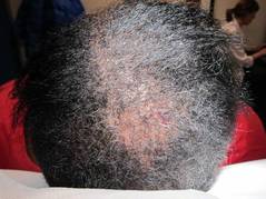
Diagnosing hair loss in black patients starts with right questions
NEW YORK – To get to the root cause of hair loss in African American patients, ask them about their chemical styling and physical styling practices, advised Dr. Roopal V. Kundu.
"Ask them about the hair products they use; how they style their hair; what kind of relaxers they use; what kind of heat, how much of it, and how often they use it; and find out if they’re wearing extensions or weaves," she said to a packed room during the American Academy of Dermatology summer meeting.
"And get their hair stylist’s input," said Dr. Kundu, director of the Center for Ethnic Skin at Northwestern University, Chicago.
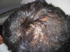
Central centrifugal cicatricial alopecia (CCCA) is one of the most common types of hair loss in African American women, Dr. Kundu said. The reason for the prevalence of CCCA in this population remains unclear, but data from several studies point not only to genetic and ethnic predispositions, but also to hair styling practices.
Data from one study showed that women with CCCA were significantly more likely to report recent traumatic hairstyling than were those who did not have the condition (J. Am. Acad. Dermatol. 2009;60:574-8).
"Educate your patients [about the effect of hairstyling practices on hair loss] as early as possible," and suspect CCCA in patients who present with a lot of breakage in their hair, even if they have no sign of alopecia or scalp inflammation, Dr. Kundu said.
Keep chemical and physical hairstyling practices in mind when examining and diagnosing hair loss in black women, she noted.
Chemical relaxers, also known as perms, are one of the most common treatments used by African American women. The relaxers are used to loosen tight curls and are applied every 6-8 weeks to new growth only. There are two types of relaxers: lye (professional product) or no lye (at-home use). The lye relaxers have more potential for irritation, Dr. Kundu said.
Physical styling, which usually pulls on hair, also can be a factor in hair loss, she explained. Some popular types of physical styling include:
• Braids, which often involve added synthetic hair and are left in place for weeks to months.
• Twisting, which involves interlocking two pieces of hair to create a loose or bound-down, three-dimensional hair style.
• Cornrows, which are braids that lie flat against the scalp, with or without added hair, that can cause excessive tension on the hair.
• Locks or dreads, which are matted coils of hair.
• Weaves, which can be clipped, glued, or sewn into place.
In addition, thermal styling (with tools such as flatirons) is a popular treatment that can damage the hair because of the high temperature of the iron, Dr. Kundu said.
When examining a skin of color patient who presents with hair loss, note their hairstyle first, Dr. Kundu emphasized. "Ask them to take their hairpiece off [if they are wearing one]. If the hairstyle doesn’t allow you to examine the scalp, the patient needs to come back between hairstyles," she said.
Grab a dermatoscope and examine the scalp, Dr. Kundu said. Look for hair loss, hair breakage, and traction. She cited a recent study suggesting that early CCCA should be considered in the differential diagnosis of these patients (Arch. Dermatol. 2012;148:1047-52).
"And don’t dismiss scalp dysesthesia* in a black woman," Dr. Kundu emphasized. "I really take this to heart," she said.
Biopsy the patient, "and always go for the periphery to see if there is active inflammation," since the central area of hair loss is usually scarred, she noted.
Dr. Kundu said that she usually prescribes potent to superpotent topical steroids, such as fluocinonide daily, and then reduces the steroid to 1-3 times a week for maintenance. She also performs intralesional injections every 6 weeks, and stops if she sees no efficacy after 3-4 serial injections.
When prescribing minoxidil, she advised taking the patient’s facial hair into consideration, and warns them about additional facial hair growth.

For patients with significant inflammation, Dr. Kundu said she first turns to doxycycline (starting at 100 mg twice a day and tapering to anti-inflammatory doses of 50 mg daily) for a total of 3-6 months.
Meanwhile, advise women with hair loss or hair loss concerns to modify their grooming practices to avoid hairstyles that pull on the scalp, she said. Also advise them to let their hair go natural completely for at least a year if possible. "It’s a hard job to do for some patients," she acknowledged, so at least encourage them to apply chemical relaxers less frequently, every 10-12 weeks instead, she said.
In addition, ask these patients to refrain from using heat on their hair more than once or twice per week, and suggest air-drying instead, she said.
Dr. Kundu had no relevant financial conflicts to disclose.
On Twitter @naseemsmiller
*CORRECTION, 10/21/13: An earlier version of this article misstated the effects of dysesthesia.
NEW YORK – To get to the root cause of hair loss in African American patients, ask them about their chemical styling and physical styling practices, advised Dr. Roopal V. Kundu.
"Ask them about the hair products they use; how they style their hair; what kind of relaxers they use; what kind of heat, how much of it, and how often they use it; and find out if they’re wearing extensions or weaves," she said to a packed room during the American Academy of Dermatology summer meeting.
"And get their hair stylist’s input," said Dr. Kundu, director of the Center for Ethnic Skin at Northwestern University, Chicago.
Central centrifugal cicatricial alopecia (CCCA) is one of the most common types of hair loss in African American women, Dr. Kundu said. The reason for the prevalence of CCCA in this population remains unclear, but data from several studies point not only to genetic and ethnic predispositions, but also to hair styling practices.
Data from one study showed that women with CCCA were significantly more likely to report recent traumatic hairstyling than were those who did not have the condition (J. Am. Acad. Dermatol. 2009;60:574-8).
"Educate your patients [about the effect of hairstyling practices on hair loss] as early as possible," and suspect CCCA in patients who present with a lot of breakage in their hair, even if they have no sign of alopecia or scalp inflammation, Dr. Kundu said.
Keep chemical and physical hairstyling practices in mind when examining and diagnosing hair loss in black women, she noted.
Chemical relaxers, also known as perms, are one of the most common treatments used by African American women. The relaxers are used to loosen tight curls and are applied every 6-8 weeks to new growth only. There are two types of relaxers: lye (professional product) or no lye (at-home use). The lye relaxers have more potential for irritation, Dr. Kundu said.
Physical styling, which usually pulls on hair, also can be a factor in hair loss, she explained. Some popular types of physical styling include:
• Braids, which often involve added synthetic hair and are left in place for weeks to months.
• Twisting, which involves interlocking two pieces of hair to create a loose or bound-down, three-dimensional hair style.
• Cornrows, which are braids that lie flat against the scalp, with or without added hair, that can cause excessive tension on the hair.
• Locks or dreads, which are matted coils of hair.
• Weaves, which can be clipped, glued, or sewn into place.
In addition, thermal styling (with tools such as flatirons) is a popular treatment that can damage the hair because of the high temperature of the iron, Dr. Kundu said.
When examining a skin of color patient who presents with hair loss, note their hairstyle first, Dr. Kundu emphasized. "Ask them to take their hairpiece off [if they are wearing one]. If the hairstyle doesn’t allow you to examine the scalp, the patient needs to come back between hairstyles," she said.
Grab a dermatoscope and examine the scalp, Dr. Kundu said. Look for hair loss, hair breakage, and traction. She cited a recent study suggesting that early CCCA should be considered in the differential diagnosis of these patients (Arch. Dermatol. 2012;148:1047-52).
"And don’t dismiss scalp dysesthesia* in a black woman," Dr. Kundu emphasized. "I really take this to heart," she said.
Biopsy the patient, "and always go for the periphery to see if there is active inflammation," since the central area of hair loss is usually scarred, she noted.
Dr. Kundu said that she usually prescribes potent to superpotent topical steroids, such as fluocinonide daily, and then reduces the steroid to 1-3 times a week for maintenance. She also performs intralesional injections every 6 weeks, and stops if she sees no efficacy after 3-4 serial injections.
When prescribing minoxidil, she advised taking the patient’s facial hair into consideration, and warns them about additional facial hair growth.
For patients with significant inflammation, Dr. Kundu said she first turns to doxycycline (starting at 100 mg twice a day and tapering to anti-inflammatory doses of 50 mg daily) for a total of 3-6 months.
Meanwhile, advise women with hair loss or hair loss concerns to modify their grooming practices to avoid hairstyles that pull on the scalp, she said. Also advise them to let their hair go natural completely for at least a year if possible. "It’s a hard job to do for some patients," she acknowledged, so at least encourage them to apply chemical relaxers less frequently, every 10-12 weeks instead, she said.
In addition, ask these patients to refrain from using heat on their hair more than once or twice per week, and suggest air-drying instead, she said.
Dr. Kundu had no relevant financial conflicts to disclose.
On Twitter @naseemsmiller
*CORRECTION, 10/21/13: An earlier version of this article misstated the effects of dysesthesia.
AT THE AAD SUMMER ACADEMY 2013

Study hints at obesity paradox in older women with coronary artery disease
AMSTERDAM – Older women with coronary artery disease who lost weight had an increased risk of death regardless of their body mass index, a finding that hints at the presence of an obesity paradox in this population, according to a registry-based retrospective study.
The unpublished abstract, which was presented at the annual congress of the European Society of Cardiology, also showed that, among patients who maintained their weight, underweight women had a twofold increase in risk of all-cause mortality while obese and overweight women had a nonsignificant but reduced risk of death, compared with normal-weight patients.
Although studies have shown that obesity is a risk factor for developing cardiovascular disease, and guidelines recommend weight loss for patients who are overweight and obese, the study’s key message is that advising weight loss to patients with heart disease is not so simple and straightforward, said Dr. Diethelm Tschoepe of Ruhr University in Bochum, Germany, who commented on the study.
Researchers analyzed data for 1,685 women with an average age of 64 years who were diagnosed with coronary artery disease (CAD) based on angiography between 2005 and 2011.
They stratified the cohort into three weight-change groups: no change (gain or loss of less than 2 kg/year), weight loss (loss of more than 2 kg/year), and weight gain (gain of more than 2 kg/year.)
Each group was then divided into four weight classes based on body mass index: underweight (BMI less than 20 kg/m2), normal weight (BMI 20-24.9 kg/m2), overweight (BMI 25-29.9 kg/m2), and obese (BMI more than 30 kg/m2).
The normal-weight group was used as reference to calculate the hazard ratios. Data were adjusted for age, smoking, diabetes, previous heart surgery, use of statins and antihypertensive drugs, degree of CAD, and previous percutaneous coronary intervention.
When comparing the BMI in weight-change groups, women who were underweight and maintained their weight had a twofold increase in death (hazard ratio, 2.15; P = .03), compared with normal-weight women. Also, when these women lost weight, their risk was increased by almost twofold, although it didn’t reach statistical significance.
Among the different BMI groups, obese women who lost weight had an eightfold increase in the risk of all-cause mortality, and obese women who gained weight had a more than fourfold risk increase, compared with obese women who maintained their weight.
Meanwhile, overweight and normal-weight women who lost weight had sixfold and threefold increases in risk of death, respectively, compared with their stable-weight counterparts.
Dr. Aziza Azimi of Gentofte Hospital, Copenhagen, who presented the study, said that the findings highlight the importance of individual-based weight management.
Researchers said that, since the study subjects had comorbidities, their weight management should be handled differently from the general population. They added that, because of the retrospective design of the study, it’s not clear why the women lost weight, although, given their condition, it was most probably unintentional and due to comorbidities or medications.
Dr. Azimi and Dr. Tschoepe had no disclosures.
On Twitter @NaseemSMiller
ESC CONGRESS 2013
Major finding: Women who were underweight and maintained their weight had a significant, 2.15-fold increase in mortality, compared with normal-weight women.
Data source: Danish registry data on 1,685 women with an average age of 64 years, diagnosed with CAD between 2005 and 2011.
Disclosures: Dr. Azimi and Dr. Tschoepe had no disclosures.

Direct Flow Medical begins TAVR trial in U.S.
Direct Flow Medical is the latest company to start a transcatheter aortic valve replacement clinical trial in the United States, bringing a new name to the field, which has so far been dominated by major players like Edwards Lifesciences and Medtronic.
The California-based company’s SALUS trial is a 30-patient, single-arm, nonrandomized, multicenter feasibility study to assess the safety and efficacy of its Transcatheter Aortic Heart Valve System, which has already received the CE Mark and is commercially available in Europe.

The device is delivered through the femoral artery, has a metal-free frame, and a "low-profile delivery system designed to eliminate aortic regurgitation," according to a company statement.
"A device that is repositionable and able to pass through smaller-diameter blood vessels is an important advance in the next generation of transcatheter aortic valve replacement [TAVR] systems," Dr. William O’Neill of Henry Ford Hospital, Detroit, who performed the first Direct Flow Medical case in the United States, said in the statement. "This could help patients who have not been good candidates for earlier TAVR devices," he added.
September was relatively eventful for TAVR. Earlier in the month, the Food and Drug Administration gave the green light to Edwards to start a clinical trial on Sapien 3. And just a day before Medicare added Direct Flow’s SALUS trial to the list of its approved studies, the FDA dropped reference to specific access points from the labeling of Edwards’ Sapien valve, which is the only FDA-approved valve in the United States.
Direct Flow is planning to seek FDA approval in 2017, according to an article in the Press Democrat, a newspaper in the company’s home town of Santa Rosa, Calif.
On Twitter @NaseemSMiller
Direct Flow Medical is the latest company to start a transcatheter aortic valve replacement clinical trial in the United States, bringing a new name to the field, which has so far been dominated by major players like Edwards Lifesciences and Medtronic.
The California-based company’s SALUS trial is a 30-patient, single-arm, nonrandomized, multicenter feasibility study to assess the safety and efficacy of its Transcatheter Aortic Heart Valve System, which has already received the CE Mark and is commercially available in Europe.
The device is delivered through the femoral artery, has a metal-free frame, and a "low-profile delivery system designed to eliminate aortic regurgitation," according to a company statement.
"A device that is repositionable and able to pass through smaller-diameter blood vessels is an important advance in the next generation of transcatheter aortic valve replacement [TAVR] systems," Dr. William O’Neill of Henry Ford Hospital, Detroit, who performed the first Direct Flow Medical case in the United States, said in the statement. "This could help patients who have not been good candidates for earlier TAVR devices," he added.
September was relatively eventful for TAVR. Earlier in the month, the Food and Drug Administration gave the green light to Edwards to start a clinical trial on Sapien 3. And just a day before Medicare added Direct Flow’s SALUS trial to the list of its approved studies, the FDA dropped reference to specific access points from the labeling of Edwards’ Sapien valve, which is the only FDA-approved valve in the United States.
Direct Flow is planning to seek FDA approval in 2017, according to an article in the Press Democrat, a newspaper in the company’s home town of Santa Rosa, Calif.
On Twitter @NaseemSMiller

Use images and analogies to explain hair disorders
NEW YORK – Dr. Paradi Mirmirani has an elegant way of explaining hair loss to her postmenopausal patients.
"Think about [hair production] like an orchestra. When your body is making hair, a whole group of musicians are coming together to make music. But when you’re postmenopausal, the estrogen isn’t there, the head violinist isn’t there, and it’s not going to be the same, but you’re still making music. It’s not going to be same sound, but it’s still there," she said.
Dr. Mirmirani of the University of California, San Francisco, is well recognized for her research in hair disorders, and during her presentation at the American Academy of Dermatology’s summer meeting, she shared her tips on diagnosing and treating different types of hair loss and alopecia in women:
• Telogen effluvium (I’m shedding gobs of hair!)
Find out whether the patient has a history of weight loss or is on new oral contraceptives, Dr. Mirmirani said. Use a hair shaft contrast card and search the scalp for scarring or scaling. Also, perform a "pull test" (for bulb) and "tug test" (for shaft), she advised. For laboratory data, she recommended ferritin and TSH, and possibly tests for antinuclear antibodies and vitamin D levels, and a biopsy of the area. Treat the underlying problem, not the hair, and assure patients that they will not go bald, she said.
• Traction alopecia (I’ve got a bald spot!)
Start by asking these patients what they do for hair care and styling, said Dr. Mirmirani. On exam, look for a telltale "fringe sign," which she and her colleagues described in a paper as "the presence of retained hairs along the frontal and/or temporal rim." (J. Clin. Exp. Dermatol. Res. 2011;2:117).
She recommended ordering ferritin and TSH tests for these patients, and possibly vitamin D. Also, treat any inflammation; consider nonspecific hair growth treatments such as minoxidil, and surgical hair restoration, and remind patients to treat their hair gently, she said.
• Alopecia areata
Ask alopecia areata patients about a personal or family history of atopic disorders, Dr. Mirmirani advised. Treatment options include intralesional corticosteroids (10 mg/cc to 2 cc total); topical corticosteroids; topical minoxidil 5% twice daily; short-contact anthralin (up to 30 minutes); topical immunotherapy; or psoralen + ultraviolet A (PUVA) treatment. Finally, remind patients that alopecia areata is an autoimmune condition that is not contagious, Dr. Mirmirani said. Compare it to an unwelcome house guest, she suggested.
• Female pattern hair loss (midlife hair crisis)
Start care for these patients by ordering (only if virilized) free and total T, dehydroepiandrosterone sulfate, and prolactin tests, Dr. Mirmirani said. Her recommended treatment protocol: minoxidil 2% or 5% solution twice daily, or 5% foam once daily; finasteride (1 mg) or spironolactone. Also, consider hair restoration or cosmetics that bring the scalp’s color closer to hair color and make hair loss less apparent, she suggested. This is when to remind patients that "the orchestra is still playing" (hair may still be produced) although estrogen (the first violinist) is absent, so it may not be quite the same.
• Acquired trichorrhexis nodosa (My hair just won’t grow!)
Acquired trichorrhexis nodosa is the most commonly reported hair shaft defect, Dr. Mirmirani said. It often results from excessive chemical processes and heat applied to hair. She described her typical acquired trichorrhexis nodosa patient as a 24-year-old black woman who washes her every 2 weeks and straightens every 6 weeks, her hair has been breaking off in the back, started after a recent color, with no symptoms.
In these patients, and exam usually shows that the overall hair density is good, no alopecia, a localized area of short hair with blunt ends, and a positive tug test (hair breaks off easily). The scalp often has mild scaling, but no pustules.
Make a hair mount, and show the patient her hair under the microscope; "it’s the easiest and most satisfying thing you can do," said Dr. Mirmirani.
Advise patients to use gentle hair care, trim unhealthy hair, and avoid heat and chemicals, she said. Wigs are fine for these patients, and most will recover the condition of their hair within a year or 2, she added.
• Cicatricial alopecia
Start with a biopsy around the margin of early active area, and then culture the pustules, said Dr. Mirmirani. Dermatopathology can show whether sebaceous glands are absent, and the degree of inflammatory infiltrate.
Explain to these patients that the hair roots or bulbs have been damaged, she added. Tell them regrowth is not possible, but they can relieve the signs and symptoms of the condition and prevent it from spreading. Describe it as "like a wildfire; we want to contain it, and halt the spread," she suggested.
Treat the predominantly lymphocytic patients with anti-inflammatories (intralesionals and topicals; antibiotics and antimalarials; systemic anti-inflammatory therapies), said Dr. Mirmirani. For lichen planopilaris and frontal fibrosing alopecia, try PPAR-gamma agonists; use topical minoxidil or finasteride to promote nonspecific hair growth; or try cosmetic or surgical therapies, she said.
Treat folliculitis decalvans with antibacterial treatments and staph eradication, and treat dissecting cellulitis with intralesionals/anti-inflammatory drugs; perform incision and drainage; use isotretinoin and antitumor-necrosis factor; and consider laser hair removal, she noted.
Dr. Mirmirani said that, in her experience, many dermatologists dread seeing hair-loss patients because of a lack of training in how to care for them. She shared several educational resources, including a reference book she coauthored, "Cicatricial Alopecia: An Approach to Diagnosis and Management," (New York: Springer, 2011) to help clinicians and residents better understand and treat hair disorders, especially the rare kinds. She also recommended the North American Hair Research Society and the Cicatricial Alopecia Research Foundation as useful resources.
Dr. Mirmirani has been an investigator and/or consultant for Johnson & Johnson, as well as Procter & Gamble.
On Twitter @NaseemSMiller
Primer on Pediatric Acne
NEW YORK – The American Acne and Rosacea Society, with the endorsement of the American Academy of Pediatrics, published evidence-based recommendations for the diagnosis and treatment of pediatric acne that incorporated mid-childhood acne, which is a sign of hyperandrogenism that must be worked up.
The guidelines "are a great reference for you for treatment algorithms and an excellent resource for treating pediatric acne," Dr. Andrea L. Zaenglein said during a presentation at the American Academy of Dermatology’s summer meeting.
Dr. Zaenglein provided a brief, but detailed summary of the classifications during her presentation, and offered some tips and practice pearls along the way.
Neonatal acne
This acne occurs in less than 20% of healthy infants who are between 2 weeks and 3 months old, and it presents in the form of papules and pustules. This common eruption rarely needs treatment, but clinicians can prescribe a topical antifungal, such as 2% ketoconazole cream, if pressed by the parents, Dr. Zaenglein noted.
Infantile acne
Unlike neonatal acne, this condition is true acne, said Dr. Zaenglein, professor of dermatology and pediatrics at Penn State/Hershey (Penn.) Medical Center. The acne, which usually presents as comedones, occurs between 3 and 6 months of age and lasts for about 1-2 years. This form of acne is usually caused by increased adrenal production of DHA, or dehydroepiandrosterone, and in boys, increased testicular androgens.
"It’s a temporary problem and usually fixes itself," said Dr. Zaenglein. "Take a full history and make sure they don’t have any signs of an underlying cause for hyperandrogenism."
Data from retrospective studies have shown that family history plays an important role, and babies with infantile acne may be at a higher risk of developing adolescent acne.
"It’s important to treat infantile acne, because it can cause scarring," said Dr. Zaenglein. The treatment principles are the same as adolescent acne. "However, remember that babies are not little adults," she said. Do not use benzoyl peroxide wash, because it can get into babies’ eyes. Instead, prescribe leave-on formulations in form of creams at lower strengths (2.5%-5%). Topical retinoids should also be used, but in a gentler formulation. Oral erythromycin is another option for more severe inflammatory acne (30 mg-50 mg/kg per day divided two or three times a day), although it might cause an upset stomach. In that case, try azithromycin, she said.
Mid-childhood acne
This new acne classification presents between 1 and 7 years of age, and requires a full exam to assess secondary sexual characteristics. Always rule out hyperandrogen states, such as Cushing’s syndrome, congenital adrenal hyperplasia, premature adrenarche, and precocious puberty, or a hormone-producing tumor, said Dr. Zaenglein. Early adrenarche also can be a risk factor for polycystic ovary syndrome. "Follow these patients closely throughout the teenage years," she advised.
The work-up for this mid-childhood acne is extensive, she said. In addition to various hormones, look at bone age and the growth chart. "And if you’re not sure, send them to a pediatric endocrinologist," she recommended.
To treat these children, apply the same principles as adolescent acne, but know that adherence is typically an issue. "Simplify the regimen, and encourage the parents to get involved," Dr. Zaenglein said.
Preadolescent acne
Acne in preadolescents is becoming more common as puberty begins earlier worldwide, said Dr. Zaenglein. The acne can present as a result of early onset of adrenarche in girls around 6 or 7 years or age, and boys between ages 7 and 8, or earlier, depending on body mass index and race. Comedonal acne in particular could be the first sign of pubertal maturation in girls, "so make sure you follow these kids," Dr. Zaenglein advised.
Preadolescent acne commonly occurs in the T-zone, and it may be a predictor of severe acne in later years, she added.
Of note, pseudoacne can occur in the nasal creases, in the form of milia, comedones, or inflamed papules. However, these lesions are not really acne and can be associated with atopic dermatitis or other conditions, said Dr. Zaenglein. They can be treated with topical antibiotics, benzoyl peroxide, or comedone extraction, she said.
The treatment principles for adolescent acne apply to preadolescent acne as well, with some modifications, Dr. Zaenglein said. Try to simplify the routines to once-daily to improve adherence, and warn parents about benzoyl peroxide staining. Also, ask about the presence of permanent teeth before considering a tetracycline, since the therapy can stain the teeth, she noted. To help kids take the pills, use a favorite breakfast spread, she suggested.
Dr. Zaenglein added that hormonal therapies should be avoided within 3 years of menarche if possible.
Dr. Zaenglein is on the advisory board for Galderma, Valeant, and Promius; a speaker for Galderma; and involved in clinical trials with Galderma, Medicis, and Valeant.
On Twitter @NaseemSMiller
EXPERT ANALYSIS FROM AAD SUMMER ACADEMY

A primer on pediatric acne
NEW YORK – In May 2013, the American Acne and Rosacea Society, with the endorsement of the American Academy of Pediatrics, published evidence-based recommendations for the diagnosis and treatment of pediatric acne that incorporated mid-childhood acne, which is a sign of hyperandrogenism that must be worked up.
The guidelines "are a great reference for you for treatment algorithms and an excellent resource for treating pediatric acne," Dr. Andrea L. Zaenglein said during a presentation at the American Academy of Dermatology’s summer meeting.

Dr. Zaenglein provided a brief, but detailed summary of the classifications during her presentation, and offered some tips and practice pearls along the way.
Neonatal acne
This acne occurs in less than 20% of healthy infants who are between 2 weeks and 3 months old, and it presents in the form of papules and pustules. This common eruption rarely needs treatment, but clinicians can prescribe a topical antifungal, such as 2% ketoconazole cream, if pressed by the parents, Dr. Zaenglein noted.
Infantile acne
Unlike neonatal acne, this condition is true acne, said Dr. Zaenglein, professor of dermatology and pediatrics at Penn State/Hershey (Penn.) Medical Center. The acne, which usually presents as comedones, occurs between 3 and 6 months of age and lasts for about 1-2 years. This form of acne is usually caused by increased adrenal production of DHA, or dehydroepiandrosterone, and in boys, increased testicular androgens.
"It’s a temporary problem and usually fixes itself," said Dr. Zaenglein. "Take a full history and make sure they don’t have any signs of an underlying cause for hyperandrogenism."
Data from retrospective studies have shown that family history plays an important role, and babies with infantile acne may be at a higher risk of developing adolescent acne.
"It’s important to treat infantile acne, because it can cause scarring," said Dr. Zaenglein. The treatment principles are the same as adolescent acne. "However, remember that babies are not little adults," she said. Do not use benzoyl peroxide wash, because it can get into babies’ eyes. Instead, prescribe leave-on formulations in form of creams at lower strengths (2.5%-5%). Topical retinoids should also be used, but in a gentler formulation. Oral erythromycin is another option for more severe inflammatory acne (30 mg-50 mg/kg per day divided two or three times a day), although it might cause an upset stomach. In that case, try azithromycin, she said.
Mid-childhood acne
This new acne classification presents between 1 and 7 years of age, and requires a full exam to assess secondary sexual characteristics. Always rule out hyperandrogen states, such as Cushing’s syndrome, congenital adrenal hyperplasia, premature adrenarche, and precocious puberty, or a hormone-producing tumor, said Dr. Zaenglein. Early adrenarche also can be a risk factor for polycystic ovary syndrome. "Follow these patients closely throughout the teenage years," she advised.
The work-up for this mid-childhood acne is extensive, she said. In addition to various hormones, look at bone age and the growth chart. "And if you’re not sure, send them to a pediatric endocrinologist," she recommended.
To treat these children, apply the same principles as adolescent acne, but know that adherence is typically an issue. "Simplify the regimen, and encourage the parents to get involved," Dr. Zaenglein said.
Preadolescent acne
Acne in preadolescents is becoming more common as puberty begins earlier worldwide, said Dr. Zaenglein. The acne can present as a result of early onset of adrenarche in girls around 6 or 7 years or age, and boys between ages 7 and 8, or earlier, depending on body mass index and race. Comedonal acne in particular could be the first sign of pubertal maturation in girls, "so make sure you follow these kids," Dr. Zaenglein advised.
Preadolescent acne commonly occurs in the T-zone, and it may be a predictor of severe acne in later years, she added.
Of note, pseudoacne can occur in the nasal creases, in the form of milia, comedones, or inflamed papules. However, these lesions are not really acne and can be associated with atopic dermatitis or other conditions, said Dr. Zaenglein. They can be treated with topical antibiotics, benzoyl peroxide, or comedone extraction, she said.
The treatment principles for adolescent acne apply to preadolescent acne as well, with some modifications, Dr. Zaenglein said. Try to simplify the routines to once-daily to improve adherence, and warn parents about benzoyl peroxide staining. Also, ask about the presence of permanent teeth before considering a tetracycline, since the therapy can stain the teeth, she noted. To help kids take the pills, use a favorite breakfast spread, she suggested.
Dr. Zaenglein added that hormonal therapies should be avoided within 3 years of menarche if possible.
Dr. Zaenglein is on the advisory board for Galderma, Valeant, and Promius; a speaker for Galderma; and involved in clinical trials with Galderma, Medicis, and Valeant.
On Twitter @NaseemSMiller
NEW YORK – In May 2013, the American Acne and Rosacea Society, with the endorsement of the American Academy of Pediatrics, published evidence-based recommendations for the diagnosis and treatment of pediatric acne that incorporated mid-childhood acne, which is a sign of hyperandrogenism that must be worked up.
The guidelines "are a great reference for you for treatment algorithms and an excellent resource for treating pediatric acne," Dr. Andrea L. Zaenglein said during a presentation at the American Academy of Dermatology’s summer meeting.
Dr. Zaenglein provided a brief, but detailed summary of the classifications during her presentation, and offered some tips and practice pearls along the way.
Neonatal acne
This acne occurs in less than 20% of healthy infants who are between 2 weeks and 3 months old, and it presents in the form of papules and pustules. This common eruption rarely needs treatment, but clinicians can prescribe a topical antifungal, such as 2% ketoconazole cream, if pressed by the parents, Dr. Zaenglein noted.
Infantile acne
Unlike neonatal acne, this condition is true acne, said Dr. Zaenglein, professor of dermatology and pediatrics at Penn State/Hershey (Penn.) Medical Center. The acne, which usually presents as comedones, occurs between 3 and 6 months of age and lasts for about 1-2 years. This form of acne is usually caused by increased adrenal production of DHA, or dehydroepiandrosterone, and in boys, increased testicular androgens.
"It’s a temporary problem and usually fixes itself," said Dr. Zaenglein. "Take a full history and make sure they don’t have any signs of an underlying cause for hyperandrogenism."
Data from retrospective studies have shown that family history plays an important role, and babies with infantile acne may be at a higher risk of developing adolescent acne.
"It’s important to treat infantile acne, because it can cause scarring," said Dr. Zaenglein. The treatment principles are the same as adolescent acne. "However, remember that babies are not little adults," she said. Do not use benzoyl peroxide wash, because it can get into babies’ eyes. Instead, prescribe leave-on formulations in form of creams at lower strengths (2.5%-5%). Topical retinoids should also be used, but in a gentler formulation. Oral erythromycin is another option for more severe inflammatory acne (30 mg-50 mg/kg per day divided two or three times a day), although it might cause an upset stomach. In that case, try azithromycin, she said.
Mid-childhood acne
This new acne classification presents between 1 and 7 years of age, and requires a full exam to assess secondary sexual characteristics. Always rule out hyperandrogen states, such as Cushing’s syndrome, congenital adrenal hyperplasia, premature adrenarche, and precocious puberty, or a hormone-producing tumor, said Dr. Zaenglein. Early adrenarche also can be a risk factor for polycystic ovary syndrome. "Follow these patients closely throughout the teenage years," she advised.
The work-up for this mid-childhood acne is extensive, she said. In addition to various hormones, look at bone age and the growth chart. "And if you’re not sure, send them to a pediatric endocrinologist," she recommended.
To treat these children, apply the same principles as adolescent acne, but know that adherence is typically an issue. "Simplify the regimen, and encourage the parents to get involved," Dr. Zaenglein said.
Preadolescent acne
Acne in preadolescents is becoming more common as puberty begins earlier worldwide, said Dr. Zaenglein. The acne can present as a result of early onset of adrenarche in girls around 6 or 7 years or age, and boys between ages 7 and 8, or earlier, depending on body mass index and race. Comedonal acne in particular could be the first sign of pubertal maturation in girls, "so make sure you follow these kids," Dr. Zaenglein advised.
Preadolescent acne commonly occurs in the T-zone, and it may be a predictor of severe acne in later years, she added.
Of note, pseudoacne can occur in the nasal creases, in the form of milia, comedones, or inflamed papules. However, these lesions are not really acne and can be associated with atopic dermatitis or other conditions, said Dr. Zaenglein. They can be treated with topical antibiotics, benzoyl peroxide, or comedone extraction, she said.
The treatment principles for adolescent acne apply to preadolescent acne as well, with some modifications, Dr. Zaenglein said. Try to simplify the routines to once-daily to improve adherence, and warn parents about benzoyl peroxide staining. Also, ask about the presence of permanent teeth before considering a tetracycline, since the therapy can stain the teeth, she noted. To help kids take the pills, use a favorite breakfast spread, she suggested.
Dr. Zaenglein added that hormonal therapies should be avoided within 3 years of menarche if possible.
Dr. Zaenglein is on the advisory board for Galderma, Valeant, and Promius; a speaker for Galderma; and involved in clinical trials with Galderma, Medicis, and Valeant.
On Twitter @NaseemSMiller
EXPERT ANALYSIS FROM AAD SUMMER ACADEMY

Teledermatology expands, but hurdles remain
NEW YORK – Teledermatology has not yet arrived at every office and institution, and reimbursement and training remain hurdles, but many researchers believe in its potential and maintain that advancements in technology will improve and expand the dermatology branch of telemedicine.
If you need a jolt of excitement, just watch our interview with Dr. Jeffrey Benabio, who is the physician director of healthcare transformation at Kaiser Permanente in San Diego. He spoke with us after a panel discussion about the role of technology in dermatology, held at the American Academy of Dermatology’s summer academy.

By January 2012, there were 37 active teledermatology programs in the United States. That number is down from 62 in 2003, according to a study by Dr. April Armstrong, director of teledermatology at the University of California, Davis, Health System, and one of the leading figures in teledermatology research. Dr. Armstrong and her colleagues published a survey of these programs in 2012.
But, "it’s not about starting a program, it’s about sustaining it,’ said Dr. Armstrong.
Her studies have shown that in practices that have sustained teledermatology over time, the median number of consultations per program nearly doubled, increasing from 184 in 2003 to 309 in 2011, albeit with a wide range.
Dr. Armstrong has reported that to sustain such practices, what’s needed are improvements in reimbursement models, technology platforms, communication with referring providers, and training.
Many of the teledermatology programs in the United States are at academic institutions (49%), Veterans Administration hospitals (27%), private practices (16%), and health maintenance organizations such as Kaiser Permanente (8%), according to Dr. Armstrong’s research. Most are concentrated in California and along the east coast; those in the middle of the country, where there are greater issues with access to health care, are few and far-between.
Meanwhile, some dermatologists are experimenting with online-only practices, such as DermatologistOnCall, DirectDermatology, and YoDerm. In addition, the AAD has sponsored a teledermatology program called AccessDerm, allowing its members in several states to provide remote care via a mobile phone.
Although the future direction of teledermatology remains unclear, Dr. Armstrong predicted that the increased use of mobile technology, greater demand for direct patient-specialist interaction, and specialists’ acceptance of telehealth as a form of care delivery will eventually lead to an expanded use of teledermatology.
Dr. Armstrong has received honoraria from several companies, including Abbott Laboratories, Amgen, and Janssen-Ortho. Dr. Benabio has received consulting fees from Dove/Unilever. He is also on the editorial advisory board for Skin & Allergy News.
On Twitter @NaseemSMiller
NEW YORK – Teledermatology has not yet arrived at every office and institution, and reimbursement and training remain hurdles, but many researchers believe in its potential and maintain that advancements in technology will improve and expand the dermatology branch of telemedicine.
If you need a jolt of excitement, just watch our interview with Dr. Jeffrey Benabio, who is the physician director of healthcare transformation at Kaiser Permanente in San Diego. He spoke with us after a panel discussion about the role of technology in dermatology, held at the American Academy of Dermatology’s summer academy.
By January 2012, there were 37 active teledermatology programs in the United States. That number is down from 62 in 2003, according to a study by Dr. April Armstrong, director of teledermatology at the University of California, Davis, Health System, and one of the leading figures in teledermatology research. Dr. Armstrong and her colleagues published a survey of these programs in 2012.
But, "it’s not about starting a program, it’s about sustaining it,’ said Dr. Armstrong.
Her studies have shown that in practices that have sustained teledermatology over time, the median number of consultations per program nearly doubled, increasing from 184 in 2003 to 309 in 2011, albeit with a wide range.
Dr. Armstrong has reported that to sustain such practices, what’s needed are improvements in reimbursement models, technology platforms, communication with referring providers, and training.
Many of the teledermatology programs in the United States are at academic institutions (49%), Veterans Administration hospitals (27%), private practices (16%), and health maintenance organizations such as Kaiser Permanente (8%), according to Dr. Armstrong’s research. Most are concentrated in California and along the east coast; those in the middle of the country, where there are greater issues with access to health care, are few and far-between.
Meanwhile, some dermatologists are experimenting with online-only practices, such as DermatologistOnCall, DirectDermatology, and YoDerm. In addition, the AAD has sponsored a teledermatology program called AccessDerm, allowing its members in several states to provide remote care via a mobile phone.
Although the future direction of teledermatology remains unclear, Dr. Armstrong predicted that the increased use of mobile technology, greater demand for direct patient-specialist interaction, and specialists’ acceptance of telehealth as a form of care delivery will eventually lead to an expanded use of teledermatology.
Dr. Armstrong has received honoraria from several companies, including Abbott Laboratories, Amgen, and Janssen-Ortho. Dr. Benabio has received consulting fees from Dove/Unilever. He is also on the editorial advisory board for Skin & Allergy News.
On Twitter @NaseemSMiller
AT THE AAD SUMMER MEETING

Advise lifestyle change to obese but metabolically healthy women
AMSTERDAM – Obese women with no metabolic disorders had the same cardiovascular disease risk as their normal-weight counterparts, according to a 5-year follow-up of more than 260,000 Danish women in their early 30s.
Meanwhile, during the same follow-up period, women with metabolic disorders had a fourfold increase in cardiovascular disease risk, regardless of their body mass index, according to Dr. Michelle D. Schmiegelow, who presented her findings at the annual congress of the European Society of Cardiology.
The absolute risks were still low in this young population, but given the findings, Dr. Schmiegelow said that physicians should have an "increased awareness of these young metabolically healthy overweight and obese women, because there might be a window of opportunity in which they could change their lifestyle and thereby reduce their risk of metabolic disorders and possibly reduce the risk of cardiovascular disease substantially."
The study was one of several presented at the meeting addressing the ongoing discussions about the obesity paradox.
"If you want to rely on the question on what is my future CVD risk, it’s not as easy to just measure your body weight,’ said Dr. Diethelm Tschöpe of Ruhr University, Bochum, Germany, in a video interview. "It’s a complex situation, which consists of body weight, risk factors, comorbidities, and the genetic background, which at the moment is hard to measure."
Dr. Schmiegelow of Gentofte Hospital, Hellerup, Denmark, and her colleagues examined the risk of myocardial infarctions, ischemic stroke, and all-cause mortality in more than 260,000 women who had given birth between 2004 and 2010, at a mean age of 31 years old. These women were identified in a nationwide registry of birth records. The median follow-up was 5.5 years after childbirth.
The women were considered metabolically unhealthy is they had a hypertensive disorder such as gestational hypertension, preeclampsia, or hypertension; an abnormal glucose metabolism such as diabetes or gestational diabetes; or dyslipidemia.
They were separated to four groups based on metabolic health status and prepregnancy body mass index (BMI): Metabolically healthy with a BMI less than 25 kg/m2; metabolically health with a BMI greater than or equal to 25; metabolically unhealthy with a BMI less than 25; and metabolically unhealthy with a BMI of greater than or equal to 25.
Data were adjusted for age and calendar year. The combined primary endpoints were the risks of MI, ischemic stroke, and all-cause mortality, using the metabolically healthy women with a BMI of less than 25 as reference.
The incidence rate (IR) of the combined endpoints for the normal weight, metabolically healthy women was 0.41 (number of events equaled 375); obese but metabolically healthy women had an IR of .45 (n = 51), and rate ratio (RR) of 1.16; normal weight but metabolically unhealthy women had an IR of .91 (n = 175), RR of 2.11; and obese and metabolically unhealthy women had an IR of 1.25 (n = 86), with an RR of 2.81.
"When you have an obese individual in front of you, that should be the first step, figuring out what their risk profile is," said Dr. Schmiegelow. "If they have metabolic disorders, that means they’re at an increased risk. If they do not have metabolic disorders, that means they’re at an increased risk of developing that, and you should really try and motivate them to change their lifestyle."
Dr. Schmiegelow and Dr. Tschöpe had no disclosures.
On Twitter @NaseemSMiller
AT THE ESC CONGRESS 2013