User login
American Academy of Dermatology (AAD): Summer Academy 2013
Diagnosing hair loss in black patients starts with right questions
NEW YORK – To get to the root cause of hair loss in African American patients, ask them about their chemical styling and physical styling practices, advised Dr. Roopal V. Kundu.
"Ask them about the hair products they use; how they style their hair; what kind of relaxers they use; what kind of heat, how much of it, and how often they use it; and find out if they’re wearing extensions or weaves," she said to a packed room during the American Academy of Dermatology summer meeting.
"And get their hair stylist’s input," said Dr. Kundu, director of the Center for Ethnic Skin at Northwestern University, Chicago.
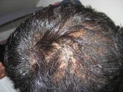
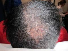
Central centrifugal cicatricial alopecia (CCCA) is one of the most common types of hair loss in African American women, Dr. Kundu said. The reason for the prevalence of CCCA in this population remains unclear, but data from several studies point not only to genetic and ethnic predispositions, but also to hair styling practices.
Data from one study showed that women with CCCA were significantly more likely to report recent traumatic hairstyling than were those who did not have the condition (J. Am. Acad. Dermatol. 2009;60:574-8).
"Educate your patients [about the effect of hairstyling practices on hair loss] as early as possible," and suspect CCCA in patients who present with a lot of breakage in their hair, even if they have no sign of alopecia or scalp inflammation, Dr. Kundu said.
Keep chemical and physical hairstyling practices in mind when examining and diagnosing hair loss in black women, she noted.
Chemical relaxers, also known as perms, are one of the most common treatments used by African American women. The relaxers are used to loosen tight curls and are applied every 6-8 weeks to new growth only. There are two types of relaxers: lye (professional product) or no lye (at-home use). The lye relaxers have more potential for irritation, Dr. Kundu said.
Physical styling, which usually pulls on hair, also can be a factor in hair loss, she explained. Some popular types of physical styling include:
• Braids, which often involve added synthetic hair and are left in place for weeks to months.
• Twisting, which involves interlocking two pieces of hair to create a loose or bound-down, three-dimensional hair style.
• Cornrows, which are braids that lie flat against the scalp, with or without added hair, that can cause excessive tension on the hair.
• Locks or dreads, which are matted coils of hair.
• Weaves, which can be clipped, glued, or sewn into place.
In addition, thermal styling (with tools such as flatirons) is a popular treatment that can damage the hair because of the high temperature of the iron, Dr. Kundu said.
When examining a skin of color patient who presents with hair loss, note their hairstyle first, Dr. Kundu emphasized. "Ask them to take their hairpiece off [if they are wearing one]. If the hairstyle doesn’t allow you to examine the scalp, the patient needs to come back between hairstyles," she said.
Grab a dermatoscope and examine the scalp, Dr. Kundu said. Look for hair loss, hair breakage, and traction. She cited a recent study suggesting that early CCCA should be considered in the differential diagnosis of these patients (Arch. Dermatol. 2012;148:1047-52).
"And don’t dismiss scalp dysesthesia* in a black woman," Dr. Kundu emphasized. "I really take this to heart," she said.
Biopsy the patient, "and always go for the periphery to see if there is active inflammation," since the central area of hair loss is usually scarred, she noted.
Dr. Kundu said that she usually prescribes potent to superpotent topical steroids, such as fluocinonide daily, and then reduces the steroid to 1-3 times a week for maintenance. She also performs intralesional injections every 6 weeks, and stops if she sees no efficacy after 3-4 serial injections.
When prescribing minoxidil, she advised taking the patient’s facial hair into consideration, and warns them about additional facial hair growth.

For patients with significant inflammation, Dr. Kundu said she first turns to doxycycline (starting at 100 mg twice a day and tapering to anti-inflammatory doses of 50 mg daily) for a total of 3-6 months.
Meanwhile, advise women with hair loss or hair loss concerns to modify their grooming practices to avoid hairstyles that pull on the scalp, she said. Also advise them to let their hair go natural completely for at least a year if possible. "It’s a hard job to do for some patients," she acknowledged, so at least encourage them to apply chemical relaxers less frequently, every 10-12 weeks instead, she said.
In addition, ask these patients to refrain from using heat on their hair more than once or twice per week, and suggest air-drying instead, she said.
Dr. Kundu had no relevant financial conflicts to disclose.
On Twitter @naseemsmiller
*CORRECTION, 10/21/13: An earlier version of this article misstated the effects of dysesthesia.
NEW YORK – To get to the root cause of hair loss in African American patients, ask them about their chemical styling and physical styling practices, advised Dr. Roopal V. Kundu.
"Ask them about the hair products they use; how they style their hair; what kind of relaxers they use; what kind of heat, how much of it, and how often they use it; and find out if they’re wearing extensions or weaves," she said to a packed room during the American Academy of Dermatology summer meeting.
"And get their hair stylist’s input," said Dr. Kundu, director of the Center for Ethnic Skin at Northwestern University, Chicago.
Central centrifugal cicatricial alopecia (CCCA) is one of the most common types of hair loss in African American women, Dr. Kundu said. The reason for the prevalence of CCCA in this population remains unclear, but data from several studies point not only to genetic and ethnic predispositions, but also to hair styling practices.
Data from one study showed that women with CCCA were significantly more likely to report recent traumatic hairstyling than were those who did not have the condition (J. Am. Acad. Dermatol. 2009;60:574-8).
"Educate your patients [about the effect of hairstyling practices on hair loss] as early as possible," and suspect CCCA in patients who present with a lot of breakage in their hair, even if they have no sign of alopecia or scalp inflammation, Dr. Kundu said.
Keep chemical and physical hairstyling practices in mind when examining and diagnosing hair loss in black women, she noted.
Chemical relaxers, also known as perms, are one of the most common treatments used by African American women. The relaxers are used to loosen tight curls and are applied every 6-8 weeks to new growth only. There are two types of relaxers: lye (professional product) or no lye (at-home use). The lye relaxers have more potential for irritation, Dr. Kundu said.
Physical styling, which usually pulls on hair, also can be a factor in hair loss, she explained. Some popular types of physical styling include:
• Braids, which often involve added synthetic hair and are left in place for weeks to months.
• Twisting, which involves interlocking two pieces of hair to create a loose or bound-down, three-dimensional hair style.
• Cornrows, which are braids that lie flat against the scalp, with or without added hair, that can cause excessive tension on the hair.
• Locks or dreads, which are matted coils of hair.
• Weaves, which can be clipped, glued, or sewn into place.
In addition, thermal styling (with tools such as flatirons) is a popular treatment that can damage the hair because of the high temperature of the iron, Dr. Kundu said.
When examining a skin of color patient who presents with hair loss, note their hairstyle first, Dr. Kundu emphasized. "Ask them to take their hairpiece off [if they are wearing one]. If the hairstyle doesn’t allow you to examine the scalp, the patient needs to come back between hairstyles," she said.
Grab a dermatoscope and examine the scalp, Dr. Kundu said. Look for hair loss, hair breakage, and traction. She cited a recent study suggesting that early CCCA should be considered in the differential diagnosis of these patients (Arch. Dermatol. 2012;148:1047-52).
"And don’t dismiss scalp dysesthesia* in a black woman," Dr. Kundu emphasized. "I really take this to heart," she said.
Biopsy the patient, "and always go for the periphery to see if there is active inflammation," since the central area of hair loss is usually scarred, she noted.
Dr. Kundu said that she usually prescribes potent to superpotent topical steroids, such as fluocinonide daily, and then reduces the steroid to 1-3 times a week for maintenance. She also performs intralesional injections every 6 weeks, and stops if she sees no efficacy after 3-4 serial injections.
When prescribing minoxidil, she advised taking the patient’s facial hair into consideration, and warns them about additional facial hair growth.
For patients with significant inflammation, Dr. Kundu said she first turns to doxycycline (starting at 100 mg twice a day and tapering to anti-inflammatory doses of 50 mg daily) for a total of 3-6 months.
Meanwhile, advise women with hair loss or hair loss concerns to modify their grooming practices to avoid hairstyles that pull on the scalp, she said. Also advise them to let their hair go natural completely for at least a year if possible. "It’s a hard job to do for some patients," she acknowledged, so at least encourage them to apply chemical relaxers less frequently, every 10-12 weeks instead, she said.
In addition, ask these patients to refrain from using heat on their hair more than once or twice per week, and suggest air-drying instead, she said.
Dr. Kundu had no relevant financial conflicts to disclose.
On Twitter @naseemsmiller
*CORRECTION, 10/21/13: An earlier version of this article misstated the effects of dysesthesia.
AT THE AAD SUMMER ACADEMY 2013

Three simple principles improve procedural safety
NEW YORK – Clinicians, think about how to eliminate – not just reduce – the risk of exposure to bloodborne pathogens in your practice, said Dr. Joseph F. Sobanko of the University of Pennsylvania, Philadelphia.
"Dermatology, with its balance of medical and surgical procedures, is a specialty with unique risks," Dr. Sobanko said in a presentation at the American Academy of Dermatology summer meeting. While the risk of exposure to bloodborne pathogens is broadly shared in health-care delivery, Dr. Sobanko cited data from a recent survey of dermatology residents showing that confirmed rates of inadvertent exposure to patient blood remain high in this population (J. Am. Acad. Dermatol. 2011;65:648-50). Most instances of exposure were needle sticks.

"The majority of those surveyed considered themselves responsible for the exposure," said Dr. Sobanko. The list of safety procedures on how to avoid exposure to bloodborne pathogens is long, and includes never recapping needles and avoiding hand-to-hand transfer of sharps, he said.
However, Dr. Sobanko condensed his "zero-risk" recommendations into three principles: mental preparedness, physical protection, and motor repetition.
The violation of any of the three principles, and especially all three simultaneously, likely explains the evidence that injury risk is highest in individuals with the least training, he said.
One example of mental preparedness is detailed preoperative planning, such as placing the instrument tray in a convenient and neutral spot so that anyone involved in the procedure can access the tools, Dr. Sobanko said. In his own center, the nurses have been supplied with photos of proper setup of the instrument tray for each procedure so that all the tools are always placed in the same spot.
For physical protection, Dr. Sobanko emphasized the importance of using shields and goggles instead of glasses. He cited a study in which shields were tested for blood after asking clinicians if there had been blood splatter. The presence of blood was only correctly predicted 10% of the time. Dr. Sobanko also cited data supporting the safety of double-gloving. Although he acknowledged that some clinicians report reduced dexterity, he said that this relative disadvantage can probably be overcome with practice.
Performing the procedure repeatedly provides its own protection, according to Dr. Sobanko, who cited data of an inverse relationship between risk of exposure to a bloodborne pathogen and experience. These data suggests the need for greatly increased vigilance during procedures with which one is less familiar, he said.
Creating a culture of safety is also important, with emphasis on safety over speed. In the survey of dermatology residents, the sense of feeling rushed was identified as an important factor in needle stick injuries, Dr. Sobanko said.
Dr. Sobanko, who is preparing an atlas of procedural safety in dermatology with Dr. Jacob Levitt of Mount Sinai School of Medicine, N.Y., added that this culture shift to an emphasis on safety falls under his category of mental preparedness.
When asked for additional perspective on safety, Dr. William K. Andersen, a dermatologist in private practice in Lancaster, Penn., agreed with Dr. Sobanko’s points, but he suggested that it is not enough to post a list of safety procedures. All health-care personnel involved in a given procedure must function together.
"Everybody has to be on board even when you have the protocols in place," Dr. Anderson noted. When everyone is cooperating, there is less risk in the face of unexpected events, he said.
Dr. Sobanko had no relevant financial conflicts to disclose.
NEW YORK – Clinicians, think about how to eliminate – not just reduce – the risk of exposure to bloodborne pathogens in your practice, said Dr. Joseph F. Sobanko of the University of Pennsylvania, Philadelphia.
"Dermatology, with its balance of medical and surgical procedures, is a specialty with unique risks," Dr. Sobanko said in a presentation at the American Academy of Dermatology summer meeting. While the risk of exposure to bloodborne pathogens is broadly shared in health-care delivery, Dr. Sobanko cited data from a recent survey of dermatology residents showing that confirmed rates of inadvertent exposure to patient blood remain high in this population (J. Am. Acad. Dermatol. 2011;65:648-50). Most instances of exposure were needle sticks.
"The majority of those surveyed considered themselves responsible for the exposure," said Dr. Sobanko. The list of safety procedures on how to avoid exposure to bloodborne pathogens is long, and includes never recapping needles and avoiding hand-to-hand transfer of sharps, he said.
However, Dr. Sobanko condensed his "zero-risk" recommendations into three principles: mental preparedness, physical protection, and motor repetition.
The violation of any of the three principles, and especially all three simultaneously, likely explains the evidence that injury risk is highest in individuals with the least training, he said.
One example of mental preparedness is detailed preoperative planning, such as placing the instrument tray in a convenient and neutral spot so that anyone involved in the procedure can access the tools, Dr. Sobanko said. In his own center, the nurses have been supplied with photos of proper setup of the instrument tray for each procedure so that all the tools are always placed in the same spot.
For physical protection, Dr. Sobanko emphasized the importance of using shields and goggles instead of glasses. He cited a study in which shields were tested for blood after asking clinicians if there had been blood splatter. The presence of blood was only correctly predicted 10% of the time. Dr. Sobanko also cited data supporting the safety of double-gloving. Although he acknowledged that some clinicians report reduced dexterity, he said that this relative disadvantage can probably be overcome with practice.
Performing the procedure repeatedly provides its own protection, according to Dr. Sobanko, who cited data of an inverse relationship between risk of exposure to a bloodborne pathogen and experience. These data suggests the need for greatly increased vigilance during procedures with which one is less familiar, he said.
Creating a culture of safety is also important, with emphasis on safety over speed. In the survey of dermatology residents, the sense of feeling rushed was identified as an important factor in needle stick injuries, Dr. Sobanko said.
Dr. Sobanko, who is preparing an atlas of procedural safety in dermatology with Dr. Jacob Levitt of Mount Sinai School of Medicine, N.Y., added that this culture shift to an emphasis on safety falls under his category of mental preparedness.
When asked for additional perspective on safety, Dr. William K. Andersen, a dermatologist in private practice in Lancaster, Penn., agreed with Dr. Sobanko’s points, but he suggested that it is not enough to post a list of safety procedures. All health-care personnel involved in a given procedure must function together.
"Everybody has to be on board even when you have the protocols in place," Dr. Anderson noted. When everyone is cooperating, there is less risk in the face of unexpected events, he said.
Dr. Sobanko had no relevant financial conflicts to disclose.
EXPERT ANALYSIS FROM THE AAD SUMMER ACADEMY 2013

Use images and analogies to explain hair disorders
NEW YORK – Dr. Paradi Mirmirani has an elegant way of explaining hair loss to her postmenopausal patients.
"Think about [hair production] like an orchestra. When your body is making hair, a whole group of musicians are coming together to make music. But when you’re postmenopausal, the estrogen isn’t there, the head violinist isn’t there, and it’s not going to be the same, but you’re still making music. It’s not going to be same sound, but it’s still there," she said.
Dr. Mirmirani of the University of California, San Francisco, is well recognized for her research in hair disorders, and during her presentation at the American Academy of Dermatology’s summer meeting, she shared her tips on diagnosing and treating different types of hair loss and alopecia in women:
• Telogen effluvium (I’m shedding gobs of hair!)
Find out whether the patient has a history of weight loss or is on new oral contraceptives, Dr. Mirmirani said. Use a hair shaft contrast card and search the scalp for scarring or scaling. Also, perform a "pull test" (for bulb) and "tug test" (for shaft), she advised. For laboratory data, she recommended ferritin and TSH, and possibly tests for antinuclear antibodies and vitamin D levels, and a biopsy of the area. Treat the underlying problem, not the hair, and assure patients that they will not go bald, she said.
• Traction alopecia (I’ve got a bald spot!)
Start by asking these patients what they do for hair care and styling, said Dr. Mirmirani. On exam, look for a telltale "fringe sign," which she and her colleagues described in a paper as "the presence of retained hairs along the frontal and/or temporal rim." (J. Clin. Exp. Dermatol. Res. 2011;2:117).
She recommended ordering ferritin and TSH tests for these patients, and possibly vitamin D. Also, treat any inflammation; consider nonspecific hair growth treatments such as minoxidil, and surgical hair restoration, and remind patients to treat their hair gently, she said.
• Alopecia areata
Ask alopecia areata patients about a personal or family history of atopic disorders, Dr. Mirmirani advised. Treatment options include intralesional corticosteroids (10 mg/cc to 2 cc total); topical corticosteroids; topical minoxidil 5% twice daily; short-contact anthralin (up to 30 minutes); topical immunotherapy; or psoralen + ultraviolet A (PUVA) treatment. Finally, remind patients that alopecia areata is an autoimmune condition that is not contagious, Dr. Mirmirani said. Compare it to an unwelcome house guest, she suggested.
• Female pattern hair loss (midlife hair crisis)
Start care for these patients by ordering (only if virilized) free and total T, dehydroepiandrosterone sulfate, and prolactin tests, Dr. Mirmirani said. Her recommended treatment protocol: minoxidil 2% or 5% solution twice daily, or 5% foam once daily; finasteride (1 mg) or spironolactone. Also, consider hair restoration or cosmetics that bring the scalp’s color closer to hair color and make hair loss less apparent, she suggested. This is when to remind patients that "the orchestra is still playing" (hair may still be produced) although estrogen (the first violinist) is absent, so it may not be quite the same.
• Acquired trichorrhexis nodosa (My hair just won’t grow!)
Acquired trichorrhexis nodosa is the most commonly reported hair shaft defect, Dr. Mirmirani said. It often results from excessive chemical processes and heat applied to hair. She described her typical acquired trichorrhexis nodosa patient as a 24-year-old black woman who washes her every 2 weeks and straightens every 6 weeks, her hair has been breaking off in the back, started after a recent color, with no symptoms.
In these patients, and exam usually shows that the overall hair density is good, no alopecia, a localized area of short hair with blunt ends, and a positive tug test (hair breaks off easily). The scalp often has mild scaling, but no pustules.
Make a hair mount, and show the patient her hair under the microscope; "it’s the easiest and most satisfying thing you can do," said Dr. Mirmirani.
Advise patients to use gentle hair care, trim unhealthy hair, and avoid heat and chemicals, she said. Wigs are fine for these patients, and most will recover the condition of their hair within a year or 2, she added.
• Cicatricial alopecia
Start with a biopsy around the margin of early active area, and then culture the pustules, said Dr. Mirmirani. Dermatopathology can show whether sebaceous glands are absent, and the degree of inflammatory infiltrate.
Explain to these patients that the hair roots or bulbs have been damaged, she added. Tell them regrowth is not possible, but they can relieve the signs and symptoms of the condition and prevent it from spreading. Describe it as "like a wildfire; we want to contain it, and halt the spread," she suggested.
Treat the predominantly lymphocytic patients with anti-inflammatories (intralesionals and topicals; antibiotics and antimalarials; systemic anti-inflammatory therapies), said Dr. Mirmirani. For lichen planopilaris and frontal fibrosing alopecia, try PPAR-gamma agonists; use topical minoxidil or finasteride to promote nonspecific hair growth; or try cosmetic or surgical therapies, she said.
Treat folliculitis decalvans with antibacterial treatments and staph eradication, and treat dissecting cellulitis with intralesionals/anti-inflammatory drugs; perform incision and drainage; use isotretinoin and antitumor-necrosis factor; and consider laser hair removal, she noted.
Dr. Mirmirani said that, in her experience, many dermatologists dread seeing hair-loss patients because of a lack of training in how to care for them. She shared several educational resources, including a reference book she coauthored, "Cicatricial Alopecia: An Approach to Diagnosis and Management," (New York: Springer, 2011) to help clinicians and residents better understand and treat hair disorders, especially the rare kinds. She also recommended the North American Hair Research Society and the Cicatricial Alopecia Research Foundation as useful resources.
Dr. Mirmirani has been an investigator and/or consultant for Johnson & Johnson, as well as Procter & Gamble.
On Twitter @NaseemSMiller
A primer on pediatric acne
NEW YORK – In May 2013, the American Acne and Rosacea Society, with the endorsement of the American Academy of Pediatrics, published evidence-based recommendations for the diagnosis and treatment of pediatric acne that incorporated mid-childhood acne, which is a sign of hyperandrogenism that must be worked up.
The guidelines "are a great reference for you for treatment algorithms and an excellent resource for treating pediatric acne," Dr. Andrea L. Zaenglein said during a presentation at the American Academy of Dermatology’s summer meeting.

Dr. Zaenglein provided a brief, but detailed summary of the classifications during her presentation, and offered some tips and practice pearls along the way.
Neonatal acne
This acne occurs in less than 20% of healthy infants who are between 2 weeks and 3 months old, and it presents in the form of papules and pustules. This common eruption rarely needs treatment, but clinicians can prescribe a topical antifungal, such as 2% ketoconazole cream, if pressed by the parents, Dr. Zaenglein noted.
Infantile acne
Unlike neonatal acne, this condition is true acne, said Dr. Zaenglein, professor of dermatology and pediatrics at Penn State/Hershey (Penn.) Medical Center. The acne, which usually presents as comedones, occurs between 3 and 6 months of age and lasts for about 1-2 years. This form of acne is usually caused by increased adrenal production of DHA, or dehydroepiandrosterone, and in boys, increased testicular androgens.
"It’s a temporary problem and usually fixes itself," said Dr. Zaenglein. "Take a full history and make sure they don’t have any signs of an underlying cause for hyperandrogenism."
Data from retrospective studies have shown that family history plays an important role, and babies with infantile acne may be at a higher risk of developing adolescent acne.
"It’s important to treat infantile acne, because it can cause scarring," said Dr. Zaenglein. The treatment principles are the same as adolescent acne. "However, remember that babies are not little adults," she said. Do not use benzoyl peroxide wash, because it can get into babies’ eyes. Instead, prescribe leave-on formulations in form of creams at lower strengths (2.5%-5%). Topical retinoids should also be used, but in a gentler formulation. Oral erythromycin is another option for more severe inflammatory acne (30 mg-50 mg/kg per day divided two or three times a day), although it might cause an upset stomach. In that case, try azithromycin, she said.
Mid-childhood acne
This new acne classification presents between 1 and 7 years of age, and requires a full exam to assess secondary sexual characteristics. Always rule out hyperandrogen states, such as Cushing’s syndrome, congenital adrenal hyperplasia, premature adrenarche, and precocious puberty, or a hormone-producing tumor, said Dr. Zaenglein. Early adrenarche also can be a risk factor for polycystic ovary syndrome. "Follow these patients closely throughout the teenage years," she advised.
The work-up for this mid-childhood acne is extensive, she said. In addition to various hormones, look at bone age and the growth chart. "And if you’re not sure, send them to a pediatric endocrinologist," she recommended.
To treat these children, apply the same principles as adolescent acne, but know that adherence is typically an issue. "Simplify the regimen, and encourage the parents to get involved," Dr. Zaenglein said.
Preadolescent acne
Acne in preadolescents is becoming more common as puberty begins earlier worldwide, said Dr. Zaenglein. The acne can present as a result of early onset of adrenarche in girls around 6 or 7 years or age, and boys between ages 7 and 8, or earlier, depending on body mass index and race. Comedonal acne in particular could be the first sign of pubertal maturation in girls, "so make sure you follow these kids," Dr. Zaenglein advised.
Preadolescent acne commonly occurs in the T-zone, and it may be a predictor of severe acne in later years, she added.
Of note, pseudoacne can occur in the nasal creases, in the form of milia, comedones, or inflamed papules. However, these lesions are not really acne and can be associated with atopic dermatitis or other conditions, said Dr. Zaenglein. They can be treated with topical antibiotics, benzoyl peroxide, or comedone extraction, she said.
The treatment principles for adolescent acne apply to preadolescent acne as well, with some modifications, Dr. Zaenglein said. Try to simplify the routines to once-daily to improve adherence, and warn parents about benzoyl peroxide staining. Also, ask about the presence of permanent teeth before considering a tetracycline, since the therapy can stain the teeth, she noted. To help kids take the pills, use a favorite breakfast spread, she suggested.
Dr. Zaenglein added that hormonal therapies should be avoided within 3 years of menarche if possible.
Dr. Zaenglein is on the advisory board for Galderma, Valeant, and Promius; a speaker for Galderma; and involved in clinical trials with Galderma, Medicis, and Valeant.
On Twitter @NaseemSMiller
NEW YORK – In May 2013, the American Acne and Rosacea Society, with the endorsement of the American Academy of Pediatrics, published evidence-based recommendations for the diagnosis and treatment of pediatric acne that incorporated mid-childhood acne, which is a sign of hyperandrogenism that must be worked up.
The guidelines "are a great reference for you for treatment algorithms and an excellent resource for treating pediatric acne," Dr. Andrea L. Zaenglein said during a presentation at the American Academy of Dermatology’s summer meeting.
Dr. Zaenglein provided a brief, but detailed summary of the classifications during her presentation, and offered some tips and practice pearls along the way.
Neonatal acne
This acne occurs in less than 20% of healthy infants who are between 2 weeks and 3 months old, and it presents in the form of papules and pustules. This common eruption rarely needs treatment, but clinicians can prescribe a topical antifungal, such as 2% ketoconazole cream, if pressed by the parents, Dr. Zaenglein noted.
Infantile acne
Unlike neonatal acne, this condition is true acne, said Dr. Zaenglein, professor of dermatology and pediatrics at Penn State/Hershey (Penn.) Medical Center. The acne, which usually presents as comedones, occurs between 3 and 6 months of age and lasts for about 1-2 years. This form of acne is usually caused by increased adrenal production of DHA, or dehydroepiandrosterone, and in boys, increased testicular androgens.
"It’s a temporary problem and usually fixes itself," said Dr. Zaenglein. "Take a full history and make sure they don’t have any signs of an underlying cause for hyperandrogenism."
Data from retrospective studies have shown that family history plays an important role, and babies with infantile acne may be at a higher risk of developing adolescent acne.
"It’s important to treat infantile acne, because it can cause scarring," said Dr. Zaenglein. The treatment principles are the same as adolescent acne. "However, remember that babies are not little adults," she said. Do not use benzoyl peroxide wash, because it can get into babies’ eyes. Instead, prescribe leave-on formulations in form of creams at lower strengths (2.5%-5%). Topical retinoids should also be used, but in a gentler formulation. Oral erythromycin is another option for more severe inflammatory acne (30 mg-50 mg/kg per day divided two or three times a day), although it might cause an upset stomach. In that case, try azithromycin, she said.
Mid-childhood acne
This new acne classification presents between 1 and 7 years of age, and requires a full exam to assess secondary sexual characteristics. Always rule out hyperandrogen states, such as Cushing’s syndrome, congenital adrenal hyperplasia, premature adrenarche, and precocious puberty, or a hormone-producing tumor, said Dr. Zaenglein. Early adrenarche also can be a risk factor for polycystic ovary syndrome. "Follow these patients closely throughout the teenage years," she advised.
The work-up for this mid-childhood acne is extensive, she said. In addition to various hormones, look at bone age and the growth chart. "And if you’re not sure, send them to a pediatric endocrinologist," she recommended.
To treat these children, apply the same principles as adolescent acne, but know that adherence is typically an issue. "Simplify the regimen, and encourage the parents to get involved," Dr. Zaenglein said.
Preadolescent acne
Acne in preadolescents is becoming more common as puberty begins earlier worldwide, said Dr. Zaenglein. The acne can present as a result of early onset of adrenarche in girls around 6 or 7 years or age, and boys between ages 7 and 8, or earlier, depending on body mass index and race. Comedonal acne in particular could be the first sign of pubertal maturation in girls, "so make sure you follow these kids," Dr. Zaenglein advised.
Preadolescent acne commonly occurs in the T-zone, and it may be a predictor of severe acne in later years, she added.
Of note, pseudoacne can occur in the nasal creases, in the form of milia, comedones, or inflamed papules. However, these lesions are not really acne and can be associated with atopic dermatitis or other conditions, said Dr. Zaenglein. They can be treated with topical antibiotics, benzoyl peroxide, or comedone extraction, she said.
The treatment principles for adolescent acne apply to preadolescent acne as well, with some modifications, Dr. Zaenglein said. Try to simplify the routines to once-daily to improve adherence, and warn parents about benzoyl peroxide staining. Also, ask about the presence of permanent teeth before considering a tetracycline, since the therapy can stain the teeth, she noted. To help kids take the pills, use a favorite breakfast spread, she suggested.
Dr. Zaenglein added that hormonal therapies should be avoided within 3 years of menarche if possible.
Dr. Zaenglein is on the advisory board for Galderma, Valeant, and Promius; a speaker for Galderma; and involved in clinical trials with Galderma, Medicis, and Valeant.
On Twitter @NaseemSMiller
EXPERT ANALYSIS FROM AAD SUMMER ACADEMY

Teledermatology expands, but hurdles remain
NEW YORK – Teledermatology has not yet arrived at every office and institution, and reimbursement and training remain hurdles, but many researchers believe in its potential and maintain that advancements in technology will improve and expand the dermatology branch of telemedicine.
If you need a jolt of excitement, just watch our interview with Dr. Jeffrey Benabio, who is the physician director of healthcare transformation at Kaiser Permanente in San Diego. He spoke with us after a panel discussion about the role of technology in dermatology, held at the American Academy of Dermatology’s summer academy.

By January 2012, there were 37 active teledermatology programs in the United States. That number is down from 62 in 2003, according to a study by Dr. April Armstrong, director of teledermatology at the University of California, Davis, Health System, and one of the leading figures in teledermatology research. Dr. Armstrong and her colleagues published a survey of these programs in 2012.
But, "it’s not about starting a program, it’s about sustaining it,’ said Dr. Armstrong.
Her studies have shown that in practices that have sustained teledermatology over time, the median number of consultations per program nearly doubled, increasing from 184 in 2003 to 309 in 2011, albeit with a wide range.
Dr. Armstrong has reported that to sustain such practices, what’s needed are improvements in reimbursement models, technology platforms, communication with referring providers, and training.
Many of the teledermatology programs in the United States are at academic institutions (49%), Veterans Administration hospitals (27%), private practices (16%), and health maintenance organizations such as Kaiser Permanente (8%), according to Dr. Armstrong’s research. Most are concentrated in California and along the east coast; those in the middle of the country, where there are greater issues with access to health care, are few and far-between.
Meanwhile, some dermatologists are experimenting with online-only practices, such as DermatologistOnCall, DirectDermatology, and YoDerm. In addition, the AAD has sponsored a teledermatology program called AccessDerm, allowing its members in several states to provide remote care via a mobile phone.
Although the future direction of teledermatology remains unclear, Dr. Armstrong predicted that the increased use of mobile technology, greater demand for direct patient-specialist interaction, and specialists’ acceptance of telehealth as a form of care delivery will eventually lead to an expanded use of teledermatology.
Dr. Armstrong has received honoraria from several companies, including Abbott Laboratories, Amgen, and Janssen-Ortho. Dr. Benabio has received consulting fees from Dove/Unilever. He is also on the editorial advisory board for Skin & Allergy News.
On Twitter @NaseemSMiller
NEW YORK – Teledermatology has not yet arrived at every office and institution, and reimbursement and training remain hurdles, but many researchers believe in its potential and maintain that advancements in technology will improve and expand the dermatology branch of telemedicine.
If you need a jolt of excitement, just watch our interview with Dr. Jeffrey Benabio, who is the physician director of healthcare transformation at Kaiser Permanente in San Diego. He spoke with us after a panel discussion about the role of technology in dermatology, held at the American Academy of Dermatology’s summer academy.
By January 2012, there were 37 active teledermatology programs in the United States. That number is down from 62 in 2003, according to a study by Dr. April Armstrong, director of teledermatology at the University of California, Davis, Health System, and one of the leading figures in teledermatology research. Dr. Armstrong and her colleagues published a survey of these programs in 2012.
But, "it’s not about starting a program, it’s about sustaining it,’ said Dr. Armstrong.
Her studies have shown that in practices that have sustained teledermatology over time, the median number of consultations per program nearly doubled, increasing from 184 in 2003 to 309 in 2011, albeit with a wide range.
Dr. Armstrong has reported that to sustain such practices, what’s needed are improvements in reimbursement models, technology platforms, communication with referring providers, and training.
Many of the teledermatology programs in the United States are at academic institutions (49%), Veterans Administration hospitals (27%), private practices (16%), and health maintenance organizations such as Kaiser Permanente (8%), according to Dr. Armstrong’s research. Most are concentrated in California and along the east coast; those in the middle of the country, where there are greater issues with access to health care, are few and far-between.
Meanwhile, some dermatologists are experimenting with online-only practices, such as DermatologistOnCall, DirectDermatology, and YoDerm. In addition, the AAD has sponsored a teledermatology program called AccessDerm, allowing its members in several states to provide remote care via a mobile phone.
Although the future direction of teledermatology remains unclear, Dr. Armstrong predicted that the increased use of mobile technology, greater demand for direct patient-specialist interaction, and specialists’ acceptance of telehealth as a form of care delivery will eventually lead to an expanded use of teledermatology.
Dr. Armstrong has received honoraria from several companies, including Abbott Laboratories, Amgen, and Janssen-Ortho. Dr. Benabio has received consulting fees from Dove/Unilever. He is also on the editorial advisory board for Skin & Allergy News.
On Twitter @NaseemSMiller
AT THE AAD SUMMER MEETING

Intensive ‘Boot Camp’ protocol improves kids’ atopic dermatitis
NEW YORK – To frustrated parents of children with severe atopic dermatitis, Dr. Sheilagh Maguiness offers a fast and effective remedy: a 2-week intensive treatment she calls Boot Camp. Instead of push-ups, however, this Boot Camp involves bleach baths, wet wraps, steroids, moisturizers, and sometimes oral antibiotics or antihistamines.
During the 2-week period, the treatment plan attempts to address all aspects of the disease – dry skin, itching, inflammation, and infection or colonization – simultaneously.

Dr. Maguiness’ instructions for the severe atopic dermatitis Boot Camp are as follows:
• For 2 weeks, bathe the child nightly in a dilute bleach bath of lukewarm water for 10 minutes. (No soap is needed, or use unscented Dove or Cetaphil for armpits, groin, hands, and feet.) After 2 weeks, use lukewarm water alone.
Data have shown that the baths are safe, said Dr. Maguiness of Harvard University, Boston, and Boston Children’s Hospital. In a recent randomized trial of 31 children, those who underwent regular, dilute bleach baths showed improvement in their atopic dermatitis with no increased susceptibility to infection (Pediatrics 2009;123:e808-14). The recipe for the bleach baths is ¼ to ½ cup of plain Clorox bleach in a full tub of bath water. (With more concentrated forms of Clorox, use 1/3 cup per full tub.) Comfort the worried parents by letting them know that the concentration is similar to that found in a swimming pool, and is safe for head and neck areas, she said in a presentation at the American Academy of Dermatology summer meeting.
• After bathing, pat the skin dry, and within 3 minutes apply triamcinolone (0.1% ointment) to affected areas on the body, and hydrocortisone (2.5% ointment) to the face.
• Follow the bath with a thick moisturizer (Vaseline, Aquaphor, or Hydrolatum ointment). Use the moisturizer on top of the medication twice daily, even if no bath is taken. Avoid lotions.
• Add wet pajama wraps nightly for 1-2 weeks as part of the Boot Camp and/or for acute flares, Dr. Maguiness noted. Studies have shown that wet wraps plus topical corticosteroids are effective, and can promote rapid improvement in the majority of patients, with very few side effects. In a recent retrospective study of 216 pediatric patients who underwent this combination, all of them showed at least 25% improvement, and 45% had 75-100% improvement (J. Am. Acad. Dermatol. 2012;67:100-6).
Advise parents to use long-sleeve cotton pajamas or Onesies for wet wraps, Dr. Maguiness added. Parents should soak the garment in warm water, wring it out until it’s slightly damp, then dress the child, and put on a dry outfit on top of the wet one. They should make sure the room is warm so the child can go to sleep.
In cases of obvious superinfection, prescribe an appropriate oral antibiotic such as cephalexin.
For itching at night, sedating antihistamines such as hydroxyzine 10 mg/5 mL, 30 minutes before bedtime can be helpful, Dr. Maguiness noted. For daytime itching, she suggested fexofenadine or cetirizine in the morning. Have a few "go-to" agents, she advised. For infants, consider fluocinolone oil or triamcinalone 0.025% ointment. For children, consider triamcinalone 0.1% ointment. For teenagers (or for use in more localized areas with younger children), consider stronger agents such as mometasone or fluocinonide ointment.
Limited studies support some systemic therapies for use in pediatric atopic dermatitis, including narrow-band ultraviolet B (nb UVB); cyclosporine and mycophenolate mofetil; and methotrexate and azathioprine, Dr. Maguiness said. Perhaps in the future, biologics such as tocilizumab may emerge as additional treatment options, she added. While cyclosporine can help with the rapid clearance of the disease, methotrexate and mycophenolate mofetil may represent longer-term options, she said.
Meanwhile, don’t be surprised if families resist the Boot Camp at first, Dr. Maguiness said.
"You have to get the families on your side," she said. "They’re terrified of bleach baths, wet pajamas, and topical steroids. Let them know there’s no faster or safer way to improve their child’s condition in 2 weeks or less."
So invest time in the first visit, educate your patients and parents about the treatment plan, and give them printed instructions and handouts, Dr. Maguiness said.
Finally, don’t forget to obtain cultures to rule out possible superinfection, as well as schedule a prompt follow-up visit in about 2 weeks.
At this visit, after following the Boot Camp regimen, the eczematous areas should be mostly cleared, with possible residual hot spots, she said. Begin to taper the topical steroids, bleach baths, and wet wraps at night.
Despite the challenges of treating severe atopic dermatitis in children, she told her audience that treating these young patients has been one of the most rewarding parts of her practice.
Dr. Maguiness had no relevant disclosures.
On Twitter @naseemsmiller
NEW YORK – To frustrated parents of children with severe atopic dermatitis, Dr. Sheilagh Maguiness offers a fast and effective remedy: a 2-week intensive treatment she calls Boot Camp. Instead of push-ups, however, this Boot Camp involves bleach baths, wet wraps, steroids, moisturizers, and sometimes oral antibiotics or antihistamines.
During the 2-week period, the treatment plan attempts to address all aspects of the disease – dry skin, itching, inflammation, and infection or colonization – simultaneously.
Dr. Maguiness’ instructions for the severe atopic dermatitis Boot Camp are as follows:
• For 2 weeks, bathe the child nightly in a dilute bleach bath of lukewarm water for 10 minutes. (No soap is needed, or use unscented Dove or Cetaphil for armpits, groin, hands, and feet.) After 2 weeks, use lukewarm water alone.
Data have shown that the baths are safe, said Dr. Maguiness of Harvard University, Boston, and Boston Children’s Hospital. In a recent randomized trial of 31 children, those who underwent regular, dilute bleach baths showed improvement in their atopic dermatitis with no increased susceptibility to infection (Pediatrics 2009;123:e808-14). The recipe for the bleach baths is ¼ to ½ cup of plain Clorox bleach in a full tub of bath water. (With more concentrated forms of Clorox, use 1/3 cup per full tub.) Comfort the worried parents by letting them know that the concentration is similar to that found in a swimming pool, and is safe for head and neck areas, she said in a presentation at the American Academy of Dermatology summer meeting.
• After bathing, pat the skin dry, and within 3 minutes apply triamcinolone (0.1% ointment) to affected areas on the body, and hydrocortisone (2.5% ointment) to the face.
• Follow the bath with a thick moisturizer (Vaseline, Aquaphor, or Hydrolatum ointment). Use the moisturizer on top of the medication twice daily, even if no bath is taken. Avoid lotions.
• Add wet pajama wraps nightly for 1-2 weeks as part of the Boot Camp and/or for acute flares, Dr. Maguiness noted. Studies have shown that wet wraps plus topical corticosteroids are effective, and can promote rapid improvement in the majority of patients, with very few side effects. In a recent retrospective study of 216 pediatric patients who underwent this combination, all of them showed at least 25% improvement, and 45% had 75-100% improvement (J. Am. Acad. Dermatol. 2012;67:100-6).
Advise parents to use long-sleeve cotton pajamas or Onesies for wet wraps, Dr. Maguiness added. Parents should soak the garment in warm water, wring it out until it’s slightly damp, then dress the child, and put on a dry outfit on top of the wet one. They should make sure the room is warm so the child can go to sleep.
In cases of obvious superinfection, prescribe an appropriate oral antibiotic such as cephalexin.
For itching at night, sedating antihistamines such as hydroxyzine 10 mg/5 mL, 30 minutes before bedtime can be helpful, Dr. Maguiness noted. For daytime itching, she suggested fexofenadine or cetirizine in the morning. Have a few "go-to" agents, she advised. For infants, consider fluocinolone oil or triamcinalone 0.025% ointment. For children, consider triamcinalone 0.1% ointment. For teenagers (or for use in more localized areas with younger children), consider stronger agents such as mometasone or fluocinonide ointment.
Limited studies support some systemic therapies for use in pediatric atopic dermatitis, including narrow-band ultraviolet B (nb UVB); cyclosporine and mycophenolate mofetil; and methotrexate and azathioprine, Dr. Maguiness said. Perhaps in the future, biologics such as tocilizumab may emerge as additional treatment options, she added. While cyclosporine can help with the rapid clearance of the disease, methotrexate and mycophenolate mofetil may represent longer-term options, she said.
Meanwhile, don’t be surprised if families resist the Boot Camp at first, Dr. Maguiness said.
"You have to get the families on your side," she said. "They’re terrified of bleach baths, wet pajamas, and topical steroids. Let them know there’s no faster or safer way to improve their child’s condition in 2 weeks or less."
So invest time in the first visit, educate your patients and parents about the treatment plan, and give them printed instructions and handouts, Dr. Maguiness said.
Finally, don’t forget to obtain cultures to rule out possible superinfection, as well as schedule a prompt follow-up visit in about 2 weeks.
At this visit, after following the Boot Camp regimen, the eczematous areas should be mostly cleared, with possible residual hot spots, she said. Begin to taper the topical steroids, bleach baths, and wet wraps at night.
Despite the challenges of treating severe atopic dermatitis in children, she told her audience that treating these young patients has been one of the most rewarding parts of her practice.
Dr. Maguiness had no relevant disclosures.
On Twitter @naseemsmiller
EXPERT ANALYSIS FROM AAD SUMMER ACADEMY 2013

PTPB formaldehyde resin proposed as pediatric allergen of the year
NEW YORK – One expert has characterized para-tertiary-butylphenol formaldehyde resin as a suitable candidate for the next pediatric contact allergen of the year, based on a growing number of case reports.
Para-tertiary-butylphenol (PTPB) formaldehyde resin is found in shin guards, push-up bras, and numerous other products used by children and adolescents.
Published case reports of contact allergies caused by PTPB formaldehyde resin date back several years, but use of this resin as an adhesive in a broad range of products – particularly leather goods, such as shoes, purses, and watch straps – appears to be increasing, according to Dr. Nanette B. Silverberg.
Dr. Silverberg presented a review of new trends in pediatric contact dermatitis at the American Academy of Dermatology summer meeting.
The causes of contact allergies in children evolve constantly because of the introduction of new products, and this should influence the order in which patch testing of specific allergens is performed, said Dr. Silverberg, a dermatologist at St. Luke’s–Roosevelt Hospital Center, New York.
In her update, she cited several recently designated "allergens of the year" from the American Contact Dermatitis Society (ACDS) and added some of her own.
Dr. Silverberg also emphasized tricks she has devised to increase the likelihood of a successful patch test, not least of which is enlisting parent cooperation. "It is important to convince parents that they are guilt free," she advised. In parents who begin to blame themselves for exposures, a sense of denial can interfere with efforts to cooperate with patch testing. To avoid this problem, she suggested a proactive effort to establish they are fault free. "Blame it on marketing if you must."
In light of the potential for lack of compliance, patch testing should be made as effortless as possible. For one approach, Dr. Silverberg suggested timing testing so it is not an excuse to miss school. Another tip was to use "heaps of tape and more tape" to keep the patch tests in place, and then provide patients with a roll of tape when they leave the office. She also recommended thinking in advance of strategies to reduce the risk of patch tests being inadvertently or intentionally removed by children, such as gauze wrap around the abdomen.
"The number one way to make patch tests work in young children is to keep your patch test panel streamlined and focused," Dr. Silverberg said. Particularly in young children, she suggested just one or two panels rather than three or four. While bribes such as lollipops and stickers "should be used judiciously," Dr. Silverberg said it is very helpful to outline each patch with highlighters that glow under Wood’s lamp to make results easier to assess.
Contact allergies are extremely common, and it is often possible to control the contact dermatitis before or even without confirming the allergen. For example, dialkyl thiourea, the ACDS allergen of the year in 2009, is another source of contact dermatitis to shin guards as well as other products, but reactions to this or PTPB formaldehyde resin can often be prevented simply by putting a layer of clothing in front of the skin. By the same principle, contact dermatitis from car seats is often caused by dimethyl fumerate, the ACDS allergen of the year in 2011, but can be prevented simply by laying fabric or creating some other barrier to prevent contact.
Methylisothiazolinone, a preservative and antifungal agent often found in moist towels and wipes, was recognized as the most recent (2013) ACDS allergen of the year. Dr. Silverberg noted that these wipes have surprisingly broad applications. While dermatitis on the hands and buttocks would be expected, she cautioned that patients use these to clean other skin surfaces, so a broad index of suspicion is needed.
The most common contact allergens, such as nickel, cobalt, and neomycin, remain relatively unchanged from year to year, but many of these products are used less frequently in children’s products in appreciation of the risks that they pose. The challenge is keeping abreast of evolving trends. While patch testing is an effective tool for identifying the source of a contact dermatitis to alert parents and patients of the products they should avoid, she said that technique is critical to patch test success.
Dr. Silverberg said she had no financial disclosures.
EXPERT ANALYSIS FROM THE AAD SUMMER ACADEMY 2013
Hemangiomas don’t always resolve, so refer
NEW YORK – Too many parents are incorrectly reassured that their child’s hemangioma will spontaneously resolve, and that no immediate treatment is needed, according to Dr. Francine Blei, medical director of the Vascular Birthmark Institute of New York at Roosevelt Hospital.
"I cannot stress enough the importance of early referral," Dr. Blei said.
Although some hemangiomas will resolve with time, those that require treatment will have the lowest risk for functional defects and the best cosmetic outcome if intervention occurs early, Dr. Blei said at the American Academy of Dermatology summer meeting.
For clinicians with experience managing these lesions, early examination can help separate those at risk of permanent disability or disfigurement from those whose lesions will resolve with an acceptable result, she said.
Infantile hemangiomas are part of an array of vascular anomalies, of which the most serious can involve internal organs and be life-threatening. Based on emerging information regarding the etiology and histopathology of vascular anomalies, the International Society for the Study of Vascular Abnormalities (ISSVA) has updated its classification system several times. Given recent progress, particularly related to the role of genetics in the pathophysiology of these lesions, new terminology is "in the works" and will be a topic of discussion at the ISSVA Workshop on Vascular Anomalies in April 2014 in Melbourne, Dr. Blei said. The two broad categories in the current terminology separate vascular tumors (such as infantile hemangiomas, which occur in up to 10% of infants) from vascular malformations, which are not necessarily congenital.
"The inconsistent terminology has been a problem," noted Dr. Blei, citing a study from the Vascular Anomalies Center at Boston Children’s Hospital. Data from this study showed that only 53% of 5,621 cases reviewed had a correct referral diagnosis (J. Pediatr. Surg. 2011;46:1784-9). Although referral diagnoses were correct for 77.8% of infantile hemangiomas, the rate was only 45.6% for vascular malformations.
For infantile hemangiomas, Dr. Blei offered a series of simple questions for clinicians to ask to help determine the risk of an adverse outcome. Although size and location are important, she emphasized looking for associated symptoms, such as stridor, ptosis, ulceration, and bleeding. While some parents may seek a conservative approach in the hope that the lesions will resolve without an invasive procedure, Dr. Blei said that she is "dogmatic" in regard to urging aggressive intervention when there is a risk of persistent functional impairment.
Other questions useful in understanding the prognosis and guiding the therapy of infantile hemangiomas include whether the lesion has subcutaneous involvement, whether there are multiple lesions, and whether the lesion is focal or segmental. For segmental lesions, a particular concern is whether the hemangioma is a manifestation of a larger syndrome, such as PHACE, a condition that includes cardiac abnormalities, and LUMBAR, which includes spinal and urogenital complications.
The age of the child at the time of evaluation is also relevant, particularly for differentiating infantile from congenital hemangiomas, Dr. Blei noted. Infantile hemangiomas begin proliferation soon after birth and, on average, reach their peak proliferation at 8-10 months of age before involuting slowly. By contrast, congenital hemangiomas are fully developed at birth and, unlike infantile hemangiomas, are negative for the immunohistochemical marker GLUT1. One kind of congenital hemangioma, called RICH (rapidly involuting congenital hemangioma), reaches an advanced stage of spontaneous resolution within the first several months of life. The other major form, called NICH (noninvoluting congenital hemangioma) persists through childhood.
If infantile hemangiomas are identified early, their treatment may be more effective and less invasive, said Dr. Blei. She noted that an adequate response can be achieved with topical beta-blockers in some cases. Oral beta-blockers and other systemic therapies also may allow for a relatively conservative resolution. Pulse dye lasers have a greater likelihood of efficacy if used early rather than after the hemangioma has become bulky, she added.
"If you treat early, you limit the size," Dr. Blei said. If you wait, the opportunity for complete or near-complete resolution may be lost, she added.
Dr. Blei had no financial conflicts to disclose.
EXPERT ANALYSIS FROM THE AAD SUMMER ACADEMY 2013
Nail complications of cancer therapies on the rise
NEW YORK – Nail complications are increasingly recognized as a problematic side effect of chemotherapies and biologic therapies for cancer patients.
In some cases these are cosmetic issues, but the side effects really do interfere with activities of daily living for many cancer patients, Dr. Patricia S. Myskowski, an attending dermatologist at Memorial Sloan Kettering Cancer Center, New York, reported at the American Academy of Dermatology summer meeting.

In a list of toxicities provided by the National Cancer Institute in 2006, only three categories of nail toxicities were listed, and these employed relatively vague descriptions, according to Dr. Myskowski. More recent summaries, including a literature review (J. Oncol. Pharm. Pract. 2009;15:143-55), have helped to classify and quantify nail complications as well as provide therapeutic guidance.
Taxanes, and specifically docetaxel, are "the worst offenders" of chemotherapies resulting in nails disorders, she said. More than 80% of patients treated with multiple cycles of docetaxel will develop some nail changes. Most are cosmetic reactions, such as depigmentation, but nearly one-third of patients have reactions that interfere with activities of daily living.
One of the most bothersome of these complications is subungual hematomas with hemopurulent discharge. In patients with nail infection, antibiotics may accelerate drainage and healing, but Dr. Myskowski suggested that this complication can be avoided by keeping the nails trimmed as short as possible.
Short nails are associated with a reduced risk of secondary infection, said Dr. Myskowski, who advised bacterial and fungal cultures when infection is suspected.
Biological therapies, particularly epidermal growth factor receptor (EGFR) inhibitors and tyrosine kinase inhibitors (TKIs), also are associated with a high rate of nail disorders, including paronychia, pyogenic granuloma, and infection. Although nail disorders are far less common than the characteristic rash associated with these agents, she cited one study suggesting a 12% incidence of symptomatic paronychia on EGFR inhibitors. Typically, nail complications emerge about 2 months after treatment is initiated.
To prevent paronychia associated with EGFR inhibitors, Dr. Myskowksi recommended following guidelines issued by the National Comprehensive Cancer Network (J. Natl. Compr. Canc. Netw. 2009;75:S5-S21).
Recommendations include avoiding frequent water immersion as well as contact with harsh chemicals. Applying petroleum jelly to the periungual soft tissue may be protective. In the event of nail infection, augmenting antibiotic therapy with topical therapies such as silver nitrate solution or white vinegar soaks may speed healing. In patients who have reinfections of toenails, disposing of shoes that may harbor bacteria sometimes resolves the problem.
Dr. Myskowski reported no financial disclosures relevant to her presentation.
NEW YORK – Nail complications are increasingly recognized as a problematic side effect of chemotherapies and biologic therapies for cancer patients.
In some cases these are cosmetic issues, but the side effects really do interfere with activities of daily living for many cancer patients, Dr. Patricia S. Myskowski, an attending dermatologist at Memorial Sloan Kettering Cancer Center, New York, reported at the American Academy of Dermatology summer meeting.
In a list of toxicities provided by the National Cancer Institute in 2006, only three categories of nail toxicities were listed, and these employed relatively vague descriptions, according to Dr. Myskowski. More recent summaries, including a literature review (J. Oncol. Pharm. Pract. 2009;15:143-55), have helped to classify and quantify nail complications as well as provide therapeutic guidance.
Taxanes, and specifically docetaxel, are "the worst offenders" of chemotherapies resulting in nails disorders, she said. More than 80% of patients treated with multiple cycles of docetaxel will develop some nail changes. Most are cosmetic reactions, such as depigmentation, but nearly one-third of patients have reactions that interfere with activities of daily living.
One of the most bothersome of these complications is subungual hematomas with hemopurulent discharge. In patients with nail infection, antibiotics may accelerate drainage and healing, but Dr. Myskowski suggested that this complication can be avoided by keeping the nails trimmed as short as possible.
Short nails are associated with a reduced risk of secondary infection, said Dr. Myskowski, who advised bacterial and fungal cultures when infection is suspected.
Biological therapies, particularly epidermal growth factor receptor (EGFR) inhibitors and tyrosine kinase inhibitors (TKIs), also are associated with a high rate of nail disorders, including paronychia, pyogenic granuloma, and infection. Although nail disorders are far less common than the characteristic rash associated with these agents, she cited one study suggesting a 12% incidence of symptomatic paronychia on EGFR inhibitors. Typically, nail complications emerge about 2 months after treatment is initiated.
To prevent paronychia associated with EGFR inhibitors, Dr. Myskowksi recommended following guidelines issued by the National Comprehensive Cancer Network (J. Natl. Compr. Canc. Netw. 2009;75:S5-S21).
Recommendations include avoiding frequent water immersion as well as contact with harsh chemicals. Applying petroleum jelly to the periungual soft tissue may be protective. In the event of nail infection, augmenting antibiotic therapy with topical therapies such as silver nitrate solution or white vinegar soaks may speed healing. In patients who have reinfections of toenails, disposing of shoes that may harbor bacteria sometimes resolves the problem.
Dr. Myskowski reported no financial disclosures relevant to her presentation.
EXPERT ANALYSIS FROM THE AAD SUMMER ACADEMY 2013

Illinois bans tanning beds for youngsters
Illinois has become the sixth state to ban indoor tanning for youth younger than 18 years. The bill, which was introduced earlier this year and signed into law by Gov. Patrick Quinn on Aug. 15, will take effect on Jan. 1, 2014.
"The state’s willingness to follow the examples set by the cities of Springfield and Chicago exemplifies a true commitment to protecting teens from the dangers of indoor tanning," Dr. Dirk M. Elston, president of the American Academy of Dermatology Association, said in a statement.
The academy said in a news release that the law is based on "significant scientific evidence that links indoor tanning to increased risk of developing melanoma and other forms of skin cancer."
Compared with previous years, 2013 has been an active year for tanning bed laws.
In a video interview earlier this month, Dr. Bruce A. Brod, the AAD’s State Policy Committee Chair, spoke about the Food and Drug Administration’s proposal to place stricter regulations on indoor tanning beds and issue stronger recommendations against their use by anyone younger than 18 years. The agency is considering the comments and may issue its final ruling by the end of 2013.
Meanwhile, in June, the Internal Revenue Service issued the final regulation on collecting a 10% tax on tanning salon receipts. The regulation was first proposed in 2010 and exempts "qualified physical fitness facilities" that offer tanning services from collecting the tax.
Groups such as the Indoor Tanning Association and the American Suntanning Association continue to lobby against the tanning ban laws.
Illinois joins California, Vermont, Oregon, Nevada, and Texas in passing age-related restrictions on the use of tanning beds. New York, New Jersey, and Connecticut have bans for youth under 17 years of age, while the District of Columbia and West Virginia have tanning bed bans for individuals younger than 14 years.
On Twitter @naseemsmiller