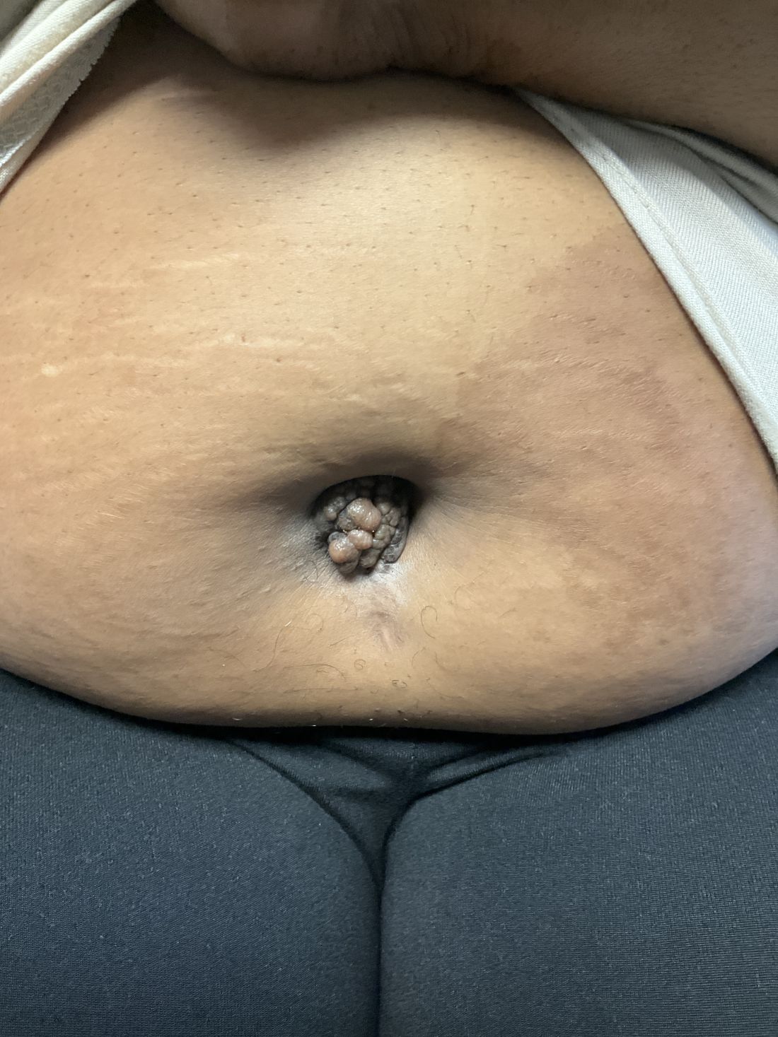
A 27-year-old Haitian woman presented with a painful umbilical mass which had been growing in size for 5 months

Article Type
Changed
Tue, 01/16/2024 - 09:24

Endometriosis is defined as the presence of endometrial tissue outside of the uterine cavity, commonly occurring in women of reproductive age. The condition usually affects the adnexa (ovaries, Fallopian tubes, and associated ligaments and connective tissue) but can also be seen in extrapelvic structures.

Cutaneous endometriosis is an uncommon subtype that accounts for 1% of endometriosis cases and occurs when endometrial tissue is found on the surface of the skin. It is divided into primary and secondary cutaneous endometriosis. The etiology of primary cutaneous endometriosis is idiopathic, while the secondary form is believed to be a consequence of abdominal or pelvic procedures that may lead to seeding of endometrial tissue on the skin. In the case of our patient, it appears that her laparoscopic procedure 2 years ago was the cause of endometrial seeding in the umbilicus.

Clinically, the condition may present with a palpable mass, cyclic pain, and bloody discharge from the affected area. Due to the rarity of cutaneous endometriosis, it may be hard to distinguish from other diagnoses such as keloids, dermatofibromas, hernias, or cutaneous metastasis of cancers (Sister Mary Joseph nodules).

BiluMartin_Donna_FLORIDA_web.jpg
Dr. Donna Bilu Martin

The definitive diagnosis can be made by biopsy and histopathological assessment showing a mixture of endometrial glands and stromal tissue. Imaging studies such as computed tomography (CT) scan and magnetic resonance imaging (MRI) are helpful in excluding more common diagnoses such as hernia or cutaneous metastasis. In this patient, the mass was surgically excised. Histopathological assessment established the diagnosis of cutaneous endometriosis.

Treatment options include surgical excision and medical therapy. Medical therapy entails the use of hormonal agents such as gonadotropin-releasing hormone agonists, danazol (a pituitary gonadotropin inhibitor), and oral contraceptives, which reduce the cyclical proliferation of endothelial tissue. These agents can be used preoperatively to reduce the size of the cutaneous mass before surgical excision, or as an alternative treatment for patients who wish to avoid surgery. The rate of recurrence is observed to be higher with medical therapy rather than surgical treatment.

The case and photo were submitted by Mina Ahmed, MBBS, Brooke Resh Sateesh MD, and Nathan Uebelhoer MD, of San Diego Family Dermatology, San Diego, California. The column was edited by Donna Bilu Martin, MD.
 

Dr. Bilu Martin is a board-certified dermatologist in private practice at Premier Dermatology, MD, in Aventura, Florida. More diagnostic cases are available at mdedge.com/dermatology. To submit a case for possible publication, send an email to dermnews@mdedge.com.

References

1. Gonzalez RH et al. Am J Case Rep. 2021;22:e932493-1–e932493-4.

2. Raffi L et al. Int J Womens Dermatol. 2019 Dec;5(5):384-386.

3. Sharma A, Apostol R. Cutaneous endometriosis. Treasure Island, Fla: Statpearls Publishing, 2023.

Publications
Topics
Sections

Endometriosis is defined as the presence of endometrial tissue outside of the uterine cavity, commonly occurring in women of reproductive age. The condition usually affects the adnexa (ovaries, Fallopian tubes, and associated ligaments and connective tissue) but can also be seen in extrapelvic structures.

Cutaneous endometriosis is an uncommon subtype that accounts for 1% of endometriosis cases and occurs when endometrial tissue is found on the surface of the skin. It is divided into primary and secondary cutaneous endometriosis. The etiology of primary cutaneous endometriosis is idiopathic, while the secondary form is believed to be a consequence of abdominal or pelvic procedures that may lead to seeding of endometrial tissue on the skin. In the case of our patient, it appears that her laparoscopic procedure 2 years ago was the cause of endometrial seeding in the umbilicus.

Clinically, the condition may present with a palpable mass, cyclic pain, and bloody discharge from the affected area. Due to the rarity of cutaneous endometriosis, it may be hard to distinguish from other diagnoses such as keloids, dermatofibromas, hernias, or cutaneous metastasis of cancers (Sister Mary Joseph nodules).

BiluMartin_Donna_FLORIDA_web.jpg
Dr. Donna Bilu Martin

The definitive diagnosis can be made by biopsy and histopathological assessment showing a mixture of endometrial glands and stromal tissue. Imaging studies such as computed tomography (CT) scan and magnetic resonance imaging (MRI) are helpful in excluding more common diagnoses such as hernia or cutaneous metastasis. In this patient, the mass was surgically excised. Histopathological assessment established the diagnosis of cutaneous endometriosis.

Treatment options include surgical excision and medical therapy. Medical therapy entails the use of hormonal agents such as gonadotropin-releasing hormone agonists, danazol (a pituitary gonadotropin inhibitor), and oral contraceptives, which reduce the cyclical proliferation of endothelial tissue. These agents can be used preoperatively to reduce the size of the cutaneous mass before surgical excision, or as an alternative treatment for patients who wish to avoid surgery. The rate of recurrence is observed to be higher with medical therapy rather than surgical treatment.

The case and photo were submitted by Mina Ahmed, MBBS, Brooke Resh Sateesh MD, and Nathan Uebelhoer MD, of San Diego Family Dermatology, San Diego, California. The column was edited by Donna Bilu Martin, MD.
 

Dr. Bilu Martin is a board-certified dermatologist in private practice at Premier Dermatology, MD, in Aventura, Florida. More diagnostic cases are available at mdedge.com/dermatology. To submit a case for possible publication, send an email to dermnews@mdedge.com.

References

1. Gonzalez RH et al. Am J Case Rep. 2021;22:e932493-1–e932493-4.

2. Raffi L et al. Int J Womens Dermatol. 2019 Dec;5(5):384-386.

3. Sharma A, Apostol R. Cutaneous endometriosis. Treasure Island, Fla: Statpearls Publishing, 2023.

Publications
Publications
Topics
Article Type
Sections
Teambase XML
<?xml version="1.0" encoding="UTF-8"?>
<!--$RCSfile: InCopy_agile.xsl,v $ $Revision: 1.35 $-->
<!--$RCSfile: drupal.xsl,v $ $Revision: 1.7 $-->
<root generator="drupal.xsl" gversion="1.7"> <header> <fileName>166503</fileName> <TBEID>0C04DF68.SIG</TBEID> <TBUniqueIdentifier>MD_0C04DF68</TBUniqueIdentifier> <newsOrJournal>News</newsOrJournal> <publisherName>Frontline Medical Communications</publisherName> <storyname>February Make the Dx</storyname> <articleType>353</articleType> <TBLocation>QC Done-All Pubs</TBLocation> <QCDate>20240112T151512</QCDate> <firstPublished>20240112T153549</firstPublished> <LastPublished>20240112T153549</LastPublished> <pubStatus qcode="stat:"/> <embargoDate/> <killDate/> <CMSDate>20240112T153549</CMSDate> <articleSource/> <facebookInfo/> <meetingNumber/> <byline>Ahmed, Sateesh, Ubelheur</byline> <bylineText>MINA AHMED, MBBS, BROOKE RESH SATEESH, MD, AND NATHAN UBELHEUR, MD</bylineText> <bylineFull>MINA AHMED, MBBS, BROOKE RESH SATEESH, MD, AND NATHAN UBELHEUR, MD</bylineFull> <bylineTitleText/> <USOrGlobal/> <wireDocType/> <newsDocType>Column</newsDocType> <journalDocType/> <linkLabel/> <pageRange/> <citation/> <quizID/> <indexIssueDate/> <itemClass qcode="ninat:text"/> <provider qcode="provider:imng"> <name>IMNG Medical Media</name> <rightsInfo> <copyrightHolder> <name>Frontline Medical News</name> </copyrightHolder> <copyrightNotice>Copyright (c) 2015 Frontline Medical News, a Frontline Medical Communications Inc. company. All rights reserved. This material may not be published, broadcast, copied, or otherwise reproduced or distributed without the prior written permission of Frontline Medical Communications Inc.</copyrightNotice> </rightsInfo> </provider> <abstract/> <metaDescription>etiology of primary cutaneous endometriosis is idiopathic, while the secondary form is believed to be a consequence of abdominal or pelvic procedures</metaDescription> <articlePDF/> <teaserImage>271802</teaserImage> <teaser>The condition may present with a palpable mass, cyclic pain, and bloody discharge from the affected area</teaser> <title>Cutaneous endometriosis</title> <deck/> <disclaimer/> <AuthorList/> <articleURL/> <doi/> <pubMedID/> <publishXMLStatus/> <publishXMLVersion>1</publishXMLVersion> <useEISSN>0</useEISSN> <urgency/> <pubPubdateYear/> <pubPubdateMonth/> <pubPubdateDay/> <pubVolume/> <pubNumber/> <wireChannels/> <primaryCMSID/> <CMSIDs/> <keywords/> <seeAlsos/> <publications_g> <publicationData> <publicationCode>skin</publicationCode> <pubIssueName/> <pubArticleType/> <pubTopics/> <pubCategories/> <pubSections/> </publicationData> <publicationData> <publicationCode>fp</publicationCode> <pubIssueName/> <pubArticleType/> <pubTopics/> <pubCategories/> <pubSections/> </publicationData> <publicationData> <publicationCode>im</publicationCode> <pubIssueName/> <pubArticleType/> <pubTopics/> <pubCategories/> <pubSections/> </publicationData> <publicationData> <publicationCode>ob</publicationCode> <pubIssueName/> <pubArticleType/> <pubTopics/> <pubCategories/> <pubSections/> </publicationData> </publications_g> <publications> <term canonical="true">13</term> <term>15</term> <term>21</term> <term>23</term> </publications> <sections> <term>52</term> <term canonical="true">87</term> </sections> <topics> <term canonical="true">39212</term> <term>203</term> <term>322</term> <term>28881</term> </topics> <links> <link> <itemClass qcode="ninat:picture"/> <altRep contenttype="image/jpeg">images/2400f1a6.jpg</altRep> <description role="drol:caption">Dr. Donna Bilu Martin</description> <description role="drol:credit"/> </link> </links> </header> <itemSet> <newsItem> <itemMeta> <itemRole>Main</itemRole> <itemClass>text</itemClass> <title>Cutaneous endometriosis</title> <deck/> </itemMeta> <itemContent> <p>Endometriosis is defined as the presence of endometrial tissue outside of the uterine cavity, commonly occurring in women of reproductive age. The condition usually affects the adnexa (ovaries, Fallopian tubes, and associated ligaments and connective tissue) but can also be seen in extrapelvic structures. </p> <p>Cutaneous endometriosis is an uncommon subtype that accounts for 1% of endometriosis cases and occurs when endometrial tissue is found on the surface of the skin. It is divided into primary and secondary cutaneous endometriosis. The <span class="tag metaDescription">etiology of primary cutaneous endometriosis is idiopathic, while the secondary form is believed to be a consequence of abdominal or pelvic procedures</span> that may lead to seeding of endometrial tissue on the skin. In the case of our patient, it appears that her laparoscopic procedure 2 years ago was the cause of endometrial seeding in the umbilicus.<br/><br/>Clinically, the condition may present with a palpable mass, cyclic pain, and bloody discharge from the affected area. Due to the rarity of cutaneous endometriosis, it may be hard to distinguish from other diagnoses such as keloids, dermatofibromas, hernias, or cutaneous metastasis of cancers (Sister Mary Joseph nodules). <br/><br/>[[{"fid":"271802","view_mode":"medstat_image_flush_right","fields":{"format":"medstat_image_flush_right","field_file_image_alt_text[und][0][value]":"Dr. Donna Bilu Martin, Premier Dermatology, MD, Aventura, Fla.","field_file_image_credit[und][0][value]":"","field_file_image_caption[und][0][value]":"Dr. Donna Bilu Martin"},"type":"media","attributes":{"class":"media-element file-medstat_image_flush_right"}}]]The definitive diagnosis can be made by biopsy and histopathological assessment showing a mixture of endometrial glands and stromal tissue. Imaging studies such as computed tomography (CT) scan and magnetic resonance imaging (MRI) are helpful in excluding more common diagnoses such as hernia or cutaneous metastasis. In this patient, the mass was surgically excised. Histopathological assessment established the diagnosis of cutaneous endometriosis.<br/><br/>Treatment options include surgical excision and medical therapy. Medical therapy entails the use of hormonal agents such as gonadotropin-releasing hormone agonists, <span class="Hyperlink"><a href="https://reference.medscape.com/drug/danazol-342756">danazol</a></span> (a pituitary gonadotropin inhibitor), and oral contraceptives, which reduce the cyclical proliferation of endothelial tissue. These agents can be used preoperatively to reduce the size of the cutaneous mass before surgical excision, or as an alternative treatment for patients who wish to avoid surgery. The rate of recurrence is observed to be higher with medical therapy rather than surgical treatment.<br/><br/>The case and photo were submitted by Mina Ahmed, MBBS, Brooke Resh Sateesh MD, and Nathan Ubelheur MD, of San Diego Family Dermatology, San Diego, California. The column was edited by Donna Bilu Martin, MD.<span class="end"/><br/><br/></p> <p> <em>Dr. Bilu Martin is a board-certified dermatologist in private practice at Premier Dermatology, MD, in Aventura, Florida. More diagnostic cases are available at <span class="Hyperlink"><a href="http://mdedge.com/dermatology">mdedge.com/dermatology</a></span>. To submit a case for possible publication, send an email to <span class="Hyperlink"><a href="mailto:dermnews%40mdedge.com?subject=">dermnews@mdedge.com</a></span>.</em> </p> <h2>References</h2> <p>1. Gonzalez RH et al. <a href="https://amjcaserep.com/abstract/full/idArt/932493"><em>Am J Case Rep.</em><span class="Hyperlink"> 2021;22:e932493-1–e932493-4</span></a>. <br/><br/>2. Raffi L et al. <a href="https://www.sciencedirect.com/science/article/pii/S2352647519300760?via%3Dihub"><em>Int J Womens Dermatol</em><span class="Hyperlink">. 2019 Dec;5(5):384-386</span></a>. <br/><br/>3. Sharma A, Apostol R. <em><a href="https://www.ncbi.nlm.nih.gov/books/NBK560918/">Cutaneous endometriosis</a></em>. Treasure Island, Fla: Statpearls Publishing, 2023. </p> </itemContent> </newsItem> <newsItem> <itemMeta> <itemRole>teaser</itemRole> <itemClass>text</itemClass> <title/> <deck/> </itemMeta> <itemContent> </itemContent> </newsItem> </itemSet></root>
Questionnaire Body

166503_photo_web.jpg
A 27-year-old Haitian woman presented with a painful umbilical mass that had been growing in size for 5 months. The mass was tender to the touch and occasionally accompanied by drainage of a malodorous discharge. On physical examination, a firm, exophytic umbilical nodule was observed. Her past medical history is significant for laparoscopic cholecystectomy 2 years ago prior to presentation.

Disallow All Ads
Content Gating
No Gating (article Unlocked/Free)
Alternative CME
Disqus Comments
Default
Use ProPublica
Hide sidebar & use full width
render the right sidebar.
Conference Recap Checkbox
Not Conference Recap
Clinical Edge
Display the Slideshow in this Article
Medscape Article
Display survey writer
Reuters content
Disable Inline Native ads
WebMD Article