User login
Considerations surrounding reconstruction after resection of musculoskeletal sarcomas
Advances in the management of soft-tissue and bone sarcomas—referred to collectively as “musculoskeletal sarcomas” hereafter—have resulted in significant improvements in survival and quality of life.1–3 Several factors have likely contributed to these advances, including improved surgical technique and the development of referral centers for sarcoma treatment that have embraced a multidisciplinary approach.1,2
The goal of treatment for musculoskeletal sarcomas is to optimize oncologic outcome and maximize functional restoration.2,3 Surgical resection has been the mainstay of therapy,1–7 as detailed earlier in this supplement. In patients with musculoskeletal sarcomas of the extremities, limb-sparing resection has been shown to be significantly superior to amputation.1,7–9 Wide local excision of the tumor along with its muscle compartment, followed by adjuvant chemotherapy and radiation therapy, has allowed limb salvage without an increased risk of recurrence in many patients.3 However, wide tumor resection can leave large defects that are not amenable to coverage by mobilization of the surrounding tissues, particularly if those tissues have been irradiated. As a result, resection can expose neurovascular structures, bone without periosteum, alloplastic materials, and internal fixation devices.
GOALS OF RECONSTRUCTION
Reconstructive surgery after musculoskeletal sarcoma resection aims to provide adequate wound coverage, preserve function, and optimize the cosmetic outcome.1–3 Tumors can be found on areas crucial to limb movement or may involve tissues vital to limb function. Reconstruction to repair these deficits can take many forms. In certain situations, amputation is still inevitable. In those cases, the reconstruction should provide stable stump coverage with durability and the ability to fit well with an external prosthesis.3
TIMING OF RECONSTRUCTION
Immediate reconstruction should be pursued if possible
Immediate reconstruction after a negative margin should always be considered and should be attempted when possible. Immediate reconstruction allows the reconstructive surgeon to benefit from better evaluation of the defect and exposed structures, as no scar tissue is present to distort the anatomy. Likewise, patients benefit from faster recovery and can receive adjuvant treatment (if necessary) sooner, as well as earlier rehabilitation. Patients may also benefit psychologically from immediate reconstruction.1,3
Indications for delayed reconstruction
Delayed reconstruction is primarily indicated when there are wound healing problems or there is uncertainty about the tumor margins. Secondary indications for delayed reconstruction are wound dehiscence and unstable soft-tissue coverage. If hardware is exposed, the recommendation is for early intervention and wound coverage with well-vascularized tissue to protect and cover the implant or prosthesis.
What about radiation therapy?
A very important consideration in reconstruction is the need for neoadjuvant or adjuvant radiation therapy.3,10,11 Irradiated wounds have a higher incidence of complications, including a tendency to dehisce. In patients who have been previously irradiated, the best practice is to perform immediate reconstruction with well-vascularized tissue, most likely a free tissue transfer.4,6,11,12 This practice reduces hospital stay, costs, and morbidity and increases limb salvage and patient satisfaction.13
SYSTEMATIC PREOPERATIVE PLANNING NEEDED
Reconstruction after musculoskeletal sarcoma resection should be planned systematically within a process that involves preoperative anticipation of the defect size and the resulting functional and cosmetic deficits that might need to be addressed. A preoperative visit to the reconstructive surgeon can be very helpful for presurgical planning.
During surgery it is usually preferable to allow the surgeon doing the tumor resection (eg, surgical oncologist or orthopedic oncologist) to complete the resection because the dimensions of the defect are not certain until negative margins are obtained.14 If tumor margins are unclear at the time of initial resection, the surgeon should consider delaying the definitive reconstruction until the permanent sections confirm negative margins. Temporary closure can be achieved with wound dressings, skin grafts (either allograft or autograft), or negative-pressure wound therapy. In the same context, if neurovascular structures are exposed it is reasonable to use a muscle flap without “tailoring” the flap to the defect. This approach allows the flap to be advanced or repositioned in case of positive margins, and the skin graft can be applied to the muscle surface in a second procedure.3
RECONSTRUCTIVE METHODS: A BRIEF OVERVIEW
Several methods can be used to close musculoskeletal sarcoma excision defects. Smaller defects can be closed primarily, although most defects are large and not amenable to primary closure. If fascia or muscle is preserved with only the skin coverage missing, the wound can be covered with either split-thickness or full-thickness skin grafts.1,4,6 Split-thickness skin grafts can be obtained in larger amounts and often heal faster than full-thickness skin grafts. However, most resections will require durable tissue coverage, particularly if adjuvant radiation therapy is planned.
In the case of long bone sarcoma resection, the resulting defect is usually large and complex and the traditional reconstruction is based on avascular allografts and local tissue flaps. However, allografts are associated with high rates of infection, nonunion, and fracture, leading to failure in about 50% of cases. Microvascular free flaps that contain bone, such as free fibula flaps, have been used instead of allografts with good success rates.2
Lately there has been growing interest in the use of the vacuum-assisted closure device (a form of negative-pressure wound therapy) to promote wound healing. It has been shown to improve the granulation and healing of open wounds by absorbing moisture, as well as to promote adherence after skin grafting, thereby reducing the risk of graft displacement.1,3 This device can be used immediately after musculoskeletal sarcoma resection while definitive tumor margin results are pending. It also can be used to prepare the wound bed for grafting in high-risk patients who would not tolerate more complex reconstructions.
Local or adjacent fascial, fasciocutaneous, and dermal flaps can also be used in lower-extremity reconstruction. However, muscle or musculocutaneous flaps are the mainstay of reconstruction after resection of musculoskeletal sarcomas. This group also includes perforator flaps, which have grown in popularity in the last few years.1,3
LOCATION-BASED WOUND RECONSTRUCTION
Musculoskeletal sarcomas can occur in virtually any region of the body, and myriad reconstructive options are available for various body sites. Since lower-extremity musculoskeletal sarcomas represent about 75% of cases,1 we will focus mainly on reconstruction of the lower extremity.
Factors driving choice of flap
Selection of an appropriate flap is essential to an optimal outcome. Flaps should be chosen with regard to donor site morbidity, functional requirements, length and diameter of the vascular pedicle, and aesthetic outcome.3 Usually physical examination, palpation of peripheral pulses, and Doppler ultrasonography are sufficient to evaluate the circulation. A preoperative angiogram should be considered in patients with severe peripheral vascular disease or previous trauma, which can potentially compromise the reconstructive outcome.15
Each region of the lower extremity possesses unique anatomic and functional characteristics that must be evaluated. It is useful to categorize the thigh, lower leg, and foot into separate anatomic units when planning reconstruction. We further divided these units into several subunits, as previously proposed by Sherman and Law15 and as outlined below.
Thigh
The thigh is usually well perfused and has several muscle groups, which facilitates reconstruction. Primary closure, skin grafts, or local flaps are acceptable options in most cases. The remaining musculature can be rotated or advanced to cover defects in the anterior or posterior thigh, providing bulk and adequate blood supply.
Hip and proximal/lateral thigh. Local muscle or myocutaneous flap options include tensor fascia lata, vastus lateralis, and rectus femoris flaps, all of which are based on the lateral circumflex femoral artery.
The tensor fascia lata flap is thin but has a long fascia extension that can be elevated from above the knee and can include a large skin paddle that is innervated by the lateral femoral cutaneous nerve. Some patients may experience knee instability after tensor fascia lata harvest.
The vastus lateralis muscle flap provides good bulk. Its arc of rotation reaches most of the inferior and posterior pelvis. It has little effect on ambulation.
The rectus femoris muscle flap is not so bulky, is easily mobilized, and has a wide arc of rotation. The donor site can be closed primarily. Harvest of this muscle can be associated with some strength loss during knee extension. For large defects of the upper third of the leg, a pedicled rectus abdominis muscle flap based on the deep inferior epigastric artery can be used. A vertically oriented skin island can be extended up to the costal margin, improving the reach. When the nature of the wound precludes use of pedicle flaps, free tissue transfer is indicated, with the latissimus dorsi muscle flap being used most commonly.15,16
Mid-thigh. Wounds in this location often can be closed with skin grafts or fasciocutaneous flaps. If the femur is exposed, however, a muscle flap will be required. As above, the tensor fascia lata, vastus lateralis, and rectus femoris can be used as flap options. If the lateral circumflex artery is unavailable, other flap options include the gracilis, vastus medialis, and rectus abdominis muscles. The gracilis muscle flap is based on the medial circumflex femoral artery and is useful for covering the medial aspect of the mid-thigh. Although this is a thin muscle, it can be used to cover long defects. The vastus medialis muscle flap is supplied by perforators from the profunda femoris and superficial femoral arteries. It can be rotated medially and advanced distally to cover patellar defects.
Supracondylar knee. The knee is a location where sarcoma resection is particularly likely to leave a defect with exposed bone, tendons, or ligaments that will need coverage. The gastrocnemius muscle flap combined with a split-thickness skin graft remains a consistent and reliable reconstructive option for this area. Other options are an extended medial gastrocnemius muscle flap or myocutaneous flap, which incorporates a random fasciocutaneous extension. For larger defects, free flaps should be considered, such as the anterior thigh flap, rectus abdominis muscle flap, or latissimus dorsi muscle flap. If tendons or ligaments need to be reconstructed, we favor autologous tissue, such as the fascia lata and plantaris tendons. These are easy to harvest and provide long-lasting joint stability.
Lower leg
Proximal third of the tibia. Defects here can usually be covered with a medial or lateral gastrocnemius muscle or myocutaneous flap, or a combination of the two. These muscles have a dominant vascular pedicle—the medial and lateral sural arteries. They can be harvested as an island for better reach, and they are reliable and have minimal donor site morbidity.15 The soleus muscle flap is another option that can be used alone or in combination with the medial or lateral gastrocnemius. Defects that are not amenable to closure by these flaps will most likely require free tissue transfer. The rectus abdominis or latissimus dorsi muscles are the first options. The latter can be combined with the serratus muscle if more bulk is needed.
Middle and lower thirds of the tibia. The soleus flap is frequently used for small or medium-sized mid-tibial defects. It is based on branches of the popliteal artery and posterior tibial artery. Larger defects require a combination of soleus and gastrocnemius muscle flaps or free tissue transfer.
Foot
Ideal reconstruction of the foot should provide thin and durable skin that will tolerate mechanical stress, and achieving this can be quite difficult. Skin grafts are seldom used for the foot, and are limited to non–weight-bearing portions with good underlying soft tissue.
Proximal non–weight-bearing areas (Achilles tendon and malleolar area). Local fasciocutaneous flaps are preferred. The lateral calcaneal artery flap, which is based on the peroneal artery branch, can cover exposed Achilles tendon, providing sensate coverage (sural nerve). The dorsalis pedis flap can be mobilized to cover the malleolar region and distal Achilles tendon, but donor site morbidity limits its use. Free tissue transfer is required for larger defects, and the the main options are flaps from the radial forearm, temporoparietal fascia, or lateral arm.
Heel and midplantar area. For heel reconstruction, the medial plantar artery flap, dorsalis pedis flap, abductor myocutaneous flap, peroneal artery flap, or anterior tibial artery flap can be used. The most versatile flap of the foot is the medial plantar artery flap, which is available only when the posterior tibial artery is intact. If local flaps are not suitable, microvascular tissue transfer is indicated. The radial forearm flap, scapular flap, lateral arm flap, or anterolateral thigh flap can be used. The radial forearm flap is usually the first choice because it is thin, has a long pedicle, and is easy to harvest.
If the foot defect is associated with a large cavity, muscle flaps are the first choices, specifically the gracilis or anterior serratus. A split latissimus muscle can also be applied. The full latissimus or the rectus abdominis are often too large for the type of defects observed.
Distal plantar area and forefoot. Most wounds in this region will require free tissue transfer. Free muscle flaps with split-thickness skin grafts provide the most stable and durable coverage.
Amputation vs limb salvage
It is important to evaluate the effects of lower-extremity salvage on ambulation. Salvage of a nonfunctional limb is of little value for the patient. Likewise, patients with severe medical problems may not be good candidates for limb salvage procedures. In those situations, amputation of the lower extremity is indicated. Adequate soft-tissue coverage and good distal perfusion are necessary to ensure healing of an amputation. If possible, local tissue rearrangement may be enough to provide a good amputation stump to fit an external prosthesis. In the case of radiation damage to the tissue, a free tissue transfer is necessary. The calcaneal-plantar unit from the amputated limb is frequently used as a free flap. Other flaps from the amputated limb, called fillet flaps, are harvested immediately and converted to flaps transferred to the defect site. Studies show that they are oncologically safe and reliable.17 Other flaps that provide good coverage for amputation defects are the latissimus dorsi muscle flap, the radial forearm flap, and the anterolateral thigh flap.
Upper extremities
POSTOPERATIVE CARE
Postoperative care following reconstruction after sarcoma resection requires a dedicated and trained team, particularly if a free flap is used for reconstruction.
Clinical evaluation of flaps includes color, temperature, and capillary refill. In cases of microsurgical reconstruction, postoperative care should include hourly examination of audible Doppler signals, at least for the first 36 hours. Free flap complications develop primarily in the first 24 hours, but they can occur during initial mobilization of the patient after a long period of bed rest. The surgical team should be aware of the potential problems and be able to act fast if necessary to reestablish blood flow to the flap.
In addition to flap monitoring, immobilization of the patient after surgery is extremely important. Postoperative swelling to the extremity should be avoided. Patients should be placed on bed rest until the postoperative swelling has subsided and the flap has adhered to the wound bed. Our protocol includes strict bed rest for about 7 days, followed by several days of dangling the extremity for short periods to ensure that dependent positioning will not alter the blood supply. A physical therapist should be involved to assist with crutches or a wheelchair. The patient should receive prophylactic anticoagulation during the resting period, in light of the high risk of deep vein thrombosis and pulmonary embolism. A compressive garment should be used to prevent lymphedema.
COMPLICATIONS ASSOCIATED WITH FLAPS
Once the flap is raised, it can still fail as a result of tension at insetting, inadequate blood flow, twisting of the pedicle, hematoma and/or infection, or the patient’s condition (eg, coagulopathy, poor nutritional status, anemia). Failure to correctly evaluate the direction of arterial flow, whether anterograde or retrograde, can cause flap loss. Instruments such as Doppler ultrasonographic equipment can be used to help to determine the flow. Partial or complete occlusion of the vascular pedicle can occur for several reasons (eg, twisting of the pedicle), and the consequences are disastrous if not recognized in time. If a pedicle problem is suspected in the case of a free flap, the patient should be taken to the operating room immediately and the flap should be explored. Rupture of the vascular anastomosis can occur as a result of technical problems, tension, and (in rare cases) infection.
Hematomas can cause mass effect, limit the venous return, and lead to flap necrosis. Hematoma formation also releases free radicals that can contribute to flap necrosis. Prevention is achieved through meticulous hemostasis. If a hematoma is suspected, the wound should be explored and the hematoma evacuated and washed out with normal saline.
The presence of an infected wound bed can also damage a flap by increasing its metabolic demand and causing the flap to be compromised by the infection itself. It is usually best to wait until the infection is controlled before planning the reconstruction.
Partial flap losses, skin graft losses, and wound dehiscence also are possible. Most of the time these require wound care, and patients’ nutrition and general health should be optimized to help the healing process. In the case of partial or complete flap loss, a new flap is often required and should be planned at a proper time.
CONCLUSIONS
Soft-tissue reconstruction following musculoskeletal sarcoma resection can be as simple as allowing the wound to heal by itself, which is less ideal, or as complex as coverage with a microsurgical osteocutaneous free flap. Limb salvage for sarcomas of the lower extremity has demonstrated good final functional outcomes without adversely affecting the oncologic results. Moreover, patients feel better psychologically and have higher quality of life.18,19
We believe that soft-tissue coverage after a wide resection is the most critical factor for avoiding postoperative complications of the tumor resection, such as infection or fractures. For this reason, we recommend the use of well-vascularized coverage at the time of the initial operation, if possible. Careful preoperative planning is especially important. We believe that reconstruction following musculoskeletal sarcoma resection can be done effectively only by using a team approach. Every such team should include, at minimum, an orthopedic surgeon and a reconstructive surgeon, with the mix of other providers dictated by the individual case.
- Misra A, Mistry N, Grimer R, Peart F. The management of soft tissue sarcoma. J Plast Reconstr Aesthet Surg 2009; 62:161–174.
- Morii T, Mochizuki K, Takushima A, Okazaki M, Satomi K. Soft tissue reconstruction using vascularized tissue transplantation following resection of musculoskeletal sarcoma: evaluation of oncologic and functional outcomes in 55 cases. Ann Plast Surg 2009; 62:252–257.
- Heller L, Kronowitz SJ. Lower extremity reconstruction. J Surg Oncol 2006; 94:479–489.
- Bannasch H, Haivas I, Momeni A, Stark GB. Oncosurgical and reconstructive concepts in the treatment of soft tissue sarcomas: a retrospective analysis. Arch Orthop Trauma Surg 2009; 129:43–49.
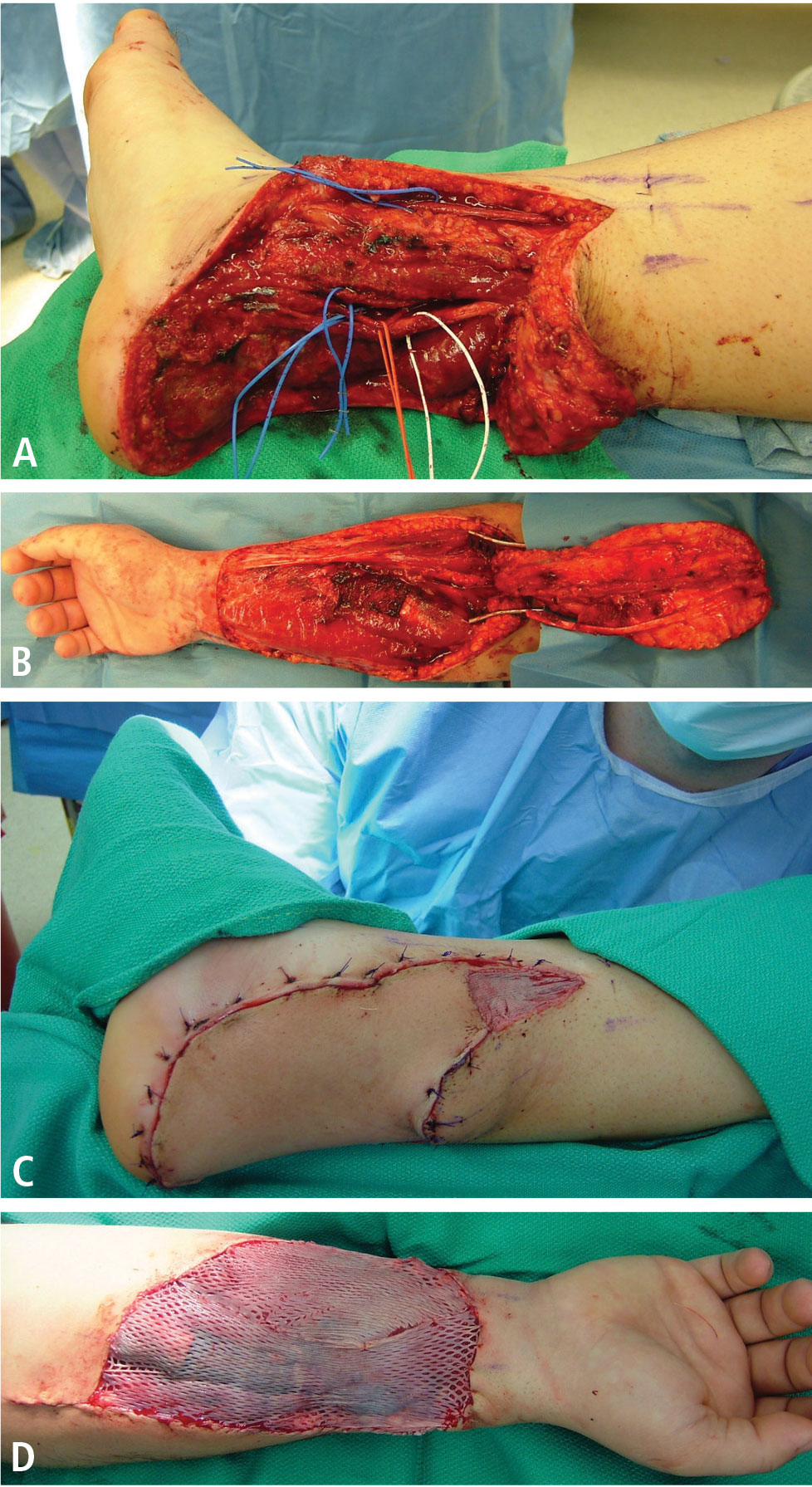
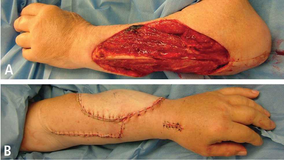
- Muramatsu K, Ihara K, Doi K, Hashimoto T, Taguchi T. Sarcoma in the forearm and hand: clinical outcomes and microsurgical reconstruction for limb salvage. Ann Plast Surg 2009; 62:28–33.
- Tukiainen E, Böhling T, Huuhtanen R. Soft tissue sarcoma of the trunk and extremities. Scand J Surg 2003; 92:257–263.
- Adelani MA, Holt GE, Dittus RS, Passman MA, Schwartz HS. Revascularization after segmental resection of lower extremity soft tissue sarcomas. J Surg Oncol 2007; 95:455–460.
- Lohman RF, Nabawi AS, Reece GP, Pollock RE, Evans GR. Soft tissue sarcoma of the upper extremity: a 5-year experience at two institutions emphasizing the role of soft tissue flap reconstruction. Cancer 2002; 94:2256–2264.
- Davis AM, Sennik S, Griffin AM, et al. Predictors of functional outcomes following limb salvage surgery for lower-extremity soft tissue sarcoma. J Surg Oncol 2000; 73:206–211.
- Heller L, Ballo MT, Cormier JN, Oates SD, Butler CE. Staged reconstruction for resection wounds in sarcoma patients treated with brachytherapy. Ann Plast Surg 2008; 60:58–63.
- Evans GR, Black JJ, Robb GL, et al. Adjuvant therapy: the effects on microvascular lower extremity reconstruction. Ann Plast Surg 1997; 39:141–144.
- Peat BG, Bell RS, Davis A, et al. Wound-healing complications after soft-tissue sarcoma surgery. Plast Reconstr Surg 1994; 93:980–987.
- Barwick WJ, Goldberg JA, Scully SP, Harrelson JM. Vascularized tissue transfer for closure of irradiated wounds after soft tissue sarcoma resection. Ann Surg 1992; 216:591–595.
- Masquelet AC, Romana MC. The medialis pedis flap: a new fasciocutaneous flap. Plast Reconstr Surg 1990; 85:765–772.
- Sherman R, Law M. Lower extremity reconstruction. In: Achauer BM, Eriksson E, Guyuron B, Coleman III JJ, Russell RC, Vander Kolk CA, eds. Plastic Surgery: Indications, Operations, and Outcomes. Vol 1. St. Louis, MO: Mosby; 2000:475–496.
- Innocenti M, Abed YY, Beltrami G, Delcroix L, Balatri A, Capanna R. Quadriceps muscle reconstruction with free functioning latissimus dorsi muscle flap after oncological resection. Microsurgery 2009; 29:189–198.
- Chiang YC, Wei FC, Wang JW, Chen WS. Reconstruction of below-knee stump using the salvaged foot fillet flap. Plast Reconstr Surg 1995; 96:731–738.
- Serletti JM, Carras AJ, O’Keefe RJ, Rosier RN. Functional outcome after soft-tissue reconstruction for limb salvage after sarcoma surgery. Plast Reconstr Surg 1998; 102:1576–1583.
- Niimi R, Matsumine A, Kusuzaki K, et al. Usefulness of limb salvage surgery for bone and soft tissue sarcomas of the distal lower leg. J Cancer Res Clin Oncol 2008; 134:1087–1095.
Advances in the management of soft-tissue and bone sarcomas—referred to collectively as “musculoskeletal sarcomas” hereafter—have resulted in significant improvements in survival and quality of life.1–3 Several factors have likely contributed to these advances, including improved surgical technique and the development of referral centers for sarcoma treatment that have embraced a multidisciplinary approach.1,2
The goal of treatment for musculoskeletal sarcomas is to optimize oncologic outcome and maximize functional restoration.2,3 Surgical resection has been the mainstay of therapy,1–7 as detailed earlier in this supplement. In patients with musculoskeletal sarcomas of the extremities, limb-sparing resection has been shown to be significantly superior to amputation.1,7–9 Wide local excision of the tumor along with its muscle compartment, followed by adjuvant chemotherapy and radiation therapy, has allowed limb salvage without an increased risk of recurrence in many patients.3 However, wide tumor resection can leave large defects that are not amenable to coverage by mobilization of the surrounding tissues, particularly if those tissues have been irradiated. As a result, resection can expose neurovascular structures, bone without periosteum, alloplastic materials, and internal fixation devices.
GOALS OF RECONSTRUCTION
Reconstructive surgery after musculoskeletal sarcoma resection aims to provide adequate wound coverage, preserve function, and optimize the cosmetic outcome.1–3 Tumors can be found on areas crucial to limb movement or may involve tissues vital to limb function. Reconstruction to repair these deficits can take many forms. In certain situations, amputation is still inevitable. In those cases, the reconstruction should provide stable stump coverage with durability and the ability to fit well with an external prosthesis.3
TIMING OF RECONSTRUCTION
Immediate reconstruction should be pursued if possible
Immediate reconstruction after a negative margin should always be considered and should be attempted when possible. Immediate reconstruction allows the reconstructive surgeon to benefit from better evaluation of the defect and exposed structures, as no scar tissue is present to distort the anatomy. Likewise, patients benefit from faster recovery and can receive adjuvant treatment (if necessary) sooner, as well as earlier rehabilitation. Patients may also benefit psychologically from immediate reconstruction.1,3
Indications for delayed reconstruction
Delayed reconstruction is primarily indicated when there are wound healing problems or there is uncertainty about the tumor margins. Secondary indications for delayed reconstruction are wound dehiscence and unstable soft-tissue coverage. If hardware is exposed, the recommendation is for early intervention and wound coverage with well-vascularized tissue to protect and cover the implant or prosthesis.
What about radiation therapy?
A very important consideration in reconstruction is the need for neoadjuvant or adjuvant radiation therapy.3,10,11 Irradiated wounds have a higher incidence of complications, including a tendency to dehisce. In patients who have been previously irradiated, the best practice is to perform immediate reconstruction with well-vascularized tissue, most likely a free tissue transfer.4,6,11,12 This practice reduces hospital stay, costs, and morbidity and increases limb salvage and patient satisfaction.13
SYSTEMATIC PREOPERATIVE PLANNING NEEDED
Reconstruction after musculoskeletal sarcoma resection should be planned systematically within a process that involves preoperative anticipation of the defect size and the resulting functional and cosmetic deficits that might need to be addressed. A preoperative visit to the reconstructive surgeon can be very helpful for presurgical planning.
During surgery it is usually preferable to allow the surgeon doing the tumor resection (eg, surgical oncologist or orthopedic oncologist) to complete the resection because the dimensions of the defect are not certain until negative margins are obtained.14 If tumor margins are unclear at the time of initial resection, the surgeon should consider delaying the definitive reconstruction until the permanent sections confirm negative margins. Temporary closure can be achieved with wound dressings, skin grafts (either allograft or autograft), or negative-pressure wound therapy. In the same context, if neurovascular structures are exposed it is reasonable to use a muscle flap without “tailoring” the flap to the defect. This approach allows the flap to be advanced or repositioned in case of positive margins, and the skin graft can be applied to the muscle surface in a second procedure.3
RECONSTRUCTIVE METHODS: A BRIEF OVERVIEW
Several methods can be used to close musculoskeletal sarcoma excision defects. Smaller defects can be closed primarily, although most defects are large and not amenable to primary closure. If fascia or muscle is preserved with only the skin coverage missing, the wound can be covered with either split-thickness or full-thickness skin grafts.1,4,6 Split-thickness skin grafts can be obtained in larger amounts and often heal faster than full-thickness skin grafts. However, most resections will require durable tissue coverage, particularly if adjuvant radiation therapy is planned.
In the case of long bone sarcoma resection, the resulting defect is usually large and complex and the traditional reconstruction is based on avascular allografts and local tissue flaps. However, allografts are associated with high rates of infection, nonunion, and fracture, leading to failure in about 50% of cases. Microvascular free flaps that contain bone, such as free fibula flaps, have been used instead of allografts with good success rates.2
Lately there has been growing interest in the use of the vacuum-assisted closure device (a form of negative-pressure wound therapy) to promote wound healing. It has been shown to improve the granulation and healing of open wounds by absorbing moisture, as well as to promote adherence after skin grafting, thereby reducing the risk of graft displacement.1,3 This device can be used immediately after musculoskeletal sarcoma resection while definitive tumor margin results are pending. It also can be used to prepare the wound bed for grafting in high-risk patients who would not tolerate more complex reconstructions.
Local or adjacent fascial, fasciocutaneous, and dermal flaps can also be used in lower-extremity reconstruction. However, muscle or musculocutaneous flaps are the mainstay of reconstruction after resection of musculoskeletal sarcomas. This group also includes perforator flaps, which have grown in popularity in the last few years.1,3
LOCATION-BASED WOUND RECONSTRUCTION
Musculoskeletal sarcomas can occur in virtually any region of the body, and myriad reconstructive options are available for various body sites. Since lower-extremity musculoskeletal sarcomas represent about 75% of cases,1 we will focus mainly on reconstruction of the lower extremity.
Factors driving choice of flap
Selection of an appropriate flap is essential to an optimal outcome. Flaps should be chosen with regard to donor site morbidity, functional requirements, length and diameter of the vascular pedicle, and aesthetic outcome.3 Usually physical examination, palpation of peripheral pulses, and Doppler ultrasonography are sufficient to evaluate the circulation. A preoperative angiogram should be considered in patients with severe peripheral vascular disease or previous trauma, which can potentially compromise the reconstructive outcome.15
Each region of the lower extremity possesses unique anatomic and functional characteristics that must be evaluated. It is useful to categorize the thigh, lower leg, and foot into separate anatomic units when planning reconstruction. We further divided these units into several subunits, as previously proposed by Sherman and Law15 and as outlined below.
Thigh
The thigh is usually well perfused and has several muscle groups, which facilitates reconstruction. Primary closure, skin grafts, or local flaps are acceptable options in most cases. The remaining musculature can be rotated or advanced to cover defects in the anterior or posterior thigh, providing bulk and adequate blood supply.
Hip and proximal/lateral thigh. Local muscle or myocutaneous flap options include tensor fascia lata, vastus lateralis, and rectus femoris flaps, all of which are based on the lateral circumflex femoral artery.
The tensor fascia lata flap is thin but has a long fascia extension that can be elevated from above the knee and can include a large skin paddle that is innervated by the lateral femoral cutaneous nerve. Some patients may experience knee instability after tensor fascia lata harvest.
The vastus lateralis muscle flap provides good bulk. Its arc of rotation reaches most of the inferior and posterior pelvis. It has little effect on ambulation.
The rectus femoris muscle flap is not so bulky, is easily mobilized, and has a wide arc of rotation. The donor site can be closed primarily. Harvest of this muscle can be associated with some strength loss during knee extension. For large defects of the upper third of the leg, a pedicled rectus abdominis muscle flap based on the deep inferior epigastric artery can be used. A vertically oriented skin island can be extended up to the costal margin, improving the reach. When the nature of the wound precludes use of pedicle flaps, free tissue transfer is indicated, with the latissimus dorsi muscle flap being used most commonly.15,16
Mid-thigh. Wounds in this location often can be closed with skin grafts or fasciocutaneous flaps. If the femur is exposed, however, a muscle flap will be required. As above, the tensor fascia lata, vastus lateralis, and rectus femoris can be used as flap options. If the lateral circumflex artery is unavailable, other flap options include the gracilis, vastus medialis, and rectus abdominis muscles. The gracilis muscle flap is based on the medial circumflex femoral artery and is useful for covering the medial aspect of the mid-thigh. Although this is a thin muscle, it can be used to cover long defects. The vastus medialis muscle flap is supplied by perforators from the profunda femoris and superficial femoral arteries. It can be rotated medially and advanced distally to cover patellar defects.
Supracondylar knee. The knee is a location where sarcoma resection is particularly likely to leave a defect with exposed bone, tendons, or ligaments that will need coverage. The gastrocnemius muscle flap combined with a split-thickness skin graft remains a consistent and reliable reconstructive option for this area. Other options are an extended medial gastrocnemius muscle flap or myocutaneous flap, which incorporates a random fasciocutaneous extension. For larger defects, free flaps should be considered, such as the anterior thigh flap, rectus abdominis muscle flap, or latissimus dorsi muscle flap. If tendons or ligaments need to be reconstructed, we favor autologous tissue, such as the fascia lata and plantaris tendons. These are easy to harvest and provide long-lasting joint stability.
Lower leg
Proximal third of the tibia. Defects here can usually be covered with a medial or lateral gastrocnemius muscle or myocutaneous flap, or a combination of the two. These muscles have a dominant vascular pedicle—the medial and lateral sural arteries. They can be harvested as an island for better reach, and they are reliable and have minimal donor site morbidity.15 The soleus muscle flap is another option that can be used alone or in combination with the medial or lateral gastrocnemius. Defects that are not amenable to closure by these flaps will most likely require free tissue transfer. The rectus abdominis or latissimus dorsi muscles are the first options. The latter can be combined with the serratus muscle if more bulk is needed.
Middle and lower thirds of the tibia. The soleus flap is frequently used for small or medium-sized mid-tibial defects. It is based on branches of the popliteal artery and posterior tibial artery. Larger defects require a combination of soleus and gastrocnemius muscle flaps or free tissue transfer.
Foot
Ideal reconstruction of the foot should provide thin and durable skin that will tolerate mechanical stress, and achieving this can be quite difficult. Skin grafts are seldom used for the foot, and are limited to non–weight-bearing portions with good underlying soft tissue.
Proximal non–weight-bearing areas (Achilles tendon and malleolar area). Local fasciocutaneous flaps are preferred. The lateral calcaneal artery flap, which is based on the peroneal artery branch, can cover exposed Achilles tendon, providing sensate coverage (sural nerve). The dorsalis pedis flap can be mobilized to cover the malleolar region and distal Achilles tendon, but donor site morbidity limits its use. Free tissue transfer is required for larger defects, and the the main options are flaps from the radial forearm, temporoparietal fascia, or lateral arm.
Heel and midplantar area. For heel reconstruction, the medial plantar artery flap, dorsalis pedis flap, abductor myocutaneous flap, peroneal artery flap, or anterior tibial artery flap can be used. The most versatile flap of the foot is the medial plantar artery flap, which is available only when the posterior tibial artery is intact. If local flaps are not suitable, microvascular tissue transfer is indicated. The radial forearm flap, scapular flap, lateral arm flap, or anterolateral thigh flap can be used. The radial forearm flap is usually the first choice because it is thin, has a long pedicle, and is easy to harvest.
If the foot defect is associated with a large cavity, muscle flaps are the first choices, specifically the gracilis or anterior serratus. A split latissimus muscle can also be applied. The full latissimus or the rectus abdominis are often too large for the type of defects observed.
Distal plantar area and forefoot. Most wounds in this region will require free tissue transfer. Free muscle flaps with split-thickness skin grafts provide the most stable and durable coverage.
Amputation vs limb salvage
It is important to evaluate the effects of lower-extremity salvage on ambulation. Salvage of a nonfunctional limb is of little value for the patient. Likewise, patients with severe medical problems may not be good candidates for limb salvage procedures. In those situations, amputation of the lower extremity is indicated. Adequate soft-tissue coverage and good distal perfusion are necessary to ensure healing of an amputation. If possible, local tissue rearrangement may be enough to provide a good amputation stump to fit an external prosthesis. In the case of radiation damage to the tissue, a free tissue transfer is necessary. The calcaneal-plantar unit from the amputated limb is frequently used as a free flap. Other flaps from the amputated limb, called fillet flaps, are harvested immediately and converted to flaps transferred to the defect site. Studies show that they are oncologically safe and reliable.17 Other flaps that provide good coverage for amputation defects are the latissimus dorsi muscle flap, the radial forearm flap, and the anterolateral thigh flap.
Upper extremities
POSTOPERATIVE CARE
Postoperative care following reconstruction after sarcoma resection requires a dedicated and trained team, particularly if a free flap is used for reconstruction.
Clinical evaluation of flaps includes color, temperature, and capillary refill. In cases of microsurgical reconstruction, postoperative care should include hourly examination of audible Doppler signals, at least for the first 36 hours. Free flap complications develop primarily in the first 24 hours, but they can occur during initial mobilization of the patient after a long period of bed rest. The surgical team should be aware of the potential problems and be able to act fast if necessary to reestablish blood flow to the flap.
In addition to flap monitoring, immobilization of the patient after surgery is extremely important. Postoperative swelling to the extremity should be avoided. Patients should be placed on bed rest until the postoperative swelling has subsided and the flap has adhered to the wound bed. Our protocol includes strict bed rest for about 7 days, followed by several days of dangling the extremity for short periods to ensure that dependent positioning will not alter the blood supply. A physical therapist should be involved to assist with crutches or a wheelchair. The patient should receive prophylactic anticoagulation during the resting period, in light of the high risk of deep vein thrombosis and pulmonary embolism. A compressive garment should be used to prevent lymphedema.
COMPLICATIONS ASSOCIATED WITH FLAPS
Once the flap is raised, it can still fail as a result of tension at insetting, inadequate blood flow, twisting of the pedicle, hematoma and/or infection, or the patient’s condition (eg, coagulopathy, poor nutritional status, anemia). Failure to correctly evaluate the direction of arterial flow, whether anterograde or retrograde, can cause flap loss. Instruments such as Doppler ultrasonographic equipment can be used to help to determine the flow. Partial or complete occlusion of the vascular pedicle can occur for several reasons (eg, twisting of the pedicle), and the consequences are disastrous if not recognized in time. If a pedicle problem is suspected in the case of a free flap, the patient should be taken to the operating room immediately and the flap should be explored. Rupture of the vascular anastomosis can occur as a result of technical problems, tension, and (in rare cases) infection.
Hematomas can cause mass effect, limit the venous return, and lead to flap necrosis. Hematoma formation also releases free radicals that can contribute to flap necrosis. Prevention is achieved through meticulous hemostasis. If a hematoma is suspected, the wound should be explored and the hematoma evacuated and washed out with normal saline.
The presence of an infected wound bed can also damage a flap by increasing its metabolic demand and causing the flap to be compromised by the infection itself. It is usually best to wait until the infection is controlled before planning the reconstruction.
Partial flap losses, skin graft losses, and wound dehiscence also are possible. Most of the time these require wound care, and patients’ nutrition and general health should be optimized to help the healing process. In the case of partial or complete flap loss, a new flap is often required and should be planned at a proper time.
CONCLUSIONS
Soft-tissue reconstruction following musculoskeletal sarcoma resection can be as simple as allowing the wound to heal by itself, which is less ideal, or as complex as coverage with a microsurgical osteocutaneous free flap. Limb salvage for sarcomas of the lower extremity has demonstrated good final functional outcomes without adversely affecting the oncologic results. Moreover, patients feel better psychologically and have higher quality of life.18,19
We believe that soft-tissue coverage after a wide resection is the most critical factor for avoiding postoperative complications of the tumor resection, such as infection or fractures. For this reason, we recommend the use of well-vascularized coverage at the time of the initial operation, if possible. Careful preoperative planning is especially important. We believe that reconstruction following musculoskeletal sarcoma resection can be done effectively only by using a team approach. Every such team should include, at minimum, an orthopedic surgeon and a reconstructive surgeon, with the mix of other providers dictated by the individual case.
- Misra A, Mistry N, Grimer R, Peart F. The management of soft tissue sarcoma. J Plast Reconstr Aesthet Surg 2009; 62:161–174.
- Morii T, Mochizuki K, Takushima A, Okazaki M, Satomi K. Soft tissue reconstruction using vascularized tissue transplantation following resection of musculoskeletal sarcoma: evaluation of oncologic and functional outcomes in 55 cases. Ann Plast Surg 2009; 62:252–257.
- Heller L, Kronowitz SJ. Lower extremity reconstruction. J Surg Oncol 2006; 94:479–489.
- Bannasch H, Haivas I, Momeni A, Stark GB. Oncosurgical and reconstructive concepts in the treatment of soft tissue sarcomas: a retrospective analysis. Arch Orthop Trauma Surg 2009; 129:43–49.
- Muramatsu K, Ihara K, Doi K, Hashimoto T, Taguchi T. Sarcoma in the forearm and hand: clinical outcomes and microsurgical reconstruction for limb salvage. Ann Plast Surg 2009; 62:28–33.
- Tukiainen E, Böhling T, Huuhtanen R. Soft tissue sarcoma of the trunk and extremities. Scand J Surg 2003; 92:257–263.
- Adelani MA, Holt GE, Dittus RS, Passman MA, Schwartz HS. Revascularization after segmental resection of lower extremity soft tissue sarcomas. J Surg Oncol 2007; 95:455–460.
- Lohman RF, Nabawi AS, Reece GP, Pollock RE, Evans GR. Soft tissue sarcoma of the upper extremity: a 5-year experience at two institutions emphasizing the role of soft tissue flap reconstruction. Cancer 2002; 94:2256–2264.
- Davis AM, Sennik S, Griffin AM, et al. Predictors of functional outcomes following limb salvage surgery for lower-extremity soft tissue sarcoma. J Surg Oncol 2000; 73:206–211.
- Heller L, Ballo MT, Cormier JN, Oates SD, Butler CE. Staged reconstruction for resection wounds in sarcoma patients treated with brachytherapy. Ann Plast Surg 2008; 60:58–63.
- Evans GR, Black JJ, Robb GL, et al. Adjuvant therapy: the effects on microvascular lower extremity reconstruction. Ann Plast Surg 1997; 39:141–144.
- Peat BG, Bell RS, Davis A, et al. Wound-healing complications after soft-tissue sarcoma surgery. Plast Reconstr Surg 1994; 93:980–987.
- Barwick WJ, Goldberg JA, Scully SP, Harrelson JM. Vascularized tissue transfer for closure of irradiated wounds after soft tissue sarcoma resection. Ann Surg 1992; 216:591–595.
- Masquelet AC, Romana MC. The medialis pedis flap: a new fasciocutaneous flap. Plast Reconstr Surg 1990; 85:765–772.
- Sherman R, Law M. Lower extremity reconstruction. In: Achauer BM, Eriksson E, Guyuron B, Coleman III JJ, Russell RC, Vander Kolk CA, eds. Plastic Surgery: Indications, Operations, and Outcomes. Vol 1. St. Louis, MO: Mosby; 2000:475–496.
- Innocenti M, Abed YY, Beltrami G, Delcroix L, Balatri A, Capanna R. Quadriceps muscle reconstruction with free functioning latissimus dorsi muscle flap after oncological resection. Microsurgery 2009; 29:189–198.
- Chiang YC, Wei FC, Wang JW, Chen WS. Reconstruction of below-knee stump using the salvaged foot fillet flap. Plast Reconstr Surg 1995; 96:731–738.
- Serletti JM, Carras AJ, O’Keefe RJ, Rosier RN. Functional outcome after soft-tissue reconstruction for limb salvage after sarcoma surgery. Plast Reconstr Surg 1998; 102:1576–1583.
- Niimi R, Matsumine A, Kusuzaki K, et al. Usefulness of limb salvage surgery for bone and soft tissue sarcomas of the distal lower leg. J Cancer Res Clin Oncol 2008; 134:1087–1095.
Breast reconstruction options following mastectomy
Patients recently diagnosed with breast cancer are distraught with concerns not only about surviving their disease but also about how its treatment will affect their body image and self-image. Although the risk of breast cancer increases with age, it is not a disease limited to the elderly. With advances in screening and awareness, breast cancers are now detected at earlier stages and in younger women. Approximately 5% of breast cancer patients are age 40 years or younger, which explains the recommendation that women be told about the benefits (and limits) of regular breast self-examinations beginning in their 20s.1 Additionally, breast cancer is the most common cancer in pregnant and postpartum women, occurring in about 1 in 3,000 pregnant women.2 Although breast conservation therapy is an attractive option, for many patients mastectomy is still the recommended surgical treatment. When mastectomy is required, it is understandable that many women are very concerned about losing their breast.
REASONS FOR RECONSTRUCTION
Mastectomies are commonly performed for women with ductal carcinoma in situ or with early or locally advanced invasive breast cancer (infiltrating ductal carcinoma) and sometimes for recurrent disease or for prophylaxis in high-risk women such as those with BRCA gene mutations or lobular carcinoma in situ. As reviewed in the preceding article in this supplement, mastectomy can be performed in various ways, using modified radical, skin-sparing, or nipple-sparing mastectomy techniques.
An emotional ‘double hit’
Following mastectomy, women are often left with what may be regarded as an emotional “double hit.” First, of course, is the anxiety from having a cancer diagnosis. Second, and perhaps equally devastating for some, is the emotional impact of losing a breast and the accompanying perception of disfigurement or loss of femininity and sexuality. These latter feelings often lead women who have undergone or will undergo mastectomy to explore the possibility of breast reconstruction.3–5
Both a medical and an emotional decision
While the reasons that women may seek breast reconstruction are many and varied (eg, to restore their self-esteem and social functioning, to help put their cancer experience behind them), it is important for primary care providers and other referring physicians to recognize that this decision is both a medical and an emotional one. Most women healthy enough to undergo extirpative surgical procedures are, in fact, healthy enough to undergo breast reconstruction if desired. Since choosing a reconstructive strategy is a complex process that takes into account many therapeutic and individual patient factors, plastic surgery consultation plays a major role in the comprehensive treatment of breast cancer.
TIMING AND TYPE OF RECONSTRUCTION
The timing of breast reconstruction can vary. In cases where the patient knows she will want reconstruction and the cancer surgery is performed at a site where a reconstructive surgery team is available, reconstruction can be performed immediately following mastectomy during a single trip to the operating room. When a reconstructive surgeon is not available locally or when systemic or local cancer therapies need to be completed first, reconstruction may need to be delayed.
Immediate reconstruction has the advantage of improved aesthetics while mitigating the sense of loss that can accompany mastectomy. Delayed reconstruction will give the patient more time for her decisions. An additional option, called “delayed-immediate” reconstruction, involves placing a tissue expander at the time of mastectomy (to preserve the breast skin envelope) and awaiting pathology results to determine whether radiation therapy is needed. If radiation is not needed, the patient undergoes reconstruction right away; if radiation is needed, the patient undergoes delayed reconstruction after radiation therapy is completed, with the breast skin envelope preserved for better aesthetic results. (The timing of reconstruction and these various timing options are discussed in detail in the final article in this supplement.) Selecting the correct timing and method of reconstruction requires good communication and coordination between the patient, her oncologist, and her multidisciplinary surgical team comprising both breast and plastic surgery specialists.
The patient and her surgeon will also discuss which reconstructive technique is best for her. Choosing a reconstructive strategy is a highly individualized process that takes into account the patient’s body characteristics, overall health, breast cancer treatment plan, and personal preferences. Consequently, a strategy offered to one patient is not necessarily valid for another. In general, options for reconstruction include use of the patient’s own tissue (autologous tissue), use of implant material (nonautologous), or a combination of an implant and autologous tissue.
IMPLANT-BASED RECONSTRUCTION
What the procedure involves
Nonautologous breast reconstruction usually involves a two-step procedure: placement of a tissue expander followed by later placement of a permanent implant.

Choice of permanent implant
Permanent implants vary by shape, texture of the implant shell, and filler material. They are typically filled with either silicone gel or saline.
Breast implants have been available for many years for use in both reconstructive breast surgery and cosmetic augmentation. A great deal of bad press and misinformation had surrounded the use of silicone gel-filled implants, with the result that they ceased to be marketed in the United States beginning in the early 1990s while the US Food and Drug Administration (FDA) reviewed additional safety information on their use. During this period when the use of silicone implants was limited, saline-filled implants became the preferred choice until the FDA approved the reintroduction of silicone implants to the market in November 2006, after what the agency described as years of rigorous scientific review of multiple clinical studies and other data.6 The FDA concluded that silicone implants are safe and effective for general use in breast reconstruction, correction of congenital breast anomalies, and breast augmentation.6 There is no evidence that silicone implants pose a significant systemic risk to women undergoing breast reconstruction.
 and postoperative (right) photos of a patient who underwent mastectomy of the right breast followed by silicone implant placement and nipple reconstruction")
Potential complications
 and postoperative (right) photos of a patient who underwent reconstruction with silicone implants after bilateral nipple-sparing mastectomy.")
Capsular contracture is another potential and more frequent complication of implant-based reconstruction. In all cases, the body forms a protective coverage, or fibrous capsule, around the implant. This process is called encapsulation. Most of the time, the capsule is relatively thin and pliable. Infrequently, however, the capsule can become thickened, hardened, and contracted, which constitutes capsular con-tracture. Although rare, severe contractures cause deformation of the reconstructed breast as well as pain. Severe contractures often require an operation to replace or remove the implant and treat the excessively thickened capsule. This can be done by exchanging the implant and either opening the capsule (capsulotomy) or removing the capsule (capsulectomy). If the contracture is significant enough or if the contracture recurs, then reconstruction using autologous tissue might be needed.
Advantages of implant reconstruction
Although nonautologous implant-based reconstruction can have some limitations, this procedure attracts many patients as a result of its advantages and good aesthetic results. The mastectomy procedure is prolonged by only about 1 hour, and most patients require only an overnight stay after the procedure. The recovery period is approximately 2 to 3 weeks, at which point tissue expansion is started.
What if radiation therapy is needed?
When treatment of the breast cancer is expected to involve radiation therapy right from the beginning, implant-based reconstruction is not an optimal choice. Radiation can affect the reconstruction in several negative ways. By design, radiation treats cancer by destroying dividing cells. Dividing cells are also required for wound healing and tissue remodeling. Without this remodeling ability, surgical scars are more susceptible to breakdown, which leads to tissue loss. In addition, because the effects of radiation are long-term, over time the thin tissue over the implant might respond poorly to the excessive stress of the implant, raising the possibility that tissue thinning could eventually lead to implant loss.7
Certainly there are instances when radiation therapy is not anticipated prior to the extirpative operation but then becomes necessary to complete the cancer treatment, based on final pathology results. Some patients in these circumstances may have had implants placed prior to the decision to give radiation. This does not doom the implant reconstruction to failure, however. Depending on the effect of the radiation and the patient’s body, there might be only a limited impact on the implant and the overall reconstruction result. We recommended close follow-up in these patients to monitor for any long-term complications such as skin discoloration, implant extrusion, or capsular contracture, which can be addressed as they arise.
AUTOLOGOUS RECONSTRUCTION
Techniques using abdominal tissue
As noted above, autologous breast reconstruction uses the patient’s own tissue. If the patient has adequate abdominal fat, the skin and fatty tissue of the lower abdomen may be used to reconstruct the missing breast. Historically, this type of reconstruction has included a portion of the abdominal muscles.
TRAM flap technique. The transverse rectus abdominis muscle (TRAM) flap technique takes advantage of the blood supply within the rectus abdominis muscle and its overlying skin and soft tissue. The muscle serves as the conduit for the blood supply of the skin and fatty tissue used in this method of reconstruction. The distal insertion of the muscle close to the pubic symphysis is cut, and the tissue receives its blood via the superior epigastric artery, which passes through the rectus muscle. This skin and soft tissue is then brought into the defect on the chest beneath the skin by tunneling it through the undermined skin flap between the abdomen and chest.
While the reconstructive results with the TRAM flap are good, this technique has been associated with increased risk of hernias or bulges in the abdominal wall. In sacrificing the rectus abdominis muscle, one of the major contributors to posture and the dynamic abdominal contour of the ventral abdomen is lost and the abdominal wall is weakened. This risk becomes even more significant when both rectus abdominis muscles are used to reconstruct both breasts.
 and postoperative (right) photos of a patient with left breast cancer who underwent mastectomy and immediate autologous reconstruction with the DIEP free flap technique.")
Limitations of techniques using abdominal tissue. Although autologous reconstruction is most commonly performed using tissue from the lower abdomen, flaps from the lower abdomen can be used only when there is sufficient fatty tissue to provide bulk for reconstructing the breast. In thin patients, using flaps from the abdomen may not be a good option. Contraindications to autologous reconstruction using the abdomen include previous abdominal surgery such as abdominoplasty, liposuction, open cholecystectomy, or other major abdominal operations that would compromise circulation to the skin and tissue over the flap. Other relative contraindications to autologous tissue reconstruction using the abdomen are obesity, smoking, a history of blood clots, and other major systemic medical conditions.
Options when abdominal tissue cannot be used
For patients who have insufficient tissue on the abdomen or have had previous abdominal surgery that compromises perfusion to the abdominal tissue, other options for autologous breast reconstruction are available. The gluteal tissue can be used, based on its superior or inferior blood supply, known as the superior gluteal artery perforator (SGAP) flap or the inferior gluteal artery perforator (IGAP) flap. Like the DIEP free flap technique, reconstruction using these flaps also requires a microsurgical procedure.
Another common option involves using skin and muscle from the back, or the latissimus dorsi myocutaneous flap. This flap does not require microsurgery; however, often the amount of tissue available to reconstruct the breast is inadequate to create a breast mound, requiring that the reconstruction be supplemented with an implant beneath the flap.8
Pros and cons of autologous reconstruction
Unlike implant-based reconstruction, autologous reconstruction obviously eliminates the need for implant replacement in the future. It also generally results in a more natural-feeling and natural-looking breast. Another advantage is that the breast reconstructed with autologous tissue will grow and decrease in size with weight fluctuations, just as a nonreconstructed breast would. Finally, in many cases the patient also essentially undergoes an abdominoplasty, or “tummy tuck” procedure, by virtue of how the tissue is harvested for reconstruction, which is likely to be welcomed by many patients.
 and postoperative (right) photos of a patient with left breast cancer who underwent mastectomy with immediate autologous reconstruction using the DIEP free flap procedure.")
COMPLETING THE RECONSTRUCTION
Nipple reconstruction
Reconstruction of the nipple and areola is important in that many patients feel that the nipple is what makes a breast. With the increased use of nipple-sparing mastectomy and improved reconstructive techniques, the aesthetic outcomes of reconstruction are often regarded as superior to many breast conservation procedures. A recent study by Cocquyt et al suggests that skin-sparing mastectomy with immediate DIEP flap reconstruction or TRAM flap reconstruction appears to yield a better cosmetic outcome than breast conservation therapy.9
Reconstruction of the nipple and areola restores the shape of the nipple, the shape of the areola, and the color of both with tattoos. Closing the autologous flap in a circular manner creates the shape of the areola, and the nipple is formed by local bilobed or trilobed skin flaps wrapped around each other to create a cone. Although nipple reconstruction can be performed at the time of immediate reconstruction, it is usually performed at a later time in the outpatient setting when the shape of the reconstructed breast is more definite after healing has occurred.
Revisional procedures
In many cases reconstructive breast surgery is not able to provide a breast that is shaped or sized exactly as desired or that perfectly matches the contralateral breast. Revisional procedures are sometimes performed to improve breast appearance and symmetry. Most revisional breast surgeries are performed on an outpatient basis and at times can be completed at the time of nipple reconstruction.
Modifying the contralateral breast
Modification of the contralateral breast is often necessary, and either a mastopexy (breast lift), reduction, or augmentation of the contralateral side may be needed for symmetry.
Mastopexy and reduction mammaplasty. Mastopexy, a skin-tightening and nipple-repositioning procedure, is performed to correct soft tissue descent without removing much breast tissue (see Figure 2), while reduction mammaplasty involves removing 400 to 2,000 grams of breast tissue (see Figure 4). A patient who has had a unilateral mastectomy without reconstruction may be a candidate for reduction mammaplasty of the contralateral breast. A unilateral large breast can cause marked neck and back pain due to the asymmetry of the weight on the chest.
Augmentation. Patients with smaller breasts often will undergo a matching augmentation procedure on the contralateral breast following completion of mastectomy and reconstruction on the other side.
Prophylactic mastectomy. For some women with a very high lifetime risk of breast cancer, such as those with BRCA1 or BRCA2 gene mutations, prophylactic mastectomy of the contralateral breast or even bilateral prophylactic mastectomy may be recommended by the oncologic surgeon. In some of these selected patients with sufficient abdominal tissue, bilateral DIEP flaps may be suitable; otherwise, the reconstruction can be completed with tissue expanders and implants.
WHAT ABOUT INSURANCE COVERAGE?
As the result of a federal law enacted 10 years ago, insurance coverage should not be a concern for women who are considering breast reconstruction following mastectomy. The Women’s Health and Cancer Rights Act of 1998 requires all medical insurers that provide coverage for mastectomy to also cover all stages of reconstruction of the affected breast as well as surgery and reconstruction of the contralateral breast to produce a symmetrical appearance.10
CONCLUSION
Although breast cancer remains a significant health risk to women and can result in significant disfigurement, breast reconstruction strategies continue to improve. These strategies offer women who have undergone mastectomy some excellent options for creating a near-normal-appearing breast. Women interested in pursuing reconstruction should meet with a plastic surgeon early in the course of their breast cancer treatment planning in order to better understand the options available and make an informed and individualized choice.
- Smith RA, Saslow D, Sawyer KA, et al. American Cancer Society guidelines for breast cancer screening: update 2003. CA Cancer J Clin 2003; 53:141–169.
- Breast cancer treatment and pregnancy. National Cancer Institute Web site. http://www.cancer.gov/cancertopics/pdq/treatment/breastcancer-and-pregnancy/HealthProfessional. Updated February 8, 2008. Accessed February 11, 2008.
- Reaby LL. Reasons why women who have mastectomy decide to have or not to have breast reconstruction. Plast Reconstr Surg 1998; 101:1810–1818.
- Nold RJ, Beamer RL, Helmer SD, McBoyle MF. Factors influencing a woman’s choice to undergo breast-conserving surgery versus modified radical mastectomy. Am J Surg 2000; 180:413–418.
- Pusic A, Thompson TA, Kerrigan CL, et al. Surgical options for the early-stage breast cancer: factors associated with patient choice and postoperative quality of life. Plast Reconstr Surg 1999; 104:1325–1333.
- FDA approves silicone gel-filled breast implants after in-depth evaluation [news release]. Rockville, MD: U.S. Food and Drug Administration; November 17, 2006. http://www.fda.gov/bbs/topics/NEWS/2006/NEW01512.html. Accessed February 7, 2008.
- Kronowitz SJ, Robb GL. Breast reconstruction with postmastectomy radiation therapy: current issues. Plast Reconstr Surg 2004; 114:950–960.
- Bostwick J III. Abdominal flap reconstruction. In: Plastic and Reconstructive Breast Surgery. 2nd ed. St. Louis, MO: Quality Medical Publishing; 2000:982–1015.
- Cocquyt VF, Blondeel PN, Depypere HT, et al. Better cosmetic results and comparable quality of life after skin-sparing mastectomy and immediate autologous breast reconstruction compared to breast conservative treatment. Br J Plast Surg 2003; 56:462–470.
- Your rights after a mastectomy...Women’s Health & Cancer Rights Act of 1998. U.S. Department of Labor Web site. http://www.dol.gov/ebsa/publications/whcra.html. Accessed February 11, 2008.
ADDITIONAL READING
Hoover SJ, Kenkel JM. Breast cancer, cancer prevention, and breast reconstruction. Selected Readings in Plastic Surgery 2002; 9:1–40.
Hartrampf CR, Scheflan M, Black PW. Breast reconstruction with a transverse abdominal island flap. Plast Reconstr Surg 1982; 69:216–225.
Hartrampf CR Jr, Bennett GK. Autogenous tissue reconstruction in the mastectomy patient: a critical review of 300 patients. Ann Surg 1987; 205:508–519.
Elliott LF, Eskenazi L, Beegle PH Jr, Podres PE, Drazan L. Immediate TRAM flap breast reconstruction: 128 consecutive cases. Plast Reconstr Surg 1993; 92:217–227.
Schusterman MA, Kroll SS, Weldon ME. Immediate breast reconstruction: why the free TRAM over the conventional TRAM flap? Plast Reconstr Surg 1992; 90:255–262.
Patients recently diagnosed with breast cancer are distraught with concerns not only about surviving their disease but also about how its treatment will affect their body image and self-image. Although the risk of breast cancer increases with age, it is not a disease limited to the elderly. With advances in screening and awareness, breast cancers are now detected at earlier stages and in younger women. Approximately 5% of breast cancer patients are age 40 years or younger, which explains the recommendation that women be told about the benefits (and limits) of regular breast self-examinations beginning in their 20s.1 Additionally, breast cancer is the most common cancer in pregnant and postpartum women, occurring in about 1 in 3,000 pregnant women.2 Although breast conservation therapy is an attractive option, for many patients mastectomy is still the recommended surgical treatment. When mastectomy is required, it is understandable that many women are very concerned about losing their breast.
REASONS FOR RECONSTRUCTION
Mastectomies are commonly performed for women with ductal carcinoma in situ or with early or locally advanced invasive breast cancer (infiltrating ductal carcinoma) and sometimes for recurrent disease or for prophylaxis in high-risk women such as those with BRCA gene mutations or lobular carcinoma in situ. As reviewed in the preceding article in this supplement, mastectomy can be performed in various ways, using modified radical, skin-sparing, or nipple-sparing mastectomy techniques.
An emotional ‘double hit’
Following mastectomy, women are often left with what may be regarded as an emotional “double hit.” First, of course, is the anxiety from having a cancer diagnosis. Second, and perhaps equally devastating for some, is the emotional impact of losing a breast and the accompanying perception of disfigurement or loss of femininity and sexuality. These latter feelings often lead women who have undergone or will undergo mastectomy to explore the possibility of breast reconstruction.3–5
Both a medical and an emotional decision
While the reasons that women may seek breast reconstruction are many and varied (eg, to restore their self-esteem and social functioning, to help put their cancer experience behind them), it is important for primary care providers and other referring physicians to recognize that this decision is both a medical and an emotional one. Most women healthy enough to undergo extirpative surgical procedures are, in fact, healthy enough to undergo breast reconstruction if desired. Since choosing a reconstructive strategy is a complex process that takes into account many therapeutic and individual patient factors, plastic surgery consultation plays a major role in the comprehensive treatment of breast cancer.
TIMING AND TYPE OF RECONSTRUCTION
The timing of breast reconstruction can vary. In cases where the patient knows she will want reconstruction and the cancer surgery is performed at a site where a reconstructive surgery team is available, reconstruction can be performed immediately following mastectomy during a single trip to the operating room. When a reconstructive surgeon is not available locally or when systemic or local cancer therapies need to be completed first, reconstruction may need to be delayed.
Immediate reconstruction has the advantage of improved aesthetics while mitigating the sense of loss that can accompany mastectomy. Delayed reconstruction will give the patient more time for her decisions. An additional option, called “delayed-immediate” reconstruction, involves placing a tissue expander at the time of mastectomy (to preserve the breast skin envelope) and awaiting pathology results to determine whether radiation therapy is needed. If radiation is not needed, the patient undergoes reconstruction right away; if radiation is needed, the patient undergoes delayed reconstruction after radiation therapy is completed, with the breast skin envelope preserved for better aesthetic results. (The timing of reconstruction and these various timing options are discussed in detail in the final article in this supplement.) Selecting the correct timing and method of reconstruction requires good communication and coordination between the patient, her oncologist, and her multidisciplinary surgical team comprising both breast and plastic surgery specialists.
The patient and her surgeon will also discuss which reconstructive technique is best for her. Choosing a reconstructive strategy is a highly individualized process that takes into account the patient’s body characteristics, overall health, breast cancer treatment plan, and personal preferences. Consequently, a strategy offered to one patient is not necessarily valid for another. In general, options for reconstruction include use of the patient’s own tissue (autologous tissue), use of implant material (nonautologous), or a combination of an implant and autologous tissue.
IMPLANT-BASED RECONSTRUCTION
What the procedure involves
Nonautologous breast reconstruction usually involves a two-step procedure: placement of a tissue expander followed by later placement of a permanent implant.
Choice of permanent implant
Permanent implants vary by shape, texture of the implant shell, and filler material. They are typically filled with either silicone gel or saline.
Breast implants have been available for many years for use in both reconstructive breast surgery and cosmetic augmentation. A great deal of bad press and misinformation had surrounded the use of silicone gel-filled implants, with the result that they ceased to be marketed in the United States beginning in the early 1990s while the US Food and Drug Administration (FDA) reviewed additional safety information on their use. During this period when the use of silicone implants was limited, saline-filled implants became the preferred choice until the FDA approved the reintroduction of silicone implants to the market in November 2006, after what the agency described as years of rigorous scientific review of multiple clinical studies and other data.6 The FDA concluded that silicone implants are safe and effective for general use in breast reconstruction, correction of congenital breast anomalies, and breast augmentation.6 There is no evidence that silicone implants pose a significant systemic risk to women undergoing breast reconstruction.
Potential complications
Capsular contracture is another potential and more frequent complication of implant-based reconstruction. In all cases, the body forms a protective coverage, or fibrous capsule, around the implant. This process is called encapsulation. Most of the time, the capsule is relatively thin and pliable. Infrequently, however, the capsule can become thickened, hardened, and contracted, which constitutes capsular con-tracture. Although rare, severe contractures cause deformation of the reconstructed breast as well as pain. Severe contractures often require an operation to replace or remove the implant and treat the excessively thickened capsule. This can be done by exchanging the implant and either opening the capsule (capsulotomy) or removing the capsule (capsulectomy). If the contracture is significant enough or if the contracture recurs, then reconstruction using autologous tissue might be needed.
Advantages of implant reconstruction
Although nonautologous implant-based reconstruction can have some limitations, this procedure attracts many patients as a result of its advantages and good aesthetic results. The mastectomy procedure is prolonged by only about 1 hour, and most patients require only an overnight stay after the procedure. The recovery period is approximately 2 to 3 weeks, at which point tissue expansion is started.
What if radiation therapy is needed?
When treatment of the breast cancer is expected to involve radiation therapy right from the beginning, implant-based reconstruction is not an optimal choice. Radiation can affect the reconstruction in several negative ways. By design, radiation treats cancer by destroying dividing cells. Dividing cells are also required for wound healing and tissue remodeling. Without this remodeling ability, surgical scars are more susceptible to breakdown, which leads to tissue loss. In addition, because the effects of radiation are long-term, over time the thin tissue over the implant might respond poorly to the excessive stress of the implant, raising the possibility that tissue thinning could eventually lead to implant loss.7
Certainly there are instances when radiation therapy is not anticipated prior to the extirpative operation but then becomes necessary to complete the cancer treatment, based on final pathology results. Some patients in these circumstances may have had implants placed prior to the decision to give radiation. This does not doom the implant reconstruction to failure, however. Depending on the effect of the radiation and the patient’s body, there might be only a limited impact on the implant and the overall reconstruction result. We recommended close follow-up in these patients to monitor for any long-term complications such as skin discoloration, implant extrusion, or capsular contracture, which can be addressed as they arise.
AUTOLOGOUS RECONSTRUCTION
Techniques using abdominal tissue
As noted above, autologous breast reconstruction uses the patient’s own tissue. If the patient has adequate abdominal fat, the skin and fatty tissue of the lower abdomen may be used to reconstruct the missing breast. Historically, this type of reconstruction has included a portion of the abdominal muscles.
TRAM flap technique. The transverse rectus abdominis muscle (TRAM) flap technique takes advantage of the blood supply within the rectus abdominis muscle and its overlying skin and soft tissue. The muscle serves as the conduit for the blood supply of the skin and fatty tissue used in this method of reconstruction. The distal insertion of the muscle close to the pubic symphysis is cut, and the tissue receives its blood via the superior epigastric artery, which passes through the rectus muscle. This skin and soft tissue is then brought into the defect on the chest beneath the skin by tunneling it through the undermined skin flap between the abdomen and chest.
While the reconstructive results with the TRAM flap are good, this technique has been associated with increased risk of hernias or bulges in the abdominal wall. In sacrificing the rectus abdominis muscle, one of the major contributors to posture and the dynamic abdominal contour of the ventral abdomen is lost and the abdominal wall is weakened. This risk becomes even more significant when both rectus abdominis muscles are used to reconstruct both breasts.
Limitations of techniques using abdominal tissue. Although autologous reconstruction is most commonly performed using tissue from the lower abdomen, flaps from the lower abdomen can be used only when there is sufficient fatty tissue to provide bulk for reconstructing the breast. In thin patients, using flaps from the abdomen may not be a good option. Contraindications to autologous reconstruction using the abdomen include previous abdominal surgery such as abdominoplasty, liposuction, open cholecystectomy, or other major abdominal operations that would compromise circulation to the skin and tissue over the flap. Other relative contraindications to autologous tissue reconstruction using the abdomen are obesity, smoking, a history of blood clots, and other major systemic medical conditions.
Options when abdominal tissue cannot be used
For patients who have insufficient tissue on the abdomen or have had previous abdominal surgery that compromises perfusion to the abdominal tissue, other options for autologous breast reconstruction are available. The gluteal tissue can be used, based on its superior or inferior blood supply, known as the superior gluteal artery perforator (SGAP) flap or the inferior gluteal artery perforator (IGAP) flap. Like the DIEP free flap technique, reconstruction using these flaps also requires a microsurgical procedure.
Another common option involves using skin and muscle from the back, or the latissimus dorsi myocutaneous flap. This flap does not require microsurgery; however, often the amount of tissue available to reconstruct the breast is inadequate to create a breast mound, requiring that the reconstruction be supplemented with an implant beneath the flap.8
Pros and cons of autologous reconstruction
Unlike implant-based reconstruction, autologous reconstruction obviously eliminates the need for implant replacement in the future. It also generally results in a more natural-feeling and natural-looking breast. Another advantage is that the breast reconstructed with autologous tissue will grow and decrease in size with weight fluctuations, just as a nonreconstructed breast would. Finally, in many cases the patient also essentially undergoes an abdominoplasty, or “tummy tuck” procedure, by virtue of how the tissue is harvested for reconstruction, which is likely to be welcomed by many patients.
COMPLETING THE RECONSTRUCTION
Nipple reconstruction
Reconstruction of the nipple and areola is important in that many patients feel that the nipple is what makes a breast. With the increased use of nipple-sparing mastectomy and improved reconstructive techniques, the aesthetic outcomes of reconstruction are often regarded as superior to many breast conservation procedures. A recent study by Cocquyt et al suggests that skin-sparing mastectomy with immediate DIEP flap reconstruction or TRAM flap reconstruction appears to yield a better cosmetic outcome than breast conservation therapy.9
Reconstruction of the nipple and areola restores the shape of the nipple, the shape of the areola, and the color of both with tattoos. Closing the autologous flap in a circular manner creates the shape of the areola, and the nipple is formed by local bilobed or trilobed skin flaps wrapped around each other to create a cone. Although nipple reconstruction can be performed at the time of immediate reconstruction, it is usually performed at a later time in the outpatient setting when the shape of the reconstructed breast is more definite after healing has occurred.
Revisional procedures
In many cases reconstructive breast surgery is not able to provide a breast that is shaped or sized exactly as desired or that perfectly matches the contralateral breast. Revisional procedures are sometimes performed to improve breast appearance and symmetry. Most revisional breast surgeries are performed on an outpatient basis and at times can be completed at the time of nipple reconstruction.
Modifying the contralateral breast
Modification of the contralateral breast is often necessary, and either a mastopexy (breast lift), reduction, or augmentation of the contralateral side may be needed for symmetry.
Mastopexy and reduction mammaplasty. Mastopexy, a skin-tightening and nipple-repositioning procedure, is performed to correct soft tissue descent without removing much breast tissue (see Figure 2), while reduction mammaplasty involves removing 400 to 2,000 grams of breast tissue (see Figure 4). A patient who has had a unilateral mastectomy without reconstruction may be a candidate for reduction mammaplasty of the contralateral breast. A unilateral large breast can cause marked neck and back pain due to the asymmetry of the weight on the chest.
Augmentation. Patients with smaller breasts often will undergo a matching augmentation procedure on the contralateral breast following completion of mastectomy and reconstruction on the other side.
Prophylactic mastectomy. For some women with a very high lifetime risk of breast cancer, such as those with BRCA1 or BRCA2 gene mutations, prophylactic mastectomy of the contralateral breast or even bilateral prophylactic mastectomy may be recommended by the oncologic surgeon. In some of these selected patients with sufficient abdominal tissue, bilateral DIEP flaps may be suitable; otherwise, the reconstruction can be completed with tissue expanders and implants.
WHAT ABOUT INSURANCE COVERAGE?
As the result of a federal law enacted 10 years ago, insurance coverage should not be a concern for women who are considering breast reconstruction following mastectomy. The Women’s Health and Cancer Rights Act of 1998 requires all medical insurers that provide coverage for mastectomy to also cover all stages of reconstruction of the affected breast as well as surgery and reconstruction of the contralateral breast to produce a symmetrical appearance.10
CONCLUSION
Although breast cancer remains a significant health risk to women and can result in significant disfigurement, breast reconstruction strategies continue to improve. These strategies offer women who have undergone mastectomy some excellent options for creating a near-normal-appearing breast. Women interested in pursuing reconstruction should meet with a plastic surgeon early in the course of their breast cancer treatment planning in order to better understand the options available and make an informed and individualized choice.
- Smith RA, Saslow D, Sawyer KA, et al. American Cancer Society guidelines for breast cancer screening: update 2003. CA Cancer J Clin 2003; 53:141–169.
- Breast cancer treatment and pregnancy. National Cancer Institute Web site. http://www.cancer.gov/cancertopics/pdq/treatment/breastcancer-and-pregnancy/HealthProfessional. Updated February 8, 2008. Accessed February 11, 2008.
- Reaby LL. Reasons why women who have mastectomy decide to have or not to have breast reconstruction. Plast Reconstr Surg 1998; 101:1810–1818.
- Nold RJ, Beamer RL, Helmer SD, McBoyle MF. Factors influencing a woman’s choice to undergo breast-conserving surgery versus modified radical mastectomy. Am J Surg 2000; 180:413–418.
- Pusic A, Thompson TA, Kerrigan CL, et al. Surgical options for the early-stage breast cancer: factors associated with patient choice and postoperative quality of life. Plast Reconstr Surg 1999; 104:1325–1333.
- FDA approves silicone gel-filled breast implants after in-depth evaluation [news release]. Rockville, MD: U.S. Food and Drug Administration; November 17, 2006. http://www.fda.gov/bbs/topics/NEWS/2006/NEW01512.html. Accessed February 7, 2008.
- Kronowitz SJ, Robb GL. Breast reconstruction with postmastectomy radiation therapy: current issues. Plast Reconstr Surg 2004; 114:950–960.
- Bostwick J III. Abdominal flap reconstruction. In: Plastic and Reconstructive Breast Surgery. 2nd ed. St. Louis, MO: Quality Medical Publishing; 2000:982–1015.
- Cocquyt VF, Blondeel PN, Depypere HT, et al. Better cosmetic results and comparable quality of life after skin-sparing mastectomy and immediate autologous breast reconstruction compared to breast conservative treatment. Br J Plast Surg 2003; 56:462–470.
- Your rights after a mastectomy...Women’s Health & Cancer Rights Act of 1998. U.S. Department of Labor Web site. http://www.dol.gov/ebsa/publications/whcra.html. Accessed February 11, 2008.
ADDITIONAL READING
Hoover SJ, Kenkel JM. Breast cancer, cancer prevention, and breast reconstruction. Selected Readings in Plastic Surgery 2002; 9:1–40.
Hartrampf CR, Scheflan M, Black PW. Breast reconstruction with a transverse abdominal island flap. Plast Reconstr Surg 1982; 69:216–225.
Hartrampf CR Jr, Bennett GK. Autogenous tissue reconstruction in the mastectomy patient: a critical review of 300 patients. Ann Surg 1987; 205:508–519.
Elliott LF, Eskenazi L, Beegle PH Jr, Podres PE, Drazan L. Immediate TRAM flap breast reconstruction: 128 consecutive cases. Plast Reconstr Surg 1993; 92:217–227.
Schusterman MA, Kroll SS, Weldon ME. Immediate breast reconstruction: why the free TRAM over the conventional TRAM flap? Plast Reconstr Surg 1992; 90:255–262.