Environmental Dermatology

What’s Eating You? Tick Bite Alopecia

Author and Disclosure Information

Tick bite alopecia is seldom reported in the literature. The condition usually presents as a solitary oval zone of alopecia with a central eschar. Histologic findings are not well described but generally indicate dense perifollicular lymphocytic inflammation. The mechanism for hair loss is poorly understood, but the prognosis for hair regrowth appears to be favorable.

Practice Points

  • Tick bite alopecia should be included in the differential diagnosis of both solitary and moth-eaten lesions of localized hair loss.
  • In most cases, hair regrowth can be expected in a lesion of tick bite alopecia.


References

Case Report

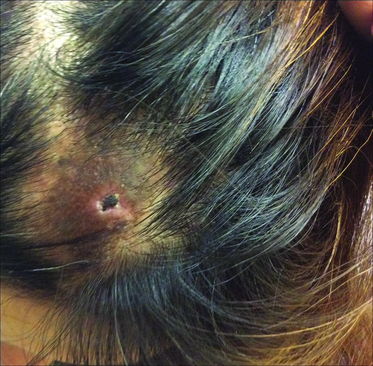
A 44-year-old woman presented with a localized patch of hair loss on the frontal scalp of several month’s duration. She had been bitten by a tick at this site during the summer. Two months later a primary care provider prescribed triamcinolone cream because of intense itching at the bite site. The patient returned to her primary care provider 2 weeks later due to persistent itching, hyperpigmentation, and hair loss. At that time, the clinician probed the central portion of the lesion because of a concern for retained tick parts. A few weeks later, a dermatologist evaluated the patient and found a roughly circular zone of alopecia measuring approximately 5 cm in diameter that was just posterior to the left frontal hairline (Figure 1). In the center of the plaque there was a small eschar surrounded by a zone of hyperpigmentation, mild induration, and almost complete loss of terminal hairs. At the periphery of the lesion, hair density gradually increased and skin pigmentation normalized.

Figure 1. A centrally located eschar that is typical of tick bite alopecia.

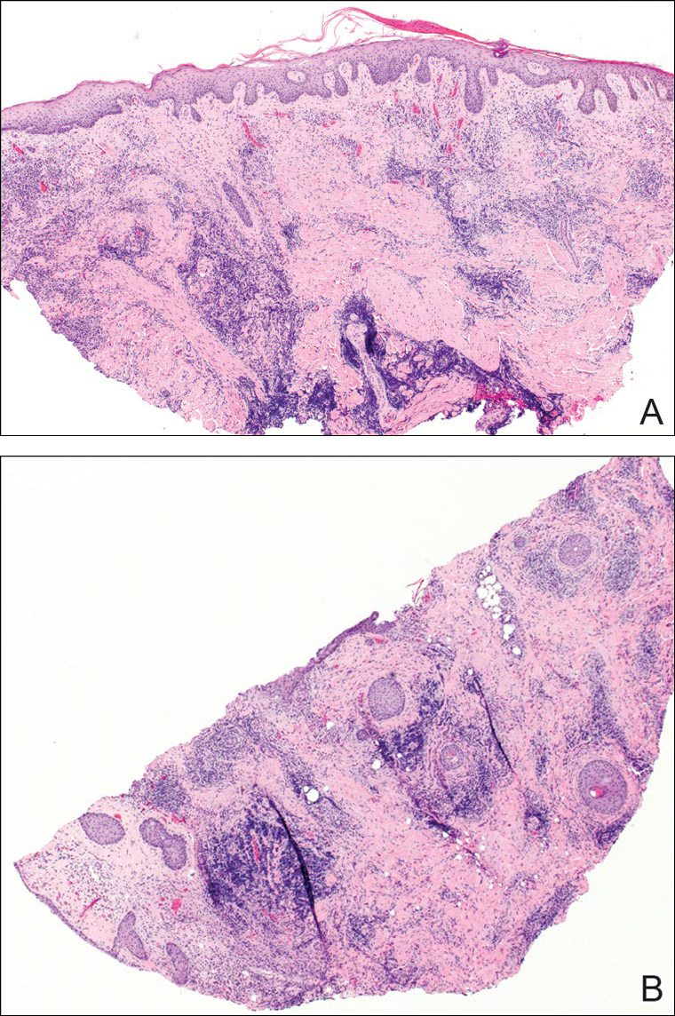
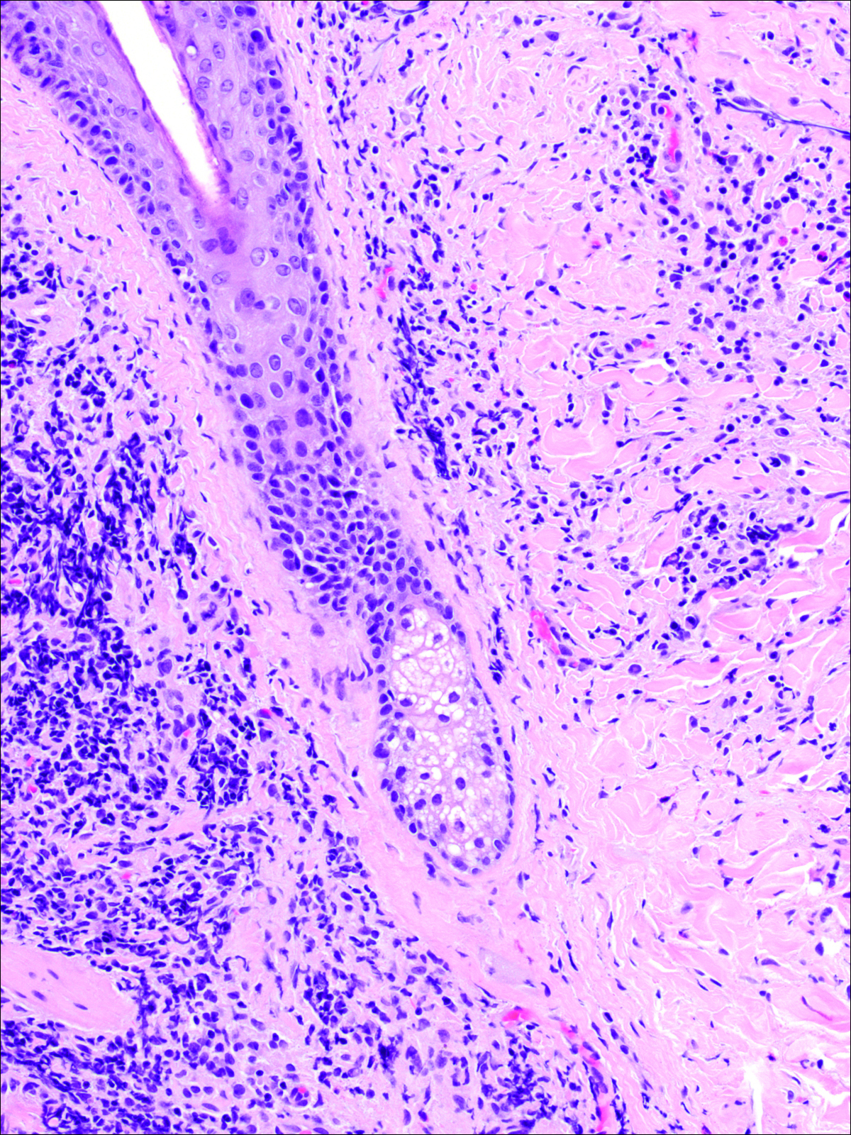
A punch biopsy was obtained from an indurated area of hyperpigmentation adjacent to the eschar. Both vertical and horizontal sections were obtained, revealing a relatively normal epidermis, a marked decrease in follicular structures with loss of sebaceous glands, and dense perifollicular lymphocytic inflammation with a few scattered eosinophils (Figures 2 and 3).

Figure 2. Vertical section (A) and horizontal section (B) of a biopsy from the lesion (both H&E, original magnification ×40).

Figure 3. Perifollicular, predominantly lymphocytic inflammation surrounding a catagen follicle. A few eosinophils also were present in the infiltrate (original magnification ×100).

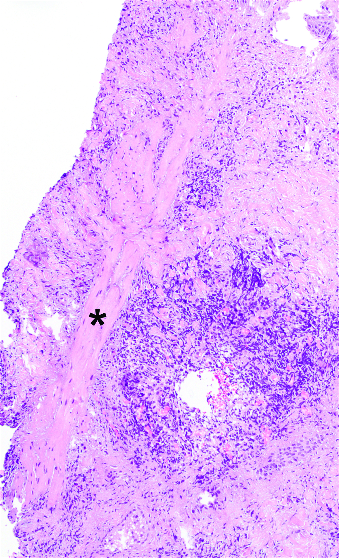
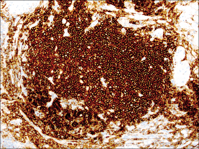
Clear-cut follicular scars surrounded by dense inflammation could be found (Figure 4). Horizontal sections revealed loss of most terminal hairs, with a few residual vellus telogen hairs present. At the sites of former follicles, some foci of dense inflammation showed evidence of germinal center formation as revealed by immunohistochemical staining (Figure 5).

Figure 4. Nodular aggregate of chronic inflammation adjacent to a follicular scar (identified with an asterisk) (original magnification ×100).

Figure 5. CD20 immunohistochemical stain of a nodular aggregate of inflammation (original magnification ×200). The dominant cells are B lymphocytes. CD4 and CD8 staining (not shown) was confirmatory.

Historical Perspective

Tick bite alopecia was first described in the French literature in 19211 and in the English-language literature in 1955.2 A few additional cases were subsequently reported.3-5 In 2008, Castelli et al6 described the histologic and immunohistochemical features of 25 tick bite cases, a few of which resulted in alopecia. Other than these reports, little original information has been written about tick bite alopecia.

Pages

Recommended Reading

Spontaneous Repigmentation of Silvery Hair in an Infant With Congenital Hydrops Fetalis and Hypoproteinemia
MDedge Dermatology
Skin Lesions in Patients Treated With Imatinib Mesylate: A 5-Year Prospective Study
MDedge Dermatology
JAK inhibitor improves alopecia, with caveats
MDedge Dermatology
Keys to alopecia areata might lie in gut microbiome
MDedge Dermatology
VIDEO: The ins and outs of JAK ihibitors for alopecia
MDedge Dermatology
Clinical Characteristics and HLA Alleles of a Family With Simultaneously Occurring Alopecia Areata
MDedge Dermatology
Patch of Hair Loss on the Scalp
MDedge Dermatology
Studies highlight diagnostic and treatment challenges in hidradenitis suppurativa
MDedge Dermatology
Subungual Exostosis
MDedge Dermatology
Fingernail Photo-onycholysis After Aminolevulinic Acid–Photodynamic Therapy Under Blue Light for Treatment of Actinic Keratoses on the Face
MDedge Dermatology