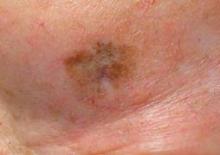
ORLANDO - Standard surgical margins often are inadequate for lentigo maligna; instead, perimeter excision techniques or Mohs surgery is preferable because these approaches consistently provide high cure rates, according to Dr. Basil S. Cherpelis.
Lentigo maligna is a subtype of melanoma in situ, characterized as an overgrowth of atypical melanocytes with the potential to become lentigo maligna melanoma. "I think of it as the problem child of the melanoma family," Dr. Cherpelis said at the annual meeting of the Florida Society of Dermatologic Surgeons.
"What margin should I use, and if I am going to do Mohs, how can I see margins on frozen sections?" Intraoperative margin control is recommended, but recent literature suggests that standard 5-mm margins are frequently inadequate (Clin. Plast. Surg. 2010;37:35-46; Int. J. Dermatol. 2010;49:482-91).
In general, traditional frozen sections permit only 1% of margins to be assessed. For that reason, Dr. Cherpelis said he prefers permanent processing or Mohs surgery for these patients. With permanent processing, a surgeon leaves a central island of tissue and removes peripheral strips. Excised samples sent to a lab for evaluation typically take several days for results.
"Permanent processing has excellent cure rates. But on the downside, a patient has to wait for pathology results, which can be inconvenient," said Dr. Cherpelis, a cutaneous oncologist at Moffitt Cancer Center at the University of South Florida in Tampa.
In contrast, Mohs micrographic excision and repair are performed on the same day. Because the entire margin is visualized, cure rates are "excellent," Dr. Cherpelis said.
As to why Mohs isn’t more widely used, one possible reason is that it can be difficult to distinguish melanocytes from keratinocytes on frozen section, he said.
Immunohistostains to the rescue: Melanoma antigen recognized by T cells (MART-1) and microphthalmia-associated transcription factor (MITF) staining each has a role.
"MART-1 is a valuable immunostain for melanoma in situ on photodamaged skin," said Dr. L. Frank Glass, a dermatopathologist at Moffitt Cancer Center. MART-1 on frozen sections can provide the same data as permanent processing. "But MART-1 is not a magic bullet," he added. "There can be false positives."
In addition, expertise and teamwork are required with immunohistostains, Dr. Glass said. "The right MART-1 staining takes a technical staff to do it right."
MITF works in frozen sections and produces results similar to permanent sections. Clinicians can use this nuclear immunostain to quantify melanocytes and assess other parameters that distinguish melanoma in situ from solar lentigines or skin with chronic sun damage, for example.
"MITF is particularly useful in combination with MART-1," Dr. Glass said.
Instead of dendritic processes that overlap and look like confluence, MITF reveals discreet melanocytic cells, Dr. Glass said. "You can feel more comfortable calling a case negative with MITF – if it shows no abnormal cytology – in the same patient where the MART-1 shows clumping."
"The bottom line is we are looking for 100% specificity for melanoma," Dr. Glass said. He and his colleagues also are developing cutoff values, based on melanocyte diameter and density, to distinguish melanoma in situ from solar lentigo or sun-damaged skin.
Dr. Cherpelis and Dr. Glass said they had no relevant financial disclosures.


