Case Reports

Systemic Epstein-Barr Virus–Positive T-cell Lymphoma of Childhood

Author and Disclosure Information

We report a case of a 7-year-old Chinese boy who presented with acute fever, multiple oral ulcers, and skin nodules. A diagnosis of systemic Epstein-Barr virus (EBV)–positive T-cell lymphoma of childhood was established using systemic laboratory examination, imaging studies, bone marrow and skin biopsy with immunohistochemistry, and in situ hybridization for EBV-encoded RNA (EBER) and gene rearrangements. Notable features of this case include the absence of pancytopenia and hemophagocytic syndrome as well as spontaneous resolution without chemotherapy for several months; however, the condition relapsed, and the patient died.

Practice Points

  • Systemic Epstein-Barr virus (EBV)–positive T-cell lymphoma of childhood is a fulminant illness with a predilection for Asians and indigenous populations from Mexico and Central and South America. In most patients, the disease has an aggressive clinical course with high mortality.
  • The disease often is complicated by hemophagocytic syndrome, coagulopathy, sepsis, and multiorgan failure. When these severe complications are absent, the prognosis might be better.
  • In situ hybridization for EBV-encoded RNA and for T-cell receptor gene rearrangements is an important tool to establish the diagnosis as well as for treatment options and predicting the prognosis.


References

Case Report

A 7-year-old Chinese boy presented with multiple painful oral and tongue ulcers of 2 weeks’ duration as well as acute onset of moderate to high fever (highest temperature, 39.3°C) for 5 days. The fever was reported to have run a relapsing course, accompanied by rigors but without convulsions or cognitive changes. At times, the patient had nasal congestion, nasal discharge, and cough. He also had a transient eruption on the back and hands as well as an indurated red nodule on the left forearm.

Before the patient was hospitalized, antibiotic therapy was administered by other physicians, but the condition of fever and oral ulcers did not improve. After the patient was hospitalized, new tender nodules emerged on the scalp, buttocks, and lower extremities. New ulcers also appeared on the palate.

History
Two months earlier, the patient had presented with a painful perioral skin ulcer that resolved after being treated as contagious eczema. Another dermatologist previously had considered a diagnosis of hand-foot-and-mouth disease.

The patient was born by normal spontaneous vaginal delivery, without abnormality. He was breastfed; feeding, growth, and the developmental history showed no abnormality. He was the family’s eldest child, with a healthy brother and sister. There was no history of familial illness. He received bacillus Calmette-Guérin and poliomyelitis vaccines after birth; the rest of the vaccine history was unclear. There was no history of immunologic abnormality.

Physical Examination
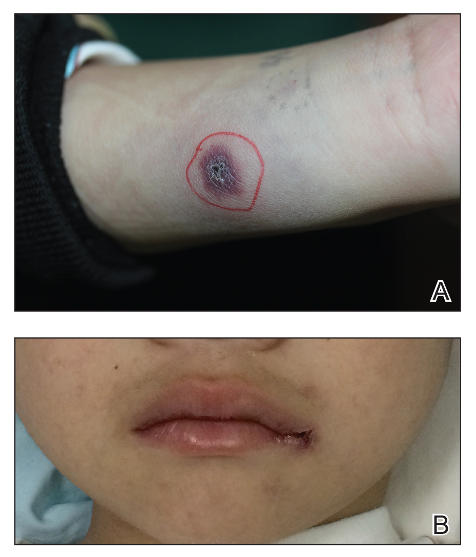
A 1.5×1.5-cm, warm, red nodule with a central black crust was noted on the left forearm (Figure 1A). Several similar lesions were noted on the buttocks, scalp, and lower extremities. Multiple ulcers, as large as 1 cm, were present on the tongue, palate, and left angle of the mouth (Figure 1B). The pharynx was congested, and the tonsils were mildly enlarged. Multiple enlarged, movable, nontender lymph nodes could be palpated in the cervical basins, axillae, and groin. No purpura or ecchymosis was detected.

Figure 1. A, A 1.5×1.5-cm, dull, red nodule with a central black crust on the left forearm. B, An ulcer on the left angle of the mouth


Pages

Recommended Reading

Pruritic, pink to violaceous, scaly papules
MDedge Dermatology
Spotting immunodeficiency in the pediatric dermatology clinic
MDedge Dermatology
Atopic Dermatitis Affects Sleep and Work Productivity
MDedge Dermatology
Flu vaccine: Larger impact on influenza burden than you thought?
MDedge Dermatology
PASI-75 with ixekizumab approaches 90% in pediatric psoriasis study
MDedge Dermatology
Psoriasis risk rises with TNF inhibitor use in children with inflammatory disorders
MDedge Dermatology
Neonatal Consultations: Vascular Lumps, Bumps, and Tumors in the Neonate
MDedge Dermatology
Patch Testing in Children: Not Just Little Adults
MDedge Dermatology
Pediatric Molluscum: An Update
MDedge Dermatology
Seborrhea Herpeticum: Cutaneous Herpes Simplex Virus Infection Within Infantile Seborrheic Dermatitis
MDedge Dermatology