Make the Diagnosis

A 45-year-old White woman with no significant medical history presented with a 1-month history of lesions on the nose and right cheek

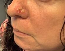
A 45-year-old White woman with no significant medical history presented with a 1-month history of lesions on the nose and right cheek. She did an online teleheath visit with primary care and was prescribed acyclovir with no improvement. She subsequently took cefadroxil with no improvement. On physical examination, erythematous papules, vesicles, and erosions with an annular crusted border were present on the nose and cheeks. The patient denied any oral, mucosal, or genital lesions. The patient had no systemic symptoms.

What's your diagnosis?

Varicella zoster virus

Herpes simplex virus

Mpox virus

Pemphigus foliaceus

Pemphigus vulgaris

Bullous impetigo

Cultures for bacteria, varicella zoster virus, herpes simplex virus, and mpox virus were all negative. A biopsy revealed suprabasilar acantholysis with follicular involvement in association with blister formation and inflammation. Direct immunofluorescence was positive for suprabasilar IgG and C3 deposition, consistent with pemphigus vulgaris (PV).

PV is an autoimmune bullous disease in which antibodies are directed against desmoglein 1 and 3 and less commonly, plakoglobin. There is likely a genetic predisposition. Medications that may induce pemphigus include penicillamine, nifedipine, or captopril.

Clinically, flaccid blistering lesions are present that may be cutaneous and/or mucosal. Bullae can progress to erosions and crusting, which then heal with pigment alteration but not scarring. The most commonly affected sites are the mouth, intertriginous areas, face, and neck. Mucosal lesions may involve the lips, esophagus, conjunctiva, and genitals.

Dr. Donna Bilu Martin

Biopsy for histology and direct immunofluorescence is important in distinguishing between PV and other blistering disorders. Up to 75% of patients with active disease also have a positive indirect immunofluorescence with circulating IgG.

Treatment is generally immunosuppressive. Systemic therapy usually begins with prednisone and then is transitioned to a steroid-sparing agent such as mycophenolate mofetil. Other steroid-sparing agents include azathioprine, methotrexate, cyclophosphamide, and intravenous immunoglobulin. Secondary infections are possible and should be treated. Topical therapies aimed at reducing pain, especially in mucosal lesions, can be beneficial.

This case and the photos are from Dr. Bilu Martin.

Dr. Bilu Martin is a board-certified dermatologist in private practice at Premier Dermatology, MD, in Aventura, Fla. More diagnostic cases are available at mdedge.com/dermatology. To submit a case for possible publication, send an email to dermnews@mdedge.com.

Recommended Reading

EULAR systemic sclerosis recommendations now include immunosuppressants
MDedge Dermatology
Patient selection key to lowering placebo response rates in lupus clinical trials
MDedge Dermatology
New EULAR lupus recommendations advise using biologics, tapering steroids
MDedge Dermatology
Long-term Remission of Pyoderma Gangrenosum, Acne, and Hidradenitis Suppurativa Syndrome
MDedge Dermatology
FDA approves ritlecitinib for ages 12 and up for alopecia areata
MDedge Dermatology
Racial Disparities in Hidradenitis Suppurativa–Related Pain: A Cross-sectional Analysis
MDedge Dermatology
Lupus flares linked to gut bacteria overgrowth
MDedge Dermatology
Porcelain White, Crinkled, Violaceous Patches on the Inner Thighs
MDedge Dermatology
Rheumatology summit tackles racial disparities in lupus trials
MDedge Dermatology
Autoantibodies could help predict cancer risk in scleroderma
MDedge Dermatology