Opinion

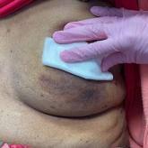
A 71-year-old White female developed erosions after hip replacement surgery 2 months prior to presentation
Direct immunofluorescence was positive for linear/granular IgG deposition throughout the epithelial cell surfaces.
Opinion

A young adult with a 1-year history of erythema, papules, and pustules on her cheeks and skin
The etiology is unknown but has been speculated to be hormone-related as it is more common in women and can be triggered by acute changes, such as...
Opinion

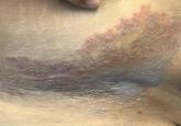
A 55-year-old female presented a with few years' history of pruritic plaques on her shins and wrists
A woman presents with thick, pruritic, erythematous plaques on her shins and wrists that come and go.
Opinion
An 88-year-old Black woman presented with 3 months duration of asymptomatic, violaceous patches on the left breast
The patient's history is significant for breast cancer.
Opinion

A 45-year-old White woman with no significant medical history presented with a 1-month history of lesions on the nose and right cheek
Erythematous papules, vesicles, and erosions with an annular crusted border were present on the nose and cheeks.
Opinion
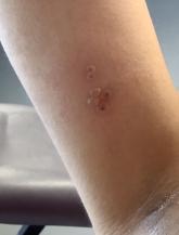
A healthy 36-year-old female presented with 4 days of itchy lesions on the right upper extremity
The patient had no systemic symptoms, but did have mild lymphadenopathy.
Opinion
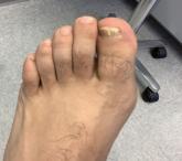
White male presents with pruritic, scaly, erythematous patches on his feet and left hand
A 47-year-old patient presents with pruritic, scaly, erythematous patches on his feet and left hand.
Opinion

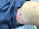
A 50-year-old woman with no significant history presented with erythematous, annular plaques, and papules on the dorsal hands and arms
A 50-year-old has had erythematous, annular plaques, and papules on her hands and arms for years.
Opinion

A 9-year old female presented with 1 day of fever, fatigue, and sore throat
A 9-year old presents with a fever of 103° F, fatigue, and sore throat, and a papular, erythematous rash on the trunk.
Opinion
A 95-year-old White male with hypertension presented with itchy patches and bullae on the trunk and extremities
The patient takes many medications, including lisinopril for his hypertension.
Opinion
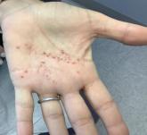
A White female presented with pustules and erythematous macules on the left palm
A 53-year-old White female presented with itchy pustules and erythematous macules on the left palm.
Opinion
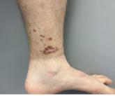
A healthy White male presented with a rash consisting of erythematous to purpuric macules
A healthy White man develops erythematous to purpuric macules on his lower extremities a few days after a visit to Disney World.
Opinion
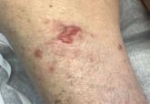
A Hispanic male presented with a 3-month history of a spreading, itchy rash
A 48-year-old Hispanic male with no significant medical history presented with a 3-month history of a spreading, itchy rash on his trunk, buttocks...
Opinion

A 64-year-old woman presents with a history of asymptomatic erythematous grouped papules on the right breast
A 64-year-old woman with a history of breast cancer presents with a 1-year history of asymptomatic erythematous grouped papules on the right...
Opinion
75-year-old White male presenting with progressive pruritus and a worsening rash
A man with dermatitis herpetiformis, well controlled on dapsone, presented with progressive pruritus and a worsening rash.