Case
A 3-year-old girl presented to the ED with a 1-week history of cough and new-onset abdominal pain. She was accompanied by her grandfather, who stated that the child had been dropped-off at his house around 5:00 pm the previous day. He noted that after putting his granddaughter to bed, she awoke around 4:30 am complaining of a stomachache. After rocking her, he said she went back to sleep but did not wake up again until 1:00 pm that afternoon. Over the first few hours of awakening, she became less active and had three episodes of posttussive emesis. The grandfather denied the child had any recent nasal congestion, fever, nausea, vomiting, or diarrhea. When questioned about possible toxic ingestion, he said there were no medications in the house and that he did not witness any substance ingestion or trauma.
Regarding history, the patient was born full-term without any complications. She did not have any significant medical or surgical history. There were no documented allergies, and she was not on any medications. All of her immunizations were up-to-date. The family history was not significant for chronic diseases except for asthma in distant relatives. Her grandfather denied any recent exposures or travel.At presentation, the patient’s vital signs were: blood pressure (BP) 86/49 mm Hg; heart rate (HR), 178 beats/minute and regular; respiratory rate (RR), 26 breaths/minute; temporal artery temperature, 104.7°F. Oxygen saturation was 99% on room air. On physical examination, she was normocephalic; there was no scleral icterus; and the throat and bilateral tympanic membranes were normal. Her extremities were warm and well perfused, with normal capillary refill. Patient’s lungs were clear, and heart sounds were normal with no detection of a murmur. The abdomen was soft and nontender; there was no evidence of organomegaly.
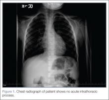
Laboratory evaluation included assessment of sodium, chloride, carbon dioxide, calcium, magnesium, amylase, lipase, and creatine levels; liver function test; complete blood count; and red-cell indices. All of the laboratory values were within normal limits, and urinalysis was negative for infection. Blood and urine cultures were also taken. A chest X-ray showed no acute intrathoracic process (Figure 1).
Secondary to the patient’s fever and tachycardia, the presumed diagnosis was systemic inflammatory response syndrome, and an intravenous (IV) bolus of normal saline 20 mL/kg along with ibuprofen and ondansetron were administered to treat fever and nausea, respectively. In addition, a dose of ceftriaxone was also given. The patient’s repeat set of vitals were: BP, 86/64 mm Hg; HR, 195 beats/minute; RR, 20 breaths/minute; and temporal artery temperature, 97.9°F; oxygen saturation was 98% on room air.During treatment, the patient became increasingly fussy with new-onset abdominal distension. Repeat physical examination revealed hepatomegaly. A bedside echocardiogram showed hyperdynamic heart with fractional shortening* (FS) of 20% and ejection fraction (EF) of 43%, but no structural abnormalities. An electrocardiogram (ECG) was then ordered, which revealed narrow complex tachycardia with inverted P-waves in inferior leads.
The patient was transferred to the intensive care unit where she was started on IV milrinone for development of secondary hypotension and reduced cardiac function. Based on inverted P waves on ECG leads II, III, and aVF, three doses of adenosine (0.1 mg/kg, 0.2 mg/kg, and 0.2 mg/kg) were given, which interrupted the tachycafrdia for only a few beats. After consultation with a cardiac electrophysiologist, a diagnosis of persistent junctional reentrant tachycardia (PJRT) was reached based on the patient’s ECG features and nonresponse to adenosine. She was started on IV amiodarone (loading dose of 5 mg/kg followed by infusion of 5 mcg/kg/min), which relieved the tachycardia (Figure 3). Her vital signs stabilized and her clinical status gradually improved. After 3 days, she was discharged on oral amiodarone and close follow-up with cardiology. A repeat echocardiogram just prior to discharge showed improvement in FS to 23% and EF to 48%.Discussion
Normal cardiac conduction involves an originating impulse from a sinus node followed by atrial muscle activation reaching the atrioventricular (AV) node. There is a necessary delay at the AV node, which is required for ventricular filling and activation of ventricles through the His-Purkinje fiber system and the bundle branches. An abnormality or interruption of this pathway results in an arrhythmia such as supraventricular tachycardia (SVT).
Supraventricular Tachycardia
Supraventricular tachycardia is the most common symptomatic abnormality in the pediatric population.1 Among the various forms of SVT, AV reentrant tachycardia (AVRT) and AV node reentrant tachycardia (AVNRT) account for most case presentations.2 Supraventricular tachycardia may be classified by duration of RP interval compared to PR interval on ECG. Short RP interval SVT includes AVNRT and AVRT through a rapidly conducting accessory path. Long RP interval SVT includes atypical AVNRT, atrial tachycardia, and PJRT.


