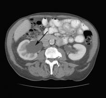
The CT scan revealed right-sided hydronephrosis. An irregular mass was also seen at the right ureterovesical junction compressing the bladder. Prostate cancer was suspected and later confirmed on biopsy.
Hydronephrosis refers to distention of the renal calyces and pelvis of one or both kidneys by urine. Hydronephrosis is not a disease but a physical result of urinary blockage that may occur at the level of the kidney, ureters, bladder, or urethra. The condition may be physiologic (eg, occurring in up to 80% of pregnant women) or pathologic. Among acquired causes in adults, pelvic tumors, renal calculi, and urethral stricture predominate. If renal colic is present, a renal stone is likely present (90% in one study). Hydronephrosis is common in pregnancy because of the compression from the enlarging uterus and functional effects of progesterone.
In this case, a urologist advised the patient of the results of the biopsy and recommended a prostatectomy. The procedure was done and the hydronephrosis resolved.
Photo courtesy of Karl T. Rew, MD. Text for Photo Rounds Friday courtesy of Richard P. Usatine, MD. This case was adapted from: Smith M. Hydronephrosis. In: Usatine R, Smith M, Mayeaux EJ, et al, eds. Color Atlas of Family Medicine. 2nd ed. New York, NY: McGraw-Hill; 2013:430-435.
To learn more about the Color Atlas of Family Medicine, see: http://www.amazon.com/Color-Family-Medicine-Richard-Usatine/dp/0071769641/
You can now get the second edition of the Color Atlas of Family Medicine as an app by clicking this link: http://usatinemedia.com/


