User login
Experts: Medical Dermatology Is Losing Ground
By 2020, the practice of dermatology will likely be narrower in scope, be less medically oriented, and place greater emphasis on dermatologic surgery and cosmetic interventions, according to two medical dermatologists.
These trends will apply worldwide but will be most pronounced in the United States, where they are already far more advanced. The dermatologists also predicted that European dermatologists will continue to practice a more traditional style of dermatology that includes inpatient care for severely ill patients.

"Cosmetic and procedural dermatology will no doubt continue to grow. You can see it growing at virtually every meeting you go to in the U.S. Why? There’s immediate gratification, and it’s the path of least resistance. You’re basically on your own, in business for yourself," said Dr. Stephen I. Katz, a dermatologist and director of the National Institute of Arthritis and Musculoskeletal and Skin Diseases.
What he finds particularly worrisome, he said, is that most cosmetic and procedural dermatology is not science based. These fields are thriving at a time of exciting advances in understanding the biologic basis of skin diseases, with the discoveries having far-reaching implications for both diagnosis and treatment. Yet aesthetic dermatology and dermatologic surgery are not pulling their fair share of the load, he said.
"With few exceptions, cosmetic and procedural dermatology is not accompanied by a research base. It’s absolutely ridiculous, for example, that we don’t have a robust research interest in wound healing," said Dr. Katz.
In an interview, Dr. Joel Schlessinger, a dermatologist in private practice in Omaha, Neb., said that although he too laments the lack of basic science research in cosmetic dermatology, he believes there is room for hope.
"Recently, over 100 residents competed for selective positions as adjunct faculty at the Third Annual Cosmetic Surgery Forum by submitting research on cosmetic dermatology," said Dr. Schlessinger.
And although several university programs have undertaken research on collagen fillers and collagen formation, "until the federal government realizes the importance that cosmetic dermatology [plays] in patients' lives, no significant basic science research will be funded at the university level. It is understandable, but not practical, for all research dollars to be in life-and-death disease states. Hopefully, the paradigm will change among strategic decisions at the government and university [levels], thereby allowing more serious research in cosmetic dermatology," he added.
"Somehow, I think, we've lost our way," said Dr. Katz. "Our specialty has already lost STDs, connective tissue disease, psoriasis, eczema, melanoma, wound healing – they’re already gone. Yes, there are psoriasis centers, but rheumatology is taking over much of psoriasis because rheumatologists are more comfortable with the more complicated biologic therapies."
The Marginalization of Academic Dermatology in the U.S.
He said that he had hoped academic dermatology would play a leadership role in keeping the specialty on track as a broad-based discipline that encompasses a wide range of subspecialty interests, but in this he has been disappointed. Academic dermatology, in his view, has undergone marginalization and trivialization.
"In the U.S., many dermatology programs are struggling to continue an academic focus. They’re basically no more than practices that happen to be located at a university, trying to meet their financial overhead with very little in the way of an academic dimension. Still, there are maybe 10-15 programs in the U.S. that have continued to focus on generating new knowledge in the university setting, often with PhD scientists leading the way," said Dr. Katz.
The National Institutes of Health now awards far more grant money for research into skin biology and skin disease to PhD scientists than to either MD or MD/PhD scientists. "The PhD scientists are so successful because they’re not encumbered by seeing patients," Dr. Katz said during a panel discussion at the World Congress of Dermatology in Seoul, South Korea.
His fellow panelist, Dr. Georg Stingl, reported that a substantial reduction in the number of hospital beds reserved for dermatology is ongoing throughout Europe, but those beds "have not yet disappeared."
"I predict the scope of dermatology in continental Europe 10 years from now will remain broader than that in the U.S., [the United Kingdom], and Asia, but it will be subject to territorial battles with other disciplines. Yet if our discipline is shrinking, it’s not the fault of others. It’s our own fault. It’s because of what we are doing," said Dr. Stingl, professor of dermatology and chairman of the division of immunology, allergy, and infectious diseases at the Medical University of Vienna.
Traditional fields of European dermatology that are now at risk of being lost to other specialties include venereology, dermato-oncology, type 1 allergy, and dermatopathology. Phlebology has already been largely taken over by vascular surgeons. On the other hand, European dermatology will see expansion of genodermatology, aesthetic dermatology, and dermatologic surgery, he added.
Dermatologic Role Changing in Japan
In Japan, as elsewhere throughout the world, the traditional dermatologic role as caregiver for severely ill patients is being taken over by other specialties.
"Traditional dermatologists are an endangered species," said Dr. Masayuki Amagai, professor of dermatology at Keio University, Tokyo. For example, Japanese dermatologists traditionally have cared for melanoma patients with terminal disease as well as for those who have early-stage disease. Now, however, more patients with advanced melanoma are being seen in integrated oncology centers.
Following the same model, it is likely that the near future will bring a new sort of integrated immunologic disease center for patients with Crohn’s disease, rheumatoid arthritis, psoriasis, and other conditions that share common inflammatory mechanisms. Dermatologists who hunger to take on the most interesting and challenging cases will want to become a part of such centers, where they will work alongside rheumatologists and gastroenterologists, Dr. Amagai said.
The good news in the United States, according to Dr. Katz, is that the recent skin biology discoveries will eventually translate into major clinical advances. And dermatology continues to attract the best and brightest medical school graduates, he said. In 2004-2007, 5.8% of U.S. dermatology residency positions were held by MD/PhDs, a rate nearly threefold greater than the average for other residency programs.
His wish list for the dermatology specialty includes a better-organized research agenda, including research programs in the cosmetic and procedural aspects of the specialty. He would also like to see the development of a clinical research consortium in dermatology, similar to the way pediatric research is conducted. "Dermatology departments are just too small to not work together," said Dr. Katz.
More effort should be placed on educating dermatologists about health services research, comparative effectiveness studies, and clinical outcomes research.
"These are areas we're not very good at. We need to be part of that whole scenario because reimbursement is going to be based on these types of studies," he said.
The speakers declared having no financial conflicts.
Dr. Katz is correct that procedural dermatology will continue to grow, as the baby boomers age, develop more cancers, and want to maintain their youthful appearance. The "immediate gratification" comment is true of any surgical subspecialty in which a physician can help a patient by performing a procedure, which generally results in a beneficial event, compared with the medical treatment of a chronic disease that may ebb and flow for decades. Either way, we all have the same objective in mind: helping patients.
Procedural and cosmetic dermatology are easy targets, but I think that in some ways, they’re straw men. Calling a spade a spade, a lot of people go into dermatology because of the lifestyle. General dermatologists work an average of 37.5 hours a week, with women physicians working fewer hours than do men. And your "average" dermatologist spends only about 10% of his or her time doing cosmetic procedures.
In addition, it’s hard to get federal funding for dermatology because the ubiquitous diseases (like acne or psoriasis) aren’t really that dangerous, and the dangerous diseases aren’t that ubiquitous.
The concept that most cosmetic and procedural dermatology is not science based is interesting, and I have two responses. Many discoveries in medicine – such as the discovery of penicillin – are not actually related to an understanding of the biological basis of disease, but rather are serendipitous and based on careful observation of various phenomena.
The extraordinary work of Dr. Jeffery A. Klein (a dermatologic surgeon who practices in San Juan Capistrano, Calif.) delineating the benefits of tumescent anesthetic – not just for liposuction, but in a wide range of surgical procedures by many specialties – was painstaking and groundbreaking work. The self-funded study has been a significant advance in modern therapeutics.
Companies like Allergan have poured tens of millions of dollars into the development of drugs that can change people’s lives. Botox might be a dirty word to some, but for a hundred medical conditions, it can be a game changer.
In the real world outside the NIH, clinicians who desire to perform research have to do so after their clinics are finished, on weekends, or on borrowed time. How many chairs of departments of dermatology allow their procedural dermatologists protected time to follow their research interests?
I don’t believe that procedural dermatology is the root cause of the degradation of our specialty or of the loss of venereal, connective tissue, and other diseases. These were being lost long before procedural dermatology saw the light of day. I would say that those who have helped develop the skin surgical specialties should be acknowledged as the saviors of the specialty at large.
Christopher Zachary, M.D., is professor and chair of the department of dermatology at the University of California, Irvine. He has received support and honoraria from Merz, Allergan, and Medicis. His comments are based on an interview with this news organization.
By 2020, the practice of dermatology will likely be narrower in scope, be less medically oriented, and place greater emphasis on dermatologic surgery and cosmetic interventions, according to two medical dermatologists.
These trends will apply worldwide but will be most pronounced in the United States, where they are already far more advanced. The dermatologists also predicted that European dermatologists will continue to practice a more traditional style of dermatology that includes inpatient care for severely ill patients.
"Cosmetic and procedural dermatology will no doubt continue to grow. You can see it growing at virtually every meeting you go to in the U.S. Why? There’s immediate gratification, and it’s the path of least resistance. You’re basically on your own, in business for yourself," said Dr. Stephen I. Katz, a dermatologist and director of the National Institute of Arthritis and Musculoskeletal and Skin Diseases.
What he finds particularly worrisome, he said, is that most cosmetic and procedural dermatology is not science based. These fields are thriving at a time of exciting advances in understanding the biologic basis of skin diseases, with the discoveries having far-reaching implications for both diagnosis and treatment. Yet aesthetic dermatology and dermatologic surgery are not pulling their fair share of the load, he said.
"With few exceptions, cosmetic and procedural dermatology is not accompanied by a research base. It’s absolutely ridiculous, for example, that we don’t have a robust research interest in wound healing," said Dr. Katz.
In an interview, Dr. Joel Schlessinger, a dermatologist in private practice in Omaha, Neb., said that although he too laments the lack of basic science research in cosmetic dermatology, he believes there is room for hope.
"Recently, over 100 residents competed for selective positions as adjunct faculty at the Third Annual Cosmetic Surgery Forum by submitting research on cosmetic dermatology," said Dr. Schlessinger.
And although several university programs have undertaken research on collagen fillers and collagen formation, "until the federal government realizes the importance that cosmetic dermatology [plays] in patients' lives, no significant basic science research will be funded at the university level. It is understandable, but not practical, for all research dollars to be in life-and-death disease states. Hopefully, the paradigm will change among strategic decisions at the government and university [levels], thereby allowing more serious research in cosmetic dermatology," he added.
"Somehow, I think, we've lost our way," said Dr. Katz. "Our specialty has already lost STDs, connective tissue disease, psoriasis, eczema, melanoma, wound healing – they’re already gone. Yes, there are psoriasis centers, but rheumatology is taking over much of psoriasis because rheumatologists are more comfortable with the more complicated biologic therapies."
The Marginalization of Academic Dermatology in the U.S.
He said that he had hoped academic dermatology would play a leadership role in keeping the specialty on track as a broad-based discipline that encompasses a wide range of subspecialty interests, but in this he has been disappointed. Academic dermatology, in his view, has undergone marginalization and trivialization.
"In the U.S., many dermatology programs are struggling to continue an academic focus. They’re basically no more than practices that happen to be located at a university, trying to meet their financial overhead with very little in the way of an academic dimension. Still, there are maybe 10-15 programs in the U.S. that have continued to focus on generating new knowledge in the university setting, often with PhD scientists leading the way," said Dr. Katz.
The National Institutes of Health now awards far more grant money for research into skin biology and skin disease to PhD scientists than to either MD or MD/PhD scientists. "The PhD scientists are so successful because they’re not encumbered by seeing patients," Dr. Katz said during a panel discussion at the World Congress of Dermatology in Seoul, South Korea.
His fellow panelist, Dr. Georg Stingl, reported that a substantial reduction in the number of hospital beds reserved for dermatology is ongoing throughout Europe, but those beds "have not yet disappeared."
"I predict the scope of dermatology in continental Europe 10 years from now will remain broader than that in the U.S., [the United Kingdom], and Asia, but it will be subject to territorial battles with other disciplines. Yet if our discipline is shrinking, it’s not the fault of others. It’s our own fault. It’s because of what we are doing," said Dr. Stingl, professor of dermatology and chairman of the division of immunology, allergy, and infectious diseases at the Medical University of Vienna.
Traditional fields of European dermatology that are now at risk of being lost to other specialties include venereology, dermato-oncology, type 1 allergy, and dermatopathology. Phlebology has already been largely taken over by vascular surgeons. On the other hand, European dermatology will see expansion of genodermatology, aesthetic dermatology, and dermatologic surgery, he added.
Dermatologic Role Changing in Japan
In Japan, as elsewhere throughout the world, the traditional dermatologic role as caregiver for severely ill patients is being taken over by other specialties.
"Traditional dermatologists are an endangered species," said Dr. Masayuki Amagai, professor of dermatology at Keio University, Tokyo. For example, Japanese dermatologists traditionally have cared for melanoma patients with terminal disease as well as for those who have early-stage disease. Now, however, more patients with advanced melanoma are being seen in integrated oncology centers.
Following the same model, it is likely that the near future will bring a new sort of integrated immunologic disease center for patients with Crohn’s disease, rheumatoid arthritis, psoriasis, and other conditions that share common inflammatory mechanisms. Dermatologists who hunger to take on the most interesting and challenging cases will want to become a part of such centers, where they will work alongside rheumatologists and gastroenterologists, Dr. Amagai said.
The good news in the United States, according to Dr. Katz, is that the recent skin biology discoveries will eventually translate into major clinical advances. And dermatology continues to attract the best and brightest medical school graduates, he said. In 2004-2007, 5.8% of U.S. dermatology residency positions were held by MD/PhDs, a rate nearly threefold greater than the average for other residency programs.
His wish list for the dermatology specialty includes a better-organized research agenda, including research programs in the cosmetic and procedural aspects of the specialty. He would also like to see the development of a clinical research consortium in dermatology, similar to the way pediatric research is conducted. "Dermatology departments are just too small to not work together," said Dr. Katz.
More effort should be placed on educating dermatologists about health services research, comparative effectiveness studies, and clinical outcomes research.
"These are areas we're not very good at. We need to be part of that whole scenario because reimbursement is going to be based on these types of studies," he said.
The speakers declared having no financial conflicts.
Oncovirus Expert: Cattle May Transmit Human Cancers
Current estimates hold that 21% of all human cancers are linked to infections. But some experts believe this figure is too low and is headed substantially higher.
Fueled by the success of vaccines for the hepatitis B virus and high-risk human papillomaviruses, which offer the first-ever means of preventing specific widespread cancers by vaccination, investigators are hunting for additional infectious causes of common malignancies. The search is focused on viruses, since they are already implicated in 64% of the known infection-related cancer burden, with bacteria and parasites accounting for the remainder.

One of the world’s most celebrated oncovirus hunters is Dr. Harald zur Hausen, winner of the 2008 Nobel Prize in Medicine for his work on human papillomaviruses as the major cause of cervical cancer. He sees a number of other malignancies as being prime candidates for potential linkage to infections, including childhood lymphoblastic leukemias, basal cell carcinomas, Epstein-Barr virus–negative Hodgkin’s lymphomas, colorectal and breast cancers, and lung cancers in nonsmokers.
The TTV (torque teno virus) family shows particular promise in this regard. The TTVs, first described by Japanese investigators in 1997 (Rev. Med. Virol. 2007;17:45-57), are an extremely heterogeneous family of single-strand DNA viruses. There are well over 100 genotypes. The torque teno viruses are widespread in all human populations. They have been found in umbilical cord blood and are vertically transmitted from mother to child, even prenatally. They frequently rearrange their genomes; and transmissibility and replication have been demonstrated to occur even for small portions of the TTV genome, according to Dr. zur Hausen, professor emeritus at the German Cancer Research Center, Heidelberg.
The TTVs are known to replicate in lymphatic and bone marrow cells – the precursor cells of leukemia. In every cell line of acute lymphoblastic leukemia and Epstein-Barr virus–negative Hodgkin’s lymphoma analyzed by Dr. zur Hausen and his coworkers, they have found the same highly conserved region of TTV DNA.
Of note, TTV load is known to be reduced by interferon. This finding is consistent with the epidemiologic observation that infants who experience multiple upper respiratory infections and other infections during their first year of life seem to be protected against childhood leukemias. The hypothesis is that bursts of increased interferon production in response to multiple infections prevents levels of an interferon-sensitive putative leukemogenic agent – perhaps a TTV – from reaching critical mass.
Dr. zur Hausen is particularly attracted to the possibility that cattle may play a role in some human cancers. He hypothesizes that some as-yet unidentified bovine virus, which is nononcogenic in its normal host, can become carcinogenic when transmitted to humans. Multiple lines of circumstantial evidence support this notion. For example, basal cell carcinomas are known to have a predisposition to arise in smallpox vaccination scars. Smallpox vaccines were prepared by inoculating vaccinia virus into the skin of calves and then harvesting the crusted skin, which could in theory contain contaminating bovine viruses.
Also, colorectal cancer, and to a lesser extent breast cancer and lung cancer in nonsmokers, have repeatedly been associated with beef consumption in epidemiologic studies. Countries with high consumption of goat or pork have relatively low rates of these malignancies.
The observed link between a red meat–rich diet and increased rates of colorectal and other cancers is often attributed to the formation of aromatic hydrocarbons and other known carcinogens during cooking or meat curing. But Dr. zur Hausen believes this interpretation might be inadequate. He noted that grilled, fried, and smoked poultry contain similar concentrations of these carcinogens, yet heavy consumption of poultry hasn’t been associated with an increased cancer risk.
It is of interest that the temperature achieved in the center of a piece of beef cooked rare is only 40 -50° C, yet torque teno viruses, papillomaviruses, and polyomaviruses are able to survive in a protein environment at temperatures of 80° C for 30 minutes or more.
"These are just suggestions. They do not prove anything, of course. But, for now, we can speculate that beef consumption plays a significant role in the development of colorectal cancer," Dr. zur Hausen said.
If even a small portion of these speculations turn out to be true, the implications for cancer prevention could be huge. It is estimated that if both the hepatitis B vaccine and the HPV vaccine were to be applied globally, the overall cancer burden in women could be reduced by 12%-14% and in men by 4%-5%, he noted.
He presented his theory at the World Congress of Dermatology in Seoul, South Korea.
Dr. zur Hausen declared having no financial conflicts.
Current estimates hold that 21% of all human cancers are linked to infections. But some experts believe this figure is too low and is headed substantially higher.
Fueled by the success of vaccines for the hepatitis B virus and high-risk human papillomaviruses, which offer the first-ever means of preventing specific widespread cancers by vaccination, investigators are hunting for additional infectious causes of common malignancies. The search is focused on viruses, since they are already implicated in 64% of the known infection-related cancer burden, with bacteria and parasites accounting for the remainder.
One of the world’s most celebrated oncovirus hunters is Dr. Harald zur Hausen, winner of the 2008 Nobel Prize in Medicine for his work on human papillomaviruses as the major cause of cervical cancer. He sees a number of other malignancies as being prime candidates for potential linkage to infections, including childhood lymphoblastic leukemias, basal cell carcinomas, Epstein-Barr virus–negative Hodgkin’s lymphomas, colorectal and breast cancers, and lung cancers in nonsmokers.
The TTV (torque teno virus) family shows particular promise in this regard. The TTVs, first described by Japanese investigators in 1997 (Rev. Med. Virol. 2007;17:45-57), are an extremely heterogeneous family of single-strand DNA viruses. There are well over 100 genotypes. The torque teno viruses are widespread in all human populations. They have been found in umbilical cord blood and are vertically transmitted from mother to child, even prenatally. They frequently rearrange their genomes; and transmissibility and replication have been demonstrated to occur even for small portions of the TTV genome, according to Dr. zur Hausen, professor emeritus at the German Cancer Research Center, Heidelberg.
The TTVs are known to replicate in lymphatic and bone marrow cells – the precursor cells of leukemia. In every cell line of acute lymphoblastic leukemia and Epstein-Barr virus–negative Hodgkin’s lymphoma analyzed by Dr. zur Hausen and his coworkers, they have found the same highly conserved region of TTV DNA.
Of note, TTV load is known to be reduced by interferon. This finding is consistent with the epidemiologic observation that infants who experience multiple upper respiratory infections and other infections during their first year of life seem to be protected against childhood leukemias. The hypothesis is that bursts of increased interferon production in response to multiple infections prevents levels of an interferon-sensitive putative leukemogenic agent – perhaps a TTV – from reaching critical mass.
Dr. zur Hausen is particularly attracted to the possibility that cattle may play a role in some human cancers. He hypothesizes that some as-yet unidentified bovine virus, which is nononcogenic in its normal host, can become carcinogenic when transmitted to humans. Multiple lines of circumstantial evidence support this notion. For example, basal cell carcinomas are known to have a predisposition to arise in smallpox vaccination scars. Smallpox vaccines were prepared by inoculating vaccinia virus into the skin of calves and then harvesting the crusted skin, which could in theory contain contaminating bovine viruses.
Also, colorectal cancer, and to a lesser extent breast cancer and lung cancer in nonsmokers, have repeatedly been associated with beef consumption in epidemiologic studies. Countries with high consumption of goat or pork have relatively low rates of these malignancies.
The observed link between a red meat–rich diet and increased rates of colorectal and other cancers is often attributed to the formation of aromatic hydrocarbons and other known carcinogens during cooking or meat curing. But Dr. zur Hausen believes this interpretation might be inadequate. He noted that grilled, fried, and smoked poultry contain similar concentrations of these carcinogens, yet heavy consumption of poultry hasn’t been associated with an increased cancer risk.
It is of interest that the temperature achieved in the center of a piece of beef cooked rare is only 40 -50° C, yet torque teno viruses, papillomaviruses, and polyomaviruses are able to survive in a protein environment at temperatures of 80° C for 30 minutes or more.
"These are just suggestions. They do not prove anything, of course. But, for now, we can speculate that beef consumption plays a significant role in the development of colorectal cancer," Dr. zur Hausen said.
If even a small portion of these speculations turn out to be true, the implications for cancer prevention could be huge. It is estimated that if both the hepatitis B vaccine and the HPV vaccine were to be applied globally, the overall cancer burden in women could be reduced by 12%-14% and in men by 4%-5%, he noted.
He presented his theory at the World Congress of Dermatology in Seoul, South Korea.
Dr. zur Hausen declared having no financial conflicts.
Paraneoplastic Autoimmune Multiorgan Syndrome Proves Rapidly Fatal
The skin may hold the key to differentiating classic pemphigus from the heterogenous autoimmune syndrome known as paraneoplastic autoimmune multiorgan syndrome.
It is a distinction of critical prognostic importance, because paraneoplastic autoimmune multiorgan syndrome (PAMS) typically is rapidly fatal, according to Dr. Sergei A. Grando, professor of dermatology and biologic chemistry at the University of California, Irvine. "The vast majority of patients die within several months of diagnosis, usually due to infections or respiratory failure, often taking the form of multiorgan system failure."

Two-thirds of patients with PAMS have a known internal malignancy at the time of their first mucocutaneous eruption. The most common of these neoplasms are non-Hodgkin’s lymphoma, which is present in more than 40% of PAMS patients; chronic lymphocytic leukemia, present in 30%; Castleman disease, present in 10%; and thymoma, present in 6%.
When a patient meets the diagnostic criteria for PAMS without having a known cancer, it is appropriate to launch a search for hidden malignancy, said Dr. Grando.
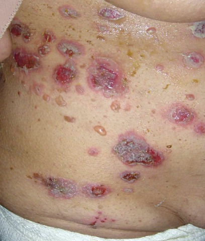
A key distinction between the skin lesions of PAMS and classic pemphigus is that PAMS involves inflammatory macules, papules, plaques, and blisters occurring on an inflammatory background over the trunk and extremities, including the palms and soles, but sparing the scalp.
In contrast, the generally more vesicular blisters and crusted erosions of pemphigus vulgaris display little erythema and usually occur on a noninflammatory background on the scalp, trunk, and extremities (but sparing the palms and soles). The most common location for skin lesions in PAMS is the palms; in pemphigus vulgaris, it is the scalp.
Also, Nikolsky's sign is positive in pemphigus vulgaris, but negative in PAMS, added Dr. Grando, who was among the investigators who first described PAMS a decade ago (Arch. Dermatol. 2001;137:193-206).
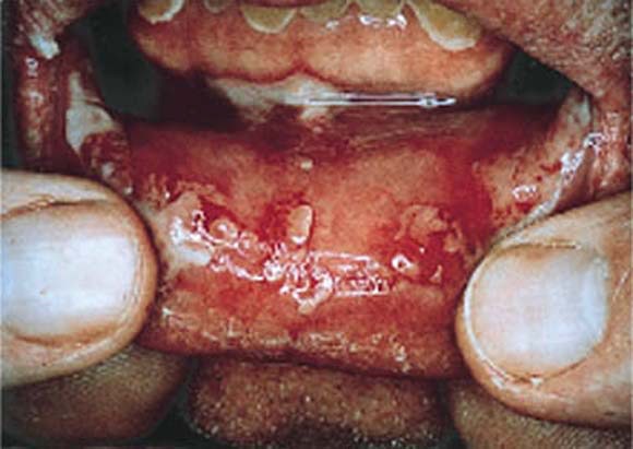
PAMS is characterized by severe and diffuse oral mucous membrane involvement, with persistent painful stomatitis because of blisters and erosions, and frequent involvement of other mucous membranes, including the eyes and genitalia. Cicatrizing conjunctivitis is particularly common in PAMS. In contrast, oral mucous membrane involvement in pemphigus is more discrete, with the eyes or other nonoral mucosa rarely involved.
Another key in the differential diagnosis: PAMS is associated with the HLA-DRB1*03 allele, whereas pemphigus vulgaris and foliaceous are strongly associated with the -04 and -14 alleles, respectively.
A hallmark of PAMS is respiratory involvement, with the sloughing of bronchial epithelial cells contributing to small airway occlusion and bronchiolitis obliterans. Classic pemphigus is free of respiratory involvement.
Both the esophagus and colon may be involved in PAMS, whereas only the esophagus is affected in pemphigus vulgaris.
In a published series of 28 PAMS patients, painful and generalized oral stomatitis was present in all 28, respiratory involvement was in 26, death from respiratory failure occurred in 22, and lichenoid skin involvement was present in 19. Only seven patients had no skin lesions (Br. J. Dermatol. 2003;149:1143-51).
At least five different subtypes of PAMS can be distinguished on the basis of the skin disease manifestations they most resemble. These subtypes of pemphiguslike PAMS include paraneoplastic pemphigus, bullous pemphigoid–like, erythema multiforme–like, lichen planus–like, and graft-vs.-host-disease–like versions of PAMS.
Dr. Grando eschews the term "paraneoplastic pemphigus" as too restrictive. After all, true pemphigus doesn’t usually affect the lungs, he noted.
Whereas most patients with classic pemphigus respond well to high-dose corticosteroids and recalcitrant disease can be effectively treated with cytotoxic agents, cyclosporine, intravenous gamma globulin, and other second-line agents, PAMS is resistant to all conventional forms of therapy.
"What can we offer? Really not much," Dr. Grando said during his presentation at the World Congress of Dermatology in Seoul, South Korea.
The preferred treatment regimen is a combination of prednisone and cyclosporine, with or without cyclophosphamide. Monthly courses of IVIG can buy patients a few extra months, he added.
Dr. Grando declared having no relevant financial interests.
The skin may hold the key to differentiating classic pemphigus from the heterogenous autoimmune syndrome known as paraneoplastic autoimmune multiorgan syndrome.
It is a distinction of critical prognostic importance, because paraneoplastic autoimmune multiorgan syndrome (PAMS) typically is rapidly fatal, according to Dr. Sergei A. Grando, professor of dermatology and biologic chemistry at the University of California, Irvine. "The vast majority of patients die within several months of diagnosis, usually due to infections or respiratory failure, often taking the form of multiorgan system failure."
Two-thirds of patients with PAMS have a known internal malignancy at the time of their first mucocutaneous eruption. The most common of these neoplasms are non-Hodgkin’s lymphoma, which is present in more than 40% of PAMS patients; chronic lymphocytic leukemia, present in 30%; Castleman disease, present in 10%; and thymoma, present in 6%.
When a patient meets the diagnostic criteria for PAMS without having a known cancer, it is appropriate to launch a search for hidden malignancy, said Dr. Grando.
A key distinction between the skin lesions of PAMS and classic pemphigus is that PAMS involves inflammatory macules, papules, plaques, and blisters occurring on an inflammatory background over the trunk and extremities, including the palms and soles, but sparing the scalp.
In contrast, the generally more vesicular blisters and crusted erosions of pemphigus vulgaris display little erythema and usually occur on a noninflammatory background on the scalp, trunk, and extremities (but sparing the palms and soles). The most common location for skin lesions in PAMS is the palms; in pemphigus vulgaris, it is the scalp.
Also, Nikolsky's sign is positive in pemphigus vulgaris, but negative in PAMS, added Dr. Grando, who was among the investigators who first described PAMS a decade ago (Arch. Dermatol. 2001;137:193-206).
PAMS is characterized by severe and diffuse oral mucous membrane involvement, with persistent painful stomatitis because of blisters and erosions, and frequent involvement of other mucous membranes, including the eyes and genitalia. Cicatrizing conjunctivitis is particularly common in PAMS. In contrast, oral mucous membrane involvement in pemphigus is more discrete, with the eyes or other nonoral mucosa rarely involved.
Another key in the differential diagnosis: PAMS is associated with the HLA-DRB1*03 allele, whereas pemphigus vulgaris and foliaceous are strongly associated with the -04 and -14 alleles, respectively.
A hallmark of PAMS is respiratory involvement, with the sloughing of bronchial epithelial cells contributing to small airway occlusion and bronchiolitis obliterans. Classic pemphigus is free of respiratory involvement.
Both the esophagus and colon may be involved in PAMS, whereas only the esophagus is affected in pemphigus vulgaris.
In a published series of 28 PAMS patients, painful and generalized oral stomatitis was present in all 28, respiratory involvement was in 26, death from respiratory failure occurred in 22, and lichenoid skin involvement was present in 19. Only seven patients had no skin lesions (Br. J. Dermatol. 2003;149:1143-51).
At least five different subtypes of PAMS can be distinguished on the basis of the skin disease manifestations they most resemble. These subtypes of pemphiguslike PAMS include paraneoplastic pemphigus, bullous pemphigoid–like, erythema multiforme–like, lichen planus–like, and graft-vs.-host-disease–like versions of PAMS.
Dr. Grando eschews the term "paraneoplastic pemphigus" as too restrictive. After all, true pemphigus doesn’t usually affect the lungs, he noted.
Whereas most patients with classic pemphigus respond well to high-dose corticosteroids and recalcitrant disease can be effectively treated with cytotoxic agents, cyclosporine, intravenous gamma globulin, and other second-line agents, PAMS is resistant to all conventional forms of therapy.
"What can we offer? Really not much," Dr. Grando said during his presentation at the World Congress of Dermatology in Seoul, South Korea.
The preferred treatment regimen is a combination of prednisone and cyclosporine, with or without cyclophosphamide. Monthly courses of IVIG can buy patients a few extra months, he added.
Dr. Grando declared having no relevant financial interests.
Thulium Laser Yields 'Dramatic' Resolution of AKs
SEOUL, SOUTH KOREA – The nonablative fractionated thulium laser at 1,927-nm wavelength is a promising new noninvasive therapy for actinic keratoses.
The laser was actually developed for superficial skin resurfacing, an application for which it is particularly well suited because the 1,927-nm wavelength minimizes patient discomfort. But while investigating the device for improvement of pigmentation, Dr. Roy G. Geronemus noted incidentally that patients were also achieving "a rather dramatic resolution" of multiple facial actinic keratoses (AKs). So he decided to conduct a formal examination of the laser’s performance for this purpose, he said at the World Congress of Dermatology
To date, in 15 patients followed for 1-6 months after the last of several thulium laser treatment sessions for multiple facial AKs, the mean clearance of the lesions was 84%-91%.
"This compares very favorably to other modalities that are out there, including the topical chemotherapies, immunomodulatory agents, and photodynamic therapy. The advantage of this is not only do you improve the AKs, but you’re also getting the cosmetic benefit simultaneously," said Dr. Geronemus, medical director of the Laser and Skin Surgery Center of New York.
Patients received up to four treatments at 2- to 6-week intervals. The laser setting was 5-20 mJ, with 30%-70% coverage per session. Topical anesthetic was utilized for 1 hour, supplemented as needed by intramuscular ketorolac.
After a single treatment a mean of 63% of AKs were cleared. After two, 84%, and after three, 85%.
The laser therapy was well tolerated. The average pain score during treatment was 2.7 on a 0-9 scale. No scarring or infections have occurred. Mild redness and peeling typically lasted 4-5 days.
Dr. Geronemus said he and his colleagues have also found that the 1,927-nm fractionated thulium laser brings about "dramatic improvement" in actinic cheilitis, and is also highly effective for the thorny problem of enlarged facial pore size.
Dr. Geronemus is a shareholder in Solta Medical, which markets the 1,927-nm fractionated thulium laser. He also is on the advisory boards of numerous dermatologic laser manufacturers.
FROM THE WORLD CONGRESS OF DERMATOLOGY
Ablative Fractional Resurfacing Smooths Atrophic Scars
SEOUL, SOUTH KOREA – Ablative fractional resurfacing with a 30-W carbon dioxide laser is a safe and effective therapy for atrophic scarring due to surgery or trauma, according to a prospective study featuring quantitative assessment of improvement in scar depth and volume.
"We’re now using this routinely on the surgical side of our practice," Dr. Roy G. Geronemus said at the World Congress of Dermatology.

He presented a study of 12 women with 19 nonacne atrophic scars, each of whom underwent three ablative fractional resurfacing (AFR) sessions at 1- to 4-month intervals. Follow-up continued for another 6 months after the final treatment, with grading of adverse events and efficacy provided both by patients and by nonblinded investigators.
The study also featured quantitative measurements of volumetric scar improvement using the Primos Imaging three-dimensional optical profiling system, which generates high-resolution topographic images of cutaneous scars. That’s a particularly noteworthy aspect of the study. The field of cosmetic dermatology is often heavily criticized for a glaring absence of this sort of objective before-and-after data.
The laser used to achieve AFR in this study was Solta Medical’s Fraxel re:pair Laser. The device delivers a pixelated pattern of microscopic ablative wounds surrounded by healthy tissue. This pattern results in zones of thermal coagulation and tissue ablation that run much deeper than those obtainable with traditional CO2 laser ablative resurfacing. This leads to more impressive neocollagenesis and dermal remodeling accompanied by shorter healing times and better safety than can be obtained with traditional broad-stroke ablative resurfacing, said Dr. Geronemus, medical director of the Laser and Skin Surgery Center of New York.
At the 6-month follow-up visit, Primos Imaging analysis demonstrated a mean 38% reduction in scar volume and a 36% decrease in maximum scar depth, compared with baseline.
Mean patient and investigator scores indicated a moderate 26%-50% improvement in scar texture, atrophy, and pigmentation. Patients and investigators respectively characterized 63% and 89% of scars as showing a 51% or greater overall improvement. A 76% or greater overall improvement was achieved in 42% and 16% of treated scars, according to patients and investigators, respectively.
Treatment-related erythema was typically rated moderate to severe, and it peaked at 72 hours after each treatment session, dropping to mild to moderate by 1 week. Edema peaked at a mild to moderate level within an hour post treatment. No incidents of delayed-onset hypopigmentation occurred during 6 months of post-AFR follow-up; in contrast, this complication has been noted in 20% or more of patients following traditional CO2 laser resurfacing, the dermatologist noted.
As to the AFR treatment settings, facial scars were typically addressed at 70 mJ per pulse, an energy density of 200 microscopic treatment zones/cm2 per pass, and 2-3 passes per session, resulting in 27%-38% coverage. Scars on the body were best addressed at 40 mJ per pulse, 200 microscopic treatment zones/cm2 per pass, and 2-3 passes, for 20%-30% coverage. Higher fluences off-face appeared to be more effective but brought prolonged erythema, which many patients find objectionable.
In an earlier study, Dr. Geronemus and his coworkers showed that the same CO2 AFR laser was effective for treatment of acne scars, achieving a mean 67% improvement in scar depth as measured using the Primos Imaging system (Lasers Surg. Med. 2008;40:381-6).
Dr. Geronemus is a shareholder in Solta Medical, and is on the advisory boards of numerous dermatologic laser manufacturers.
SEOUL, SOUTH KOREA – Ablative fractional resurfacing with a 30-W carbon dioxide laser is a safe and effective therapy for atrophic scarring due to surgery or trauma, according to a prospective study featuring quantitative assessment of improvement in scar depth and volume.
"We’re now using this routinely on the surgical side of our practice," Dr. Roy G. Geronemus said at the World Congress of Dermatology.
He presented a study of 12 women with 19 nonacne atrophic scars, each of whom underwent three ablative fractional resurfacing (AFR) sessions at 1- to 4-month intervals. Follow-up continued for another 6 months after the final treatment, with grading of adverse events and efficacy provided both by patients and by nonblinded investigators.
The study also featured quantitative measurements of volumetric scar improvement using the Primos Imaging three-dimensional optical profiling system, which generates high-resolution topographic images of cutaneous scars. That’s a particularly noteworthy aspect of the study. The field of cosmetic dermatology is often heavily criticized for a glaring absence of this sort of objective before-and-after data.
The laser used to achieve AFR in this study was Solta Medical’s Fraxel re:pair Laser. The device delivers a pixelated pattern of microscopic ablative wounds surrounded by healthy tissue. This pattern results in zones of thermal coagulation and tissue ablation that run much deeper than those obtainable with traditional CO2 laser ablative resurfacing. This leads to more impressive neocollagenesis and dermal remodeling accompanied by shorter healing times and better safety than can be obtained with traditional broad-stroke ablative resurfacing, said Dr. Geronemus, medical director of the Laser and Skin Surgery Center of New York.
At the 6-month follow-up visit, Primos Imaging analysis demonstrated a mean 38% reduction in scar volume and a 36% decrease in maximum scar depth, compared with baseline.
Mean patient and investigator scores indicated a moderate 26%-50% improvement in scar texture, atrophy, and pigmentation. Patients and investigators respectively characterized 63% and 89% of scars as showing a 51% or greater overall improvement. A 76% or greater overall improvement was achieved in 42% and 16% of treated scars, according to patients and investigators, respectively.
Treatment-related erythema was typically rated moderate to severe, and it peaked at 72 hours after each treatment session, dropping to mild to moderate by 1 week. Edema peaked at a mild to moderate level within an hour post treatment. No incidents of delayed-onset hypopigmentation occurred during 6 months of post-AFR follow-up; in contrast, this complication has been noted in 20% or more of patients following traditional CO2 laser resurfacing, the dermatologist noted.
As to the AFR treatment settings, facial scars were typically addressed at 70 mJ per pulse, an energy density of 200 microscopic treatment zones/cm2 per pass, and 2-3 passes per session, resulting in 27%-38% coverage. Scars on the body were best addressed at 40 mJ per pulse, 200 microscopic treatment zones/cm2 per pass, and 2-3 passes, for 20%-30% coverage. Higher fluences off-face appeared to be more effective but brought prolonged erythema, which many patients find objectionable.
In an earlier study, Dr. Geronemus and his coworkers showed that the same CO2 AFR laser was effective for treatment of acne scars, achieving a mean 67% improvement in scar depth as measured using the Primos Imaging system (Lasers Surg. Med. 2008;40:381-6).
Dr. Geronemus is a shareholder in Solta Medical, and is on the advisory boards of numerous dermatologic laser manufacturers.
FROM THE WORLD CONGRESS OF DERMATOLOGY
Real-Time Monitoring of Melanoma Markers Predicts Relapse
SEOUL, SOUTH KOREA – Serial monitoring of melanoma tumor marker levels in peripheral blood using a novel quantitative real-time reverse-transcriptase polymerase chain reaction method after surgical resection of melanoma has shown promise for the early detection of patients at high risk for disease progression.
The real-time polymerase chain reaction (PCR) assay measures circulating levels of five markers unique to melanoma cells: glycoprotein 100 (gp100), melanoma antigen gene-3 (MAGE-3), tyrosinase, melanoma marker A (Melan-A), and melanoma inhibitory activity (MIA) protein, Dr. Spyridon Gkalpakiotis explained at the World Congress of Dermatology.
He reported on 65 patients who underwent peripheral blood testing and analysis of the five markers every 3 months for the first 2 years after resection of their stage II or III melanoma, for a total of 2,925 PCR assays.
Twenty-six patients experienced elevated test results. All 26 relapsed during 5 years of follow-up; the 5-year survival rate in this group was 65%.
In contrast, only 1 of 39 patients with consistently negative real-time PCR assays experienced disease progression; 5-year survival in PCR-negative patients was 97%, reported Dr. Gkalpakiotis of Charles University in Prague.
MAGE-3 was expressed in 21 patients with disease progression. The next most sensitive markers of melanoma progression were MIA and gp100.
Dr. Gkalpakiotis declared having no financial conflicts.
FROM THE WORLD CONGRESS OF DERMATOLOGY
Major Finding: Twenty-six patients experienced
elevated test results. All 26 relapsed during 5 years of follow-up.
Data Source: Patients (n = 65) who underwent peripheral blood testing and analysis of the
five markers every 3 months for the first 2 years after resection of
their stage II or III melanoma, for a total of 2,925 PCR assays.
Disclosures: Dr. Gkalpakiotis declared having no financial conflicts.
Cutaneous Lupus Over Age 50 Probably Drug Induced
SEOUL, SOUTH KOREA – Onset of subacute cutaneous lupus erythematosus after the age of 50 years is a good clue it is more likely to be drug induced, findings from two recent studies indicate.
Prompt recognition of drug-induced subacute cutaneous lupus erythematosus (SCLE) by physicians prevents significant morbidity, both by quelling the number and activity of skin lesions and by sparing affected patients from exposure to the potent immunosuppressive agents that are often used in idiopathic SCLE.
In drug-induced SCLE, the treatment of choice is withdrawal of the offending medication and nothing more, Dr. Christopher B. Hansen said at the World Congress of Dermatology.
"The important thing is stopping the medication. Most patients don't require additional therapy," emphasized Dr. Hansen of the University of Utah in Salt Lake City.
He and his coinvestigators recently combed the medical literature and published a systematic review of 117 cases of drug-induced SCLE, the largest series to date (Br. J. Dermatol. 2011;164:465-72). One of the standout findings was that the average age of onset was 58 years, compared with just 43 years in patients with idiopathic SCLE culled from the literature.
Strikingly, this 15-year age gap was identical to that reported in a recent study by investigators at the University of Milan, Dr. Hansen noted. In the Italian series, the average age of onset of drug-induced SCLE was 67 years, or 15 years more than the 52 years in 79 patients with idiopathic SCLE.
The Italian investigators described several key cutaneous features that helped differentiate drug-induced from idiopathic SCLE. Lesions were widespread in 82% of patients with drug-induced SCLE, compared with just 6% of those with the idiopathic form. Malar rash occurred in 45% of drug-induced SCLE patients versus 6% with idiopathic SCLE. Bullous and erythema multiforme-like lesions were present in 45% of the drug-induced SCLE cohort, compared with just 1% of those with idiopathic SCLE. Vasculitic lesions were noted in 45% of patients with drug-induced SCLE but only 3% of those with idiopathic SCLE (Br. J. Dermatol. 2011 May 12 [doi:10.1111/j.1365-2133.2011.10397.x]).
Internal organ involvement was notably absent in patients with drug-induced SCLE, both in Dr. Hansen’s study and in the Italian investigation. That's an important point of distinction between drug-induced SCLE and drug-induced systemic lupus erythematosus (SLE).
Also, the drugs implicated in causing SCLE are generally different from those that trigger drug-induced SLE, which is probably an indication of fundamentally different underlying mechanisms of disease, he continued.
The list of medications linked to drug-induced SCLE is lengthy. Topping the list are antihypertensive agents, responsible for 34% of cases in the study by Dr. Hansen and his colleagues, and antifungals, which triggered another 26%. Terbinafine, thiazide diuretics, and calcium channel blockers were particularly common offenders. Chemotherapeutic agents were a distant third category of triggering medications, responsible for 9% of cases, followed closely by antihistamines at 8% and immunomodulators at 7%.
A defining characteristic of drug-induced SCLE is the lengthy incubation and resolution times compared with most drug eruptions. After stopping the offending drug, it took a median of 4 weeks and a mean of 7.3 weeks for the skin lesions to go away, according to Dr. Hansen.
Anti-Ro/SSA autoantibodies were present in 81% of patients at diagnosis. Two-thirds of patients remained autoantibody-positive at testing conducted 6 weeks to 3 years after resolution of the rash.
Several theories have been put forth regarding the pathogenesis of drug-induced SCLE. One is that it is a photosensitivity reaction, given that many of the drugs linked to the condition are known to be photosensitizers.
Another theory is that the skin disease results from a drug-induced disruption of T-cell function and immune tolerance. Still another theory holds that the drug or its metabolite causes direct cytotoxicity.
Asked for tips on how to discover the offending medication in older patients on many drugs, he said the best approach is to start by trying to eliminate any medications that have been started within the past month or two. Secondly, he added, take a close look at any remaining medications the patient is on that have been reported as triggers most frequently in the literature.
Dr. Hansen declared having no financial conflicts of interest.
EXPERT ANALYSIS FROM THE WORLD CONGRESS OF DERMATOLOGY
Consider Infliximab for Refractory Cutaneous Sarcoidosis
SEOUL, SOUTH KOREA – Infliximab appears to be an effective corticosteroid-sparing treatment for patients with refractory cutaneous sarcoidosis, albeit an off-label one.
"Infliximab seems to be a good candidate as third-line therapy for refractory cutaneous sarcoidosis. We recommend a continuous evaluation of clinical response and an increase in infliximab dosing up to 10 mg/kg every 4 weeks in cases of insufficient response [as well as] the use of concomitant low-dose methotrexate to increase infliximab efficacy and prevent the development of anti-infliximab antibodies," noted Dr. Fabien Guibal at the World Congress of Dermatology.
Skin involvement is common in sarcoidosis, affecting up to 30% of patients. It carries substantial morbidity, and treatment options are limited. Systemic corticosteroids are the mainstay, but the associated side effects are well known. Methotrexate is a good second-line therapy, although the response is highly variable. Azathioprine, antimalarials, and thalidomide are among the alternative options, noted Dr. Guibal of Hôpital Saint-Louis, Paris.
He presented a retrospective, single-center study involving nine patients placed on infliximab (Remicade) for refractory cutaneous sarcoidosis. Participants had failed to improve on a median of five prior immunosuppressive drugs.
The subjects (eight women and one man) were a median of 43 years old when diagnosed with sarcoidosis, and 51 years of age when they started on infliximab. The cutaneous lesions consisted of lupus pernio in eight patients, disseminated papules in seven, and a large facial plaque in one. All patients had extracutaneous disease, including pulmonary sarcoidosis in eight individuals and CNS involvement in one.
The impetus for turning to the tumor necrosis factor–alpha inhibitor in these nine patients was the fact that TNF-alpha is known to be a critical cytokine in the formation and maintenance of granulomas. Plus, there have been several reports of favorable treatment responses in patients with pulmonary sarcoidosis and other internal organ involvement.
Three of nine patients experienced resolution of cutaneous lesions on their first course of infliximab. Another four had near-resolution, and the remaining two patients showed partial improvement. The median lag time until maximal response was 12 weeks (range, 2-22 weeks).
When infliximab was eventually discontinued in six patients, five relapsed after a median 9 weeks off the drug. The sole patient who didn’t relapse had successfully replaced the biologic agent with methotrexate.
The five relapsers underwent a second course of infliximab. Four experienced near-resolution of their cutaneous disease, and the fifth showed lesion stabilization. Three of them eventually discontinued infliximab, one because of loss of efficacy, another who remained relapse free while replacing the biologic with methotrexate, and a third who was lost to follow-up.
After a median 34 months of follow-up, five of the nine patients remain on infliximab. Their average oral prednisone dose is half of the 12 mg/day it was before they started taking infliximab. No serious adverse effects have occurred.
Dr. Guibal declared having no relevant financial relationships.
FROM THE WORLD CONGRESS OF DERMATOLOGY
Major Finding: Lesions of cutaneous sarcoidosis resolved in three of nine patients after a course of infliximab therapy.
Data Source: A retrospective, single-center study involving nine patients who were placed on infliximab for refractory cutaneous sarcoidosis.
Disclosures: Dr. Guibal declared having no relevant financial relationships.
Tretinoin, Isotretinoin Found Equally Effective for Photoaging
SEOUL, SOUTH KOREA – Low-dose oral isotretinoin and alternate-day tretinoin proved similarly effective for treatment of photoaging in a prospective randomized trial.
Treatment of photoaged skin is an off-label application for isotretinoin, but there have been a number of published favorable case series (J. Eur. Acad. Dermatol. Venereol. 2009;23:115-23).
Some dermatologists are prescribing it in more severe cases where teratogenicity is a nonissue. So Dr. Edileia Bagatin decided to put the potent oral retinoid to the test in a randomized comparison with tretinoin (Renova), a topical retinoid that does have a regulatory indication for photoaging.
The result of the 12-month study was a draw in terms of efficacy. The two treatment groups showed similar improvements in photoaging based upon blinded dermatologic evaluations, patient self-ratings, quality of life scores, and histologic findings.
And because the topical retinoid doesn't come with the baggage for which isotretinoin is notorious – including dyslipidemia, birth defects, liver dysfunction, depression, and inflammatory bowel disease – tretinoin has the clear advantage in most situations, said Dr. Bagatin at the World Congress of Dermatology.
The trial involved 22 patients, aged 50-75 years, with moderate to advanced photoaging. Nine of 11 subjects in each treatment arm were smokers.
Patients were assigned to either 20 mg/day of isotretinoin for 6 months followed by 0.05% tretinoin cream applied every other day for 6 months, or to alternate-day tretinoin for the full 12 months. All patients were instructed to use a moisturizing sunscreen twice daily.
The number of actinic keratoses on the face was reduced to a similar extent via both therapies: by an average of 74% after 6 months and 54% after 12 months. The number of AKs on the forearms dropped by an average of 42% with either therapy after 6 months, and by 61% after 12 months, according to Dr. Bagatin, of the Federal University of Sao Paulo (Brazil).
The patients themselves rated their wrinkles, skin elasticity, actinic keratoses, and freckles as significantly improved at both 6 and 12 months, with no significant difference between treatment arms. Median Dermatology Life Quality Index scores showed significant improvement over time, again with no difference between the two groups.
On histology, both groups displayed reductions in corneal layer thickness and elastosis, along with increased epidermal thickness; these changes were greater with time. There was also a significant reduction in p53 expression and an increase in collagen I, with both changes being greater at 12 months than 6 months.
The study was funded by the Sao Paulo State Research Foundation. Dr. Bagatin reported having no financial conflicts.
FROM THE WORLD CONGRESS OF DERMATOLOGY
Major Finding: Oral isotretinoin and topical tretinoin both reduced facial actinic keratoses by 74% after 6 months and 54% after 12 months.
Data Source: A 1-year randomized trial comparing the efficacy of low-dose oral isotretinoin to alternate-day tretinoin in 22 patients with photoaging.
Disclosures: The study was funded by the Sao Paulo State Research Foundation. Dr. Bagatin reported having no financial conflicts.
Real-Time Monitoring of Melanoma Markers Predicts Relapse
SEOUL, SOUTH KOREA – Serial monitoring of melanoma tumor marker levels in peripheral blood using a novel quantitative real-time reverse-transcriptase polymerase chain reaction method after surgical resection of melanoma has shown promise for the early detection of patients at high risk for disease progression.
The real-time polymerase chain reaction (PCR) assay measures circulating levels of five markers unique to melanoma cells: glycoprotein 100 (gp100), melanoma antigen gene-3 (MAGE-3), tyrosinase, melanoma marker A (Melan-A), and melanoma inhibitory activity (MIA) protein, Dr. Spyridon Gkalpakiotis explained at the World Congress of Dermatology.
He reported on 65 patients who underwent peripheral blood testing and analysis of the five markers every 3 months for the first 2 years after resection of their stage II or III melanoma, for a total of 2,925 PCR assays.
Twenty-six patients experienced elevated test results. All 26 relapsed during 5 years of follow-up; the 5-year survival rate in this group was 65%.
In contrast, only 1 of 39 patients with consistently negative real-time PCR assays experienced disease progression; 5-year survival in PCR-negative patients was 97%, reported Dr. Gkalpakiotis of Charles University in Prague.
MAGE-3 was expressed in 21 patients with disease progression. The next most sensitive markers of melanoma progression were MIA and gp100.
Dr. Gkalpakiotis declared having no financial conflicts.
SEOUL, SOUTH KOREA – Serial monitoring of melanoma tumor marker levels in peripheral blood using a novel quantitative real-time reverse-transcriptase polymerase chain reaction method after surgical resection of melanoma has shown promise for the early detection of patients at high risk for disease progression.
The real-time polymerase chain reaction (PCR) assay measures circulating levels of five markers unique to melanoma cells: glycoprotein 100 (gp100), melanoma antigen gene-3 (MAGE-3), tyrosinase, melanoma marker A (Melan-A), and melanoma inhibitory activity (MIA) protein, Dr. Spyridon Gkalpakiotis explained at the World Congress of Dermatology.
He reported on 65 patients who underwent peripheral blood testing and analysis of the five markers every 3 months for the first 2 years after resection of their stage II or III melanoma, for a total of 2,925 PCR assays.
Twenty-six patients experienced elevated test results. All 26 relapsed during 5 years of follow-up; the 5-year survival rate in this group was 65%.
In contrast, only 1 of 39 patients with consistently negative real-time PCR assays experienced disease progression; 5-year survival in PCR-negative patients was 97%, reported Dr. Gkalpakiotis of Charles University in Prague.
MAGE-3 was expressed in 21 patients with disease progression. The next most sensitive markers of melanoma progression were MIA and gp100.
Dr. Gkalpakiotis declared having no financial conflicts.
SEOUL, SOUTH KOREA – Serial monitoring of melanoma tumor marker levels in peripheral blood using a novel quantitative real-time reverse-transcriptase polymerase chain reaction method after surgical resection of melanoma has shown promise for the early detection of patients at high risk for disease progression.
The real-time polymerase chain reaction (PCR) assay measures circulating levels of five markers unique to melanoma cells: glycoprotein 100 (gp100), melanoma antigen gene-3 (MAGE-3), tyrosinase, melanoma marker A (Melan-A), and melanoma inhibitory activity (MIA) protein, Dr. Spyridon Gkalpakiotis explained at the World Congress of Dermatology.
He reported on 65 patients who underwent peripheral blood testing and analysis of the five markers every 3 months for the first 2 years after resection of their stage II or III melanoma, for a total of 2,925 PCR assays.
Twenty-six patients experienced elevated test results. All 26 relapsed during 5 years of follow-up; the 5-year survival rate in this group was 65%.
In contrast, only 1 of 39 patients with consistently negative real-time PCR assays experienced disease progression; 5-year survival in PCR-negative patients was 97%, reported Dr. Gkalpakiotis of Charles University in Prague.
MAGE-3 was expressed in 21 patients with disease progression. The next most sensitive markers of melanoma progression were MIA and gp100.
Dr. Gkalpakiotis declared having no financial conflicts.
FROM THE WORLD CONGRESS OF DERMATOLOGY