User login
Maximize cosmetic procedures for men
MIAMI BEACH – More men are seeking cosmetic dermatologic procedures and products, and successfully engaging and treating this segment of the population require recognition of a number of gender-specific differences with respect to skin biology, skin aging, behaviors, and rejuvenation goals, according to Dr. Ivan Camacho.
"We need to be able to understand all of these the differences in order to be able to provide tailored treatments for our patients, so they can get the best results possible," Dr. Camacho said during a focus session on men’s aesthetics at the annual meeting of the American Academy of Dermatology.
Skin biology
When it comes to skin biology, men and women share a number of characteristics, but there are important differences driven by genetic or hormonal specificity that can affect treatment outcomes, Dr. Camacho said. For example, although the number of cell layers and thickness of the stratum corneum is similar in men and women, the dermis is about 20% thicker in men than in women, across the age spectrum and in all anatomic locations, he noted.
Men also have higher collagen density early in life, but they experience skin thinning at a younger age than women. For women, skin thickness generally remains constant until menopause, and then the skin begins to thin rapidly. This difference is most likely because of the role of testosterone in maintaining collagen content, said Dr. Camacho of the University of Miami.
Men also have less subcutaneous fat, greater distribution of body fat in the abdomen and trunk, and higher facial bone density mass than women, Dr. Camacho explained. In addition, men tend to have higher transepidermal water loss because of the lack of estrogen, which has positive effects in the stratum corneum, he said.
Dr. Camacho described other functional differences in men’s skin compared with women’s skin, including:
• A fourfold increase in sebum generation.
• A 30% overall increase in sweating.
• Different hair distribution as determined by androgens, but also by genetics.
• Stronger skin tone.
• Greater sensitivity to ultraviolet radiation, and thus a lower minimal erythema dose threshold and increased rate of skin cancers.
• Greater susceptibility to bacterial and viral infections and slower healing rates as a result of the inhibitory effects of testosterone and hydrotestosterone with respect to wound healing.
• Greater susceptibility to stress-induced immunosuppression, which may explain the higher skin cancer rates and delayed wound healing.
Skin aging
As for the aging process, men age differently from women in that their higher collagen density leads to better maintenance of elasticity, and their higher facial bone density provides better overall support, said Dr. Camacho.
However, thicker skin and stronger muscles make men more prone to develop deeper expression lines, as opposed to the "superficial wrinkles that women complain about," Dr. Camacho noted.
Also, the reduced level of subcutaneous fat in men can lead to more dramatic volume loss.
"Men are ‘sinkers.’ We sink more than wrinklers or saggers, because we have good elasticity, but we actually tend to lose quite a bit of subcutaneous fat," Dr. Camacho explained.
As a result of other differences related to skin aging, he said, men’s skin may be:
• More prone to acne and enlarged pores due to the higher sebaceous gland count.
• More prone to darker and/or redder complexions because of the increased tone and vascularity.
• More likely to have dull areas due to the epidermal water loss.
• Less prone to perioral lines and wrinkles due to facial hair distribution, which acts as a structural support.
• More likely to have unwanted fat in the abdominal and trunk region.
• More likely to develop both melanoma and nonmelanoma skin cancers and to experience photoaging because of greater sensitivity to ultraviolet radiation.
Behavior
In Dr. Camacho’s experience, men tend to be very goal oriented, and that carries over to cosmetic procedures.
"Men are very results-oriented, so we want to have a very clear purpose of what we want to achieve with a given treatment or product," he added, noting that male patients often prefer a lot of detail about procedures and processes.
Providing the extra details requires a greater educational effort on the part of the physician, he said, but "that’s a great thing, because they are going to be well informed about the pros and cons of a given treatment."
Male patients also want fast results. For these reasons, injectables and laser therapies are probably a good fit, he noted.
Men also tend to prefer simplicity, minimal discomfort, and minimal downtime, making noninvasive procedures and multifunctional skin care products ideal, he added.
Rejuvenation goals
Men often have rejuvenation goals that are different from those of women, Dr. Camacho said.
The most common reason that men seek cosmetic treatment is for a "tired, sinking face"; they want to look refreshed and confident, but they want subtle, natural-looking results, he said.
And, of course, there’s hair.
"Hair, hair, hair. Hair is a huge concern for men," Dr. Camacho said.
Sometimes men think they have too much hair, sometimes they have too little. Men drive the market for hair loss treatments, and also are increasingly seeking hair removal treatments, Dr. Camacho noted. Since traditional methods for hair removal, such as waxing, shaving, and epilation, are temporary and can cause irritation, laser hair removal is increasing in popularity among men. In fact, according to the American Society of Aesthetic Plastic Surgery, it was the second most common nonsurgical cosmetic intervention for men in 2011, Dr. Camacho said.
Other useful cosmetic procedures
One of the most popular noninvasive cosmetic interventions for both men and women is neuromodulation injections, for softening of expression lines and treating areas including the glabella, forehead, and periocular area, that can contribute to an angry-, tired-, or sad-looking face, Dr. Camacho said.
In 2011, men accounted for 9.1% of botulinum toxin treatment patients, and this represented a 258% increase from the year 2000, he noted.
Skin resurfacing treatments are increasing in popularity among men as well. In 2011, men accounted for 7.3% of skin resurfacing treatments, making skin resurfacing the fourth most common nonsurgical cosmetic intervention in men, Dr. Camacho said.
Other treatments for skin issues of concern to men include laser and light therapies to improve the complexion, and skin-tightening procedures, such as radiofrequency and ultrasound, to help improve skin laxity
In addition, data from 2011 showed that men accounted for about 20% of patients undergoing CoolSculpting, which is both effective and appealing to men seeking treatment for abdominal and trunk fat deposits, Dr. Camacho said.
Treatment pearls
When providing facial treatments for men, preservation of a masculine appearance is essential, Dr. Camacho said.
"The last thing we want to do is have a man’s face looking like a woman’s face; we need to be able to preserve the masculine appearance," Dr. Camacho said, noting that in his experience, a combination of neurotoxins and dermal fillers is excellent for achieving desired results.
For neurotoxins, remember that men require doses at about 1.5 to 2 times as much as women, he said
"But also remember that men want to preserve some animation, so it’s always important to keep a balance between being effective with our treatment, but also keeping the patient happy with what they want," he said.
In Dr. Camacho’s experience, most men are less concerned with periocular lines, but some are, so be sure to ask them exactly what they want at a treatment session.
"You can’t assume they want three areas treated at a time as we usually do with women," he said.
Be sure to preserve the masculine position of the brow, which in men is very subtle, located right at the supraorbital rim, and has no major arching, said Dr. Camacho.
By avoiding injecting at the superior portion of the orbicularis ocular muscle, the subtle arch at the supraorbital rim can be maintained, and feminization of the brow avoided, he said.
"Also, when treating the frontalis major make sure to go all the way lateral, because you don’t want a little bit of frontalis pulling and giving some arching to the eyebrows," he added.
As for fillers, keep in mind that men probably will require higher volumes, which is an important consideration when discussing finances during the initial consultation.
Fillers are particularly useful in the tear trough area for men seeking treatment of tired-looking sunken eyes.
"Very easily, we can create a smooth transition between the lower eyelid and cheek. We can also use (fillers) for prominent nasolabial folds," Dr. Camacho explained.
Before filling the lines, however, consider restoring the cheek to correct for the midfacial fat loss common in men, he noted.
Also, if treatment involves the lip area, avoid overfilling the vermilion border – this will feminize the lips, he said.
Breaking into the men’s market
Consider developing a marketing strategy that shows your practice’s appeal to both genders – by launching a website and developing marketing materials that feature both male and female models and patients, and by offering cosmeceutical product lines developed for men, Dr. Camacho suggested.
"The modern man is here, and the modern man is actively looking for advice on cosmetic procedures and recommendations for skin care. Dermatologists will have a very important role in male aesthetics," he said, noting that a dermatologist’s role can include enhancing awareness and cultural acceptance of cosmetic interventions for men.
"We have every day in our practices, an opportunity to educate our male patients on the multiple treatment options available for them. We also have a responsibility for being knowledgeable about the specificities of male skin ... to be able to formulate tailored treatments, and to be able to, therefore, obtain the best results for our male patients," he said.
Dr. Camacho reported having no disclosures.
AT THE AAD ANNUAL MEETING
More men seek skin care products
MIAMI BEACH – American men are showing an increased interest in skin care products, according to Dr. Ivan Camacho.
Men are becoming more aware of the importance of skin care, and they are actively seeking information and products. In fact, the men’s skin care market increased 9% from 2009 to 2010, and is expected to grow 16% by 2014, Dr. Camacho said at the annual meeting of the American Academy of Dermatology.
One point for dermatologists to keep in mind when it comes to introducing men to a skin care routine is that simple is best. Using multipurpose products, keeping regimens to one or two steps – three at most – and incorporating new products into an established routine, such as shaving, will likely improve compliance and results, said Dr. Camacho of the University of Miami.
Dr. Camacho’s additional tips for better skin care for men include:
• Recommend multifunctional products, such as those that combine antioxidants and botanicals, as well as other cosmeceuticals that can enhance anti-aging, provide anti-inflammatory effects, and hydrate the skin.
• Suggest fragrance-free or subtly scented products.
• Incorporate a broad-spectrum sunscreen (also unscented or subtly scented) with a sun protection factor (SPF) of at least 30. A product with botanical and other cosmeceutical ingredients or with anti-inflammatory properties may be a good choice, especially in patients with inflammatory conditions like acne or rosacea.
• Incorporate a moisturizer, which is very important for restoring facial hydration and improving the skin barrier. A moisturizer can be included in the sunscreen or other products.
"Tell patients to moisturize, moisturize, moisturize," Dr. Camacho said, noting: "If you tell them they need to do it three times a day, they will probably do it once a day, because this is a practice very neglected by many men. As we know, moisturizers will benefit all skin types."
For men with oily skin, recommend an oil-absorbing or mattifying formulation; for those with drier skin, recommend a lipid-based formulation. Given that more men are seeking information about skin care, dermatologists would do well to become knowledgeable about the various products available that may be most appealing to and effective for men, said Dr. Camacho.
Products currently attracting attention include moisturizers with topical caffeine, which has been shown to reduce the transepidermal water loss that is greater in men than in women, he noted.
Also, glycerin-based and niacinamide-based moisturizers have been shown in several studies to reduce transepidermal water loss, which may increase after shaving, he said.
In addition, many men can benefit from cleansers and toners developed for their particular skin types, shaving products that prevent or relieve irritation, oil-absorbing primers to provide temporary relief for skin oiliness, and exfoliating products and retinoids to improve an uneven complexion, said Dr. Camacho.
In addition, antiaging formulations containing alpha-hydroxy acids, retinoids, growth factors, antioxidants, peptides, and/or botanicals can be used to help reverse ultraviolet-related damage and help improve the appearance of fine lines, he said.
Dr. Camacho reported having no disclosures.
AT THE AAD ANNUAL MEETING
Technique is key for filler injection in darker skin
MIAMI BEACH – When considering dermal fillers for skin of color patients, remember that fewer injections can help reduce the risk of keloid formation and pigmentary changes, Dr. Valerie D. Callender said at the annual meeting of the American Academy of Dermatology.
People with skin of color made up 20% of the patient population seeking cosmetic procedures in 2011, and they are the fastest-growing demographic group in the U.S. population, Dr. Callender said. Dermatologists can expect to see more patients with ethnic skin in their practices, and it’s important for them to remember that not all aging skin is created equal, and that different techniques come into play for different skin types, she added.

Aging in ethnic facial skin differs from aging in lighter skin, mainly because of the photoprotective effect of melanin against UV radiation, said Dr. Callender. The effects of photodamage usually appear 10-20 years later in skin of color patients and with less severity.
"Your typical skin of color patient is 45 years old, has some volume loss, some infraorbital hollowing, and is definitely concerned about laugh lines," said Dr. Callender.
"The No. 1 tip is to minimize the number of injections to minimize the risk of postinflammatory hyperpigmentation," she emphasized. "If there is erythema, apply a topical corticosteroid."
Hyaluronic acid fillers are among the top five nonsurgical cosmetic procedures in the United States, but there is a paucity of published studies in skin of color patients, said Dr. Callender of Howard University, Washington. The population included in large, pivotal clinical studies is composed mainly of white patients, and even if these studies include a subset of skin of color patients, they don’t specifically report on treatment and safety outcomes in those patients, Dr. Callender said.
Dr. Callender listed several published and unpublished studies including data on the use of fillers in patients with Fitzpatrick Skin Types IV to VI. The products studied included Restylane, Perlane, Juvéderm Ultra and Ultra Plus, Hylaform, Hylaform Plus, Captique, Belotero Balance, and Radiesse.
Pigmentary changes were common throughout the studies, but they did not affected more than 9% of the study population, according to Dr. Callender. In the Radiesse study, the authors concluded that lack of pigmentary changes may have occurred from a deeper injection level, compared with HA fillers, she noted. No keloids or scarring were reported at 6 months’ follow-up and the investigators used a 25- to 27-gauge needle (Dermatol. Surg. 2009; 35:1641-5).
There have been no formal clinical trials evaluating safety of Sculptra in skin of color patients, said Dr. Callender, director of Callender Dermatology & Cosmetic Center, Glenn Dale, Md. However, the investigators in a 2010 study advised clinicians to lower the injection time, use proper product reconstitution and proper produce placement, perform immediate and postoperative massage, and avoid Sculptra on patients with a history of keloids in order to reduce the risk of adverse events (J. Drugs Dermatol. 2010;9:451-6).
Dr. Callender has been a consultant and investigator for Allergan, Galderma, Medicis, and Merz.
On Twitter @NaseemSMiller
MIAMI BEACH – When considering dermal fillers for skin of color patients, remember that fewer injections can help reduce the risk of keloid formation and pigmentary changes, Dr. Valerie D. Callender said at the annual meeting of the American Academy of Dermatology.
People with skin of color made up 20% of the patient population seeking cosmetic procedures in 2011, and they are the fastest-growing demographic group in the U.S. population, Dr. Callender said. Dermatologists can expect to see more patients with ethnic skin in their practices, and it’s important for them to remember that not all aging skin is created equal, and that different techniques come into play for different skin types, she added.
Aging in ethnic facial skin differs from aging in lighter skin, mainly because of the photoprotective effect of melanin against UV radiation, said Dr. Callender. The effects of photodamage usually appear 10-20 years later in skin of color patients and with less severity.
"Your typical skin of color patient is 45 years old, has some volume loss, some infraorbital hollowing, and is definitely concerned about laugh lines," said Dr. Callender.
"The No. 1 tip is to minimize the number of injections to minimize the risk of postinflammatory hyperpigmentation," she emphasized. "If there is erythema, apply a topical corticosteroid."
Hyaluronic acid fillers are among the top five nonsurgical cosmetic procedures in the United States, but there is a paucity of published studies in skin of color patients, said Dr. Callender of Howard University, Washington. The population included in large, pivotal clinical studies is composed mainly of white patients, and even if these studies include a subset of skin of color patients, they don’t specifically report on treatment and safety outcomes in those patients, Dr. Callender said.
Dr. Callender listed several published and unpublished studies including data on the use of fillers in patients with Fitzpatrick Skin Types IV to VI. The products studied included Restylane, Perlane, Juvéderm Ultra and Ultra Plus, Hylaform, Hylaform Plus, Captique, Belotero Balance, and Radiesse.
Pigmentary changes were common throughout the studies, but they did not affected more than 9% of the study population, according to Dr. Callender. In the Radiesse study, the authors concluded that lack of pigmentary changes may have occurred from a deeper injection level, compared with HA fillers, she noted. No keloids or scarring were reported at 6 months’ follow-up and the investigators used a 25- to 27-gauge needle (Dermatol. Surg. 2009; 35:1641-5).
There have been no formal clinical trials evaluating safety of Sculptra in skin of color patients, said Dr. Callender, director of Callender Dermatology & Cosmetic Center, Glenn Dale, Md. However, the investigators in a 2010 study advised clinicians to lower the injection time, use proper product reconstitution and proper produce placement, perform immediate and postoperative massage, and avoid Sculptra on patients with a history of keloids in order to reduce the risk of adverse events (J. Drugs Dermatol. 2010;9:451-6).
Dr. Callender has been a consultant and investigator for Allergan, Galderma, Medicis, and Merz.
On Twitter @NaseemSMiller
EXPERT ANALYSIS FROM THE AAD ANNUAL MEETING

Agencies continue push for indoor tanning regulations
MIAMI BEACH – Calls for a ban on the use of tanning beds by minors in the United States have thus far gone unheeded, but medical organizations are increasingly supporting such a ban – and with good reason, according to Alan Geller of the Harvard School of Public Health, Boston.
The data linking tanning bed use and melanoma are consistent and convincing. A 2010 University of Minnesota case-control study, for example, demonstrated that melanoma risk was significantly increased among users, compared with nonusers, of UVB-enhanced tanning devices (adjusted odds ratio, 2.86) and primarily UVA-emitting devices (AOR, 4.44), Mr. Geller said at the annual meeting of the American Academy of Dermatology.

The risk increased as tanning bed use increased (Cancer Epidemiol. Biomarkers Prev. 2010;19:1557-68).
A more recent study demonstrated that with every visit to a tanning bed, the risk of melanoma increased by 1.8% – and the risk was even greater among those who started tanning at a younger age (BMJ 2012;345:e4757).
"We are clearly in the throes of a modern-day epidemic, particularly among teenage white girls and young women between the ages of 18 and 25," Mr. Geller said, noting that study after study shows that about a third of white teenage girls and about 20% of all teenage girls use a tanning bed by the age of 17.
And yet only five states restrict the use of tanning beds by those under age 18. Others have parental consent restrictions, but these have been shown to have no effect on tanning bed use by minors. That means that in 45 states, children aged 15 years and younger are free to visit tanning salons with no restrictions, he said, noting that a Washington University in St. Louis survey released in February showed that 65% of Missouri tanning salon owners would allow preteens aged 10-12 years to use their tanning beds – and that 43% of tanning salon employees believe indoor tanning poses no health risks.
Data show that 7% of girls use tanning beds by age 14 years. This doubles from age 14 to 15, and doubles again from age 15 to 17, he said, noting that girls are about five to six times more likely than boys to use tanning beds.
Of particular concern, not only are girls using tanning beds early, but they are using them more often.
A Centers for Disease Control and Prevention survey showed that while the rate of use (20% among all girls) has remained constant in recent years, the "prom phenomenon" – the occasional use of tanning beds before a special event – is no longer the norm; the average yearly number of uses of tanning beds among those surveyed was 28.
"We’re way past the prom phenomenon," Mr. Geller said, noting that one reason for this is that tanning salons "do a wonderful job of selling giant packages of use for very little money."
"When people are beginning to think of some kind of restrictions on the tanning bed industry, that would be one we could surely consider," he said, noting that based on the data showing a 1.8% increase in melanoma risk with each tanning bed use, the risk would be 54%-90% in a teen who starts tanning at age 18 and quits at age 19.
That’s a conservative estimate, because most teens start before age 18 and don’t stop at age 19, he said.
Surveillance, Epidemiology, and End Results (SEER) data from the National Cancer Institute show that the risk of melanoma has doubled among women aged 20-24 years since the 1980s, while the risk in men has declined in some age groups, and remained the same in others.
"You have to ask what’s happened during that time," Mr. Geller said, adding that there is concern about the late effects of tanning bed use, especially given that sun exposure time hasn’t changed in that age group over time.
As for what can be done from a public health perspective to reduce tanning bed use, Mr. Geller said a number of research, legislative, and public health campaigns are underway.
"We know from doing qualitative work, that indoor tanning is largely socially driven. "When [girls] are not tanning, they talk about tanning, they blog about tanning," he said, explaining that "the tanning culture involves some kind of socially driven bonds."
The key is to figure out how to break up those bonds.
"If one girl in a social group quits tanning, will this have an effect on the others? We don’t know," he said, adding that this is among the areas that require further research.
Researchers are also studying the effects of antitanning campaigns and legislation in other countries, a number of which have restricted access to tanning beds for minors. A recent web-based advertising campaign in Denmark targeted teens, and, along with legislation restricting access, resulted in a substantial drop in tanning bed use there, he said.
The results of campaigns and legislative efforts like these are being closely monitored so that the lessons learned about if and how they work can be incorporated into efforts here.
Lessons from the campaign against smoking launched three decades ago also are being incorporated into the current effort to reduce tanning, he said.
Although the link between tanning and melanoma isn’t quite as strong as the link between smoking and lung cancer, the seven key principles that made the antismoking campaign a success can be adapted for this purpose. These are surveillance, taxation, legal strategies, public health advertising campaigns, educational programs, legislation, and "some move to mandate enforcement," he said.
Some progress has been made with respect to these principles. For example, state-by-state surveillance and scoring of states’ level of compliance with existing regulations are underway, a 10% tax has been imposed on tanning salons, cost-efficacy studies are being planned, and lawsuits have been filed in multiple states. However, most of these efforts are in their infancy, Mr. Geller said.
For now, what exists across the United States is a "patchwork quilt of pretty crummy regulations," he said.
While intense pressure is on the Food and Drug Administration to ban tanning bed use by those under age 18 – including pressure from the American Academy of Dermatology – and while the agency is cognizant of the risks and has acknowledged a need for more regulations, "politics have prevailed, and at this point we don’t have the ban," he said.
The FDA website does, however, indicate plans for revising regulations and strengthening warning labels to make consumers more aware of the risks, he noted.
"This is good, but I think it’s a really faulty response to everything that we know about the link between tanning beds and melanoma," he said.
Despite the slow progress toward a ban for those under age 18, there have been some successes in the antitanning campaign. For one, numerous organizations have taken up the cause, including the World Health Organization, the American Academy of Pediatrics, the American Medical Association, the Society of Surgical Oncology, and the Canadian Pediatric Society.
Also, thanks to a Federal Trade Commission crackdown in 2010, the tanning industry is no longer allowed to claim that tanning has certain health benefits, such as reducing the risks of some types of cancers. And in 2012, the U.S. Preventive Services Task Force issued its first guidelines on tanning, stating that the evidence is strong enough to recommend that women aged 10-24 years who have fair skin should avoid prime-time sun exposure and tanning beds.
Additionally, a wellness provision of the Patient Protection and Affordable Care Act that will go into effect in May provides for full reimbursement to health care providers for counseling about skin cancer prevention and tanning bed reduction.
"We want to study this because we think this will have a huge effect on increasing the rate of counseling," he said.
Mr. Geller reported having no disclosures.
MIAMI BEACH – Calls for a ban on the use of tanning beds by minors in the United States have thus far gone unheeded, but medical organizations are increasingly supporting such a ban – and with good reason, according to Alan Geller of the Harvard School of Public Health, Boston.
The data linking tanning bed use and melanoma are consistent and convincing. A 2010 University of Minnesota case-control study, for example, demonstrated that melanoma risk was significantly increased among users, compared with nonusers, of UVB-enhanced tanning devices (adjusted odds ratio, 2.86) and primarily UVA-emitting devices (AOR, 4.44), Mr. Geller said at the annual meeting of the American Academy of Dermatology.
The risk increased as tanning bed use increased (Cancer Epidemiol. Biomarkers Prev. 2010;19:1557-68).
A more recent study demonstrated that with every visit to a tanning bed, the risk of melanoma increased by 1.8% – and the risk was even greater among those who started tanning at a younger age (BMJ 2012;345:e4757).
"We are clearly in the throes of a modern-day epidemic, particularly among teenage white girls and young women between the ages of 18 and 25," Mr. Geller said, noting that study after study shows that about a third of white teenage girls and about 20% of all teenage girls use a tanning bed by the age of 17.
And yet only five states restrict the use of tanning beds by those under age 18. Others have parental consent restrictions, but these have been shown to have no effect on tanning bed use by minors. That means that in 45 states, children aged 15 years and younger are free to visit tanning salons with no restrictions, he said, noting that a Washington University in St. Louis survey released in February showed that 65% of Missouri tanning salon owners would allow preteens aged 10-12 years to use their tanning beds – and that 43% of tanning salon employees believe indoor tanning poses no health risks.
Data show that 7% of girls use tanning beds by age 14 years. This doubles from age 14 to 15, and doubles again from age 15 to 17, he said, noting that girls are about five to six times more likely than boys to use tanning beds.
Of particular concern, not only are girls using tanning beds early, but they are using them more often.
A Centers for Disease Control and Prevention survey showed that while the rate of use (20% among all girls) has remained constant in recent years, the "prom phenomenon" – the occasional use of tanning beds before a special event – is no longer the norm; the average yearly number of uses of tanning beds among those surveyed was 28.
"We’re way past the prom phenomenon," Mr. Geller said, noting that one reason for this is that tanning salons "do a wonderful job of selling giant packages of use for very little money."
"When people are beginning to think of some kind of restrictions on the tanning bed industry, that would be one we could surely consider," he said, noting that based on the data showing a 1.8% increase in melanoma risk with each tanning bed use, the risk would be 54%-90% in a teen who starts tanning at age 18 and quits at age 19.
That’s a conservative estimate, because most teens start before age 18 and don’t stop at age 19, he said.
Surveillance, Epidemiology, and End Results (SEER) data from the National Cancer Institute show that the risk of melanoma has doubled among women aged 20-24 years since the 1980s, while the risk in men has declined in some age groups, and remained the same in others.
"You have to ask what’s happened during that time," Mr. Geller said, adding that there is concern about the late effects of tanning bed use, especially given that sun exposure time hasn’t changed in that age group over time.
As for what can be done from a public health perspective to reduce tanning bed use, Mr. Geller said a number of research, legislative, and public health campaigns are underway.
"We know from doing qualitative work, that indoor tanning is largely socially driven. "When [girls] are not tanning, they talk about tanning, they blog about tanning," he said, explaining that "the tanning culture involves some kind of socially driven bonds."
The key is to figure out how to break up those bonds.
"If one girl in a social group quits tanning, will this have an effect on the others? We don’t know," he said, adding that this is among the areas that require further research.
Researchers are also studying the effects of antitanning campaigns and legislation in other countries, a number of which have restricted access to tanning beds for minors. A recent web-based advertising campaign in Denmark targeted teens, and, along with legislation restricting access, resulted in a substantial drop in tanning bed use there, he said.
The results of campaigns and legislative efforts like these are being closely monitored so that the lessons learned about if and how they work can be incorporated into efforts here.
Lessons from the campaign against smoking launched three decades ago also are being incorporated into the current effort to reduce tanning, he said.
Although the link between tanning and melanoma isn’t quite as strong as the link between smoking and lung cancer, the seven key principles that made the antismoking campaign a success can be adapted for this purpose. These are surveillance, taxation, legal strategies, public health advertising campaigns, educational programs, legislation, and "some move to mandate enforcement," he said.
Some progress has been made with respect to these principles. For example, state-by-state surveillance and scoring of states’ level of compliance with existing regulations are underway, a 10% tax has been imposed on tanning salons, cost-efficacy studies are being planned, and lawsuits have been filed in multiple states. However, most of these efforts are in their infancy, Mr. Geller said.
For now, what exists across the United States is a "patchwork quilt of pretty crummy regulations," he said.
While intense pressure is on the Food and Drug Administration to ban tanning bed use by those under age 18 – including pressure from the American Academy of Dermatology – and while the agency is cognizant of the risks and has acknowledged a need for more regulations, "politics have prevailed, and at this point we don’t have the ban," he said.
The FDA website does, however, indicate plans for revising regulations and strengthening warning labels to make consumers more aware of the risks, he noted.
"This is good, but I think it’s a really faulty response to everything that we know about the link between tanning beds and melanoma," he said.
Despite the slow progress toward a ban for those under age 18, there have been some successes in the antitanning campaign. For one, numerous organizations have taken up the cause, including the World Health Organization, the American Academy of Pediatrics, the American Medical Association, the Society of Surgical Oncology, and the Canadian Pediatric Society.
Also, thanks to a Federal Trade Commission crackdown in 2010, the tanning industry is no longer allowed to claim that tanning has certain health benefits, such as reducing the risks of some types of cancers. And in 2012, the U.S. Preventive Services Task Force issued its first guidelines on tanning, stating that the evidence is strong enough to recommend that women aged 10-24 years who have fair skin should avoid prime-time sun exposure and tanning beds.
Additionally, a wellness provision of the Patient Protection and Affordable Care Act that will go into effect in May provides for full reimbursement to health care providers for counseling about skin cancer prevention and tanning bed reduction.
"We want to study this because we think this will have a huge effect on increasing the rate of counseling," he said.
Mr. Geller reported having no disclosures.
EXPERT ANALYSIS FROM THE AAD ANNUAL MEETING

New data improve characterization of pediatric melanoma
MIAMI BEACH – Data from several recently published studies have shed new light on the behavior and natural history of melanoma in children and adolescents – and much of the news is good, according to Dr. Sheilagh M. Maguiness.
For example, the findings suggest that the risk of malignant melanoma arising in large congenital nevi is lower than previously thought, at about 2%. Also, outside of the neonatal period the prognosis is excellent for most children diagnosed with melanoma, said Dr. Maguiness of Boston Children’s Hospital.
Data from three of the studies, taken together, show that 36 deaths occurred in 278 cases involving melanoma during childhood or adolescence, for a mortality rate of 13%.
"There was only one death in a child under 10, and that was in the setting of a large congenital nevus," she said at the annual meeting of the American Academy of Dermatology.
Furthermore, the presentation of melanoma in adolescents is similar to that in adults, and the outcomes seem to parallel – and perhaps exceed – those of adults with similar stage tumors, she noted.
The studies do little, however, to clear up controversy about the value of sentinel lymph node biopsy for predicting outcomes in children with melanoma, she said.
Pediatric melanoma represents only about 1%-3% of all melanomas and about 2% of all pediatric malignancies, and it is best considered based on the timing of presentation – presentation during the congenital period, during childhood up to the age of 10 years, and during adolescence – because findings during these stages differ substantially, she said.
Melanoma during the congenital and neonatal period is extremely rare. Four cases involving transplacental metastases from maternal malignant melanoma, nine cases involving large or giant congenital melanocytic nevi, and seven de novo cases have been reported in the literature.
"Unfortunately, when melanoma presents this early, it really does have a poor prognosis, with greater than 50% of the patients dying from their disease," Dr. Maguiness said.
The outlook is much better for older children with melanoma, but any discussion of these cases must include consideration of cases that arise in large congenital melanocytic nevi (LCMN). Controversy has existed over the actual risk of melanoma in these cases, with reports citing risks of anywhere from 7% to 40%, but the studies reviewed by Dr. Maguiness demonstrate that the risk is actually quite low.
A retrospective analysis of data on this topic published in March in the Journal of the American Academy of Dermatology, for example, showed that melanoma developed in only 2% of 2,578 cases of LCMN in 14 studies (J. Am. Acad. Dermatol. 2013;68:493-8.e14).
Other "very, very valuable points" coming out of this data include a finding that 14% of the melanomas occurred viscerally or noncutaneously, and a finding that the mortality rate is high in these patients at about 55%.
"And one of the most important points that I had never appreciated before is a finding that in over 90% of the cases where melanoma developed in these children, satellite nevi were present. Historically we’ve known that the risk of melanoma arising within the satellite nevi itself is quite low – it almost never occurs – but (the finding) that they confer an elevated risk of development of cutaneous melanoma is very interesting," she said.
These findings suggest that a careful examination and regular follow-up is crucial in patients with LCMN, she said.
Findings from three large single-institution studies also have provided interesting new information about pediatric melanoma, she said.
Researchers at the University of Texas Health Science Center at Houston reviewed data on 109 patients under age 19 years with melanoma, including 25 under age 10 years. Seven of 82 patients with adequate follow-up died from their disease, and none of those were in the group under age 10, Dr. Maguiness said.
Among other notable findings from this study: Patients were more likely to be nonwhite, 44% had spitzoid histology, and 52% of those under age 10 who underwent sentinel lymph node biopsy had positive results, compared with 26% in the adolescent cohort (Ann. Surg. 2011;253:1211-15).
The rate of positivity decreased by 13% for each year of increasing age, and because those under age 10 did paradoxically well, the authors suggested that sentinel lymph node positivity does not predict prognosis or outcome in childhood melanoma, Dr. Maguiness noted.
However, investigators from the Moffitt Cancer Center in Tampa concluded, conversely, that sentinel lymph node biopsy does predict outcomes in pediatric melanoma.
Of 126 patients with pediatric melanoma in that retrospective review, 62 underwent sentinel lymph node biopsy and 29% had positive findings. Overall, 19 melanoma-related deaths occurred, for a rate of 16%.
Six patients under the age of 12 were included in the study, but these patients did not undergo sentinel lymph node biopsy. All survived, but there were several late recurrences – after 5 years – even in the node-negative patients, she said.
When the investigators looked at recurrence-free survival, they found that node-positive patients had significantly worse recurrence-free survival and melanoma-specific survival than that of node-negative patients (60% vs.94% and 78% vs. 97%, respectively) at a median follow-up of 5 years (Ann. Surg. Oncol. 2012;19:3888-95).
A study by investigators at the University of California, San Francisco, focused more on historical data, finding that many of the 70 patients included in the study had putative risk factors for melanoma. For example, 20% had numerous nevi, 27% had a positive family history, and 25% had a history of sunburn. Also, this study was the only one of the three to address the presence of LCMN, and only three patients had melanoma arising in a nevus, providing further evidence of a low risk of melanoma in LCMN.
The diagnosis of pediatric melanoma was delayed by about a year in more than 60% of the patients.
The investigators noted that primary lesion characteristics differed from those seen in adults, and they concluded that the conventional ABCDE criteria used to help in the diagnosis of melanoma did not capture melanoma in about 60% of the childhood cases and 40% of the adolescent cases.
Lesions in this study were much more likely to be amelanotic in children, and of uniform color in adolescents. Lesional evolution was nearly universal, and bleeding, bumps, variable diameter, and de novo development were common.
On histopathology, a majority of tumors were not superficial spreading type; more were unclassified spitzoid and other histopathologic subtypes, she noted.
Ten patients (14%) died from their melanoma, and of these, 7 had amelanotic melanoma. Only 1 patient under age 10 years died, and that was in the setting of a large congenital melanocytic nevus.
Based on their findings, the investigators suggested pediatric-specific ABCD criteria (A = amelanotic, B = bleeding, bumps, C = color uniformity, and D = de novo and any diameter) to be used along with the conventional ABCDE criteria to facilitate earlier recognition and treatment of pediatric melanoma (J. Am. Acad. Dermatol. 2013 [doi:10.1016/j.jaad.2012.12.953]).
Although there are some conflicting findings in these three studies – including differing conclusions with respect to the value of sentinel lymph node biopsy for predicting outcomes – there also are some consistent findings, Dr. Maguiness said.
Prepubertal melanoma tends to involve thicker tumors, and darker skin types are overrepresented. Also, lesions in all ages in the pediatric population tend to be amelanotic with spitzoid histology, and tend to have higher rates of positive sentinel lymph node biopsies, compared with adult cases. Prepubertal cases have the highest rates of node positivity, she said.
"So, in conclusion, the risk of malignant melanoma within large congenital nevi seems to be lower than we thought, and the diagnosis of malignant melanoma of childhood has excellent prognosis – speaking to the unique natural history and biology of these tumors, which we probably don’t fully understand," she said, adding that adolescent presentations of melanoma seem to be similar to those in adults, with a slightly better overall prognosis.
Dr. Maguiness reported having no disclosures.
EXPERT ANALYSIS FROM THE AAD ANNUAL MEETING
Laser choice enhances hair removal for darker skin
MIAMI BEACH – Choosing the right laser and the correct parameters, along with proper patient selection and counseling, can reduce the risk of complications and promote safe and effective hair removal in skin of color patients, according to Dr. H. Ray Jalian of the University of California, Los Angeles.
Patient selection and counseling come first, Dr. Jalian said at the annual meeting of the American Academy of Dermatology. Explain to darker-skinned patients that they may need more treatments than patients with lighter skin (Fitzpatrick types I to III), he said.

Both the 800-810-nm long-pulsed diode laser and the long-pulsed 1064 Nd:YAG laser have proven safe and effective in laser hair removal in darker skin types, but Dr. Jalian said he prefers the long-pulsed 1064 Nd:YAG.
To improve safety, pay attention to the laser parameters, Dr. Jalian advised. Use longer wavelengths to ensure less melanin absorption, he said. In addition, the pulse duration should to be longer than the thermal relaxation time (TRT) of the epidermal melanosomes. For example, the TRT for a melanosome is 250 ns, and a typical laser pulse duration is 10-100 ns; the TRT for a terminal hair follicle is 100 ms, and a typical pulse duration is 3-100 ms, he said.
Before a procedure, Dr. Jalian advises his patients to use sun protection and to shave the area, because the burning hair can act like a "hot coal." He also puts patients on oral antibiotics if they report a history of pseudofolliculitis barbae flares.
In addition, "perform the procedure on a test spot that’s representative of the area for hair removal, and reevaluate it in 4 weeks before treating the entire area," said Dr. Jalian.
During the procedure, look for desired endpoints, including perifollicular erythema and singed hairs. But also look for undesired endpoints, including epidermal graying, blisters, and excessive pain, Dr. Jalian said.
He also recommended epidermal cooling to minimize epidermal damage caused by the absorption of light by melanin. Cooling strategies include using passive cooling methods such as cold gel, and using the cooling tools available on many lasers, such as the sapphire tip, cryogen spray, and forced chilled air, he added.
"And remember that there can be too much of a good thing," in terms of cooling, said Dr. Jalian. "There should be a balance between heating and cooling of the skin to achieve best results."
After a laser hair removal procedure, he recommends a single application of a midpotency topical steroid, and sun protection.
Common complications of laser hair removal in darker skin types include hyperpigmentation and hypopigmentation, infections and folliculitis, scarring, and eye injury. Dr. Jalian advised against using an Nd:YAG laser close to the orbit of the eye to reduce the odds of such an injury.
Paradoxical hypertrichosis after laser hair removal has been reported, mostly in darker skin types, and with all light sources. Some risk factors include Mediterranean, Middle Eastern, and Indian ethnicities, a low-set frontal hair line, and fine or intermediate hair. Subtherapeutic fluence also may cause induction of hair cycle at the edge of a laser spot, he said.
Dr. Jalian had no financial conflicts to disclose.
On Twitter @naseemsmiller
MIAMI BEACH – Choosing the right laser and the correct parameters, along with proper patient selection and counseling, can reduce the risk of complications and promote safe and effective hair removal in skin of color patients, according to Dr. H. Ray Jalian of the University of California, Los Angeles.
Patient selection and counseling come first, Dr. Jalian said at the annual meeting of the American Academy of Dermatology. Explain to darker-skinned patients that they may need more treatments than patients with lighter skin (Fitzpatrick types I to III), he said.
Both the 800-810-nm long-pulsed diode laser and the long-pulsed 1064 Nd:YAG laser have proven safe and effective in laser hair removal in darker skin types, but Dr. Jalian said he prefers the long-pulsed 1064 Nd:YAG.
To improve safety, pay attention to the laser parameters, Dr. Jalian advised. Use longer wavelengths to ensure less melanin absorption, he said. In addition, the pulse duration should to be longer than the thermal relaxation time (TRT) of the epidermal melanosomes. For example, the TRT for a melanosome is 250 ns, and a typical laser pulse duration is 10-100 ns; the TRT for a terminal hair follicle is 100 ms, and a typical pulse duration is 3-100 ms, he said.
Before a procedure, Dr. Jalian advises his patients to use sun protection and to shave the area, because the burning hair can act like a "hot coal." He also puts patients on oral antibiotics if they report a history of pseudofolliculitis barbae flares.
In addition, "perform the procedure on a test spot that’s representative of the area for hair removal, and reevaluate it in 4 weeks before treating the entire area," said Dr. Jalian.
During the procedure, look for desired endpoints, including perifollicular erythema and singed hairs. But also look for undesired endpoints, including epidermal graying, blisters, and excessive pain, Dr. Jalian said.
He also recommended epidermal cooling to minimize epidermal damage caused by the absorption of light by melanin. Cooling strategies include using passive cooling methods such as cold gel, and using the cooling tools available on many lasers, such as the sapphire tip, cryogen spray, and forced chilled air, he added.
"And remember that there can be too much of a good thing," in terms of cooling, said Dr. Jalian. "There should be a balance between heating and cooling of the skin to achieve best results."
After a laser hair removal procedure, he recommends a single application of a midpotency topical steroid, and sun protection.
Common complications of laser hair removal in darker skin types include hyperpigmentation and hypopigmentation, infections and folliculitis, scarring, and eye injury. Dr. Jalian advised against using an Nd:YAG laser close to the orbit of the eye to reduce the odds of such an injury.
Paradoxical hypertrichosis after laser hair removal has been reported, mostly in darker skin types, and with all light sources. Some risk factors include Mediterranean, Middle Eastern, and Indian ethnicities, a low-set frontal hair line, and fine or intermediate hair. Subtherapeutic fluence also may cause induction of hair cycle at the edge of a laser spot, he said.
Dr. Jalian had no financial conflicts to disclose.
On Twitter @naseemsmiller
EXPERT ANALYSIS FROM THE AAD ANNUAL MEETING

Emerging enterovirus strain causes severe HFMD
An emerging enterovirus strain known as coxsackievirus A6 is associated with cases of severe hand, foot, and mouth disease with a more extensive and varied exanthem than usual, according to data from a review of recent cases.
As a result, some coxsackievirus A6 (CVA6) cases have been confused with bullous impetigo, eczema herpeticum vasculitis, and primary immunobullous diseases, Dr. Vikash Oza of the University of California, San Francisco, reported at the annual meeting of the American Academy of Dermatology.
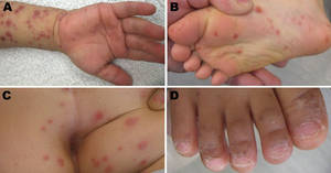
The Centers for Disease Control and Prevention reported a CVA6 outbreak in March 2012, during which several states reported cases of hand, foot, and mouth disease (HFMD) with atypical cutaneous features. Dr. Oza and his colleagues reviewed 80 cases from seven academic centers and identified five distinct morphologies associated with the outbreak:
• Widespread vesiculobullous and erosive exanthema involving more than 10% of the body surface area. Some level of this condition was seen in nearly every patient, Dr. Oza said, noting that vesicles occurred on the face and trunk, and were much more widespread than with classic HFMD. Patients also had a marked predilection for perioral involvement, which led to an initial diagnosis of bullous impetigo in a number of cases, and some had "quite impressive development of bulli." These bulli occurred mainly in children under age 1, and "often evoked differentials that included primary immunobullous disorders."
• Eczema herpeticum–like eruption in atopic dermatitis patients. Dr. Oza and his colleagues termed this condition, seen in 55% of cases in the series, "eczema coxsackiuma." This eruption likely reflected the fact that cases were pulled from pediatric dermatology clinics where atopic dermatitis rates are high, he noted. The clinical morphology closely mirrors that of eczema herpeticum, with abrupt development of hollow ulcerations on areas of atopic dermatitis. Of note, none of the patients with this morphology developed serious systemic illness, unlike patients with eczema herpeticum, who may develop serious systemic sequelae, he said.
• Gianotti-Crosti–like eruption. This papular eruption on the face and extremities occurred in more than a third of patients, Dr. Oza said.
• Petechial/purpuric eruption. This eruption on acral surfaces was seen in 17% of the cases – mainly in older children, and often evoked a differential diagnosis including vasculitis or stocking-glove purpura.
• Delayed onychomadesis and palm and sole desquamation. In a subset of patients followed after their illnesses resolved, cases of onychomadesis were noted between 3 and 8 weeks after resolution, and cases of palm and sole desquamation were noted within 3 weeks after resolution.
No serious systemic complications, such as the meningoencephalitis, myocarditis, or pneumonitis seen with other enterovirus strains, were observed in these cases, but the findings underscore a need for awareness about CVA6 and the potentially severe cases of HFMD that may be associated with outbreaks, Dr. Oza said.
In the past 5 years, the number of reported cases has increased, beginning in Finland in 2008, with more recent reports from China and Japan, he added.
Patients in this study were children with a median age of 1.5 years who were treated at one of seven academic dermatology centers between July 2011 and June 2012. Of the 80 cases identified, 17 had polymerase chain reaction (PCR) confirmation of the disease, and 63 met predefined clinical criteria for inclusion.
Although the low median age is typical for HFMD, the case series spanned a range of ages, including adolescents and older teens, which likely reflects a lack of acquired immunity to this novel viral pathogen, Dr. Oza noted.
The findings demonstrate the evolving role of viral pathogens in dermatologic illness, and should alert physicians to the fact that CVA6-related disease can closely mimic other dermatologic conditions in children, he said.
In addition, the data suggest that enterovirus PCR testing is the best approach for identifying CVA6, said Dr. Oza. It is important to send samples in suspected cases, but it remains prudent to continue testing for other more common causes of vesicular eruptions in children as well, he said.
Dr. Oza had no financial conflicts to disclose.
An emerging enterovirus strain known as coxsackievirus A6 is associated with cases of severe hand, foot, and mouth disease with a more extensive and varied exanthem than usual, according to data from a review of recent cases.
As a result, some coxsackievirus A6 (CVA6) cases have been confused with bullous impetigo, eczema herpeticum vasculitis, and primary immunobullous diseases, Dr. Vikash Oza of the University of California, San Francisco, reported at the annual meeting of the American Academy of Dermatology.
The Centers for Disease Control and Prevention reported a CVA6 outbreak in March 2012, during which several states reported cases of hand, foot, and mouth disease (HFMD) with atypical cutaneous features. Dr. Oza and his colleagues reviewed 80 cases from seven academic centers and identified five distinct morphologies associated with the outbreak:
• Widespread vesiculobullous and erosive exanthema involving more than 10% of the body surface area. Some level of this condition was seen in nearly every patient, Dr. Oza said, noting that vesicles occurred on the face and trunk, and were much more widespread than with classic HFMD. Patients also had a marked predilection for perioral involvement, which led to an initial diagnosis of bullous impetigo in a number of cases, and some had "quite impressive development of bulli." These bulli occurred mainly in children under age 1, and "often evoked differentials that included primary immunobullous disorders."
• Eczema herpeticum–like eruption in atopic dermatitis patients. Dr. Oza and his colleagues termed this condition, seen in 55% of cases in the series, "eczema coxsackiuma." This eruption likely reflected the fact that cases were pulled from pediatric dermatology clinics where atopic dermatitis rates are high, he noted. The clinical morphology closely mirrors that of eczema herpeticum, with abrupt development of hollow ulcerations on areas of atopic dermatitis. Of note, none of the patients with this morphology developed serious systemic illness, unlike patients with eczema herpeticum, who may develop serious systemic sequelae, he said.
• Gianotti-Crosti–like eruption. This papular eruption on the face and extremities occurred in more than a third of patients, Dr. Oza said.
• Petechial/purpuric eruption. This eruption on acral surfaces was seen in 17% of the cases – mainly in older children, and often evoked a differential diagnosis including vasculitis or stocking-glove purpura.
• Delayed onychomadesis and palm and sole desquamation. In a subset of patients followed after their illnesses resolved, cases of onychomadesis were noted between 3 and 8 weeks after resolution, and cases of palm and sole desquamation were noted within 3 weeks after resolution.
No serious systemic complications, such as the meningoencephalitis, myocarditis, or pneumonitis seen with other enterovirus strains, were observed in these cases, but the findings underscore a need for awareness about CVA6 and the potentially severe cases of HFMD that may be associated with outbreaks, Dr. Oza said.
In the past 5 years, the number of reported cases has increased, beginning in Finland in 2008, with more recent reports from China and Japan, he added.
Patients in this study were children with a median age of 1.5 years who were treated at one of seven academic dermatology centers between July 2011 and June 2012. Of the 80 cases identified, 17 had polymerase chain reaction (PCR) confirmation of the disease, and 63 met predefined clinical criteria for inclusion.
Although the low median age is typical for HFMD, the case series spanned a range of ages, including adolescents and older teens, which likely reflects a lack of acquired immunity to this novel viral pathogen, Dr. Oza noted.
The findings demonstrate the evolving role of viral pathogens in dermatologic illness, and should alert physicians to the fact that CVA6-related disease can closely mimic other dermatologic conditions in children, he said.
In addition, the data suggest that enterovirus PCR testing is the best approach for identifying CVA6, said Dr. Oza. It is important to send samples in suspected cases, but it remains prudent to continue testing for other more common causes of vesicular eruptions in children as well, he said.
Dr. Oza had no financial conflicts to disclose.
EXPERT ANALYSIS FROM THE AAD ANNUAL MEETING

Prevent pigment problems in skin of color
MIAMI BEACH – When it comes to procedures such as chemical peels, microdermabrasion, and laser therapies, one size doesn’t fit all, and dermatologists should take special precautions when treating patients with darker skin.
Dr. Marta Rendon, a dermatologist in Boca Raton, Florida, said she sees at least two patients a week who are seeking treatment for pigmentary complications that have been caused by prior cosmetic procedures performed by other physicians.
Some she can treat, and some are beyond repair.
"If I were to sum up my presentation, I would tell you that above all, be conservative and don’t be aggressive," especially in patients with ethnic skin, she told her audience at the annual meeting of the American Academy of Dermatology.
Dr. Rendon, who is also the president of the Skin of Color Society, urged physicians to take precautions because with the increasing diversity of the United States, their patient population is only going to get more diverse.
"Do the patient history. Take your time," advised Dr. Rendon. "Find out what their ethnic background is. Ask them about their grandmother, and where they are from."
The second most important part of history, she said, is asking about reaction to prior procedures or surgeries and prior history of postinflammatory hyperpigmentation (PIH).
"Ask them about what they do, what their hobbies are, or if they play a sport," she said. And take into account the season. During warmer temperatures, consider superficial peels and be careful with lasers. Medium peels and laser resurfacing are more appropriate during cooler and cloudier seasons, she said.
Take caution
Dr. Rendon had the following advice for various procedures in ethnic skin:
• Don’t perform chemical peels on patients on tretinoin. Don’t start with a high concentration. Don’t do excessive layers. And don’t combine surgical procedures with peels in the same visit.
• When performing microdermabrasion, don’t be too aggressive to avoid PIH, streaking, and scratch marks. Don\'t perform the procedure too close to the eyes. Avoid extremely sensitive skin and pressure urticaria. And always start with the lowest strength and time interval.
• With fillers, be careful with superficial placement since the colored material can be seen through the skin. Hyaluronic acid is safer, because it’s colorless and less risky to use. Be mindful that severe bruising can lead to hemosiderin. And be sure that all your patients are using sunscreen.
• For laser and light therapies, always do a test spot. Have a solid understanding of laser-tissue interaction. And be prepared if pigmentation problems develop, so that you can treat them early.
Treatment
Aggressive and early intervention is crucial in treating side effects from cosmetic procedures.
One of the keys to prevent hyperpigmentation is UV protection, whether it’s with sunscreens, cosmetics, antioxidants such as topical vitamin C and E, or systemic agents such as chloroquine, fish oil, or green-tea extract, said Dr. Rendon.
Several topical agents can be used to treat hyperpigmentation. They include hydroquinone, hydroquinone combination, glycolic/retinoid/steroid combination, or antioxidants.
Dr. Rendon also listed several combination bleaching agents including hydroquinone 4%, tretinoin 0.05%, and fluocinolone 0.01%; hydroquinone 4% and retinol 0.3%; hydroquinone microentrapped 4% and retinol 0.15%; hydroquinone and glycolic acid 10%; hydroquinone, glycolic acid 10% + and hyaluronic acid; mequinol and tretinoin 0.01%; retinaldehyde and glycolic acid; and compounded hydroquinone 6%-8%.
Niacinamide is one of the cosmeceutical skin-lightening agents that act as a vitamin exfoliant, reducing melanosome transfer. Soy-protease inhibitors and glutathione also have skin-lightening effects. Dr. Rendon also listed several skin lightening products including Melanozyme, Melaplex, Lumixyl (oligopeptide), retinaldehyde, lactic acid, ferrulic acid, and sunscreen.
She said her favorite method of treating pigmentary complications is to combine treatments. For instance, she combines peels with microdermabrasion; peels with laser; microdermabrasion with IPL; and fractional resurfacing with topical regimens. She added that she maximizes the procedures with topical regimens.
The bottom line is aggressive and early intervention for side effects, Dr. Rendon said.
As a result of growing ethnic population, the treatment options for pigmented skin is expanding, but in the meantime, dermatologists should ensure that their procedures are specific and individualized, Dr. Rendon advised.
"There’s no way of predicting who will hyperpigment, unless you take a good history," she said. And remember two pearls to stay out of trouble: Be conservative, and don’t use aggressive techniques.
Dr. Rendon has performed clinical research for and/or served as a consultant for several companies, including Amgen, Aveeno, Galderma, J&J, Neutrogena, and Sanofi-Aventis. She is a global spokesperson for the H&S brand.
On Twitter @naseemsmiller
EXPERT ANALYSIS FROM THE AAD ANNUAL MEETING

Propranolol safely clears most infantile hemangiomas
MIAMI BEACH – Approximately 60% of infants with proliferating hemangiomas showed significant improvement after treatment with oral propranolol, compared with a placebo, in a randomized, double-blind adaptive phase 2/3 study.
After 24 weeks, 61 of 101 patients (60.4%) treated daily with 3 mg/kg of propranolol had complete or nearly complete resolution of the hemangiomas, compared with only 2 of 55 patients (3.6%) who received a placebo, Dr. Christine Léauté-Labrèze of University Hospital of Bordeaux (France) reported at the annual meeting of the American Academy of Dermatology.
The complete or nearly complete resolution of hemangiomas was assessed by blinded independent investigators, based on a comparison of baseline photographs of the target lesions and photographs taken at week 24.
Patients in the international, multicenter study included infants aged 1-5 months with proliferating infantile hemangiomas requiring systemic therapy. Initially, 456 infants from 60 sites in 15 countries were randomized to receive one of four treatment regimens with oral propranolol: 1 or 3 mg/kg per day for either 3 or 6 months. After an interim efficacy and safety analysis based on the first 188 patients, only the treatment group receiving 3 mg/kg per day for 6 months was retained for trial completion.
The outcomes in both 3-month treatment arms were similar to those in the placebo group. However, the outcomes in the patients who received 3 mg/kg per day for 6 months were better – with no evidence of an increased risk of adverse events – than those in the patients who received 1 mg/kg per day for 6 months, reported Dr. Léauté-Labrèze, principal investigator in the study.
Of note, about half of the placebo patients dropped out of the study after 1 month because of nonefficacy; at the end of the study only about a third of the babies remained in the placebo arm. Similarly, in both of the 3-month treatment arms, dropout rates were high when patients switched from active treatment to placebo. However, approximately 80% of the patients in the group receiving 3 mg/kg per day for 6 months completed the study.
Treatment was well tolerated, and no unexpected safety signals emerged during the course of the study.
The findings of "a highly significant efficacy level" in this randomized, controlled study are important, because 5% of all babies born in the United States are affected by infantile hemangiomas, Dr. Léauté-Labrèze said. Approximately 12% of those babies will likely have complications such as ulceration, impairment of vision, or risk of disfigurement; therefore, effective treatment is needed, she said.
Although several smaller studies and open-label case series involving propranolol have demonstrated efficacy, no treatment has yet received Food and Drug Administration approval for the treatment of infantile hemangiomas, she added.
The study was sponsored by Pierre Fabre Dermatologie.
AT THE AAD ANNUAL MEETING
Major finding: Among children treated with propranolol, 60% had complete or nearly complete resolution of hemangiomas after 24 weeks, compared with 4% of children treated with placebo.
Data source: A randomized, double-blind, placebo-controlled adaptive phase 2/3 study.
Disclosures: This study was sponsored by Pierre Fabre Dermatologie.

Vismodegib shrinks BCCs, reduces Mohs defect size
MIAMI BEACH – Vismodegib treatment for 3 months prior to Mohs surgery for operable basal cell carcinomas shrunk tumors by 46% and reduced the Mohs defect size by 38%, based on data from an open-label intervention trial. The findings were reported at the annual meeting of the American Academy of Dermatology.
The estimated Mohs surgical defect area (based on tumor size) in the first five patients treated in the single-arm study decreased by a mean of 1.4 cm2 from baseline, and actual Mohs surgical defect size decreased by 1.1 cm2 after a mean of 3.4 months of treatment with vismodegib. The changes were statistically significant.
The Mohs defect size was considered a secondary endpoint, because actual defect size can be influenced by skin tension and lesion location, said Dr. Mina Ally, a research fellow at Stanford (Calif.) University.
Patients included in the study were five adults with a total of seven basal cell carcinomas (BCCs) of varying histologic subtypes. One occurred on the shoulder, and the rest occurred on the face; one was a recurrence. All patients were treated for at least 3 months at a vismodegib dosage of 150 mg daily, and all required only a single Mohs stage of excision.
Vismodegib, an oral hedgehog pathway inhibitor approved for the indefinite treatment of advanced and metastatic basal cell carcinomas (that result from aberrant hedgehog pathway signaling), was generally safe, said Dr. Ally.
"All patients experienced mild grade 1 side effects, including muscle cramps, hair loss, and taste loss, and we only needed to discontinue the medication early – after 2 months – in one patient due to a grade 2 elevation in liver enzymes," she said, noting that all of the adverse events resolved after treatment discontinuation.
Further sectioning of Mohs specimens revealed no evidence of residual BCC in three cases and residual BCC in one case. The diagnosis was equivocal in the remaining cases.
"This was because we were seeing an increased number of aberrant follicular structures after (vismodegib) treatment, which were sometimes difficult to differentiate from residual BCC," Dr. Ally explained.
Further staining using a panel that included pleckstrin homology-like domain, family A, member 1, a hair follicle stem cell marker, helped differentiate the follicular structures from BCC, she noted.
None of the patients experienced tumor recurrence during a median 5 months of follow-up.
The findings are of interest because about 1 million people in the United States are affected by BCCs each year, said Dr. Ally. While vismodegib appears to represent a useful adjuvant therapy when given for 3 months prior to Mohs surgery in some cases, certain challenges must be overcome, she said.
"Suppression of the Hedgehog pathway does appear to alter normal follicular development, and this was causing us difficulty in analyzing our histology specimens," Dr. Ally noted. "Future challenges really lie in interpreting these histology specimens and being able to differentiate these follicular structures from residual BCC, which can confound tumor margin clearance."
Dr. Ally reported having no disclosures.
AT THE AAD ANNUAL MEETING
Major finding: Vismodegib reduced tumor size by an average of 46% after 3 months.
Data source: Interim results from an open-label single-arm intervention study.
Disclosures: Dr. Ally reported having no disclosures.