User login
TAVR quickly dominates high-risk aortic stenosis
SAN FRANCISCO – It’s been barely half a year since U.S. cardiologists and cardiac surgeons first became able to routinely offer operable, high-risk patients with aortic stenosis the option of transcatheter valve replacement, yet in the first few months the transcatheter approach quickly rivaled open surgery.
But for the time being in U.S. practice, transcatheter aortic valve replacement (TAVR) remains boxed into the high-risk niche, along with the subgroup of patients who are not suitable for open surgery, king of a pair of relatively small hills.
And no matter how well TAVR performs in the current pair of trials that are comparing it with open surgical aortic valve replacement (SAVR) for intermediate-risk patients, it will remain relegated to niche status for years to come. That’s because roughly two-thirds of all operable patients with aortic stenosis who need valve replacement fall into the low-risk category, with a Society of Thoracic Surgeons (STS) risk score of less than 4%, which experts agree will remain SAVR’s exclusive territory for the foreseeable future.
The high-risk stratum of operable patients, which TAVR now dominates, constitutes about 10% of all patients who need a new aortic valve and can undergo open surgery, patients with an STS score greater than 8%. The intermediate-risk category – an STS score of 4%-8%, where TAVR now vies against open SAVR in two high-profile trials – makes up the final quarter of the operable-patient pie.
Within the high-risk and operable universe, TAVR’s rise has been meteoric, starting last October when the Food and Drug Administration gave Edwards, marketer of the SAPIEN valve system, approval for these patients. A small survey of operators from U.S. TAVR programs in March at the annual scientific session of the American College of Cardiology (ACC) revealed a uniform perception that by early 2013 a sizable majority of U.S. patients with severe aortic stenosis who are deemed operable and are at high surgical risk will wind up being treated by TAVR instead of SAVR. The cardiac surgeons who collaborate on TAVR seem to have fully conceded the advantages of TAVR for these patients.
"We generally go with TAVR. Most patients want it, and with the equivalence" in outcomes from the first PARTNER (Placement of Aortic Transcatheter Valves) cohort A (operable patients) trial (New Engl. J. Med. 2010:364:2187-98), "you usually go with the less invasive procedure," said Dr. Joseph E. Bavaria, professor of surgery and director of thoracic aortic surgery at the University of Pennsylvania in Philadelphia. The small number of high-risk patients who go to open surgery tend to be men, "because they do better with surgery, especially if their life expectancy is greater than 5-8 years," or patients with high stroke risk, Dr. Bavaria said in an interview at the annual meeting of the American College of Cardiology.
"Operable, high-risk patients get TAVR. I’m a surgeon saying that. I’ve already done the [PARTNER cohort A] trial, and I don’t want to do it again," said Dr. Michael Mack, a cardiothoracic surgeon at the Heart Hospital in Plano, Texas. "The results are the same [from TAVR and SAVR] at 30 days, 1 year, and 2 years, but boy do we beat up patients with open surgery. If it was my mom, she’d get TAVR," he said in an interview. "The tie goes to the less invasive treatment."
When high-risk, operable patients are seen by the heart team Dr. Mack works with, the only ones who go to SAVR are patients – generally men – who have a large aortic annulus and need a 29-mm-diameter valve, which is not available for the time being to U.S. TAVR patients; women with septal hypertrophy causing significant left ventricular outflow-tract obstruction; and the small percent of patients who opt for open surgery, usually because it’s the more established approach or because they fear a higher stroke risk from TAVR.
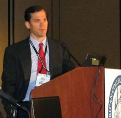
"The majority of high-risk, operable patients now go to TAVR; I think that’s pretty much true across the United States," said Dr. Jeffrey J. Popma, a cardiologist who does TAVR and is a professor of medicine at Harvard University in Boston.
Where TAVR stood in 2010 and 2011
How is TAVR performing? Performance can only be completely assessed months or years after the fact, so the impact that high-risk, operable U.S. patients received from TAVR’s use in routine practice in late 2012 and the first months of 2013 remains to be seen. The most recently treated patients now available for meaningful analysis in large numbers come from 2010 and 2011: new data reported at the ACC meeting from a U.S. program of continued TAVR access that began in late 2009 following the end of recruitment into the first PARTNER trial, and 1-year follow-up of nearly 14,000 patients who underwent TAVR in 2011 as part of routine practice in Germany and were entered into the country’s national TAVR registry.
Both databases had good news for high-risk or inoperable patients. TAVR outcomes in the first couple of years immediately following PARTNER in both the United States and Germany showed clinically meaningful improvements over the way TAVR performed during the first PARTNER trial. Less optimistic news for the low- and intermediate-risk patients who underwent TAVR in Germany, where wider device use is possible, was that in broad terms open surgery outperformed TAVR in these lower-risk patients, although TAVR’s defenders are quick to point out how tricky it is to make cross-treatment comparisons with registry data.
The nonrandomized, continued-access cohort that followed the first PARTNER trial at 22 U.S. centers, 3 sites in Canada, and 1 site in Germany included 1,017 inoperable or high-risk patients who had successful transfemoral TAVR between August 2009 and December 2011. When compared with the 415 inoperable or high-risk patients who underwent transfemoral TAVR in both cohorts of PARTNER, the more recently treated patients had a statistically significant decrease in all-cause 1-year mortality, from a 25% death rate in PARTNER to a 20% rate during the continued-access period, Dr. William F. Fearon reported at the meeting.
The continued-access patients also showed significant cuts in their rates of major vascular complications, which dropped from 15% of patients in PARTNER to 6% during continued access; and in rates of major bleeding complications, which fell from 15% in PARTNER to 7% during continued access. Strokes were also down during continued access, 5% compared with 7% during PARTNER, but this was not a statistically significant drop, reported Dr. Fearon, a cardiologist at Stanford (Calif.) University.
"Maybe these improvements are due to better patient selection, and maybe we have also gotten better at what we do," commented Dr. Popma.
The latest German experience
The 1-year German Aortic Valve Registry (GARY) results from 2011 show similar improvements compared with PARTNER. Among the 2,689 patients who underwent transvascular TAVR, the 1-year total mortality rate was 21%, and in the subset of patients with stroke data the combined rate of major and minor stroke was 5%.
But it was the way that transvascular TAVR (which includes both transfemoral and other vascular approaches but excludes the 1,181 patients who underwent transapical TAVR) stacked up against open surgical replacement that raised concern.
Of the 13,860 total patients entered into GARY during 2011, 9,985 underwent SAVR, with 6,523 of these patients undergoing an isolated procedure (the rest had valve replacement combined with coronary artery bypass). One-year mortality was 7% in patients who had isolated SAVR, dramatically below the 21% rate among the transvascular TAVR patients, Dr. Friedrich-Wilhelm Mohr reported at the meeting. The transapical TAVR patients had a 28% 1-year mortality rate.
To address the issue of between-treatment differences in patients’ underlying risk, Dr. Mohr presented two sets of analyses that stratified patients with two different risk-scoring systems, the EuroSCORE and the AKL (aortic valve surgery) score, also known as the German aortic valve score. The results showed how disparate the outcomes were when patients were subgrouped by their underlying risk. Among the patients who had lone SAVR, 80% fell into the lowest-risk level, with an AKL score of less than 3. Among the transvascular TAVR patients, only 17% had AKL scores below 3.
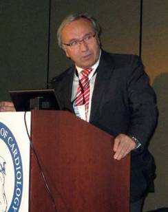
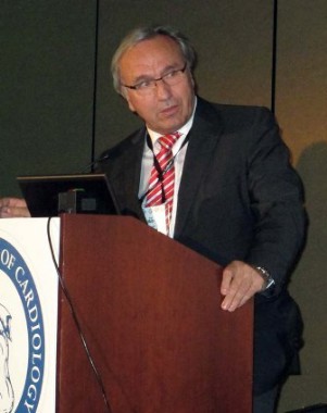
Among patients with AKL scores less than 3, those who underwent SAVR without bypass had a roughly 5% mortality rate after 1 year, compared with about a 15% mortality rate among the transvascular TAVR patients. Analyses in higher-risk patient subgroups showed that the survival gap between SAVR and TAVR patients progressively shrank, until in high-risk patients – as in those studied in PARTNER – the mortality rates were about the same in the SAVR and TAVR groups, said Dr. Mohr, professor and director of heart surgery at Leipzig University, Germany. He also reported similar findings when patients were stratified by their baseline EuroSCORE.
A big factor behind the worse survival among lower-risk TAVR patients is the problem of aortic regurgitation, Dr. Mohr said. Following valve replacement, 56% of the transvascular TAVR patients had grade 1 regurgitation, 7% had grade 2, and less than 1% had grade 3 leakage. These rates are way too high, he said. "Our major concern is the incidence of aortic regurgitation; more than half of the [TAVR] patients had some kind of regurgitation. Regurgitation matters whether it is mild or severe."
Despite his concern, TAVR use in Germany is accelerating. The total number of aortic valve replacements done in Germany jumped from just less than 14,000 in 2011 to more than 22,000 last year; TAVR cases more than doubled, from fewer than 4,000 in 2011 to nearly 10,000 in 2012, with a "move to intermediate-risk groups," said Dr. Mohr, who made it clear that he does not endorse this trend. "TAVR must be proven better than or equal to surgery before it’s used on a large scale," he said. The GARY results show "a clear difference [between TAVR and SAVR] in lower-risk patients. The GARY results do not support going into intermediate-risk patients."
Dr. Martin B. Leon agreed with Dr. Mohr that a definitive determination of whether TAVR is at least as good as SAVR in intermediate-risk patients must await results from the two major, ongoing multicenter trials testing this hypothesis, the PARTNER II trial, using the Edwards balloon-expandable SAPIEN XT valve system, and the SURTAVI (Safety and Efficacy Study of the Medtronic CoreValve System in the Treatment of Severe, Symptomatic Aortic Stenosis in Intermediate Risk Subjects Who Need Aortic Valve Replacement) trial. But Dr. Leon also cautioned that possible confounders might be distorting the GARY results, creating what he calls the "GARY Fallacy."
"The GARY Fallacy is the absurd notion that you can compare SAVR and TAVR in various risk strata without formal risk-adjustment methods to account for imbalances in baseline variables that are not captured in standard risk scores," he said in a talk at the meeting. "Aortic-stenosis patients were selected for TAVR based on their presumed increased risk, including many variables that are not represented in the risk algorithms, such as frailty, liver disease, porcelain aorta, a hostile chest, dementia, and severe chronic obstructive pulmonary disease. Therefore, the risk scores for TAVR patients underrepresent their true risk," Dr. Leon said.
But it’s also possible that TAVR needs more refinement before it completely catches up with SAVR, especially in patients who have the best outcomes from SAVR.

"I think SAVR outperforms TAVR in low-risk patients in the German registry because SAVR is a more mature procedure," said Dr. Raj R. Makkar, a TAVR operator and director of interventional cardiology at Cedars-Sinai Medical Center in Los Angeles. "Maybe this will change, as TAVR becomes safer with fewer valve leaks, strokes, and vascular complications."
Assessing patient risks
Experts also realize that the EuroSCORE, the AKL score, and the other risk-stratification tools now available have flaws when applied to TAVR patients. "Risk stratification for TAVR and SAVR is very problematic; the EuroSCORE has shown poor predictive value for mortality," Dr. Leon said. And while the AKL score was developed specifically for SAVR patients, its relevance to TAVR patients is suspect. In the first PARTNER trial, the multivariate predictors of mortality in the TAVR patients were "completely different" from the predictors in the SAVR patients, he noted.
"It’s tricky calculating scores," agreed Dr. Makkar. Both he and Dr. Mohr cited comorbidities such as pulmonary hypertension, cirrhosis, knee replacement producing impaired mobility, and porcelain aorta that each ratchet up a patient’s risk but have no effect whatsoever on a patient’s EuroSCORE or STS score.
The German cardiology and cardiac surgery societies recognize the limitations of current risk-scoring formulas and are developing a risk-stratification tool specifically designed for TAVR patients, Dr. Mohr said.
The way patients are assessed before, during, and after TAVR is receiving careful scrutiny from some investigators who reported their findings at the ACC meeting, with a particular focus on efforts to characterize and minimize aortic-valve regurgitation following TAVR.
One report, for example, reviewed 2,679 patients who underwent TAVR at any of 33 French centers and 1 in Monaco between January 2010 and October 2011, and were enrolled in the French Aortic National CoreValve and Edwards (FRANCE 2) Registry, established by the French cardiology and thoracic and cardiovascular surgery societies (N. Engl. J. Med. 2012;366:1705-15). FRANCE 2 includes nearly 1,900 patients who received the balloon-expandable Edwards SAPIEN valve, and nearly 900 treated with the self-expanding Medtronic CoreValve device.
Following TAVR, 60% of all patients in the registry had paravalvular aortic regurgitation: 45% with grade 1 regurgitation, 14% with grade 2, and 1% with grade 3 or 4. In a multivariate analysis, the self-expandable device was linked to twice the rate of higher-grade aortic regurgitation, grade 2 or higher, compared with the balloon-expandable valve; and TAVRs done via the femoral artery approach were also about twice as likely to result in higher-grade regurgitations compared with other catheterization routes, reported Dr. Eric Van Belle, a professor at the Cardiology Hospital in Lille, France.
Postprocedural paravalvular regurgitation of grade 2 or higher "was associated with a twofold increase in 1-year mortality, and was the strongest independent predictor of mortality," said Dr. Van Belle in his talk at the meeting. In addition, "annulus diameter and prosthesis diameter were major determinants of aortic regurgitation" in patients who received a balloon-expandable valve.
The FRANCE 2 results showed that postprocedural aortic regurgitation at grade 2 or higher "is a major issue and should be avoided, especially when there is no significant aortic regurgitation at baseline, or when a nonfemoral delivery approach is used." The link between nonfemoral delivery approaches and lower rates of aortic regurgitation suggests "good control of the depth of device delivery and improved catheter technology are key to reducing regurgitation rates," Dr. Van Belle said. "In addition, the prosthesis diameter relative to annulus diameter is key to preventing regurgitation with balloon-expandable devices. Prevention of aortic regurgitation is a major challenge for developing the next-generation device technology."
Minimizing aortic regurgitation
Two ways to cut aortic regurgitation rates following TAVR are to better match the valve to the annulus size, and when possible not finish a TAVR procedure until regurgitation has been minimized.
Two-dimensional transthoracic echocardiography had been the standard approach for annulus sizing as recently as 3 years ago, but it has been replaced with more accurate approaches, either three-dimensional transthoracic echo or CT. "Two-dimensional measurements are seriously limited due to variations and noncircular annular anatomy," Dr. Makkar said in a talk at the meeting. "CT provides the best overall assessment, because in addition to the cross-sectional measurement of the annulus it provides the best assessment of calcification," and three-dimensional transthoracic echo is now standard for intraprocedural assessments, he said.
The impact that a concerted effort to minimize aortic regurgitation can have on outcomes was examined in a single-center study reported by Dr. Jan-Malte Sinning, a cardiologist at University Hospital in Bonn, Germany. Last year, Dr. Sinning and his associates reported developing a quantitative measure of aortic regurgitation immediately following TAVR, the aortic regurgitation (AR) index, based on the difference between a patient’s diastolic blood pressure in the aorta and the left ventricular end-diastolic pressure (J. Am. Coll. Cardiol. 2012;59:1134-41). They then calculated the AR index immediately after TAVR in a prospective series of 167 patients, and set themselves the goal of immediately taking whatever steps were needed to bring the AR index above 25, a cutoff that seemed to correspond to no worse than mild regurgitation.
In their series, 62 patients underwent immediate post-TAVR corrective steps to reduce aortic regurgitation and bring their AR index above 25, most commonly additional balloon dilatation of the valve. The result was that while 16 patients had severe regurgitation before these steps, no patient had severe regurgitation following the corrective measures. The adjustments also cut the incidence of moderate regurgitations from 41 patients to 10, Dr. Sinning reported.
The consequence was that the 30-day stroke rate in the new cohort was 1%, compared with a 6% rate in a historical TAVR cohort at University Hospital in Bonn. The need for pacemaker implants was also cut in half by the intervention compared with the historical group, and 30-day mortality was 3% with these interventions, compared with 7% in the historical controls.
The results suggest that using the AR index as a trigger for taking corrective measures can help improve survival, and that post-TAVR dilatation can successfully decrease regurgitation without increasing patients’ stroke risk, Dr. Sinning concluded.
U.S. operators who perform TAVR say they take similar steps these days to deal with aortic regurgitation after TAVR. "We all understand that you don’t want patients to leave with a leak," said Dr. Popma. "We do everything we can to minimize leaks. When a patient leaves the lab with a moderate or severe leak, there wasn’t anything we could do about it."
Finding TAVR’s limits
Even as TAVR has become the go-to method for high-risk patients, operators have also tried to define the procedure’s outer limit, the point of disease severity when a patient is dying with aortic stenosis rather than because of aortic stenosis, and performing TAVR doesn’t make sense.
Dr. Makkar analyzed 369 inoperable patients who underwent TAVR in both the PARTNER I trial and the nonrandomized continued-access phase that followed. The patients fell into three groups: those who were inoperable because of technical reasons, such as a porcelain aorta or prior chest irradiation; patients who were inoperable because of comorbidities, such as frailty or severe lung disease; and those with both limitations. The 85 patients who were inoperable only because of a technical limitation had a 2-year mortality rate of 23% following TAVR; the other 284 patients who all had comorbidities had a 2-year mortality rate of about 43%.
Last year, Dr. Makkar reported that analysis of these 369 inoperable patients showed a 2-year mortality rate of 20% in the subgroup with an STS score of less than 5%, a mortality rate of 40% among those with a score of 5%-14.5% (but still significantly below the 60% 2-year mortality among similar inoperable patients managed by standard medical therapy only), and a 60% mortality rate among patients with a baseline STS score of 15% or more, an outcome that was no better than that of patients managed without TAVR (N. Engl. J. Med. 2012;366:1696-704).
In a multivariate analysis he recently ran on the data from these 369 patients, the likelihood of 2-year mortality rose by a statistically significant 3% for every 1% increase in the patient’s baseline STS risk score.
"It is important to remember that there are diminishing returns from TAVR in patients with more comorbidities, especially when their STS risk score is greater than 15%," said Dr. Makkar.
Dr. Sinning and Dr. Van Belle had no relevant disclosures. Dr. Mohr is a PARTNER investigator but had no other disclosures. Dr. Bavaria has been a speaker for Edwards and is a PARTNER investigator. Dr. Mack has received travel support from Edwards and is a PARTNER investigator. Dr. Popma has been a consultant to Boston Scientific, Abbott Vascular, and Covidien, and received research support from Boston Scientific, Abbott Vascular, Abiomed, Medtronic, and Cordis. Dr. Fearon has been a consultant to Heart Flow and received research support from St. Jude Medical. Dr. Leon has been a consultant to Symetis; has a major equity stake in Sadra, Claret, Valve Medical, and Apica; has received research support from Boston Scientific, Edwards, and Medtronic; and is a PARTNER investigator. Dr. Makkar has been a consultant to Cordis, Medtronic, Abbott, Entourage Medical, and Abiomed; has been a speaker for Lilly; has received research and travel support from Edwards; and is a PARTNER investigator.
On Twitter @mitchelzoler
SAN FRANCISCO – It’s been barely half a year since U.S. cardiologists and cardiac surgeons first became able to routinely offer operable, high-risk patients with aortic stenosis the option of transcatheter valve replacement, yet in the first few months the transcatheter approach quickly rivaled open surgery.
But for the time being in U.S. practice, transcatheter aortic valve replacement (TAVR) remains boxed into the high-risk niche, along with the subgroup of patients who are not suitable for open surgery, king of a pair of relatively small hills.
And no matter how well TAVR performs in the current pair of trials that are comparing it with open surgical aortic valve replacement (SAVR) for intermediate-risk patients, it will remain relegated to niche status for years to come. That’s because roughly two-thirds of all operable patients with aortic stenosis who need valve replacement fall into the low-risk category, with a Society of Thoracic Surgeons (STS) risk score of less than 4%, which experts agree will remain SAVR’s exclusive territory for the foreseeable future.
The high-risk stratum of operable patients, which TAVR now dominates, constitutes about 10% of all patients who need a new aortic valve and can undergo open surgery, patients with an STS score greater than 8%. The intermediate-risk category – an STS score of 4%-8%, where TAVR now vies against open SAVR in two high-profile trials – makes up the final quarter of the operable-patient pie.
Within the high-risk and operable universe, TAVR’s rise has been meteoric, starting last October when the Food and Drug Administration gave Edwards, marketer of the SAPIEN valve system, approval for these patients. A small survey of operators from U.S. TAVR programs in March at the annual scientific session of the American College of Cardiology (ACC) revealed a uniform perception that by early 2013 a sizable majority of U.S. patients with severe aortic stenosis who are deemed operable and are at high surgical risk will wind up being treated by TAVR instead of SAVR. The cardiac surgeons who collaborate on TAVR seem to have fully conceded the advantages of TAVR for these patients.
"We generally go with TAVR. Most patients want it, and with the equivalence" in outcomes from the first PARTNER (Placement of Aortic Transcatheter Valves) cohort A (operable patients) trial (New Engl. J. Med. 2010:364:2187-98), "you usually go with the less invasive procedure," said Dr. Joseph E. Bavaria, professor of surgery and director of thoracic aortic surgery at the University of Pennsylvania in Philadelphia. The small number of high-risk patients who go to open surgery tend to be men, "because they do better with surgery, especially if their life expectancy is greater than 5-8 years," or patients with high stroke risk, Dr. Bavaria said in an interview at the annual meeting of the American College of Cardiology.
"Operable, high-risk patients get TAVR. I’m a surgeon saying that. I’ve already done the [PARTNER cohort A] trial, and I don’t want to do it again," said Dr. Michael Mack, a cardiothoracic surgeon at the Heart Hospital in Plano, Texas. "The results are the same [from TAVR and SAVR] at 30 days, 1 year, and 2 years, but boy do we beat up patients with open surgery. If it was my mom, she’d get TAVR," he said in an interview. "The tie goes to the less invasive treatment."
When high-risk, operable patients are seen by the heart team Dr. Mack works with, the only ones who go to SAVR are patients – generally men – who have a large aortic annulus and need a 29-mm-diameter valve, which is not available for the time being to U.S. TAVR patients; women with septal hypertrophy causing significant left ventricular outflow-tract obstruction; and the small percent of patients who opt for open surgery, usually because it’s the more established approach or because they fear a higher stroke risk from TAVR.
"The majority of high-risk, operable patients now go to TAVR; I think that’s pretty much true across the United States," said Dr. Jeffrey J. Popma, a cardiologist who does TAVR and is a professor of medicine at Harvard University in Boston.
Where TAVR stood in 2010 and 2011
How is TAVR performing? Performance can only be completely assessed months or years after the fact, so the impact that high-risk, operable U.S. patients received from TAVR’s use in routine practice in late 2012 and the first months of 2013 remains to be seen. The most recently treated patients now available for meaningful analysis in large numbers come from 2010 and 2011: new data reported at the ACC meeting from a U.S. program of continued TAVR access that began in late 2009 following the end of recruitment into the first PARTNER trial, and 1-year follow-up of nearly 14,000 patients who underwent TAVR in 2011 as part of routine practice in Germany and were entered into the country’s national TAVR registry.
Both databases had good news for high-risk or inoperable patients. TAVR outcomes in the first couple of years immediately following PARTNER in both the United States and Germany showed clinically meaningful improvements over the way TAVR performed during the first PARTNER trial. Less optimistic news for the low- and intermediate-risk patients who underwent TAVR in Germany, where wider device use is possible, was that in broad terms open surgery outperformed TAVR in these lower-risk patients, although TAVR’s defenders are quick to point out how tricky it is to make cross-treatment comparisons with registry data.
The nonrandomized, continued-access cohort that followed the first PARTNER trial at 22 U.S. centers, 3 sites in Canada, and 1 site in Germany included 1,017 inoperable or high-risk patients who had successful transfemoral TAVR between August 2009 and December 2011. When compared with the 415 inoperable or high-risk patients who underwent transfemoral TAVR in both cohorts of PARTNER, the more recently treated patients had a statistically significant decrease in all-cause 1-year mortality, from a 25% death rate in PARTNER to a 20% rate during the continued-access period, Dr. William F. Fearon reported at the meeting.
The continued-access patients also showed significant cuts in their rates of major vascular complications, which dropped from 15% of patients in PARTNER to 6% during continued access; and in rates of major bleeding complications, which fell from 15% in PARTNER to 7% during continued access. Strokes were also down during continued access, 5% compared with 7% during PARTNER, but this was not a statistically significant drop, reported Dr. Fearon, a cardiologist at Stanford (Calif.) University.
"Maybe these improvements are due to better patient selection, and maybe we have also gotten better at what we do," commented Dr. Popma.
The latest German experience
The 1-year German Aortic Valve Registry (GARY) results from 2011 show similar improvements compared with PARTNER. Among the 2,689 patients who underwent transvascular TAVR, the 1-year total mortality rate was 21%, and in the subset of patients with stroke data the combined rate of major and minor stroke was 5%.
But it was the way that transvascular TAVR (which includes both transfemoral and other vascular approaches but excludes the 1,181 patients who underwent transapical TAVR) stacked up against open surgical replacement that raised concern.
Of the 13,860 total patients entered into GARY during 2011, 9,985 underwent SAVR, with 6,523 of these patients undergoing an isolated procedure (the rest had valve replacement combined with coronary artery bypass). One-year mortality was 7% in patients who had isolated SAVR, dramatically below the 21% rate among the transvascular TAVR patients, Dr. Friedrich-Wilhelm Mohr reported at the meeting. The transapical TAVR patients had a 28% 1-year mortality rate.
To address the issue of between-treatment differences in patients’ underlying risk, Dr. Mohr presented two sets of analyses that stratified patients with two different risk-scoring systems, the EuroSCORE and the AKL (aortic valve surgery) score, also known as the German aortic valve score. The results showed how disparate the outcomes were when patients were subgrouped by their underlying risk. Among the patients who had lone SAVR, 80% fell into the lowest-risk level, with an AKL score of less than 3. Among the transvascular TAVR patients, only 17% had AKL scores below 3.
Among patients with AKL scores less than 3, those who underwent SAVR without bypass had a roughly 5% mortality rate after 1 year, compared with about a 15% mortality rate among the transvascular TAVR patients. Analyses in higher-risk patient subgroups showed that the survival gap between SAVR and TAVR patients progressively shrank, until in high-risk patients – as in those studied in PARTNER – the mortality rates were about the same in the SAVR and TAVR groups, said Dr. Mohr, professor and director of heart surgery at Leipzig University, Germany. He also reported similar findings when patients were stratified by their baseline EuroSCORE.
A big factor behind the worse survival among lower-risk TAVR patients is the problem of aortic regurgitation, Dr. Mohr said. Following valve replacement, 56% of the transvascular TAVR patients had grade 1 regurgitation, 7% had grade 2, and less than 1% had grade 3 leakage. These rates are way too high, he said. "Our major concern is the incidence of aortic regurgitation; more than half of the [TAVR] patients had some kind of regurgitation. Regurgitation matters whether it is mild or severe."
Despite his concern, TAVR use in Germany is accelerating. The total number of aortic valve replacements done in Germany jumped from just less than 14,000 in 2011 to more than 22,000 last year; TAVR cases more than doubled, from fewer than 4,000 in 2011 to nearly 10,000 in 2012, with a "move to intermediate-risk groups," said Dr. Mohr, who made it clear that he does not endorse this trend. "TAVR must be proven better than or equal to surgery before it’s used on a large scale," he said. The GARY results show "a clear difference [between TAVR and SAVR] in lower-risk patients. The GARY results do not support going into intermediate-risk patients."
Dr. Martin B. Leon agreed with Dr. Mohr that a definitive determination of whether TAVR is at least as good as SAVR in intermediate-risk patients must await results from the two major, ongoing multicenter trials testing this hypothesis, the PARTNER II trial, using the Edwards balloon-expandable SAPIEN XT valve system, and the SURTAVI (Safety and Efficacy Study of the Medtronic CoreValve System in the Treatment of Severe, Symptomatic Aortic Stenosis in Intermediate Risk Subjects Who Need Aortic Valve Replacement) trial. But Dr. Leon also cautioned that possible confounders might be distorting the GARY results, creating what he calls the "GARY Fallacy."
"The GARY Fallacy is the absurd notion that you can compare SAVR and TAVR in various risk strata without formal risk-adjustment methods to account for imbalances in baseline variables that are not captured in standard risk scores," he said in a talk at the meeting. "Aortic-stenosis patients were selected for TAVR based on their presumed increased risk, including many variables that are not represented in the risk algorithms, such as frailty, liver disease, porcelain aorta, a hostile chest, dementia, and severe chronic obstructive pulmonary disease. Therefore, the risk scores for TAVR patients underrepresent their true risk," Dr. Leon said.
But it’s also possible that TAVR needs more refinement before it completely catches up with SAVR, especially in patients who have the best outcomes from SAVR.
"I think SAVR outperforms TAVR in low-risk patients in the German registry because SAVR is a more mature procedure," said Dr. Raj R. Makkar, a TAVR operator and director of interventional cardiology at Cedars-Sinai Medical Center in Los Angeles. "Maybe this will change, as TAVR becomes safer with fewer valve leaks, strokes, and vascular complications."
Assessing patient risks
Experts also realize that the EuroSCORE, the AKL score, and the other risk-stratification tools now available have flaws when applied to TAVR patients. "Risk stratification for TAVR and SAVR is very problematic; the EuroSCORE has shown poor predictive value for mortality," Dr. Leon said. And while the AKL score was developed specifically for SAVR patients, its relevance to TAVR patients is suspect. In the first PARTNER trial, the multivariate predictors of mortality in the TAVR patients were "completely different" from the predictors in the SAVR patients, he noted.
"It’s tricky calculating scores," agreed Dr. Makkar. Both he and Dr. Mohr cited comorbidities such as pulmonary hypertension, cirrhosis, knee replacement producing impaired mobility, and porcelain aorta that each ratchet up a patient’s risk but have no effect whatsoever on a patient’s EuroSCORE or STS score.
The German cardiology and cardiac surgery societies recognize the limitations of current risk-scoring formulas and are developing a risk-stratification tool specifically designed for TAVR patients, Dr. Mohr said.
The way patients are assessed before, during, and after TAVR is receiving careful scrutiny from some investigators who reported their findings at the ACC meeting, with a particular focus on efforts to characterize and minimize aortic-valve regurgitation following TAVR.
One report, for example, reviewed 2,679 patients who underwent TAVR at any of 33 French centers and 1 in Monaco between January 2010 and October 2011, and were enrolled in the French Aortic National CoreValve and Edwards (FRANCE 2) Registry, established by the French cardiology and thoracic and cardiovascular surgery societies (N. Engl. J. Med. 2012;366:1705-15). FRANCE 2 includes nearly 1,900 patients who received the balloon-expandable Edwards SAPIEN valve, and nearly 900 treated with the self-expanding Medtronic CoreValve device.
Following TAVR, 60% of all patients in the registry had paravalvular aortic regurgitation: 45% with grade 1 regurgitation, 14% with grade 2, and 1% with grade 3 or 4. In a multivariate analysis, the self-expandable device was linked to twice the rate of higher-grade aortic regurgitation, grade 2 or higher, compared with the balloon-expandable valve; and TAVRs done via the femoral artery approach were also about twice as likely to result in higher-grade regurgitations compared with other catheterization routes, reported Dr. Eric Van Belle, a professor at the Cardiology Hospital in Lille, France.
Postprocedural paravalvular regurgitation of grade 2 or higher "was associated with a twofold increase in 1-year mortality, and was the strongest independent predictor of mortality," said Dr. Van Belle in his talk at the meeting. In addition, "annulus diameter and prosthesis diameter were major determinants of aortic regurgitation" in patients who received a balloon-expandable valve.
The FRANCE 2 results showed that postprocedural aortic regurgitation at grade 2 or higher "is a major issue and should be avoided, especially when there is no significant aortic regurgitation at baseline, or when a nonfemoral delivery approach is used." The link between nonfemoral delivery approaches and lower rates of aortic regurgitation suggests "good control of the depth of device delivery and improved catheter technology are key to reducing regurgitation rates," Dr. Van Belle said. "In addition, the prosthesis diameter relative to annulus diameter is key to preventing regurgitation with balloon-expandable devices. Prevention of aortic regurgitation is a major challenge for developing the next-generation device technology."
Minimizing aortic regurgitation
Two ways to cut aortic regurgitation rates following TAVR are to better match the valve to the annulus size, and when possible not finish a TAVR procedure until regurgitation has been minimized.
Two-dimensional transthoracic echocardiography had been the standard approach for annulus sizing as recently as 3 years ago, but it has been replaced with more accurate approaches, either three-dimensional transthoracic echo or CT. "Two-dimensional measurements are seriously limited due to variations and noncircular annular anatomy," Dr. Makkar said in a talk at the meeting. "CT provides the best overall assessment, because in addition to the cross-sectional measurement of the annulus it provides the best assessment of calcification," and three-dimensional transthoracic echo is now standard for intraprocedural assessments, he said.
The impact that a concerted effort to minimize aortic regurgitation can have on outcomes was examined in a single-center study reported by Dr. Jan-Malte Sinning, a cardiologist at University Hospital in Bonn, Germany. Last year, Dr. Sinning and his associates reported developing a quantitative measure of aortic regurgitation immediately following TAVR, the aortic regurgitation (AR) index, based on the difference between a patient’s diastolic blood pressure in the aorta and the left ventricular end-diastolic pressure (J. Am. Coll. Cardiol. 2012;59:1134-41). They then calculated the AR index immediately after TAVR in a prospective series of 167 patients, and set themselves the goal of immediately taking whatever steps were needed to bring the AR index above 25, a cutoff that seemed to correspond to no worse than mild regurgitation.
In their series, 62 patients underwent immediate post-TAVR corrective steps to reduce aortic regurgitation and bring their AR index above 25, most commonly additional balloon dilatation of the valve. The result was that while 16 patients had severe regurgitation before these steps, no patient had severe regurgitation following the corrective measures. The adjustments also cut the incidence of moderate regurgitations from 41 patients to 10, Dr. Sinning reported.
The consequence was that the 30-day stroke rate in the new cohort was 1%, compared with a 6% rate in a historical TAVR cohort at University Hospital in Bonn. The need for pacemaker implants was also cut in half by the intervention compared with the historical group, and 30-day mortality was 3% with these interventions, compared with 7% in the historical controls.
The results suggest that using the AR index as a trigger for taking corrective measures can help improve survival, and that post-TAVR dilatation can successfully decrease regurgitation without increasing patients’ stroke risk, Dr. Sinning concluded.
U.S. operators who perform TAVR say they take similar steps these days to deal with aortic regurgitation after TAVR. "We all understand that you don’t want patients to leave with a leak," said Dr. Popma. "We do everything we can to minimize leaks. When a patient leaves the lab with a moderate or severe leak, there wasn’t anything we could do about it."
Finding TAVR’s limits
Even as TAVR has become the go-to method for high-risk patients, operators have also tried to define the procedure’s outer limit, the point of disease severity when a patient is dying with aortic stenosis rather than because of aortic stenosis, and performing TAVR doesn’t make sense.
Dr. Makkar analyzed 369 inoperable patients who underwent TAVR in both the PARTNER I trial and the nonrandomized continued-access phase that followed. The patients fell into three groups: those who were inoperable because of technical reasons, such as a porcelain aorta or prior chest irradiation; patients who were inoperable because of comorbidities, such as frailty or severe lung disease; and those with both limitations. The 85 patients who were inoperable only because of a technical limitation had a 2-year mortality rate of 23% following TAVR; the other 284 patients who all had comorbidities had a 2-year mortality rate of about 43%.
Last year, Dr. Makkar reported that analysis of these 369 inoperable patients showed a 2-year mortality rate of 20% in the subgroup with an STS score of less than 5%, a mortality rate of 40% among those with a score of 5%-14.5% (but still significantly below the 60% 2-year mortality among similar inoperable patients managed by standard medical therapy only), and a 60% mortality rate among patients with a baseline STS score of 15% or more, an outcome that was no better than that of patients managed without TAVR (N. Engl. J. Med. 2012;366:1696-704).
In a multivariate analysis he recently ran on the data from these 369 patients, the likelihood of 2-year mortality rose by a statistically significant 3% for every 1% increase in the patient’s baseline STS risk score.
"It is important to remember that there are diminishing returns from TAVR in patients with more comorbidities, especially when their STS risk score is greater than 15%," said Dr. Makkar.
Dr. Sinning and Dr. Van Belle had no relevant disclosures. Dr. Mohr is a PARTNER investigator but had no other disclosures. Dr. Bavaria has been a speaker for Edwards and is a PARTNER investigator. Dr. Mack has received travel support from Edwards and is a PARTNER investigator. Dr. Popma has been a consultant to Boston Scientific, Abbott Vascular, and Covidien, and received research support from Boston Scientific, Abbott Vascular, Abiomed, Medtronic, and Cordis. Dr. Fearon has been a consultant to Heart Flow and received research support from St. Jude Medical. Dr. Leon has been a consultant to Symetis; has a major equity stake in Sadra, Claret, Valve Medical, and Apica; has received research support from Boston Scientific, Edwards, and Medtronic; and is a PARTNER investigator. Dr. Makkar has been a consultant to Cordis, Medtronic, Abbott, Entourage Medical, and Abiomed; has been a speaker for Lilly; has received research and travel support from Edwards; and is a PARTNER investigator.
On Twitter @mitchelzoler
AT ACC 13
Meta-analysis backs same-day discharge after PCI
SAN FRANCISCO – Sending patients home the same day that they undergo percutaneous coronary intervention appears to be as safe as watching patients overnight in the hospital, based on a meta-analysis of 37 studies involving 12,803 patients.
Most patients underwent PCI for stable angina: 66% in the seven randomized, controlled trials and 98% in the 30 observational studies in the meta-analysis.
In a meta-analysis of results for 2,738 of the patients who were in seven randomized, controlled trials, rates for a composite of death, myocardial infarction (MI), and target lesion revascularization did not differ significantly between the same-day discharge group and the overnight observation group, Dr. Kimberly Brayton and her associates reported.
The two groups also did not differ significantly in the rate of another composite endpoint in the randomized trials: major bleeding or vascular complications, she said at the annual meeting of the American College of Cardiology.
Pooled results for 10,065 of the patients in 30 observational studies showed a 1% rate of the combined endpoint of death, MI, and target lesion revascularization and a 0.7% rate of major bleeding or vascular complications, said Dr. Brayton, of Stanford (Calif.) University.
Of the 15 deaths in the observational studies, 11 occurred more than 24 hours after PCI and "presumably were not modifiable by overnight observation," she added. Data on the remaining four deaths did not specify when they occurred and did not suggest the patients died between 6 and 24 hours following PCI.
The similarities of results between groups in the randomized controlled trials held steady in a secondary analysis that excluded three trials of patients with acute coronary syndrome, and in another secondary analysis that excluded studies conducted outside the United States.
Restricting the analysis to studies using only transfemoral access for PCI did not produce any significant differences between groups in major bleeding or vascular complications.
The findings support consideration of same-day discharge for carefully selected patients undergoing PCI, though larger studies are needed for more robust comparisons. "PCI is now much safer than it was in the past," she said.
Complications from PCI predominantly happen in the first 6 hours after the procedure, previous studies have shown. Outside the United States, patients commonly go home the day of PCI, but only approximately 1% of U.S. patients get same-day discharge after PCI, one study suggests (JAMA 2011;306:1461-1467).
Dr. Brayton and her associates developed a sample protocol for deciding which patients undergoing PCI to send home the same day and which to admit for observation, based on existing PCI guidelines and the protocols used in the studies in the meta-analyses.
Some preprocedure criteria would exclude patients from same-day discharge, including age older than 75 years, a glomerular filtration rate less than 60 mL/min per 1.73 m2, an ejection fraction less than 30%, the presence of acute coronary syndrome, allergy to contrast media, inadequate social support at home, or living more than 30-60 minutes away from the hospital.
If none of those factors are present, consider procedural factors that would exclude same-day discharge. These include significant left main disease, use of a glycoprotein IIb/IIIa inhibitor, performance of a complex PCI, or procedure-related complications.
Barring any of those, observe the patient for 6 hours and admit for overnight observation if there is any recurrent chest pain, hemodynamic instability, or bleeding or vascular complications. If not, the patient may go home the same day, but schedule a follow-up visit or phone call for 24 hours after PCI, Dr. Brayton suggested.
Patients had a mean age of 61 years in the randomized controlled trials and 62 years in the observational studies. Before PCI, 27% in the randomized studies and 29% in the observational studies had multivessel disease. As part of PCI, 39% in the randomized studies and 17% in the observational studies received glycoprotein inhibitors, and 29% in the observational studies received bivalirudin (with no data on bivalirudin for the randomized trials). A closure device was used in 4% of patients in the randomized trials and 54% in the observational studies.
The findings were limited by the heterogeneity of the studies in the meta-analysis, limited statistical power in the small numbers studied, the unavailability of patient-level data, and possible bias, especially in the observational studies, she said.
Dr. Brayton reported having no financial disclosures.
On Twitter @sherryboschert
AT ACC 13
Major finding: The rate of the combined endpoint of death, MI, and target lesion revascularization was 1% after PCI, and the rate for major bleeding or vascular complications was 0.7%. Rates were similar for patients discharged on the same day and those hospitalized overnight.
Data source: Meta-analysis of 37 studies involving 12,803 patients.
Disclosures: Dr. Brayton reported having no financial disclosures.

REMINDER: Early eplerenone reduces BNP in STEMI
SAN FRANCISCO – Early administration of the aldosterone blocker eplerenone improved cardiovascular outcomes in patients presenting with ST-elevation myocardial infarction without concomitant heart failure.
In the randomized, double-blind REMINDER trial, starting eplerenone within 24 hours after symptom onset without first checking serum potassium, as per the study protocol, proved reassuringly safe. There was no significant increase in hyperkalemia compared with placebo. Hypokalemia, defined as a serum potassium level less than 3.5 mEq/L, occurred in 1.4% of the eplerenone group and in 5.5% of controls (P = .0002). Rates of other adverse events were similar in the two study arms, Dr. Gilles Montalescot reported at the annual meeting of the American College of Cardiology.
REMINDER was an international clinical trial involving 1,012 patients presenting with acute STEMI with no history or current signs or symptoms of heart failure and with a left ventricular ejection fraction of at least 40%. A series of studies have shown that the more elevated the early aldosterone in acute STEMI, the worse the prognosis in terms of mortality, new or worsening heart failure, and ventricular fibrillation.
This was a low-risk STEMI population. Patients with renal failure were ineligible. Rates of guideline-recommended medications were strikingly high. Primary percutaneous coronary intervention was the means of myocardial reperfusion in 85% of participants.
The primary study endpoint during a mean 10.5 months of follow-up was a composite of cardiovascular death, hospitalization due to diagnosed heart failure, sustained ventricular tachycardia or fibrillation, an LVEF of 40% after more than 1 month, and an elevated level of brain natriuretic peptide (BNP) or N-terminal prohormone of BNP (NT-proBNP) occurring after 1 month of follow-up.
The rate of the primary endpoint was 18.4% in the eplerenone group, compared with 29.6% with placebo, for a significant 43% relative risk reduction, said Dr. Montalescot, chair of the REMINDER steering committee and professor of cardiology at Pitié-Salpêtrière Hospital, Paris.
The difference in the composite outcome was driven by a sharply lower incidence of elevated BNP/NT-proBNP in the eplerenone group: 16% as compared to 25.9% in controls. The other elements of the composite endpoint didn’t differ significantly between the two groups, although they tended to be lower in the eplerenone group.
Aldosterone has a multitude of cardiotoxic effects. It increases sodium retention, arrhythmogenic depletion of potassium and calcium, cardiac myocyte necrosis, myocardial remodeling, and endothelial dysfunction.
Earlier, in the landmark, randomized EPHESUS trial, investigators showed that prescribing eplerenone in patients with left ventricular systolic dysfunction and heart failure present 3-14 days after an acute MI resulted in a 17% reduction in cardiovascular mortality relative to placebo (N. Engl. J. Med. 2003;348:1309-21). Subsequently, in the EMPHASIS-HF trial eplerenone achieved a 24% reduction in cardiovascular mortality when prescribed for patients with left ventricular systolic dysfunction and mild, NYHA class II heart failure post-MI (N. Engl. J. Med. 2011;364:11-21). The goal in REMINDER was to preempt that systolic dysfunction from arising.
Dr. Montalescot noted that the ongoing 1,600-patient ALBATROSS (Aldosterone Blockade Early After Acute MI) trial, sponsored by the Public Assistance Hospital of Paris, is looking at the use of spironolactone in moderate-risk acute STEMI and has 6-month hard clinical endpoints.
The REMINDER trial was sponsored by Pfizer. Dr. Montalescot reported having no financial conflicts.
The long-term clinical relevance of an elevated BNP is questionable and constitutes "the fly in the ointment" in the REMINDER trial. The biomarker is not a rock-solid predictor of later major adverse clinical events.
 |
|
Eplerenone is already recommended in the European Society of Cardiology and American heart failure guidelines based upon firm evidence of clinical benefit. Yet this recommendation is the least followed, and hardly ever used in practice. In light of that situation, it’s unlikely that reliance on a biomarker endpoint will persuade many physicians to extend use of the aldosterone blocking agent to acute STEMI patients who don’t actually have heart failure.
Dr. Montalescot has said that to demonstrate a 20% reduction in the dual endpoint of mortality or onset of heart failure in low-risk STEMI patients would require 10,000 patients in each study arm, a preclusively costly endeavor.
The combined rate of the ‘hard’ clinical endpoints of cardiovascular mortality, heart failure, or severe ventricular arrhythmia in REMINDER was 3.2% in the control group and 1.8% with eplerenone therapy. Given eplerenone’s cost, prescribing it early on in all acute STEMI patients is unlikely when 97% of them do not appear to benefit.
Dr. E. Magnus Ohman is professor of medicine and director of the program for advanced coronary disease at Duke University, Durham, N.C. Dr. Ohman was the discussant of the study at ACC 13.
The long-term clinical relevance of an elevated BNP is questionable and constitutes "the fly in the ointment" in the REMINDER trial. The biomarker is not a rock-solid predictor of later major adverse clinical events.
|
|
Eplerenone is already recommended in the European Society of Cardiology and American heart failure guidelines based upon firm evidence of clinical benefit. Yet this recommendation is the least followed, and hardly ever used in practice. In light of that situation, it’s unlikely that reliance on a biomarker endpoint will persuade many physicians to extend use of the aldosterone blocking agent to acute STEMI patients who don’t actually have heart failure.
Dr. Montalescot has said that to demonstrate a 20% reduction in the dual endpoint of mortality or onset of heart failure in low-risk STEMI patients would require 10,000 patients in each study arm, a preclusively costly endeavor.
The combined rate of the ‘hard’ clinical endpoints of cardiovascular mortality, heart failure, or severe ventricular arrhythmia in REMINDER was 3.2% in the control group and 1.8% with eplerenone therapy. Given eplerenone’s cost, prescribing it early on in all acute STEMI patients is unlikely when 97% of them do not appear to benefit.
Dr. E. Magnus Ohman is professor of medicine and director of the program for advanced coronary disease at Duke University, Durham, N.C. Dr. Ohman was the discussant of the study at ACC 13.
SAN FRANCISCO – Early administration of the aldosterone blocker eplerenone improved cardiovascular outcomes in patients presenting with ST-elevation myocardial infarction without concomitant heart failure.
In the randomized, double-blind REMINDER trial, starting eplerenone within 24 hours after symptom onset without first checking serum potassium, as per the study protocol, proved reassuringly safe. There was no significant increase in hyperkalemia compared with placebo. Hypokalemia, defined as a serum potassium level less than 3.5 mEq/L, occurred in 1.4% of the eplerenone group and in 5.5% of controls (P = .0002). Rates of other adverse events were similar in the two study arms, Dr. Gilles Montalescot reported at the annual meeting of the American College of Cardiology.
REMINDER was an international clinical trial involving 1,012 patients presenting with acute STEMI with no history or current signs or symptoms of heart failure and with a left ventricular ejection fraction of at least 40%. A series of studies have shown that the more elevated the early aldosterone in acute STEMI, the worse the prognosis in terms of mortality, new or worsening heart failure, and ventricular fibrillation.
This was a low-risk STEMI population. Patients with renal failure were ineligible. Rates of guideline-recommended medications were strikingly high. Primary percutaneous coronary intervention was the means of myocardial reperfusion in 85% of participants.
The primary study endpoint during a mean 10.5 months of follow-up was a composite of cardiovascular death, hospitalization due to diagnosed heart failure, sustained ventricular tachycardia or fibrillation, an LVEF of 40% after more than 1 month, and an elevated level of brain natriuretic peptide (BNP) or N-terminal prohormone of BNP (NT-proBNP) occurring after 1 month of follow-up.
The rate of the primary endpoint was 18.4% in the eplerenone group, compared with 29.6% with placebo, for a significant 43% relative risk reduction, said Dr. Montalescot, chair of the REMINDER steering committee and professor of cardiology at Pitié-Salpêtrière Hospital, Paris.
The difference in the composite outcome was driven by a sharply lower incidence of elevated BNP/NT-proBNP in the eplerenone group: 16% as compared to 25.9% in controls. The other elements of the composite endpoint didn’t differ significantly between the two groups, although they tended to be lower in the eplerenone group.
Aldosterone has a multitude of cardiotoxic effects. It increases sodium retention, arrhythmogenic depletion of potassium and calcium, cardiac myocyte necrosis, myocardial remodeling, and endothelial dysfunction.
Earlier, in the landmark, randomized EPHESUS trial, investigators showed that prescribing eplerenone in patients with left ventricular systolic dysfunction and heart failure present 3-14 days after an acute MI resulted in a 17% reduction in cardiovascular mortality relative to placebo (N. Engl. J. Med. 2003;348:1309-21). Subsequently, in the EMPHASIS-HF trial eplerenone achieved a 24% reduction in cardiovascular mortality when prescribed for patients with left ventricular systolic dysfunction and mild, NYHA class II heart failure post-MI (N. Engl. J. Med. 2011;364:11-21). The goal in REMINDER was to preempt that systolic dysfunction from arising.
Dr. Montalescot noted that the ongoing 1,600-patient ALBATROSS (Aldosterone Blockade Early After Acute MI) trial, sponsored by the Public Assistance Hospital of Paris, is looking at the use of spironolactone in moderate-risk acute STEMI and has 6-month hard clinical endpoints.
The REMINDER trial was sponsored by Pfizer. Dr. Montalescot reported having no financial conflicts.
AT ACC 13
Major finding: The rate of the primary endpoint was 18.4% in the eplerenone group, compared with 29.6% with placebo, for a significant 43% relative risk reduction based on more than 10 months of follow-up.
Data source: REMINDER is an international, randomized, double-blind, placebo-controlled phase III trial involving 1,012 patients presenting with acute STEMI and no heart failure.
Disclosures: The REMINDER trial was funded by Pfizer. Dr. Montalescot reported having no financial conflicts.
DK crush edges culotte for left main PCI
SAN FRANCISCO – The double-kissing crush approach to stenting an unprotected left main coronary artery at the distal bifurcation produced clearly better 1-year clinical outcomes than did an alternative way to stent at the bifurcation, the culotte approach, in a multicenter, randomized trial in China with 419 patients.
The survival rate of patients free of major adverse coronary events 1 year after treatment was 94% for the double kissing (DK) crush technique and 84% for the culotte technique, a statistically significant benefit for DK crush for the study’s primary endpoint, Dr. Jun-Jie Zhang reported at the American College of Cardiology/Cardiovascular Research Foundation Innovation in Intervention Summit.

This definitive clinical comparison, DKCRUSH-III (DK Crush Versus Culotte Stenting for the Treatment of Unprotected Left Main Bifurcation Lesions), is the third in a series of large randomized trials led by Dr. Zhang and his associates to compare the DK crush method of coronary bifurcation stenting with other stenting approaches. In DKCRUSH-1 they found that DK crush worked better than classical crush for treating coronary bifurcations of all types, not just in unprotected left main coronaries (Eur. J. Clin. Invest. 2008;38:361-71). In DKCRUSH-II they compared DK crush with provisional side-branch stenting in a variety of coronary artery types, and found that while DK crush was associated with significantly less restenosis it produced no significant difference in 1-year major adverse coronary events compared with provisional stenting (J. Amer. Coll. Cardiol. 2011;57:914-20).
Dr. Zhang, in collaboration with Dr. Shao-Liang Chen and their associates at Nanjing First Hospital in China, pioneered the DK crush technique, reporting results from their first 20 patients in 2005 (Chinese Med. J. 2005;118:1746-50).
Their new trial notably focused exclusively on distal bifurcations in the unprotected left main coronary artery.
"We know that left main midshaft lesions respond very well to PCI [percutaneous coronary intervention], but distal left main lesions, which represent the majority of left main lesions, have a higher 1-year event rate, mostly related to the ostium circumflex," commented Dr. Gary S. Mintz, chief medical officer of the Cardiovascular Research Foundation in Washington. "There is growing interest in stenting left main lesions among U.S. interventionalists. It is the last frontier for U.S. interventionalists but is routinely done in Asia. The goal is to get the largest cross-sectional area throughout the bifurcation, but the question is how to do that, and what produces the least amount of restenosis at the circumflex ostium? That is the Achilles heel of using two stents at the bifurcation."
While the new results seem to clearly establish the superiority of DK crush over the culotte method, the trial did not address whether stenting of the side branch is better than leaving it unstented or using a different PCI approach.
"When the bifurcation angle is more than 70 degrees, most would do neither DK crush nor culotte; if they felt they had to stent [the side branch] they would use a T stent technique," commented Dr. Gregg W. Stone, professor of medicine and director of cardiovascular research and education at Columbia University in New York.

The DKCRUSH-III trial enrolled patients between March 2009 and October 2011 at 18 Chinese centers. The patients averaged about 64 years old, and nearly four-fifths were men. Their average SYNTAX score was about 31, and their New Risk Stratification Score averaged 26. Almost two-thirds of the patients received everolimus-eluting coronary stents (J. Amer. Coll. Cardiol. Intv. 2010;3:632-41).
Quantitative coronary angiography done about 7 months after PCI in about 84% of the patients in both arms showed no significant differences in patency of the left main coronary, but the side-branch patients in the culotte group had a significantly higher rate of late lumen loss. The overall rate of side-branch restenosis at 7 months was 7% in the DK crush group and 13% in the culotte group.
The 1-year combined rate of cardiac death, myocardial infarction, or clinically driven need for target-lesion or target-vessel revascularization – the primary endpoint – was 6% in the DK crush group and 16% in the culotte group, a significant difference, reported Dr. Zhang, an interventional cardiologist at Nanjing First Hospital. The difference in the rates between the two treatment arms was primarily driven by a difference in the need for revascularization.
A set of four prespecified subgroup analyses showed that the DK crush method was significantly better than the culotte method in three of the high-risk subgroups: patients with a bifurcation angle of 70 degrees or greater, patients with a SYNTAX score of 23 or greater, and patients with a New Risk Stratification score of 20 or greater. In the fourth high-risk subgroup – patients with diabetes – DK crush was also superior, but the difference just missed statistical significance.
Concurrent with Dr. Zhang’s report at the meeting, the results were also published online (J. Am. Coll. Cardiol. 2013;61:1482-88).
Dr. Zhang said that he and his associates had no disclosures. Dr. Mintz and Dr. Stone had no relevant disclosures.
On Twitter @mitchelzoler
 |
Mitchel L. Zoler/IMNG Medical Media
|
The interventionalists who participated in the DKCRUSH-III trial are to be congratulated for their excellent patient outcomes. Patients treated in this study had a 1% cardiac death rate after 1 year despite having unprotected left main disease, with 70% having triple-vessel disease. These outcomes prompt us to ask whether interventionalists should be doing more of these types of cases.
Despite this success, I am not a big fan of the crush technique. I sometimes find it difficult to assess the side branch after stenting due to x-ray artifact caused by having so much metal in place when we crush a stent placed in a side branch. My practice is to do more single-vessel stenting and leave the side branch alone.
The "one stent" technique involves stenting the main branch and just rescuing the side branch, going through the struts of the main stent to enter the circumflex and push the struts out of the way. Results from several studies have shown that this approach is just as good as using two stents and causes fewer complications, although it has not been examined specifically at the distal left main coronary bifurcation. The one-stent approach is what U.S. interventionalists most commonly use today to treat lesions at coronary bifurcations.
Dr. Cindy L. Grines, an interventional cardiologist at Detroit Medical Center, made these comments as a designated discussant for the report. She had no relevant disclosures.
|
Mitchel L. Zoler/IMNG Medical Media
|
The interventionalists who participated in the DKCRUSH-III trial are to be congratulated for their excellent patient outcomes. Patients treated in this study had a 1% cardiac death rate after 1 year despite having unprotected left main disease, with 70% having triple-vessel disease. These outcomes prompt us to ask whether interventionalists should be doing more of these types of cases.
Despite this success, I am not a big fan of the crush technique. I sometimes find it difficult to assess the side branch after stenting due to x-ray artifact caused by having so much metal in place when we crush a stent placed in a side branch. My practice is to do more single-vessel stenting and leave the side branch alone.
The "one stent" technique involves stenting the main branch and just rescuing the side branch, going through the struts of the main stent to enter the circumflex and push the struts out of the way. Results from several studies have shown that this approach is just as good as using two stents and causes fewer complications, although it has not been examined specifically at the distal left main coronary bifurcation. The one-stent approach is what U.S. interventionalists most commonly use today to treat lesions at coronary bifurcations.
Dr. Cindy L. Grines, an interventional cardiologist at Detroit Medical Center, made these comments as a designated discussant for the report. She had no relevant disclosures.
SAN FRANCISCO – The double-kissing crush approach to stenting an unprotected left main coronary artery at the distal bifurcation produced clearly better 1-year clinical outcomes than did an alternative way to stent at the bifurcation, the culotte approach, in a multicenter, randomized trial in China with 419 patients.
The survival rate of patients free of major adverse coronary events 1 year after treatment was 94% for the double kissing (DK) crush technique and 84% for the culotte technique, a statistically significant benefit for DK crush for the study’s primary endpoint, Dr. Jun-Jie Zhang reported at the American College of Cardiology/Cardiovascular Research Foundation Innovation in Intervention Summit.
This definitive clinical comparison, DKCRUSH-III (DK Crush Versus Culotte Stenting for the Treatment of Unprotected Left Main Bifurcation Lesions), is the third in a series of large randomized trials led by Dr. Zhang and his associates to compare the DK crush method of coronary bifurcation stenting with other stenting approaches. In DKCRUSH-1 they found that DK crush worked better than classical crush for treating coronary bifurcations of all types, not just in unprotected left main coronaries (Eur. J. Clin. Invest. 2008;38:361-71). In DKCRUSH-II they compared DK crush with provisional side-branch stenting in a variety of coronary artery types, and found that while DK crush was associated with significantly less restenosis it produced no significant difference in 1-year major adverse coronary events compared with provisional stenting (J. Amer. Coll. Cardiol. 2011;57:914-20).
Dr. Zhang, in collaboration with Dr. Shao-Liang Chen and their associates at Nanjing First Hospital in China, pioneered the DK crush technique, reporting results from their first 20 patients in 2005 (Chinese Med. J. 2005;118:1746-50).
Their new trial notably focused exclusively on distal bifurcations in the unprotected left main coronary artery.
"We know that left main midshaft lesions respond very well to PCI [percutaneous coronary intervention], but distal left main lesions, which represent the majority of left main lesions, have a higher 1-year event rate, mostly related to the ostium circumflex," commented Dr. Gary S. Mintz, chief medical officer of the Cardiovascular Research Foundation in Washington. "There is growing interest in stenting left main lesions among U.S. interventionalists. It is the last frontier for U.S. interventionalists but is routinely done in Asia. The goal is to get the largest cross-sectional area throughout the bifurcation, but the question is how to do that, and what produces the least amount of restenosis at the circumflex ostium? That is the Achilles heel of using two stents at the bifurcation."
While the new results seem to clearly establish the superiority of DK crush over the culotte method, the trial did not address whether stenting of the side branch is better than leaving it unstented or using a different PCI approach.
"When the bifurcation angle is more than 70 degrees, most would do neither DK crush nor culotte; if they felt they had to stent [the side branch] they would use a T stent technique," commented Dr. Gregg W. Stone, professor of medicine and director of cardiovascular research and education at Columbia University in New York.
The DKCRUSH-III trial enrolled patients between March 2009 and October 2011 at 18 Chinese centers. The patients averaged about 64 years old, and nearly four-fifths were men. Their average SYNTAX score was about 31, and their New Risk Stratification Score averaged 26. Almost two-thirds of the patients received everolimus-eluting coronary stents (J. Amer. Coll. Cardiol. Intv. 2010;3:632-41).
Quantitative coronary angiography done about 7 months after PCI in about 84% of the patients in both arms showed no significant differences in patency of the left main coronary, but the side-branch patients in the culotte group had a significantly higher rate of late lumen loss. The overall rate of side-branch restenosis at 7 months was 7% in the DK crush group and 13% in the culotte group.
The 1-year combined rate of cardiac death, myocardial infarction, or clinically driven need for target-lesion or target-vessel revascularization – the primary endpoint – was 6% in the DK crush group and 16% in the culotte group, a significant difference, reported Dr. Zhang, an interventional cardiologist at Nanjing First Hospital. The difference in the rates between the two treatment arms was primarily driven by a difference in the need for revascularization.
A set of four prespecified subgroup analyses showed that the DK crush method was significantly better than the culotte method in three of the high-risk subgroups: patients with a bifurcation angle of 70 degrees or greater, patients with a SYNTAX score of 23 or greater, and patients with a New Risk Stratification score of 20 or greater. In the fourth high-risk subgroup – patients with diabetes – DK crush was also superior, but the difference just missed statistical significance.
Concurrent with Dr. Zhang’s report at the meeting, the results were also published online (J. Am. Coll. Cardiol. 2013;61:1482-88).
Dr. Zhang said that he and his associates had no disclosures. Dr. Mintz and Dr. Stone had no relevant disclosures.
On Twitter @mitchelzoler
AT THE ACC/CRF I2 SUMMIT
Major finding: After 1 year, the survival rate of patients free from major coronary events was 94% for DK crush and 84% for culotte stenting.
Data source: The DKCRUSH-III trial, which randomized 419 patients with unprotected left main coronary distal bifurcation lesions to stenting by the DK crush or culotte techniques.
Disclosures: Dr. Zhang said that he and his associates had no disclosures. Dr. Mintz and Dr. Stone had no relevant disclosures.

Inclacumab reduces troponin in NSTEMI
SAN FRANCISCO – Inclacumab, an inhibitor of the P-selectin pathway, may be a novel treatment for reducing myocardial damage after percutaneous coronary intervention for non–ST-elevation myocardial infarction, based on results from a phase-II study.
The trial, SELECT-ACS, is best viewed as a proof-of-concept study utilizing a reduction in biomarkers of myocardial damage as the endpoint, Dr. Jean-Claude Tardif said at the annual meeting of the American College of Cardiology.

Periprocedural myocardial damage, albeit often mild in nature, is common in PCI patients and is due in part to platelet activation and inflammation. The clinical relevance of post-PCI changes in biomarkers of myocardial damage is uncertain. What is clear from SELECT-ACS is that inclacumab is biologically active in NSTEMI patients. Future phase III studies with hard clinical endpoints conducted in patients with or without PCI will be critical to determining the drug’s relevance, he said.
In addition, the results of the ongoing SELECT-CABG study, in which the effects of inclacumab are being studied in patients undergoing surgical revascularization, will be presented later this year, according to Dr. Tardif, professor of medicine and director of research at the Montreal Heart Institute.
Inclacumab is a human recombinant monoclonal antibody that is a highly specific P-selectin antagonist. P-selectin is a cell adhesion molecule known to play a critical role in communication between activated platelets, WBCs, and the arterial wall. In animal studies, P-selectin inhibition reduces platelet stickiness, macrophage accumulation, and neointimal formation after injury.
"The P-selectin pathway is really at the crossroads of thrombosis and inflammation," Dr. Tardif explained. "It’s probably important to see this drug, inclacumab, not only as an antithrombotic but as an anti-inflammatory agent."
In SELECT-ACS, 544 patients with NSTEMI scheduled for coronary angiography and possible PCI were randomized to receive a single 1-hour-long infusion of inclacumab at 5 or 20 mg/kg or a placebo infusion up to 24 hours before their procedure. Most patients received the infusion just a few hours before angiography.
The primary study endpoint in SELECT-ACS was change in troponin I level from baseline at 16 and 24 hours post-PCI, compared with placebo. Thus, the analysis was restricted to the 322 study participants who underwent PCI. The medically managed SELECT-ACS participants treated with inclacumab will be the subject of a future report.
In the inclacumab 20 mg/kg group, the drop in troponin I was 22% greater than seen in placebo-treated controls at 16 hours and 24% greater at 24 hours. Peak troponin I level was reduced by 24% relative to placebo, and the area under the curve over a 24-hour span was reduced by 34%.
In addition, the decrease in creatine kinase MB (CK-MB) fraction was 16% greater with inclacumab 20 mg/kg than with placebo at 16 hours, and it also showed a 17% greater decrease at 24 hours. The incidence of a CK-MB rise greater than three times the upper limit of normal within the first 24 hours following PCI was 8.9% with inclacumab 20 mg/kg, compared with 18% with placebo. Moreover, soluble P-selectin levels were 22% lower in the inclacumab 20 mg/kg group than in controls.
Inclacumab at 5 mg/kg had no effect.
The pattern and intensity of adverse events were similar in the inclacumab and placebo groups. Given the dual antithrombotic and anti-inflammatory effects of P-selectin inhibition, it’s encouraging to note that the inclacumab-treated patients had no increase in bleeding or infections, Dr. Tardif said.
The study was funded by F. Hoffmann-La Roche. Dr. Tardif reported having no relevant financial interests.
Simultaneous with Dr. Tardif’s presentation of the SELECT-ACS findings in San Francisco, the study was published online (J. Am. Coll. Cardiol. 2013 [doi:10.1016/j.jaac.2013.03.003]).
The SELECT-ACS study is a great phase II trial – and I emphasize "phase II." It’s an early look at a very new and exciting thing. We haven’t been down this road before.
 |
|
I’m struck by the disconnect between the inclacumab-induced reduction in troponin release and clinical events. There were no deaths in the placebo group, but four in the low-dose and two in the high-dose inclacumab groups. Also, nonfatal MI occurred in two placebo-treated patients, compared with four on low-dose and seven on high-dose inclacumab.
Whether this is a real signal, is due to chance, or is simply the way the investigators reported periprocedural MIs in this study is not entirely certain. More definitive understanding is likely to come from the phase-III trials.
Dr. Erik Magnus Ohman is professor of medicine and director of the program for advanced coronary disease at Duke University in Durham, N.C. Dr. Ohman was the study discussant at ACC 13.
The SELECT-ACS study is a great phase II trial – and I emphasize "phase II." It’s an early look at a very new and exciting thing. We haven’t been down this road before.
|
|
I’m struck by the disconnect between the inclacumab-induced reduction in troponin release and clinical events. There were no deaths in the placebo group, but four in the low-dose and two in the high-dose inclacumab groups. Also, nonfatal MI occurred in two placebo-treated patients, compared with four on low-dose and seven on high-dose inclacumab.
Whether this is a real signal, is due to chance, or is simply the way the investigators reported periprocedural MIs in this study is not entirely certain. More definitive understanding is likely to come from the phase-III trials.
Dr. Erik Magnus Ohman is professor of medicine and director of the program for advanced coronary disease at Duke University in Durham, N.C. Dr. Ohman was the study discussant at ACC 13.
SAN FRANCISCO – Inclacumab, an inhibitor of the P-selectin pathway, may be a novel treatment for reducing myocardial damage after percutaneous coronary intervention for non–ST-elevation myocardial infarction, based on results from a phase-II study.
The trial, SELECT-ACS, is best viewed as a proof-of-concept study utilizing a reduction in biomarkers of myocardial damage as the endpoint, Dr. Jean-Claude Tardif said at the annual meeting of the American College of Cardiology.
Periprocedural myocardial damage, albeit often mild in nature, is common in PCI patients and is due in part to platelet activation and inflammation. The clinical relevance of post-PCI changes in biomarkers of myocardial damage is uncertain. What is clear from SELECT-ACS is that inclacumab is biologically active in NSTEMI patients. Future phase III studies with hard clinical endpoints conducted in patients with or without PCI will be critical to determining the drug’s relevance, he said.
In addition, the results of the ongoing SELECT-CABG study, in which the effects of inclacumab are being studied in patients undergoing surgical revascularization, will be presented later this year, according to Dr. Tardif, professor of medicine and director of research at the Montreal Heart Institute.
Inclacumab is a human recombinant monoclonal antibody that is a highly specific P-selectin antagonist. P-selectin is a cell adhesion molecule known to play a critical role in communication between activated platelets, WBCs, and the arterial wall. In animal studies, P-selectin inhibition reduces platelet stickiness, macrophage accumulation, and neointimal formation after injury.
"The P-selectin pathway is really at the crossroads of thrombosis and inflammation," Dr. Tardif explained. "It’s probably important to see this drug, inclacumab, not only as an antithrombotic but as an anti-inflammatory agent."
In SELECT-ACS, 544 patients with NSTEMI scheduled for coronary angiography and possible PCI were randomized to receive a single 1-hour-long infusion of inclacumab at 5 or 20 mg/kg or a placebo infusion up to 24 hours before their procedure. Most patients received the infusion just a few hours before angiography.
The primary study endpoint in SELECT-ACS was change in troponin I level from baseline at 16 and 24 hours post-PCI, compared with placebo. Thus, the analysis was restricted to the 322 study participants who underwent PCI. The medically managed SELECT-ACS participants treated with inclacumab will be the subject of a future report.
In the inclacumab 20 mg/kg group, the drop in troponin I was 22% greater than seen in placebo-treated controls at 16 hours and 24% greater at 24 hours. Peak troponin I level was reduced by 24% relative to placebo, and the area under the curve over a 24-hour span was reduced by 34%.
In addition, the decrease in creatine kinase MB (CK-MB) fraction was 16% greater with inclacumab 20 mg/kg than with placebo at 16 hours, and it also showed a 17% greater decrease at 24 hours. The incidence of a CK-MB rise greater than three times the upper limit of normal within the first 24 hours following PCI was 8.9% with inclacumab 20 mg/kg, compared with 18% with placebo. Moreover, soluble P-selectin levels were 22% lower in the inclacumab 20 mg/kg group than in controls.
Inclacumab at 5 mg/kg had no effect.
The pattern and intensity of adverse events were similar in the inclacumab and placebo groups. Given the dual antithrombotic and anti-inflammatory effects of P-selectin inhibition, it’s encouraging to note that the inclacumab-treated patients had no increase in bleeding or infections, Dr. Tardif said.
The study was funded by F. Hoffmann-La Roche. Dr. Tardif reported having no relevant financial interests.
Simultaneous with Dr. Tardif’s presentation of the SELECT-ACS findings in San Francisco, the study was published online (J. Am. Coll. Cardiol. 2013 [doi:10.1016/j.jaac.2013.03.003]).
AT ACC 13
Major finding: In the inclacumab 20 mg/kg group, the drop in troponin I was 22% greater than seen in placebo-treated controls at 16 hours and 24% greater at 24 hours. Peak troponin I level was reduced by 24%, relative to placebo, and the area under the curve over a 24-hour span was reduced by 34%.
Data source: SELECT-ACS was an international, prospective, placebo-controlled, randomized, double-blind trial involving 544 NSTEMI patients assigned to low- or high-dose inclacumab or placebo up to 24 hours prior to coronary angiography.
Disclosures: The study was funded by F. Hoffmann-La Roche. The presenter reported having no financial conflicts.

High-sensitivity troponin T assay shows prognostic superiority
SAN FRANCISCO – An investigational high-sensitivity troponin T assay provides prognostically meaningful information beyond that yielded by its commercially available sibling test.
The Roche high-sensitivity troponin T assay (hsTnT) excelled over the commercially available Roche Elecys fourth-generation cardiac troponin T test (cTnT) in patients presenting with high-risk non–ST-elevation acute coronary syndrome, Dr. Jonathan Grinstein reported at the annual meeting of the American College of Cardiology.

The investigational test results prove strikingly useful for the subset of patients who test hsTnT-positive and cTnT-negative. In this group, the adjusted odds ratio of 30-day cardiovascular death or new MI is 6.7-fold greater than that of patients who were hsTnT negative and cTnT positive.
"Saying it another way, among this patient subgroup, who represent about 3.5% of our total study population, if one were to analyze the troponins using only the fourth-generation assay, we’d be falsely reassured regarding the likelihood of recurrent cardiovascular events in the next 30 days. In reality, this is a high-risk group," Dr. Grinstein observed.
Both assays were utilized for comparative purposes in 4,160 patients with suspected high-risk non–ST-elevation acute coronary syndrome who participated in two completed and previously reported randomized clinical trials. The studies, known as SEPIA-ACS1 TIMI 42 and EARLY ACS, had as their primary purpose the investigation of novel anticoagulants and optimal timing of administration. All subjects included in this secondary analysis had cardiac ischemia at rest for at least 10 minutes and one or more other high-risk features and underwent dual troponin T testing within 24 hours following symptom onset, explained Dr. Grinstein, a cardiology fellow and TIMI study investigator at Brigham and Women’s Hospital, Boston.
The 99th percentile standard for the investigational hsTnT assay is 14 ng/L. The 3,697 patients in the two clinical trials who met or exceeded this standard had a 30-day incidence of cardiovascular death or new MI of 9.1%, compared with a much more modest rate of 1.9% in the 463 subjects with an hsTnT level below 14 ng/L.
The relationship between hsTnT and 30-day adverse outcome was continuous: The adverse event rate was 1.9% in the 463 patients with an hsTnT below 14 ng/L, 6.4% in 532 subjects with an hsTnT of 14-50 ng/L, 8.6% in 383 patients with a level of 50-100 ng/L, 9.5% in 2,417 patients with an hsTnT of 100-1,500 ng/L, and 11% in the 365 patients with a level in excess of 1,500 ng/L, according to Dr. Grinstein.
Fifteen percent of the subjects with an hsTnT below 14 ng/L had a high TIMI risk score, compared with 36% of those with an hsTnT of 14 ng/L or more.
Overall, two-thirds of the 30-day adverse events were centrally adjudicated acute MIs and one-third were cardiovascular deaths, but the proportion of cardiovascular deaths rose in patients with higher initial hsTnT values.
In the side-by-side analysis including both the hsTnT and fourth-generation cTnT assays, 463 patients had sufficiently low values on both tests that they were categorized as dual negative; their 30-day event rate was 1.9%. Another 378 patients were hsTnT negative but cTnT positive, with an associated 1.3% event rate.
More interesting were the 146 patients who were hsTnT positive and cTnT negative; their event rate was 8.2%. The adjusted odds ratio of 30-day cardiovascular death or new MI in such patients was 6.7-fold greater than in those who were hsTnT negative and fourth-generation positive.
Among the 3,551 subjects with a high-risk score on both assays, the 30-day cardiovascular event rate was 9.1%, he added.
The hsTnT assay, now under Food and Drug Administration review, is widely available in other countries. Compared with commercially available assays in the United States, it provides greatly increased sensitivity in the detection of myocardial necrosis. There have been concerns, however, that this enhanced sensitivity might come at the expense of reduced specificity, leaving the clinical significance of low-level elevations in hsTnT debatable. This study provides reassurance on that score, Dr. Grinstein said.
The SEPIA-ACS1 TIMI 42 trial was sponsored by Sanofi-Aventis. EARLY ACS was sponsored by Schering-Plough. The troponin T assays are manufactured by Roche. Dr. Grinstein reported having no financial conflicts.
SAN FRANCISCO – An investigational high-sensitivity troponin T assay provides prognostically meaningful information beyond that yielded by its commercially available sibling test.
The Roche high-sensitivity troponin T assay (hsTnT) excelled over the commercially available Roche Elecys fourth-generation cardiac troponin T test (cTnT) in patients presenting with high-risk non–ST-elevation acute coronary syndrome, Dr. Jonathan Grinstein reported at the annual meeting of the American College of Cardiology.
The investigational test results prove strikingly useful for the subset of patients who test hsTnT-positive and cTnT-negative. In this group, the adjusted odds ratio of 30-day cardiovascular death or new MI is 6.7-fold greater than that of patients who were hsTnT negative and cTnT positive.
"Saying it another way, among this patient subgroup, who represent about 3.5% of our total study population, if one were to analyze the troponins using only the fourth-generation assay, we’d be falsely reassured regarding the likelihood of recurrent cardiovascular events in the next 30 days. In reality, this is a high-risk group," Dr. Grinstein observed.
Both assays were utilized for comparative purposes in 4,160 patients with suspected high-risk non–ST-elevation acute coronary syndrome who participated in two completed and previously reported randomized clinical trials. The studies, known as SEPIA-ACS1 TIMI 42 and EARLY ACS, had as their primary purpose the investigation of novel anticoagulants and optimal timing of administration. All subjects included in this secondary analysis had cardiac ischemia at rest for at least 10 minutes and one or more other high-risk features and underwent dual troponin T testing within 24 hours following symptom onset, explained Dr. Grinstein, a cardiology fellow and TIMI study investigator at Brigham and Women’s Hospital, Boston.
The 99th percentile standard for the investigational hsTnT assay is 14 ng/L. The 3,697 patients in the two clinical trials who met or exceeded this standard had a 30-day incidence of cardiovascular death or new MI of 9.1%, compared with a much more modest rate of 1.9% in the 463 subjects with an hsTnT level below 14 ng/L.
The relationship between hsTnT and 30-day adverse outcome was continuous: The adverse event rate was 1.9% in the 463 patients with an hsTnT below 14 ng/L, 6.4% in 532 subjects with an hsTnT of 14-50 ng/L, 8.6% in 383 patients with a level of 50-100 ng/L, 9.5% in 2,417 patients with an hsTnT of 100-1,500 ng/L, and 11% in the 365 patients with a level in excess of 1,500 ng/L, according to Dr. Grinstein.
Fifteen percent of the subjects with an hsTnT below 14 ng/L had a high TIMI risk score, compared with 36% of those with an hsTnT of 14 ng/L or more.
Overall, two-thirds of the 30-day adverse events were centrally adjudicated acute MIs and one-third were cardiovascular deaths, but the proportion of cardiovascular deaths rose in patients with higher initial hsTnT values.
In the side-by-side analysis including both the hsTnT and fourth-generation cTnT assays, 463 patients had sufficiently low values on both tests that they were categorized as dual negative; their 30-day event rate was 1.9%. Another 378 patients were hsTnT negative but cTnT positive, with an associated 1.3% event rate.
More interesting were the 146 patients who were hsTnT positive and cTnT negative; their event rate was 8.2%. The adjusted odds ratio of 30-day cardiovascular death or new MI in such patients was 6.7-fold greater than in those who were hsTnT negative and fourth-generation positive.
Among the 3,551 subjects with a high-risk score on both assays, the 30-day cardiovascular event rate was 9.1%, he added.
The hsTnT assay, now under Food and Drug Administration review, is widely available in other countries. Compared with commercially available assays in the United States, it provides greatly increased sensitivity in the detection of myocardial necrosis. There have been concerns, however, that this enhanced sensitivity might come at the expense of reduced specificity, leaving the clinical significance of low-level elevations in hsTnT debatable. This study provides reassurance on that score, Dr. Grinstein said.
The SEPIA-ACS1 TIMI 42 trial was sponsored by Sanofi-Aventis. EARLY ACS was sponsored by Schering-Plough. The troponin T assays are manufactured by Roche. Dr. Grinstein reported having no financial conflicts.
AT ACC 13
Major Finding: Patients with suspected high-risk non–ST-elevation acute coronary syndrome had a 30-day incidence of cardiovascular death or new MI of 8.2% if they had a positive result on an investigational high-sensitivity troponin T assay and a negative result on a commercially available fourth-generation cardiac troponin T test.
Data Source: A secondary analysis of data on 4,160 participants in EARLY ACS and SEPIA-ACS1 TIMI 42.
Disclosures: The SEPIA-ACS1 TIMI 42 trial was sponsored by Sanofi-Aventis. EARLY ACS was sponsored by Schering-Plough. The troponin T assays are manufactured by Roche. The presenter reported having no financial conflicts.

For hypertension, pair CPAP with weight loss
SAN FRANCISCO– A 24-week program combining weight-loss efforts with continuous positive airway pressure produced significantly greater reductions in systolic blood pressure in obese patients with obstructive sleep apnea, compared with either intervention alone, a study of 181 adults found.
In an intent-to-treat analysis, all three groups reduced systolic pressures to a statistically similar extent, compared with baseline – decreases of approximately 8 mm Hg in the combination group and 4 mm Hg with either monotherapy. Among 136 patients who adhered to therapy by completing the trial, however, systolic blood pressure decreased by a mean of 14 mm Hg in the 62 patients randomized to the combination of weight-loss efforts and continuous positive airway pressure (CPAP), a significantly greater decrease than the reductions of approximately 7 mm Hg in 61 patients randomized to weight-loss efforts alone and 3 mm Hg in 58 patients who got CPAP alone.

A 14 mm Hg–drop in systolic blood pressure is an "important reduction" with potentially significant clinical benefits in obese patients with obstructive sleep apnea, Dr. Julio A. Chirinos said at the annual meeting of the American College of Cardiology.
Previous observational studies have shown strong associations in obesity, obstructive sleep apnea, and hypertension but have not been able to assess whether there is any incremental blood pressure benefit to combining treatments for obesity and sleep apnea.
Dr. Chirinos and his associates conducted an ancillary analysis of data from the Cardiovascular Consequences of Obstructive Sleep Apnea trial, which focused primarily on the treatments’ effects on C-reactive protein levels. The study randomized adults with at least moderate obesity and moderate to severe obstructive sleep apnea and C-reactive protein levels greater than 1 mg/L to the three intervention groups. Baseline characteristics were similar among groups. Approximately 41% of patients were hypertensive at baseline.
In the per-protocol analysis of patients who adhered to therapy, the decreases in systolic blood pressure, compared with baseline, were statistically significant in the combination and weight-loss groups but not the CPAP group, reported Dr. Chirinos of the University of Pennsylvania, Philadelphia.
Body weight and body mass index decreased significantly in the weight-loss and combination groups compared with baseline but did not change significantly in the CPAP group. Patients in the weight-loss and combination groups dropped approximately 7 kg in the intent-to-treat analysis and 10-11 kg in the per-protocol analysis. Body mass index decreased by a mean of two to three points in the intent-to-treat analysis and by approximately four points among those who adhered to therapy. No details were provided about the specifics of the weight-loss efforts.
Mean arterial pressure decreased significantly in all three groups, compared with baseline, in both intent-to-treat and per-protocol analyses, but fell significantly more in the combination group, compared with monotherapy, in the per-protocol analysis. Among patients adherent to treatment, mean arterial pressure decreased by more than 10 mm Hg in the combination group, compared with approximately 4 mm Hg–declines with either monotherapy.
Only combination therapy significantly reduced brachial pulse pressure, compared with baseline (by approximately 3 mm Hg), in the intent-to-treat analysis. In the per-protocol analysis, brachial pulse pressure dropped significantly, compared with baseline, in the combination group (a 6 mm Hg decrease) and the weight-loss group (a 4 mm Hg–decrease) but not in the CPAP group.
Brachial pulse pressure decreased significantly with combination therapy in the intent-to-treat analysis but not with either treatment alone. Mean brachial pulse pressure in the per-protocol analysis decreased significantly with combination therapy (a 6 mm Hg–reduction) or weight-loss therapy (a 4 mm Hg–decrease) but increased in the CPAP group.
Carotid-radial pulse pressure amplification measurements did not change significantly from baseline in any group, suggesting that the study’s results were not brought about by missing some changes in central pressure that were not being picked up by brachial measurements, Dr. Chirinos said.
In general, approximately 50% of people with obstructive sleep apnea have hypertension, and around 30% of hypertensive patients have obstructive sleep apnea, he said.
The findings are limited by the study’s strict criteria for patient inclusion or exclusion, its lack of arterial blood pressure monitoring, the use of noninvasive estimates of central pressure measurements, and its duration of only 6 months. The study used carotid tonometry to measure central pulse pressure.
The study excluded patients with blood pressures greater than 160/95 mm Hg, those with predominantly central sleep apnea, patients using supplemental oxygen, and anyone in a high-risk occupation or with a record of dangerous driving. Also excluded were patients with confounders of systemic inflammation, sustained arrhythmia, unstable cardiopulmonary disease, severe restless leg syndrome or chronic pain causing frequent night awakenings, pregnancy, severe depression or other serious medical or psychological conditions that might compromise their safety in the study, and prior CPAP within 8 weeks of the study.
The American Heart Association funded Dr. Chirinos’s analysis. Dr. Chirinos reported having no financial disclosures. Some of his associates reported financial relationships with multiple pharmaceutical companies.
On Twitter @sherryboschert
SAN FRANCISCO– A 24-week program combining weight-loss efforts with continuous positive airway pressure produced significantly greater reductions in systolic blood pressure in obese patients with obstructive sleep apnea, compared with either intervention alone, a study of 181 adults found.
In an intent-to-treat analysis, all three groups reduced systolic pressures to a statistically similar extent, compared with baseline – decreases of approximately 8 mm Hg in the combination group and 4 mm Hg with either monotherapy. Among 136 patients who adhered to therapy by completing the trial, however, systolic blood pressure decreased by a mean of 14 mm Hg in the 62 patients randomized to the combination of weight-loss efforts and continuous positive airway pressure (CPAP), a significantly greater decrease than the reductions of approximately 7 mm Hg in 61 patients randomized to weight-loss efforts alone and 3 mm Hg in 58 patients who got CPAP alone.
A 14 mm Hg–drop in systolic blood pressure is an "important reduction" with potentially significant clinical benefits in obese patients with obstructive sleep apnea, Dr. Julio A. Chirinos said at the annual meeting of the American College of Cardiology.
Previous observational studies have shown strong associations in obesity, obstructive sleep apnea, and hypertension but have not been able to assess whether there is any incremental blood pressure benefit to combining treatments for obesity and sleep apnea.
Dr. Chirinos and his associates conducted an ancillary analysis of data from the Cardiovascular Consequences of Obstructive Sleep Apnea trial, which focused primarily on the treatments’ effects on C-reactive protein levels. The study randomized adults with at least moderate obesity and moderate to severe obstructive sleep apnea and C-reactive protein levels greater than 1 mg/L to the three intervention groups. Baseline characteristics were similar among groups. Approximately 41% of patients were hypertensive at baseline.
In the per-protocol analysis of patients who adhered to therapy, the decreases in systolic blood pressure, compared with baseline, were statistically significant in the combination and weight-loss groups but not the CPAP group, reported Dr. Chirinos of the University of Pennsylvania, Philadelphia.
Body weight and body mass index decreased significantly in the weight-loss and combination groups compared with baseline but did not change significantly in the CPAP group. Patients in the weight-loss and combination groups dropped approximately 7 kg in the intent-to-treat analysis and 10-11 kg in the per-protocol analysis. Body mass index decreased by a mean of two to three points in the intent-to-treat analysis and by approximately four points among those who adhered to therapy. No details were provided about the specifics of the weight-loss efforts.
Mean arterial pressure decreased significantly in all three groups, compared with baseline, in both intent-to-treat and per-protocol analyses, but fell significantly more in the combination group, compared with monotherapy, in the per-protocol analysis. Among patients adherent to treatment, mean arterial pressure decreased by more than 10 mm Hg in the combination group, compared with approximately 4 mm Hg–declines with either monotherapy.
Only combination therapy significantly reduced brachial pulse pressure, compared with baseline (by approximately 3 mm Hg), in the intent-to-treat analysis. In the per-protocol analysis, brachial pulse pressure dropped significantly, compared with baseline, in the combination group (a 6 mm Hg decrease) and the weight-loss group (a 4 mm Hg–decrease) but not in the CPAP group.
Brachial pulse pressure decreased significantly with combination therapy in the intent-to-treat analysis but not with either treatment alone. Mean brachial pulse pressure in the per-protocol analysis decreased significantly with combination therapy (a 6 mm Hg–reduction) or weight-loss therapy (a 4 mm Hg–decrease) but increased in the CPAP group.
Carotid-radial pulse pressure amplification measurements did not change significantly from baseline in any group, suggesting that the study’s results were not brought about by missing some changes in central pressure that were not being picked up by brachial measurements, Dr. Chirinos said.
In general, approximately 50% of people with obstructive sleep apnea have hypertension, and around 30% of hypertensive patients have obstructive sleep apnea, he said.
The findings are limited by the study’s strict criteria for patient inclusion or exclusion, its lack of arterial blood pressure monitoring, the use of noninvasive estimates of central pressure measurements, and its duration of only 6 months. The study used carotid tonometry to measure central pulse pressure.
The study excluded patients with blood pressures greater than 160/95 mm Hg, those with predominantly central sleep apnea, patients using supplemental oxygen, and anyone in a high-risk occupation or with a record of dangerous driving. Also excluded were patients with confounders of systemic inflammation, sustained arrhythmia, unstable cardiopulmonary disease, severe restless leg syndrome or chronic pain causing frequent night awakenings, pregnancy, severe depression or other serious medical or psychological conditions that might compromise their safety in the study, and prior CPAP within 8 weeks of the study.
The American Heart Association funded Dr. Chirinos’s analysis. Dr. Chirinos reported having no financial disclosures. Some of his associates reported financial relationships with multiple pharmaceutical companies.
On Twitter @sherryboschert
AT ACC 13
Major finding: Mean systolic blood pressure decreased by 14 mm Hg in the combination group at 24 weeks, compared with 7 mm Hg with weight loss alone and 3 mm Hg with CPAP alone in patients who adhered to therapy.
Data source: Secondary analysis of a randomized study of 181 obese adults with obstructive sleep apnea.
Disclosures: The American Heart Association funded Dr. Chirinos’s analysis. Dr. Chirinos reported having no financial disclosures. Some of his associates reported financial relationships with multiple pharmaceutical companies.

Lipid levels vary seasonally
SAN FRANCISCO – Cholesterol levels fluctuated significantly by season in a prospective study of 227,359 Brazilians.
Compared with lipid levels in the summer, in the winter the mean plasma LDL cholesterol level increased by 7 mg/dL, resulting in an 8% increase in the prevalence of high LDL cholesterol (>130 mg/dL) in the winter, Dr. Filipe A. Moura and his associates reported at the annual meeting of the American College of Cardiology.

The variations are pronounced enough that clinicians may want to keep a closer eye on patients who are borderline hypercholesterolemic in the summer months, because they may be at higher risk than expected once winter comes, said Dr. Moura, a doctoral candidate at the State University of Campinas, Brazil. He and his associates plan to study patients who present with an MI to see if lipid profiles at presentation vary seasonally in a similar fashion.
The current cross-sectional study prospectively compared lipid levels in patients seen for health checkups in five primary care centers in Campinas from 2008 to 2010. The variations between maximum and minimum lipid levels reached 7 mg/dL for LDL cholesterol, 3 mg/dL for HDL, and 12 mg/dL for triglycerides.
Even greater variations might be seen in nontropical countries that experience more extreme climate changes between seasons, such as the United States or Europe, Dr. Moura speculated. Campinas has mild, dry winters and is located at 1,821-2,559 feet above sea level.
Other lipid levels varied as well. Mean HDL cholesterol levels below 40 mg/dL were 9% more prevalent in summer than in winter, and mean triglyceride levels higher than 150 mg/dL were 5% more prevalent in summer than in winter,
Previous, smaller studies had suggested that there might be seasonal variations in LDL cholesterol. Those studies did not find the increased rates of low HDL and high triglycerides in summer seen in the current study, perhaps because they were not done in tropical climates, he said.
The current study found more pronounced seasonal lipid changes in women and middle-aged adults, but this is likely due to larger sample sizes in these subgroups, Dr. Moura said. Patients ranged in age from less than 1 year to 110 years old, and 64% were female.
Besides the seasonal climate changes, seasonal behavior changes may have affected lipid levels, he speculated. In winter, people tend to exercise less and eat more food in general and fattier foods in particular. Less exposure to sunshine in winter may lower serum concentrations of vitamin D, and vitamin D status has been associated with the ratio of bad to good cholesterol.
Dr. Moura reported having no financial disclosures.
Twitter: @sherryboschert
SAN FRANCISCO – Cholesterol levels fluctuated significantly by season in a prospective study of 227,359 Brazilians.
Compared with lipid levels in the summer, in the winter the mean plasma LDL cholesterol level increased by 7 mg/dL, resulting in an 8% increase in the prevalence of high LDL cholesterol (>130 mg/dL) in the winter, Dr. Filipe A. Moura and his associates reported at the annual meeting of the American College of Cardiology.
The variations are pronounced enough that clinicians may want to keep a closer eye on patients who are borderline hypercholesterolemic in the summer months, because they may be at higher risk than expected once winter comes, said Dr. Moura, a doctoral candidate at the State University of Campinas, Brazil. He and his associates plan to study patients who present with an MI to see if lipid profiles at presentation vary seasonally in a similar fashion.
The current cross-sectional study prospectively compared lipid levels in patients seen for health checkups in five primary care centers in Campinas from 2008 to 2010. The variations between maximum and minimum lipid levels reached 7 mg/dL for LDL cholesterol, 3 mg/dL for HDL, and 12 mg/dL for triglycerides.
Even greater variations might be seen in nontropical countries that experience more extreme climate changes between seasons, such as the United States or Europe, Dr. Moura speculated. Campinas has mild, dry winters and is located at 1,821-2,559 feet above sea level.
Other lipid levels varied as well. Mean HDL cholesterol levels below 40 mg/dL were 9% more prevalent in summer than in winter, and mean triglyceride levels higher than 150 mg/dL were 5% more prevalent in summer than in winter,
Previous, smaller studies had suggested that there might be seasonal variations in LDL cholesterol. Those studies did not find the increased rates of low HDL and high triglycerides in summer seen in the current study, perhaps because they were not done in tropical climates, he said.
The current study found more pronounced seasonal lipid changes in women and middle-aged adults, but this is likely due to larger sample sizes in these subgroups, Dr. Moura said. Patients ranged in age from less than 1 year to 110 years old, and 64% were female.
Besides the seasonal climate changes, seasonal behavior changes may have affected lipid levels, he speculated. In winter, people tend to exercise less and eat more food in general and fattier foods in particular. Less exposure to sunshine in winter may lower serum concentrations of vitamin D, and vitamin D status has been associated with the ratio of bad to good cholesterol.
Dr. Moura reported having no financial disclosures.
Twitter: @sherryboschert
AT ACC 13
Major finding: Mean LDL levels were 7 mg/dL higher, for an 8% increase in prevalence of high LDL (greater than130 mg/dL), in winter compared with summer.
Data source: Prospective cross-sectional study of lipid measurements in 227,359 Brazilians during 2008-2010.
Disclosures: Dr. Moura reported having no financial disclosures.

Handheld ultrasound outperforms cardiologist's physical exam
SAN FRANCISCO – Handheld ultrasound proved "vastly superior" to physical examination conducted by cardiologists for the evaluation of a variety of cardiovascular complaints in a head-to-head prospective trial.
All 250 study participants underwent a clinically indicated standard 2-D and Doppler transthoracic echocardiography exam. But first they received an initial clinical assessment with both a point-of-care, handheld ultrasound scan and a physical exam performed by randomly assigned cardiologists who had widely varying degrees of experience. The cardiologists’ physical exam took an average of 5 minutes, while the limited ultrasound evaluation performed with the commercially available VScan device took a mean of 8.2 minutes, Dr. Manish Mehta reported at the annual meeting of the American College of Cardiology.
Ultrasound had a far higher correct-diagnosis rate than did cardiologists’ physical exam for nearly all of the heart conditions the cardiologists encountered. For example, ultrasound correctly diagnosed moderate or severe mitral regurgitation as confirmed by standard echocardiography in 20 of 20 affected patients, compared with 12 of 20 diagnosed correctly as a result of the physical exam. Ultrasound was threefold more accurate than was physical exam in diagnosis of left ventricular dysfunction in the 54 affected patients. It was also significantly more accurate in the diagnosis of right ventricular dysfunction, tricuspid regurgitation, moderate or severe valve abnormalities, and a miscellaneous category comprising 107 patients with pleural, pericardial, aortic, or congenital heart disease.
In the other two diagnostic categories – pulmonary hypertension and elevated right atrial pressure – ultrasound outperformed physical exam, but not by a statistically significant margin, added Dr. Mehta of Oregon Health and Science University, Portland.
"Routine incorporation of a hand-carried ultrasound device into the physical exam facilitates early and accurate diagnosis of cardiac pathology, which can result in more efficient delivery of care and [can] potentially reduce cost," he concluded.
Dr. Mehta said he and his coinvestigators conducted this study because hand-carried ultrasound has not caught on in cardiology practice to date the way they feel it should. They wanted to provide further supporting evidence for its routine use.
The study was funded by GE Healthcare, which markets the VScan system. Dr. Mehta reported having no financial conflicts.
AT ACC 13
Major Finding: Handheld, point-of-care echocardiography proved far more accurate than did physical examination by cardiologists in the initial assessment of patients with a variety of cardiovascular conditions.
Data Source: A prospective head-to-head comparative study in which 250 cardiology inpatients and outpatients underwent a limited scan with a hand-carried ultrasound device as well as a physical exam by a cardiologist prior to a clinically indicated standard 2-D and Doppler transthoracic echo exam, which provided the definitive diagnosis.
Disclosures: The study was funded by GE Healthcare. The presenter reported having no financial conflicts.
Exercise ABI aids PAD risk stratification
SAN FRANCISCO – A postexercise ankle-brachial index measure provides useful, additional risk stratification to patients with peripheral arterial disease, according to a review of more than 2,800 patients tested at one U.S. center.
"Postexercise ABI [ankle brachial index], which is not routinely tested in most vascular labs, is a very effective tool to identify patients at risk of peripheral disease," Jason Strefling, D.O., said while presenting a poster at the annual meeting of the American College of Cardiology. "Resting ABI is a pretty sensitive test, but when you add exercise you can significantly increase the sensitivity," said Dr. Strefling, a cardiologist at Texas Tech University in Lubbock.
"Patients with diabetes or renal dysfunction can have an unusually normal resting ABI," but measuring a patient’s ABI a second time, after exercise, can better stratify patients who have some level of peripheral arterial disease (PAD), he said in an interview. "Postexercise ABI should probably be measured in all patients having their ABI measured, and on all PAD patients, but most vascular labs don’t do it." Patients with depressed ABIs both at rest and after exercise have the worst outcomes, but depressed exercise ABI with a normal rest ABI may identify patients with milder but nonetheless clinically significant PAD.
The exercise can be in the form of a treadmill protocol, or as simple as a few minutes of toe lifts, he said.
Dr. Strefling and his associates reviewed 2,842 patients seen at the vascular laboratory at the Cleveland Clinic during 2005-2009 who underwent both resting and postexercise ABI measurements. "A lot of vascular surgeons and cardiologists [at the Cleveland Clinic] order an exercise ABI," he explained. The ABI, the difference in systolic pressure between the ankle and arm, is measured with a handheld continuous wave Doppler ultrasound device and a blood pressure cuff. An ABI measurement of less than 0.90 is considered diagnostic of PAD. Dr. Strefling was on the Cleveland Clinic staff when he did this analysis.
The cohort included 1,383 patients with a normal ABI, greater than 0.9, at rest and post exercise, 479 with a normal rest ABI but a depressed ABI post exercise, 0.9 or less, and 980 patients with a low ABI at both measurements.
During follow-up of up to 5 years, the mortality rate was 9% in patients with both ABIs normal, 13% in patients whose ABI was only depressed post exercise, and 21% in patients with low rest and exercise ABIs, statistically significant between-group differences. The researchers reported a similar, statistically significant risk stratification pattern for the combined outcome of death, stroke, and myocardial infarction.
A third combined outcome – death, stroke, myocardial infarction, or lower extremity revascularization or amputation – had rates of 14% in patients with normal rest and exercise ABIs, 27% in patients with a normal rest ABI but reduced postexercise ABI, and 51% in patients with a low ABI on both measures, statistically significant differences. This combined endpoint was primarily driven by episodes of revascularization or amputation, Dr. Strefling noted.
No study results have yet documented the best way to manage patients with a normal resting ABI and a depressed exercise ABI, but Dr. Strefling suggested treating these patients with at least one antiplatelet drug and possibly two of these drugs (such as aspirin and clopidogrel), aggressive statin treatment, and other treatments for risk-factor modification.
Dr. Strefling said that he had no disclosures.
Twitter: @mitchelzoler
AT ACC 2013
Major finding: Mortality was 9% in patients with ABIs normal at rest and post exercise and 21% in patients with low rest and low postexercise ABI.
Data source: A review of 2,842 patients at one U.S. center who underwent both rest and postexercise ankle-brachial index measurements.
Disclosures: Dr. Strefling said that he had no disclosures.