User login
Novel model predicts disease transitions in RA
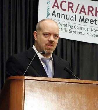
WASHINGTON – What variables predict the course of an individual patient’s rheumatoid arthritis? It’s a question two research groups are beginning to answer by mining data from a large patient database.
Using data from CORRONA (the Consortium of Rheumatology Researchers of North America), a multicenter, longitudinal, prospective database in the United States with more than 30,000 patients, George W. Reed, Ph.D., of the University of Massachusetts in Worcester, and his colleagues have constructed a Markov model as a framework for analyzing the probabilities of transition between disease states in RA.
It’s a way to "talk about the probability of what’s next [in the course of a patient’s disease], and what’s associated with what’s next," Dr. Reed reported in a poster presentation at the annual meeting of the American College of Rheumatology.
Dr. Reed and his colleagues began by defining disease activity states: Low disease activity was defined as a clinical disease activity index (CDAI) score of 10 or less, moderate disease activity as a CDAI score between 11 and 22, and severe disease activity as a CDAI score of 23 or greater. Then states were determined at each visit, and variables were measured to see if they correlated with any changes. Covariates included initiation of disease-modifying antirheumatic drugs (DMARDs), duration of RA, patient age, and insurance status.
The covariates are just examples of "some possible variables that could be plugged in" to future models, he said.
Overall, the investigators examined 160,262 visits from 24,136 RA patients in the CORRONA database who had CDAI measures at the current and the prior visit.
The data imply that RA management, in general, is improving. Transition from any disease state to a low disease state improved from 2001-2005 to 2009-2012, said Dr. Reed.
If a DMARD was not initiated at the prior visit, "a patient in a prior state of moderate disease had a relative risk ratio of 7.6 (95% confidence interval, 7.08-8.21) to still have moderate disease at the current visit." However, if a DMARD was initiated, CDAI declined, and the relative risk of remaining in a moderate disease state was cut to 4.1 (95% CI, 3.59-4.63).
A longer duration of RA affected transitions in disease states, but patient age and insurance status did not.
In a separate CORRONA-based study, patients with moderate disease were found to transition frequently in and out of severe and low disease states.
In that study, led by Sameer V. Kotak of Pfizer in New York, 60% of 4,118 RA patients who exhibited moderate disease activity at baseline had transitioned to a low CDAI by 6 months. About 31% continued to have a moderate CDAI, and 9% transitioned to a severe CDAI.
At 12 months, about 75% of the patients with a low CDAI at the 6-month benchmark remained in that category, with 20% transitioning back to a moderate CDAI and the remaining 5% transitioning to severe disease.
Similarly, 46% of patients with a moderate CDAI at baseline remained at that level at 6 months and transitioned to a low CDAI by 12 months, while 11% of these patients transitioned to a severe CDAI.
The data show transition potential and disease instability in an understated population, "even within a short follow-up window of 6 months," Mr. Kotak said.
Dr. Reed disclosed that he is supported by a research contract with CORRONA through the University of Massachusetts. Mr. Kotak, along with another coinvestigator, is an employee of Pfizer. Employment with, and other financial relationships to, additional pharmaceutical companies were disclosed by multiple investigators in the studies.
AT THE ANNUAL MEETING OF THE AMERICAN COLLEGE OF RHEUMATOLOGY
Major Finding: Among RA patients, a novel model was used to calculate a 7.6 relative risk of remaining in a moderate disease activity state at two consecutive doctor’s visits, versus patients who started out in a low disease state; initiating a DMARD lessened the risk.
Data Source: A cohort of patients culled from the CORRONA registry.
Disclosures: Dr. Reed disclosed that he is supported by a research contract with CORRONA through the University of Massachusetts. Mr. Kotak, along with another coinvestigator, is an employee of Pfizer. Employment with, and other financial relationships to, additional pharmaceutical companies were disclosed by multiple investigators in the studies.
VIDEO: Model predicts lifetime risk for knee OA
A computer simulation model called the Osteoarthritis Policy Model (OAPol) is being used to estimate risks of symptomatic knee OA starting at 40 years of age, stratified by gender and ethnicity.
In an interview at the annual meeting of the American College of Rheumatology in Washington, Elena Losina, Ph.D., of Brigham and Women's Hospital in Boston, describes how the model can be used to develop a risk score that doctors and patients can use to calculate an individual's risk of developing knee OA and take preventive action.
The study was funded in part by the National Institute of Arthritis, Musculoskeletal and Skin Diseases of the National Institutes of Health. Dr. Losina had no financial conflicts to disclose.

Rheumatology education, in 140 characters or less
WASHINGTON – Do you Twitter?
That was the question posed by Dr. Christopher Collins at the annual meeting of the American College of Rheumatology, and it wasn’t an idle query. According to Dr. Collins, the social media site and other mobile, Web-based media tools are poised to become the next big thing in rheumatology education.
"The next generation of rheumatology fellows has already embraced mobile technology as an important aspect of not only everyday life, but as a tool to augment their education," said Dr. Collins, director of the fellowship program for the division of rheumatology at Washington (D.C.) Hospital.

"Social media outlets such as Twitter provide a means for interactions which are not limited by geography, and provide a potential means to enhance the education experience."
At the meeting, Dr. Collins presented proof of concept: His Twitter feed, @RheumPearls, offers a (roughly) daily, 140-character rheumatology "pearl of wisdom" on topics including arthritis, lupus, and gout.
The development of his feed was supported by a grant from the ACR’s Rheumatology Research Foundation, awarded to Dr. Collins in 2010. The Clinician Scholar Educator award aims to further the development of new teaching strategies in rheumatology, according to the ACR’s website.
Founded in March 2011, Dr. Collins’s Twitter feed has nearly 1,000 followers from 57 different countries, with 434 tweets posted at the time of his presentation.
In an analysis of his first 100 followers, Dr. Collins calculated that 25% were physicians (61% of whom were rheumatologists), 23% were patients with self-identified rheumatic conditions (especially lupus and rheumatoid arthritis), 18% were professional medical organizations, and 11% were medical trainees, including medical students, residents, and fellows.
The remainder were unidentified or affiliated with the pharmaceutical industry.
A website Dr. Collins created to complement the Twitter feed has also garnered attention, with 8,125 page views and 6,500 unique visitors from 102 different countries at the time of his presentation.
According to Dr. Collins, his Twitter feed represents just one of the myriad ways in which the social media site, and sites like it, could be used to educate and inform students as well as practitioners of rheumatology.
For example, he pointed to the ways in which Twitter is already used at the ACR’s annual meeting.
"With the use of hashtags, tweets can be ‘tagged’ for searching relevant topics," said Dr. Collins in an e-mail interview.
"During the ACR, searching ‘#ACR2012’ revealed all the relevant tweets being posted about the conference for participants to follow."
In the future, "after a presentation on a particular topic has ended, discussion points can be posted to Twitter for comment by the participants," he said.
In addition, "the participants can continue to be part of the discussion even after the original presentation has ended, as long as they have access to the Internet (as most do with their mobile devices)," Dr. Collins noted.
The technology can also foster more individualized education, even as it reaches out readily to the vast network of Twitter users.
"A mentor who is willing to engage their learners through social media like Twitter can increase their availability to students and augment interactions through direct conversations, and provide links to literature, etc.," he said.
"In addition to purely informative educational Twitter feeds like mine, I see Twitter as having the potential to engage a global population interested in rheumatology and rheumatology education through live interactive discussions."
Dr. Collins disclosed being on a speakers bureau for GlaxoSmithKline and Abbott Laboratories. He stated that he had nothing to disclose relevant to the development and implementation of RheumPearls.
WASHINGTON – Do you Twitter?
That was the question posed by Dr. Christopher Collins at the annual meeting of the American College of Rheumatology, and it wasn’t an idle query. According to Dr. Collins, the social media site and other mobile, Web-based media tools are poised to become the next big thing in rheumatology education.
"The next generation of rheumatology fellows has already embraced mobile technology as an important aspect of not only everyday life, but as a tool to augment their education," said Dr. Collins, director of the fellowship program for the division of rheumatology at Washington (D.C.) Hospital.
"Social media outlets such as Twitter provide a means for interactions which are not limited by geography, and provide a potential means to enhance the education experience."
At the meeting, Dr. Collins presented proof of concept: His Twitter feed, @RheumPearls, offers a (roughly) daily, 140-character rheumatology "pearl of wisdom" on topics including arthritis, lupus, and gout.
The development of his feed was supported by a grant from the ACR’s Rheumatology Research Foundation, awarded to Dr. Collins in 2010. The Clinician Scholar Educator award aims to further the development of new teaching strategies in rheumatology, according to the ACR’s website.
Founded in March 2011, Dr. Collins’s Twitter feed has nearly 1,000 followers from 57 different countries, with 434 tweets posted at the time of his presentation.
In an analysis of his first 100 followers, Dr. Collins calculated that 25% were physicians (61% of whom were rheumatologists), 23% were patients with self-identified rheumatic conditions (especially lupus and rheumatoid arthritis), 18% were professional medical organizations, and 11% were medical trainees, including medical students, residents, and fellows.
The remainder were unidentified or affiliated with the pharmaceutical industry.
A website Dr. Collins created to complement the Twitter feed has also garnered attention, with 8,125 page views and 6,500 unique visitors from 102 different countries at the time of his presentation.
According to Dr. Collins, his Twitter feed represents just one of the myriad ways in which the social media site, and sites like it, could be used to educate and inform students as well as practitioners of rheumatology.
For example, he pointed to the ways in which Twitter is already used at the ACR’s annual meeting.
"With the use of hashtags, tweets can be ‘tagged’ for searching relevant topics," said Dr. Collins in an e-mail interview.
"During the ACR, searching ‘#ACR2012’ revealed all the relevant tweets being posted about the conference for participants to follow."
In the future, "after a presentation on a particular topic has ended, discussion points can be posted to Twitter for comment by the participants," he said.
In addition, "the participants can continue to be part of the discussion even after the original presentation has ended, as long as they have access to the Internet (as most do with their mobile devices)," Dr. Collins noted.
The technology can also foster more individualized education, even as it reaches out readily to the vast network of Twitter users.
"A mentor who is willing to engage their learners through social media like Twitter can increase their availability to students and augment interactions through direct conversations, and provide links to literature, etc.," he said.
"In addition to purely informative educational Twitter feeds like mine, I see Twitter as having the potential to engage a global population interested in rheumatology and rheumatology education through live interactive discussions."
Dr. Collins disclosed being on a speakers bureau for GlaxoSmithKline and Abbott Laboratories. He stated that he had nothing to disclose relevant to the development and implementation of RheumPearls.
EXPERT ANALYSIS FROM THE ANNUAL MEETING OF THE AMERICAN COLLEGE OF RHEUMATOLOGY
Late stop to antirheumatics may risk postsurgical infection
WASHINGTON – The risk of infection following orthopedic surgery is increased in patients with inflammatory rheumatic disease, compared with those with degenerative disease or traumatic injury, according to findings from a review of more than 50,000 surgical procedures.
The risk is greatest in those inflammatory rheumatic disease patients treated with conventional disease-modifying anti-rheumatic drugs (cDMARDs) or tumor necrosis factor (TNF) inhibitors, especially those receiving more than one cDMARD or TNF inhibitor with a long administration interval, and when surgery takes place without discontinuation of treatment, Catrina B. Scherrer reported at the annual meeting of the American College of Rheumatology.
Of 50,359 surgical procedures performed in 37,137 patients from a hospital surgery registry, 422 resulted in surgery-related infections. Of these infections, 49 occurred in 2,472 patients with an inflammatory rheumatic disease (IRD; 2%), and 373 occurred in 47,887 patients with degenerative disease/posttraumatic injury (0.8%). The difference was statistically significant, even after adjustment for other risk factors, including age, gender, diabetes, being overweight, cardiovascular disease, smoking, and type of surgery, said Ms. Scherrer of the Schulthess Clinic, Zürich.
The lowest rates of infection occurred with hand and shoulder surgery; the highest rates occurred with elbow surgery, she noted.
In 1,329 patients in the IRD group for whom complete information about medication was available, 171 (13%) had documented use of TNF inhibitors, and 49 of these (29%) discontinued treatment more than three administration intervals before surgery. Of the remaining 122 TNF inhibitor users, the time lag was three or fewer administration intervals.
An increased infection rate was seen in those who used more than one cDMARD (odds ratio, 2.425) and more than one TNF inhibitor (OR, 2.627) prior to surgery, and the risk of infection was increased tenfold when surgery was performed within one administration interval (OR, 10.047).
Patients who had their last treatment within one administration interval before surgery included 81% of infliximab users, compared with only 33% of adalimumab users and 24% of etanercept users, Ms. Scherrer noted.
Surgery patients in this study were followed over 8 years as part of a single-center surgery registry. The findings, which are limited by the study’s retrospective design and thus require confirmation in prospective studies, are nonetheless important because patients with aggressive disease such as IRDs frequently require orthopedic surgery.
The study findings suggest that IRD patients are, in general, at high risk of postoperative infection, that special attention should be paid to patients using more than one cDMARD or TNF inhibitor with long administration intervals, and that the last intake of TNF inhibitors – particularly infliximab – should be at least more than one administration interval before planned surgery, as the risk of postoperative infection is significantly increased if surgery occurs within this period, she concluded.
Ms. Scherrer reported having no disclosures.
AT THE ANNUAL MEETING OF THE AMERICAN COLLEGE OF RHEUMATOLOGY
Major Finding: An increased infection rate was seen in patients who used more than one cDMARD (odds ratio, 2.425) and more than one TNF inhibitor (OR, 2.627) prior to surgery, and the risk of infection was increased 10-fold when surgery was performed within one treatment administration interval (OR, 10.047).
Data Source: A retrospective study of surgery cases.
Disclosures: Ms. Scherrer reported having no disclosures.
In select RA patients, it's okay to taper therapy
WASHINGTON – Autoantibody seronegativity may be the best predictor of which patients will achieve remission in rheumatoid arthritis, but the question of whether and when to withdraw treatment still looms.
"Drug-free remission is possible. Sustained remission is less common," Dr. Stanley Cohen said at the annual meeting of the American College of Rheumatology. "But certainly seronegativity seems to be a strong predictor of the likelihood of sustained remission."
First, Dr. Cohen of Metroplex Clinical Research Center, Dallas, cited several "prebiologic era" studies, including one 1996 randomized controlled trial of 285 patients who stopped nonbiological disease-modifying antirheumatic drug (DMARD) therapy after achieving the "quite stringent" 1981 ACR/ARA remission definitions for 1 year.
In that study, after the year of remission, patients were randomized to either placebo or to continue their treatment regimen: by 52 weeks, 38% of the placebo group had flared, compared with 22% of the continued therapy group (P = .002) (Lancet 1996;347:347-52).
The authors then looked at the predictors of maintaining remission.
"They determined that having rheumatoid factor activity [there were no CCP (cyclic citrullinated peptide) antibodies at that time] was a poor prognostic factor for maintaining remission," said Dr. Cohen.
"More aggressive treatment prior to remission also boded poorly for maintaining that remission," he noted.
A similar finding was seen with the Leiden Early Arthritis Clinic trial of 454 patients and the British Early Rheumatoid Arthritis Study (ERAS) of 895 patients, both reviewed in 2009.
Sustained drug-free remission was seen in 15% of the former cohort and in 9% in the latter. The absences of CCP autoantibodies and rheumatoid factor were the sole two independent predictors of remission in multivariate analysis (Arthritis Rheum. 2009;60:2262-71).
Having early disease also plays a role in the ability to withdraw treatment, Dr. Cohen said. He pointed to the COMET trial (Combination of Methotrexate and Etanercept in Active Early Rheumatoid Arthritis), which looked at etanercept plus methotrexate versus methotrexate alone in moderate-to-severe rheumatoid arthritis for a duration of 3-24 months.
"What I’m interested in is year 2," Dr. Cohen said, when 111 patients who were on methotrexate plus etanercept in year 1 stopped methotrexate (Arthritis Rheum. 2010;62:674-82).
"Compared to the group that continued etanercept plus methotrexate, the majority continued to be in DAS28 remission without methotrexate" at 2 years, he said. "So, in patients with very early disease, it looks like we can withdraw or simplify the therapy in a significant number of those patients."
Treatment withdrawal, however, also can be achieved in patients with more longstanding disease.
Dr. Cohen cited a 2003 long-term extension of a clinical registration trial in which 79 patients with very long and very severe disease (mean duration of disease, 14 years) received etanercept and methotrexate for a median of 44 months (Arthritis Rheum. 2003;48:1493-9).
Among the 36 patients assessed at 3 years, 30 (83%) were able to decrease their dosages of corticosteroids and 20 (56%) could stop corticosteroids altogether.
"This certainly follows what we see in the clinic, in that we can frequently lower our doses of corticosteroids or withdraw them," Dr. Cohen said.
"What about methotrexate?" he asked. At 3 years, the dosage of methotrexate was decreased in 41 of 66 patients (62%), and methotrexate therapy was discontinued in 19 patients (29%).
"We try to wean our therapy in the majority of these patients," said Dr. Cohen. "Some are successful, some are not."
Dr. Cohen stated that he had no disclosures related to this presentation; he previously disclosed relationships with Abbott Laboratories, Amgen, Astellas, Bristol-Myers Squibb, Janssen, Pfizer, and Roche.
EXPERT ANALYSIS FROM THE ANNUAL MEETING OF THE AMERICAN COLLEGE OF RHEUMATOLOGY
Seropositivity predicts progressive joint damage in established RA
WASHINGTON – Seropositivity for either rheumatoid factor or anti-cyclic citrullinated peptide was significantly associated with progressive joint damage in adults with established rheumatoid arthritis, based on a single-center, observational cohort study of 390 patients.
Most rheumatoid arthritis (RA) patients in clinical practice have established disease, but the predictors and proportion of disease progression in these patients has not been well studied, Dr. Siri Lillegraven of Diakonhjemmet Hospital in Oslo said at the annual meeting of the American College of Rheumatology.
Dr. Lillegraven and her colleagues reviewed data from BRASS (the Brigham and Women’s Hospital Rheumatoid Arthritis Sequential Study). Joint damage was assessed using baseline and 2-year radiographic data for patients with disease duration of at least 5 years. The average age of the patients was 60 years, 84% were women, and 44% received biologic treatment in the form of disease-modifying antirheumatic drugs (DMARDs). The median disease duration was 17 years. Disease progression was defined as a change in Sharp/van der Heijde score of 1 or more units per year.
Overall, 44% of the patients showed disease progression after 2 years. A total of 68% of the patients were positive for both rheumatoid factor (RF) and anticyclic citrullinated peptide (anti-CCP), 16% were positive for either RF or anti-CCP, and 16% were negative for both.
Seropositivity was the only significant independent predictor of disease progression after controlling for factors including age, gender, body-mass index, smoking status, treatment, DMARD use, disease duration, and presence of subcutaneous nodules, Dr. Lillegraven said.
Patients who were seropositive for either RF or anti-CCP were five times more likely than were seronegative patients to have disease progression (odds ratio 5.0), while seropositivity for both was associated with four times greater odds for disease progression (odds ratio 4.1).
"Although the odds ratios for progressive joint damage were similar if patients were positive for one or both of RF and anti-CCP, patients who were positive for both RF and anti-CCP tended to experience more joint damage," Dr. Lillegraven said. Rapid progression of joint damage (defined as a change in van der Heijde-Sharp score of 5 or more units per year) was noted in 16% patients who were seropositive for both RF and anti-CCP, compared with 9% of those who were positive for either RF or anti-CCP, although this difference was not statistically significant.
The results were limited by the use of data from a single center and by the challenge of fully adjusting for treatment in patients with established RA, Dr. Lillegraven noted. However, the findings suggest that seropositivity could be used to inform treatment decisions for these patients, she said.
Dr. Lillegraven had no financial conflicts to disclose. Several of the study coauthors disclosed relationships with multiple pharmaceutical companies, including Amgen, Abbott, Merck, and MedImmune.
AT THE ANNUAL MEETING OF THE AMERICAN COLLEGE OF RHEUMATOLOGY
Major Finding: Patients with established rheumatoid arthritis and seropositivity for either RF or anti-CCP were five times more likely to have disease progression after 2 years than were seronegative patients.
Data Source: The Brigham and Women’s Hospital Rheumatoid Arthritis Sequential Study (BRASS), a single-center, observational cohort study of 390 patients.
Disclosures: Dr. Lillegraven had no financial conflicts to disclose. Several of the study coauthors disclosed relationships with multiple pharmaceutical companies including Amgen, Abbott, Merck, and MedImmune.
Liver function monitoring for methotrexate needs more study
WASHINGTON – "Blanket" monitoring guidelines for liver toxicity in patients taking methotrexate may do more harm than good.
That’s according to Dr. Gabriela Schmajuk, who found that up to a third of patients taking the drug for rheumatoid arthritis may have the drug discontinued unnecessarily in the presence of mildly elevated liver function tests (LFTs).
"We need more research to say what the best strategies are [when it comes to] LFT monitoring, and what our response to abnormal LFTs should be," she said at the annual meeting of the American College of Rheumatology.
In a poster presented at the meeting, Dr. Schmajuk of the University of California, San Francisco, and her colleagues looked at 899 new methotrexate users (97% male, mean age 71 years) aged 65 years and older treated at the Veterans Health Administration (VHA) during 2007-2008.
The study was conducted in response to the recent measure issued by the National Quality Forum, which assesses whether rheumatoid arthritis patients prescribed methotrexate received an LFT within 120 days of their prescription claim. The measure is for public quality reporting purposes.
First, Dr. Schmajuk looked at how many patients met the National Quality Forum’s measure.
Patients all had at least a 28-day supply of methotrexate with follow-up at the VHA for 3 months after the drug was dispensed.
Patients were excluded from the study if they had any diagnosis of inflammatory bowel disease or if they sought medical care outside the VHA, and the researchers correlated the first abnormal LFT with the rate of methotrexate discontinuation.
Overall, they found that 148 patients (16.5%) did not have any LFT testing after methotrexate was initiated.
Among the patients who did have their LFTs monitored, 136 (15.1%) had any abnormality of AST or ALT, and only 49 patients (5%) had an elevation of at least 1.5 times the upper limit of normal; among these, methotrexate was discontinued in 27 patients.
However, the researchers also found that methotrexate was discontinued in 28 (32%) of 87 patients with LFT elevations less than 1.5 times the upper limit of normal.
In an e-mail interview, Dr. Schmajuk conceded that a full chart review on these patients has not yet been completed, and that "it is entirely possible that patients were discontinuing [methotrexate] for reasons not related to the LFTs."
However, "We need more research to help decide what a safe cutoff of discontinuation should be – it is possible that it is higher than previously thought."
She added: "In the future, we should be able to tailor monitoring frequency to individual patients."
A second poster, also presented at the meeting, offered a step forward toward just that.
In that study, Dr. Monica Ramirez of Brigham and Women’s Hospital, Boston, attempted to identify clinical predictors of liver enzyme elevation in methotrexate-treated rheumatoid arthritis patients.
Dr. Ramirez and her colleagues conducted detailed chart evaluations of 500 rheumatoid arthritis patients treated at a single center since 1992 who were identified as ever having received treatment with methotrexate and who had LFTs greater than two times the upper limit of normal.
Patients were excluded if they had ever been diagnosed with hepatitis or any other liver disease, or if they had any concurrent hospital or clinic visits for cardiac disease, sepsis, acute kidney disease, metastatic cancer, trauma, or surgery.
A total of 90 (18%) cases were confirmed as having LFT elevations attributed to methotrexate usage.
The authors then assessed the relationship between several known risk factors for liver toxicity to see whether any of them predicted methotrexate dose reduction or discontinuation: age, sex, obesity, hyperlipidemia, methotrexate dose (2.5-7.5 mg, 10-17.5 mg, or 20-25 mg), alcohol use, statin use, and NSAID use.
In univariate analysis, only obesity was significantly associated with a likelihood of methotrexate discontinuation or dose reduction (odds ratio, 2.89; 95% confidence interval, 1.20-7.00; P = .02). Similarly, in multivariate analysis, obesity was the lone predictor (OR, 2.60; 95% CI, 1.01-6.66; P = .05).
"This suggests a potentially heightened risk of hepatotoxicity among obese patients," the authors concluded.
Dr. Schmajuk and her fellow researchers reported no disclosures and no outside funding for their study. The authors of the second study, led by Dr. Ramirez, reported funding from the National Institutes of Health, and one of the investigators reported financial support from Amgen, Abbott, and other companies.
AT THE ANNUAL MEETING OF THE AMERICAN COLLEGE OF RHEUMATOLOGY
Major Finding: Methotrexate was discontinued in 28 (32%) of 87 patients with liver function test elevations less than 1.5 times the upper limit of normal.
Data Source: A cohort of 899 patients with rheumatoid arthritis in the Veterans Health Administration.
Disclosures: Dr. Schmajuk and her fellow researchers reported no disclosures and no outside funding for their study. The authors of the second study, led by Dr. Ramirez, reported funding from the National Institutes of Health, and one of the investigators reported financial support from Amgen, Abbott, and other companies.
Antibodies May Link to Lung Disease in RA
WASHINGTON – Specific anticitrullinated peptide antibody levels were significantly higher in rheumatoid arthritis patients with interstitial lung disease than in those without lung disease, based on data from 177 patients.
Complications and death are common in rheumatoid arthritis (RA) patients with interstitial lung disease (ILD), and the findings "may implicate the lung as a site in which protein citrullination initiates epitope spreading and propagation of RA," said Dr. Jon T. Giles of Columbia University in New York.

To determine the association of anticitrullinated peptide antibodies (ACPA) with ILD, Dr. Giles and his colleagues reviewed data from multidetector computed tomography images and concurrent serum samples for 177 RA patients. The mean age of the patients was 59 years, 60% were women, and 11% were smokers.
A total of 57 patients (32%) showed some evidence of ILD on imaging, and 32 (18%) had ILD scores of 3 U or higher, Dr. Giles said at the meeting. Overall, levels of anticyclic citrullinated peptide (anti-CCP) and 17 specific ACPAs ranged from 46% to 273% higher in patients with ILD than in those without ILD. Patients with ILD were more likely to be male, to smoke, and to have a history of prednisone and leflunomide use as well as rheumatoid factor and anti-CCP seropositivity compared with patients without ILD, said Dr. Giles.
Anti-CCP seropositivity was significantly more common among patients with any ILD vs. those without ILD (89% vs. 69%), and the seropositivity was even more common (94%) in patients with ILD scores of 3 U or higher.
Higher levels of seven or more ACPAs were significantly more common in patients with reticulation, honeycombing, or traction bronchiectasis than in those with no ILD (40% vs. 18%).
In addition, higher levels of seven or more ACPAs were significantly more common in patients with evidence of restriction on a pulmonary function test and/or decreased results on a diffusing capacity of the lungs for carbon monoxide test compared with those without ILD (39% vs. 20%). The differences were significant after adjustment for age, sex, smoking status, disease activity score (using DAS28), and current prednisone and leflunomide use.
Levels of antibodies targeting noncitrullinated proteins were not significantly higher in patients with ILD, which "suggests a specificity for ACPA in the association," Dr. Giles said.
"Another mechanistic possibility is that ACPA[s] targeting citrullinated synovial antigens are generated and mediate remote pathogenic effects upon circulating to the lungs, where their cognate citrullinated proteins may also be present," he said.
The findings were limited in part by the use of multidetector computed tomography, which differs in slice thickness from high-resolution CT, Dr. Giles noted. But the strengths of the research include the multiple measures of pulmonary disease, he said.
Dr. Giles said he had no financial conflicts to disclose. The study was supported in part by the National Institute of Arthritis and Musculoskeletal and Skin Diseases and the American College of Rheumatology’s Within Our Reach Campaign.
WASHINGTON – Specific anticitrullinated peptide antibody levels were significantly higher in rheumatoid arthritis patients with interstitial lung disease than in those without lung disease, based on data from 177 patients.
Complications and death are common in rheumatoid arthritis (RA) patients with interstitial lung disease (ILD), and the findings "may implicate the lung as a site in which protein citrullination initiates epitope spreading and propagation of RA," said Dr. Jon T. Giles of Columbia University in New York.
To determine the association of anticitrullinated peptide antibodies (ACPA) with ILD, Dr. Giles and his colleagues reviewed data from multidetector computed tomography images and concurrent serum samples for 177 RA patients. The mean age of the patients was 59 years, 60% were women, and 11% were smokers.
A total of 57 patients (32%) showed some evidence of ILD on imaging, and 32 (18%) had ILD scores of 3 U or higher, Dr. Giles said at the meeting. Overall, levels of anticyclic citrullinated peptide (anti-CCP) and 17 specific ACPAs ranged from 46% to 273% higher in patients with ILD than in those without ILD. Patients with ILD were more likely to be male, to smoke, and to have a history of prednisone and leflunomide use as well as rheumatoid factor and anti-CCP seropositivity compared with patients without ILD, said Dr. Giles.
Anti-CCP seropositivity was significantly more common among patients with any ILD vs. those without ILD (89% vs. 69%), and the seropositivity was even more common (94%) in patients with ILD scores of 3 U or higher.
Higher levels of seven or more ACPAs were significantly more common in patients with reticulation, honeycombing, or traction bronchiectasis than in those with no ILD (40% vs. 18%).
In addition, higher levels of seven or more ACPAs were significantly more common in patients with evidence of restriction on a pulmonary function test and/or decreased results on a diffusing capacity of the lungs for carbon monoxide test compared with those without ILD (39% vs. 20%). The differences were significant after adjustment for age, sex, smoking status, disease activity score (using DAS28), and current prednisone and leflunomide use.
Levels of antibodies targeting noncitrullinated proteins were not significantly higher in patients with ILD, which "suggests a specificity for ACPA in the association," Dr. Giles said.
"Another mechanistic possibility is that ACPA[s] targeting citrullinated synovial antigens are generated and mediate remote pathogenic effects upon circulating to the lungs, where their cognate citrullinated proteins may also be present," he said.
The findings were limited in part by the use of multidetector computed tomography, which differs in slice thickness from high-resolution CT, Dr. Giles noted. But the strengths of the research include the multiple measures of pulmonary disease, he said.
Dr. Giles said he had no financial conflicts to disclose. The study was supported in part by the National Institute of Arthritis and Musculoskeletal and Skin Diseases and the American College of Rheumatology’s Within Our Reach Campaign.
AT THE ANNUAL MEETING OF THE AMERICAN COLLEGE OF RHEUMATOLOGY
Major Finding: Levels of anticyclic citrullinated peptide and 17 specific anticitrullinated peptide antibodies ranged from 46% to 273% higher in rheumatoid arthritis patients with interstitial lung disease than in those without.
Data Source: The data come from chest imaging and serum samples from 177 adults with RA.
Disclosures: Dr. Giles said he had no relevant financial conflicts to disclose. The study was supported in part by the National Institute of Arthritis and Musculoskeletal and Skin Diseases and the American College of Rheumatology’s Within Our Reach Campaign.

Ischemic Stroke Rates Same for Anti-TNFs vs. nbDMARDs in RA
WASHINGTON – Exposure to anti-tumor necrosis factor therapy does not appear to be associated with a greater short-term risk of ischemic stroke in patients with rheumatoid arthritis when compared with exposure to nonbiologic disease-modifying antirheumatic drug therapy, according to findings from the British Society for Rheumatology Biologics Register-Rheumatoid Arthritis.
Of 130 verified incident ischemic cerebrovascular accidents that occurred among participants in that large prospective cohort study of RA patients, 109 occurred in 11,642 patients treated with anti–tumor necrosis factor drugs (anti-TNFs) for an incidence rate of 178/100,000 person-years, and 21 occurred in 3,271 patients in a comparator group of biologic-naive patients who had been treated with only nonbiologic disease-modifying antirheumatic drugs (nbDMARDs) for an incident rate of 175/100,000 person-years, Dr. Audrey S. Low reported at the annual meeting of the American College of Rheumatology.
The incident rates did not differ significantly, and no association was seen between ever exposure to anti-TNF drugs and ischemic stroke risk after adjustment for confounders using a propensity score based on age, sex, ethnicity, body mass index, disease activity score, disease duration, Health Assessment Questionnaire score, prior nbDMARD use, steroid use at baseline, year of entry to the study, smoking, baseline drugs, and history of cancer, hypertension, ischemic heart disease, diabetes, depression, and chronic lung disease (hazard ratio, 0.88), said Dr. Low of the University of Manchester (England).
Patients in the British Society for Rheumatology Biologics Register–Rheumatoid Arthritis (BSRBR-RA) were recruited during 2001-2008 and were followed semiannually for 3 years, then annually thereafter. All serious adverse events, such as stroke, and all drug therapy were reported. Events reported through Oct. 21, 2010, were included in this analysis.
The investigators also linked study subjects to the national death register and collected information from medical records; a concerted effort was made to classify stroke types.
The anti-TNF and nbDMARD groups differed with respect to several factors: The anti-TNF cohort was younger, included more women, and had higher disease activity, longer disease duration, and higher levels of disability. That cohort also used more DMARDs, more steroids, and more nonsteroidal cyclo-oxygenase-2 (COX-2) inhibitors. However, the rates of hypertension and diabetes were similar in the anti-TNF and nbDMARD cohorts.
This study is among the largest to show that anti-TNF therapy does not increase stroke risk in RA patients, when compared with nbDMARDS, Dr. Low said.
"We know that patients with RA are at risk of increased cardiovascular morbidity and mortality, and that includes stroke," she said, noting that several other observational studies and meta-analyses have demonstrated an overall increase in that risk.
However, outcomes have varied in studies comparing anti-TNF–treated RA patients and controls, with some showing as much as a 60% increase in ischemic stroke risk and others demonstrating a 30% reduction in risk.
In this large cohort of European patients with RA, no association was seen between the short-term risk of ischemic stroke and the use of anti-TNF therapy, she said, adding that additional follow-up is needed to assess risk over time.
Dr. Low reported having no relevant financial disclosures.
AT THE ANNUAL MEETING OF THE AMERICAN COLLEGE OF RHEUMATOLOGY
Major Finding: Of 130 verified incident ischemic cerebrovascular accidents, 109 occurred in 11,642 patients treated with anti–tumor necrosis factor drugs for an incident rate of 178/100,000 person-years, and 21 occurred in 3,271 patients who had been treated with only nonbiologic disease-modifying antirheumatic drugs for an incident rate of 175/100,000 person-years. The incident rates did not differ significantly after adjustment for possible confounders (hazard ratio, 0.88).
Data Source: Data are from the British Society for Rheumatology Biologics Register–Rheumatoid Arthritis.
Disclosures: Dr. Low reported having no relevant financial disclosures.
Osteoporosis, Vertebral Fractures May Flag Heart Risk in RA
WASHINGTON – Low bone mineral density and the presence of vertebral fractures in patients with rheumatoid arthritis not only signal the likely presence of osteoporosis but also appear to signal increased cardiovascular disease risk, according to findings from a cross-sectional study.*
The findings suggest that dual-energy x-ray absorptiometry (DEXA) scans commonly performed in rheumatoid arthritis (RA) patients to assess for osteoporosis could also serve as an assessment of cardiovascular disease risk, Dr. Ausaf Mohammad reported at the annual meeting of the American College of Rheumatology.
Of 603 patients from a convenience sample of adults who met 1987 ACR criteria for RA classification, 230 had at least one documented cardiovascular disease event, and those patients, compared with patients without a cardiovascular disease event, had significantly lower total hip and lumbar spine bone mineral density, and were four times as likely to have osteoporosis (60% vs. 15%) and twice as likely to have a vertebral fracture (24% vs. 12%), said Dr. Mohammad of Galway (Ireland) University Hospitals.
Low bone mineral density and the presence of vertebral fractures were independently associated with cardiovascular disease after adjustment for age, sex, smoking, hypertension, diabetes, and hypercholesterolemia. These measures outperformed traditional risk factors and RA disease activity scores for predicting cardiovascular disease.*
For example, the age- and sex-adjusted odds ratios for osteoporosis and vertebral fractures in those with cardiovascular disease were 2.70 and 2.67; the odds ratios for diabetes mellitus, smoking history, hypertension, hyperlipidemia, C-reactive protein, and Disease Activity Score (DAS-28) indicative of active RA were 1.61, 1.18, 1.58, 1.02, 1.73, and 1.63, respectively.
Patients in the RA cohort included 446 women and 157 men over age 40 years (mean, 56 years) who had a prior DEXA scan with vertebral fracture assessment available for analysis. The scans were evaluated by blinded musculoskeletal radiologists, and osteoporosis diagnoses were made using World Health Organization DEXA criteria; 32% of patients had osteoporosis by bone mineral density criteria, and 13% had one or more vertebral fractures on vertebral fracture assessment.
Cardiovascular events in the cohort included myocardial infarction in 45 patients, stent in 145, heart failure in 33, and stroke in 7.
The findings are of note given that traditional risk factors for cardiovascular disease tend to underperform in patients with RA, and that cardiovascular disease remains the leading cause of mortality in this population, Dr. Mohammad said.
"The risk of cardiovascular disease rises shortly after onset of arthritis, but does not precede it, and the risk appears similar to what is seen in non-RA subjects with diabetes and non-RA subjects who are 5-10 years older, so this tells us we are looking at a very high-risk population," he said.
However, despite a similar distribution in RA and non-RA patients, risk factors for cardiovascular disease "behave differently" in RA patients, and multiple studies have shown that methods for assessing risk, such as the Framingham risk score, underestimate risk in those with RA.
Indeed, the Framingham risk score did not differ significantly between those with and without cardiovascular disease events in the current study, Dr. Mohammad said, adding: "This poses a significant challenge. Therefore we need better markers and better prediction tools."
It appears, based on these findings, that bone mineral density could serve as such a marker, and that DEXA scans could be a good prediction tool, he said.
Since RA patients are also at increased risk for osteoporosis and fractures, many are already referred for DEXA scans.
"So DEXA may provide an opportunity to assess RA subjects at the time of osteoporosis for cardiovascular disease without the need for additional testing," he said.
Although this study has inherent limitations associated with its cross-sectional design, it does have several strengths, including the large cohort size and the fact that participating radiologists were blinded to patient status. Also, the findings are in keeping with those from multiple prior studies in non-RA populations, demonstrating strong associations between osteoporosis and cardiovascular disease, he added, noting that the findings underscore a need for interventions to reduce cardiovascular disease risk in RA patients with osteoporosis.
Dr. Mohammad reported having no relevant financial disclosures.
*Correction: 12/04/2012: An earlier version of this story misstated Dr. Mohammad's findings.
AT THE ANNUAL MEETING OF THE AMERICAN COLLEGE OF RHEUMATOLOGY
Major Finding: Of 603 patients from a convenience sample of adults who met 1987 ACR criteria for RA classification, 230 had at least one documented cardiovascular disease event, and those patients, compared with patients without a cardiovascular disease event, had significantly lower total hip and lumbar spine bone mineral density, and were four times as likely to have osteoporosis (60% vs. 15%) and twice as likely to have a vertebral fracture (24% vs. 12%).
Data Source: These findings come from a cross-sectional study of 603 adults with RA.
Disclosures: Dr. Mohammad said he had no relevant financial disclosures to report.