User login
American Neurological Association (ANA): Annual Meeting 2014
CNS infections may respond with antimicrobials and time
BALTIMORE – Patients with seemingly desperate illnesses of the central nervous system can make a dramatic recovery if they receive appropriate, early antimicrobials and have enough time to respond.
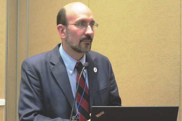
When these illnesses strike, “Without timely, appropriate antimicrobials, we are in trouble,” Dr. Claude Hemphill said at the annual meeting of the American Neurological Association. “All is usually lost. But patients can look very sick and still make a good recovery if we treat early. However, good neurocritical care is crucial.”
As an example, he cited the case of a 28-year-old nurse who had symptoms of an upper respiratory infection, including fever and headache. Her mother found her unresponsive and with left arm flexure. She required suctioning, which revealed pus in her throat. She did open her eyes to loud voices, but couldn’t follow commands. She had severe right-sided hemiparesis.

Imaging showed a severe empyema with brain inflammation and cerebral venous sinus thrombosis. Further testing revealed an Staphylococcus aureus brain infection.
“This was an emergency neurosurgical situation and she went emergently to the operating room,” where she had a decompressive craniectomy and postoperative angiography, said Dr. Hemphill, the Kenneth Rainin Chair in Neurocritical Care at the University of California, San Francisco. She was started on antibiotics and had several days of tissue plasminogen activator infusion into the superior sagittal sinus. But she remained in a coma.”
Intracranial pressure (ICP) monitoring and brain oxygen monitoring were not favorable. By hospital day 29, “You could see swelling everywhere. The middle cerebral artery was pushed up, and there was brain herniation through the craniectomy. At this time, we were weaning her off barbiturates and strongly urged to give up. But we didn’t. We finished her treatment and sent her to a rehab facility.”
Ten weeks later, Dr. Hemphill got a call from the young woman, requesting a note to return to work as soon as her craniectomy skull flap healed. “I wrote the note and chalked it up to a good lesson for both me and my team.”
In addition to discovering the root cause of the primary injury (in this case, infective illness), a key to the nurse’s good recovery was the limiting of secondary brain injury, he said. ICP monitoring was crucial during treatment and the long waiting period, Dr. Hemphill said. But ICP monitoring for these patients is often viewed as an unusual tactic.
“If you ask the neurosurgeon for an ICP, you might get a funny look,” he warned. “In the U.S. at least, not doing so is merely an accepted practice pattern – it’s not anything based on the pathophysiology of the disease.”
Even if the situation looks grim, Dr. Hemphill advised optimizing treatment to give patients with encephalitis a fighting chance. Antimicrobials may be helpful even if no infective agent can be identified. Corticosteroids, especially dexamethasone, appear beneficial if given concomitantly. “The reason we shouldn’t defer dexamethasone is that the blood-brain barrier may be less permeable after antibiotics,” Dr. Hemphill noted.
Glycerol, anti-inflammatory drugs, and paracetamol have not been proven helpful.
Because nonconvulsive status epilepticus isn’t uncommon, he also recommended continuous EEG monitoring, although prophylactic anticonvulsants are not usually indicated.
Dr. Hemphill had no financial disclosures.
On Twitter @alz_gal
BALTIMORE – Patients with seemingly desperate illnesses of the central nervous system can make a dramatic recovery if they receive appropriate, early antimicrobials and have enough time to respond.
When these illnesses strike, “Without timely, appropriate antimicrobials, we are in trouble,” Dr. Claude Hemphill said at the annual meeting of the American Neurological Association. “All is usually lost. But patients can look very sick and still make a good recovery if we treat early. However, good neurocritical care is crucial.”
As an example, he cited the case of a 28-year-old nurse who had symptoms of an upper respiratory infection, including fever and headache. Her mother found her unresponsive and with left arm flexure. She required suctioning, which revealed pus in her throat. She did open her eyes to loud voices, but couldn’t follow commands. She had severe right-sided hemiparesis.
Imaging showed a severe empyema with brain inflammation and cerebral venous sinus thrombosis. Further testing revealed an Staphylococcus aureus brain infection.
“This was an emergency neurosurgical situation and she went emergently to the operating room,” where she had a decompressive craniectomy and postoperative angiography, said Dr. Hemphill, the Kenneth Rainin Chair in Neurocritical Care at the University of California, San Francisco. She was started on antibiotics and had several days of tissue plasminogen activator infusion into the superior sagittal sinus. But she remained in a coma.”
Intracranial pressure (ICP) monitoring and brain oxygen monitoring were not favorable. By hospital day 29, “You could see swelling everywhere. The middle cerebral artery was pushed up, and there was brain herniation through the craniectomy. At this time, we were weaning her off barbiturates and strongly urged to give up. But we didn’t. We finished her treatment and sent her to a rehab facility.”
Ten weeks later, Dr. Hemphill got a call from the young woman, requesting a note to return to work as soon as her craniectomy skull flap healed. “I wrote the note and chalked it up to a good lesson for both me and my team.”
In addition to discovering the root cause of the primary injury (in this case, infective illness), a key to the nurse’s good recovery was the limiting of secondary brain injury, he said. ICP monitoring was crucial during treatment and the long waiting period, Dr. Hemphill said. But ICP monitoring for these patients is often viewed as an unusual tactic.
“If you ask the neurosurgeon for an ICP, you might get a funny look,” he warned. “In the U.S. at least, not doing so is merely an accepted practice pattern – it’s not anything based on the pathophysiology of the disease.”
Even if the situation looks grim, Dr. Hemphill advised optimizing treatment to give patients with encephalitis a fighting chance. Antimicrobials may be helpful even if no infective agent can be identified. Corticosteroids, especially dexamethasone, appear beneficial if given concomitantly. “The reason we shouldn’t defer dexamethasone is that the blood-brain barrier may be less permeable after antibiotics,” Dr. Hemphill noted.
Glycerol, anti-inflammatory drugs, and paracetamol have not been proven helpful.
Because nonconvulsive status epilepticus isn’t uncommon, he also recommended continuous EEG monitoring, although prophylactic anticonvulsants are not usually indicated.
Dr. Hemphill had no financial disclosures.
On Twitter @alz_gal
EXPERT ANALYSIS AT ANA 2014
Don’t make hasty decisions in postarrest hypothermia
BALTIMORE – A few days’ delay in prognostication can make the difference between life and death for patients who are treated with hypothermia after cardiac arrest.*
There’s no doubt now that the cooling technique improves survival and neurologic outcomes after cardiac arrest, Dr. Romergryko Geocadin said at the annual meeting of the American Neurological Association. But there isn’t yet firm consensus on the optimal target temperature, how long cooling and rewarming should last, or how long to wait before recovery can be adequately assessed, said Dr. Geocadin of Johns Hopkins University, Baltimore.

Because these questions remain open, both clinicians and families can make what he called a possibly tragic mistake: assuming too early that all hope is lost.
“The concept of waiting and allowing the tincture of time to work is very straightforward,” said Dr. Geocadin, who is also president of the Neurocritical Care Society. “It’s the problem of nihilism that is so immense. Once we as physicians say a disease is fatal – it is. Because if you predict the patient will die, then you will give up. Then patient will die, and then you will be correct.”
Dr. Geocadin argues that many cardiac arrest patients don’t get the time they need to begin recovery after hypothermic treatment. “The forces against this are sadly aligned” against patience, he said in an interview. Families are in distress and crave answers to difficult questions. Doctors feel pressured to give them, and predict outcomes too early. And, unfortunately, medical economics always lurks in the background. “In the ICU, you are always in a bed crunch. People demand to know why we’re waiting on this patient who is obviously going to have a poor outcome. I would say, what’s more unethical? Sentencing a patient to prolonged existence with little hope, or using insufficient information to predict a fatal outcome? I’m not saying sustain someone for 3 months – but just give it a week before making the decision that treatment is futile.”
No one knows exactly why postarrest hypothermia improves overall survival and good neurologic outcomes, Dr. Geocadin said. Treatment guidelines from the American Academy of Neurology and American Heart Association are scheduled to be released sometime in 2015. But for now, some important questions remain. What’s the best target temperature and how long should it be sustained? How about rewarming? The latest research isn’t finding definitive answers.
In 2013, a randomized trial of 950 patients added to the confusion. Mortality and neurologic recovery were similar whether patients had been cooled to 33° C or a near-normothermic 36° C.
The study suggests that cooling itself might not be the only key outcomes driver, Dr. Geocadin said. A combination of several other factors could also be at play. The real benefit could be in avoiding postresuscitation fever, which is associated with poor outcomes. It’s also possible that hypothermia’s therapeutic bang might actually be because of the improved multidisciplinary critical care protocols that accompany it – stabilizing blood pressure and blood glucose, controlling seizures, and increased clinician vigilance. Or it could simply be a marker of facility quality: A hospital equipped to offer hypothermia will likely be offering other advanced care practices and expert personnel as well.
“But one thing needs to be emphasized,” he said. “Temperature management at either 33° C or 36° C is superior to no temperature management [normothermia or more than 36° C] in comatose, postcardiac arrest patients."
Hypothermia slows down everything, including drug metabolism. “It’s not just cell death that slows or stops. Metabolism of drugs, such as sedatives, slows as well, and this could be related to the delayed brain recovery that we saw in the earlier trials, where neurologic function started to return by about 5-6 days. This could be because of the brain itself being slowed, or the being affected by slowly metabolized drugs. And this is why we need that delay before we can make prognostication about outcomes. We just need to give people a chance to come out of it.”
Dr. Geocadin had no financial disclosures. However, he is a member of the task force creating the new American Academy of Neurology/American Heart Association hypothermia treatment guidelines.
*Correction, 11/14/2014: A portion of the article that incorrectly stated the cardiac arrest hypothermia protocol at Johns Hopkins has been deleted.
On Twitter @alz_gal
BALTIMORE – A few days’ delay in prognostication can make the difference between life and death for patients who are treated with hypothermia after cardiac arrest.*
There’s no doubt now that the cooling technique improves survival and neurologic outcomes after cardiac arrest, Dr. Romergryko Geocadin said at the annual meeting of the American Neurological Association. But there isn’t yet firm consensus on the optimal target temperature, how long cooling and rewarming should last, or how long to wait before recovery can be adequately assessed, said Dr. Geocadin of Johns Hopkins University, Baltimore.
Because these questions remain open, both clinicians and families can make what he called a possibly tragic mistake: assuming too early that all hope is lost.
“The concept of waiting and allowing the tincture of time to work is very straightforward,” said Dr. Geocadin, who is also president of the Neurocritical Care Society. “It’s the problem of nihilism that is so immense. Once we as physicians say a disease is fatal – it is. Because if you predict the patient will die, then you will give up. Then patient will die, and then you will be correct.”
Dr. Geocadin argues that many cardiac arrest patients don’t get the time they need to begin recovery after hypothermic treatment. “The forces against this are sadly aligned” against patience, he said in an interview. Families are in distress and crave answers to difficult questions. Doctors feel pressured to give them, and predict outcomes too early. And, unfortunately, medical economics always lurks in the background. “In the ICU, you are always in a bed crunch. People demand to know why we’re waiting on this patient who is obviously going to have a poor outcome. I would say, what’s more unethical? Sentencing a patient to prolonged existence with little hope, or using insufficient information to predict a fatal outcome? I’m not saying sustain someone for 3 months – but just give it a week before making the decision that treatment is futile.”
No one knows exactly why postarrest hypothermia improves overall survival and good neurologic outcomes, Dr. Geocadin said. Treatment guidelines from the American Academy of Neurology and American Heart Association are scheduled to be released sometime in 2015. But for now, some important questions remain. What’s the best target temperature and how long should it be sustained? How about rewarming? The latest research isn’t finding definitive answers.
In 2013, a randomized trial of 950 patients added to the confusion. Mortality and neurologic recovery were similar whether patients had been cooled to 33° C or a near-normothermic 36° C.
The study suggests that cooling itself might not be the only key outcomes driver, Dr. Geocadin said. A combination of several other factors could also be at play. The real benefit could be in avoiding postresuscitation fever, which is associated with poor outcomes. It’s also possible that hypothermia’s therapeutic bang might actually be because of the improved multidisciplinary critical care protocols that accompany it – stabilizing blood pressure and blood glucose, controlling seizures, and increased clinician vigilance. Or it could simply be a marker of facility quality: A hospital equipped to offer hypothermia will likely be offering other advanced care practices and expert personnel as well.
“But one thing needs to be emphasized,” he said. “Temperature management at either 33° C or 36° C is superior to no temperature management [normothermia or more than 36° C] in comatose, postcardiac arrest patients."
Hypothermia slows down everything, including drug metabolism. “It’s not just cell death that slows or stops. Metabolism of drugs, such as sedatives, slows as well, and this could be related to the delayed brain recovery that we saw in the earlier trials, where neurologic function started to return by about 5-6 days. This could be because of the brain itself being slowed, or the being affected by slowly metabolized drugs. And this is why we need that delay before we can make prognostication about outcomes. We just need to give people a chance to come out of it.”
Dr. Geocadin had no financial disclosures. However, he is a member of the task force creating the new American Academy of Neurology/American Heart Association hypothermia treatment guidelines.
*Correction, 11/14/2014: A portion of the article that incorrectly stated the cardiac arrest hypothermia protocol at Johns Hopkins has been deleted.
On Twitter @alz_gal
EXPERT ANALYSIS FROM ANA 2014

Decompression can save lives in ventricular trapping
BALTIMORE –Aggressive decompression dramatically improved survival in patients who had trapped ventricle syndrome as a result of tumor or intracerebral hemorrhage in a retrospective study.
Overall mortality in the cohort was 70% among those who had no decompression, Dr. Gabriel L. Pagani-Estevez said at the annual meeting of the American Neurological Association. But it dropped to 19% among those who underwent some form of decompression therapy. Even after controlling for confounding factors like age, etiology, and hemorrhage volume, decompression remained a significant independent predictor of survival, said Dr. Pagani-Estevez, a neurology resident at the Mayo Clinic, Rochester, Minn.
Despite all the methodological issues inherent in a retrospective study, the findings “provide at least a suggestion that neurosurgical intervention can markedly reduce mortality in trapped ventricle syndrome,” he said. “Now, research needs to clarify the ideal intervention, the effect of decompression on functional outcome, and which patients might derive the most benefit from treatment.”
The cohort comprised 392 patients who developed ventricular trapping and were treated during 2002-2010. They were a mean of 58 years old. Most (223) were not on anticoagulation therapy. A total of 80 patients were taking aspirin, and the remainder were taking other anticoagulants. The median midline shift was about 10 mm.
Trapping was caused by a tumor in 177 patients. Other etiologies included intracerebral hemorrhage (80), subdural hematoma (55), trauma (26), and stroke (18). Unspecified causes made up the remainder.
The left lateral ventricle was most often involved (176). The right lateral ventricle was trapped in 159 patients and both were involved in 32. Thirteen patients had a trapped fourth ventricle, and 12 had unspecified trapping.
Some kind of decompression procedure was performed on 221 patients. These included craniotomy (126), craniectomy (26), external ventricular drain (30), ventricular-peritoneal shunt (23), and endoscopic septum pellucidum fenestration (16).
Comparisons showed significantly decreased mortality for intervention vs. nonintervention in groups with various causes of ventricular trapping: intracerebral hemorrhage (48% vs. 95%), tumor (12% vs. 47%), and subdural hematoma (20% vs. 90%).
There were nonsignificant declines in mortality among patients who underwent intervention for ventricular trapping caused by trauma or ischemic stroke, but the number of patients in those subgroups were small, which probably confounded the results, Dr. Pagani-Estevez said.
He then conducted a multivariate analysis to determine patient characteristics that might have contributed to survival. Patients who had a decompression procedure were 87% less likely to die than were those who had not – a highly significant finding (P = .0001). A midline shift conferred a slight increase in the risk of death, while having intracerebral hemorrhage as the trapping etiology increased the risk fourfold.

Trapped ventricle carries a notoriously poor prognosis, said Dr. Alejandro A. Rabinstein, a coauthor on the study. “By the time you develop it, it’s a very bad situation, so whatever way you can achieve decompression may improve the situation,” said Dr. Rabinstein, a critical care neurologist who is also at the Mayo Clinic in Rochester. “If you don’t think the patient has enough left to merit the intervention, then you just don’t do it. But despite that limitation, if you think the patient can recover some function, it’s appropriate. An intervention will make patients survive way more often than no intervention. Without something, though, the prospect of survival is bleak.”
Neither Dr. Pagani-Estevez nor Dr. Rabinstein had any financial disclosures.
On Twitter @alz_gal
BALTIMORE –Aggressive decompression dramatically improved survival in patients who had trapped ventricle syndrome as a result of tumor or intracerebral hemorrhage in a retrospective study.
Overall mortality in the cohort was 70% among those who had no decompression, Dr. Gabriel L. Pagani-Estevez said at the annual meeting of the American Neurological Association. But it dropped to 19% among those who underwent some form of decompression therapy. Even after controlling for confounding factors like age, etiology, and hemorrhage volume, decompression remained a significant independent predictor of survival, said Dr. Pagani-Estevez, a neurology resident at the Mayo Clinic, Rochester, Minn.
Despite all the methodological issues inherent in a retrospective study, the findings “provide at least a suggestion that neurosurgical intervention can markedly reduce mortality in trapped ventricle syndrome,” he said. “Now, research needs to clarify the ideal intervention, the effect of decompression on functional outcome, and which patients might derive the most benefit from treatment.”
The cohort comprised 392 patients who developed ventricular trapping and were treated during 2002-2010. They were a mean of 58 years old. Most (223) were not on anticoagulation therapy. A total of 80 patients were taking aspirin, and the remainder were taking other anticoagulants. The median midline shift was about 10 mm.
Trapping was caused by a tumor in 177 patients. Other etiologies included intracerebral hemorrhage (80), subdural hematoma (55), trauma (26), and stroke (18). Unspecified causes made up the remainder.
The left lateral ventricle was most often involved (176). The right lateral ventricle was trapped in 159 patients and both were involved in 32. Thirteen patients had a trapped fourth ventricle, and 12 had unspecified trapping.
Some kind of decompression procedure was performed on 221 patients. These included craniotomy (126), craniectomy (26), external ventricular drain (30), ventricular-peritoneal shunt (23), and endoscopic septum pellucidum fenestration (16).
Comparisons showed significantly decreased mortality for intervention vs. nonintervention in groups with various causes of ventricular trapping: intracerebral hemorrhage (48% vs. 95%), tumor (12% vs. 47%), and subdural hematoma (20% vs. 90%).
There were nonsignificant declines in mortality among patients who underwent intervention for ventricular trapping caused by trauma or ischemic stroke, but the number of patients in those subgroups were small, which probably confounded the results, Dr. Pagani-Estevez said.
He then conducted a multivariate analysis to determine patient characteristics that might have contributed to survival. Patients who had a decompression procedure were 87% less likely to die than were those who had not – a highly significant finding (P = .0001). A midline shift conferred a slight increase in the risk of death, while having intracerebral hemorrhage as the trapping etiology increased the risk fourfold.
Trapped ventricle carries a notoriously poor prognosis, said Dr. Alejandro A. Rabinstein, a coauthor on the study. “By the time you develop it, it’s a very bad situation, so whatever way you can achieve decompression may improve the situation,” said Dr. Rabinstein, a critical care neurologist who is also at the Mayo Clinic in Rochester. “If you don’t think the patient has enough left to merit the intervention, then you just don’t do it. But despite that limitation, if you think the patient can recover some function, it’s appropriate. An intervention will make patients survive way more often than no intervention. Without something, though, the prospect of survival is bleak.”
Neither Dr. Pagani-Estevez nor Dr. Rabinstein had any financial disclosures.
On Twitter @alz_gal
AT ANA 2014
Key clinical point: Decompression for a trapped ventricle can be a life-saving procedure.
Major finding: Mortality significantly declined from 70% in patients without decompression to 19% in those who underwent decompression via a variety of methods.
Data source: The retrospective study comprised 392 patients.
Disclosures: Neither Dr. Pagani-Estevez nor Dr. Rabinstein had any financial disclosures.
ICU meds can bring on serotonin syndrome
BALTIMORE – Serotonin syndrome can easily develop in the intensive care unit, particularly when patients receive opiates and antiemetic medications in addition to the serotonin-enhancing medications they may already be taking.
“These medications are pervasively present, and they are notorious for drug-drug interactions,” Dr. Alejandro Rabinstein said at the annual meeting of the American Neurological Association. “And, in the ICU, we often use them without even realizing it. The combination can be enough to cause serotonin syndrome, which is sometimes recognized too late, and can have serious consequences.”
Because there are scant data on the phenomenon, Dr. Rabinstein, a critical care neurologist at the Mayo Clinic, Rochester, Minn., conducted a literature search to identify cases and examine their outcomes. His series comprised 33 patients, 22 of whom were seen at the Mayo facility. The patients were a median of 42 years old. Four of them had been admitted with serotonin syndrome as their primary diagnosis, but it was either unsuspected or had not yet developed in the remainder.
For 17 patients (52%), the primary reason for admission was a change in mental status or altered consciousness. Four were admitted for sepsis syndrome. Other reasons included resuscitation after cardiac arrest (three), severe pneumonia (two), tumor-related complications (two), emergency surgery (two), graft vs. host disease (one), liver failure (one), and trauma (one).
At admission, the mean APACHE-III score was 67, although the range was wide (11-146). A total of 18 patients required mechanical ventilation, and 6 had undergone surgery before serotonin syndrome developed.
All of the patients showed altered mental status, a key characteristic of serotonin syndrome. Agitation was present in 14 patients. Other common signs were tachypnea (23), tachycardia (20), clonus (29), hyperreflexia (24), and rigidity (26). Mydriasis was present in 14 and tremor in 12. Seven patients were markedly diaphoretic. Fever was present in 14.
About 40% of the group (13 patients) developed signs of serotonin syndrome only after they were hospitalized (mean day 4). Dr. Rabinstein said 22 drugs with a direct serotonin-enhancing action were involved in the cases. In fact, almost all were taking at least one such drug on admission, and 70% got new serotonergic medications while in the ICU.
About 75% of the patients were taking a selective serotonin reuptake inhibitor when admitted (citalopram, fluoxetine, sertraline, escitalopram, and paroxetine ). Almost a third were taking a selective norepinephrine reuptake inhibitor (venlafaxine and duloxetine). About a fourth were taking some other antidepressant with serotonergic activity (trazodone, mirtazapine, buspirone, clomipramine). While in the ICU, three patients received a new serotonergic antidepressant.
Four patients were taking an antiemetic upon admission, and seven started a new antiemetic when admitted. Three were taking an opioid upon admission, and 21 started an opioid when admitted.
All of the patients received benzodiazepine treatment, and 24% had to be paralyzed while their serotonergic symptoms resolved. The anticholinergic antihistamine cyproheptadine was given to the 13 most severely affected patients. However, Dr. Rabinstein said, its efficacy couldn’t be reliably assessed because of this selection bias.
It took a mean of 56 hours for patients to recover to the point where they could interact normally, but the range of recovery time was very wide (8-288 hours). Despite some increases in creatinine kinase and lactic acid, none of the patients developed rhabdomyolysis-related kidney failure. Although serotonin syndrome can be fatal, there were no related deaths in this group.
The review points to how easily ICU treatment can precipitate a very serious neurologic disorder. “Almost everyone came into the ICU on one of these medications, and almost everyone got at least one more after arriving,” Dr. Rabinstein said. “It’s something we have to be on the alert for. The reason I did this study was simply to raise awareness, because this diagnosis is blown all the time. You don’t have to be a neurologist to make this diagnosis, but you need to be very attentive to these signs. ”
He had no financial disclosures.
On Twitter @alz_gal
EXPERT ANALYSIS FROM ANA 2014
Clopidogrel seems safe in ischemic stroke with microbleeds
BALTIMORE – The presence of cerebral microbleeds was not associated with a higher frequency of hemorrhagic transformation among patients who received a loading dose of the antiplatelet drug, compared with patients who had no evidence of microbleeds, in a retrospective study of 1,060 patients who presented with acute ischemic stroke.
“Clopidogrel seems like it can be a safe treatment for patients who are not eligible for tissue plasminogen activator, even if the stroke severity is higher than is being tested in recent clinical trials (CHANCE and POINT, for example),” Melisa Valmoria said at the annual meeting of the American Neurological Association.

Ms. Valmoria, a clinical research assistant at the Tulane University stroke research program, New Orleans, presented a retrospective study of 1,060 patients who presented with acute ischemic stroke. Of 1,060 patients in the study, 324 received a loading dose of at least 300 mg clopidogrel within 6 hours of admission, in addition to aspirin. A total of 51 of those patients had evidence of prior cerebral microbleeds on imaging.
The patients were similar in age (average, 63.5 years). Those with microbleeds were more likely to have had a prior stroke (63% vs. 49%; P = .05). The median NIH Stroke Scale (NIHSS) score was also higher among those patients (6 vs. 4).
There were no significant between-group differences in stroke events, including rates of inpatient complications (33% with microbleeds vs. 28% without), hemorrhagic transformation (6% vs. 8%), and recurrent thrombotic events (16% vs. 14%). Neurologic worsening, defined as an increase of at least 2 points in the NIHSS score over any 24-hour period, was not significantly different between the groups (39% vs. 27%). None of the patients with microbleeds developed a symptomatic hemorrhage.
Patients with microbleeds started off with a higher NIHSS score, but did not have significantly worse discharge outcomes. Poor functional outcome (a modified Rankin Scale score of more than 2) occurred in 59% of those with microbleeds and 47% of those without. An unfavorable discharge disposition (discharge to anywhere except the patient’s home or to inpatient rehabilitation) occurred in 16% of those with microbleeds and 10% of those without, which was not a significant difference.
A subanalysis of patients with more than 10 microbleeds showed that clopidogrel did not increase their risk of hemorrhagic transformation or poor functional outcomes, compared with patients who did not have microbleeds.
“Some trials (MATCH and CHARISMA) have raised concern about bleeding risk, specifically brain bleeding, in patients who receive dual antiplatelets, including clopidogrel, after stroke,” Ms. Valmoria said. “This risk may be higher among patients who have evidence of prior brain bleeding, such as microbleeds. Fortunately, our study did not find support for increased short-term bleeding in patients with ischemic stroke who were treated with a loading dose of clopidogrel with aspirin. Our study included many patients with moderatetosevere strokes, as measured by the NIHSS score who are not wellrepresented in modern clinical trials examining the safety and efficacy of clopidogrel loading, raising the question of whether enrollment criteria could be safely loosened.”
She said she had no relevant financial disclosures.
On Twitter @alz_gal
BALTIMORE – The presence of cerebral microbleeds was not associated with a higher frequency of hemorrhagic transformation among patients who received a loading dose of the antiplatelet drug, compared with patients who had no evidence of microbleeds, in a retrospective study of 1,060 patients who presented with acute ischemic stroke.
“Clopidogrel seems like it can be a safe treatment for patients who are not eligible for tissue plasminogen activator, even if the stroke severity is higher than is being tested in recent clinical trials (CHANCE and POINT, for example),” Melisa Valmoria said at the annual meeting of the American Neurological Association.
Ms. Valmoria, a clinical research assistant at the Tulane University stroke research program, New Orleans, presented a retrospective study of 1,060 patients who presented with acute ischemic stroke. Of 1,060 patients in the study, 324 received a loading dose of at least 300 mg clopidogrel within 6 hours of admission, in addition to aspirin. A total of 51 of those patients had evidence of prior cerebral microbleeds on imaging.
The patients were similar in age (average, 63.5 years). Those with microbleeds were more likely to have had a prior stroke (63% vs. 49%; P = .05). The median NIH Stroke Scale (NIHSS) score was also higher among those patients (6 vs. 4).
There were no significant between-group differences in stroke events, including rates of inpatient complications (33% with microbleeds vs. 28% without), hemorrhagic transformation (6% vs. 8%), and recurrent thrombotic events (16% vs. 14%). Neurologic worsening, defined as an increase of at least 2 points in the NIHSS score over any 24-hour period, was not significantly different between the groups (39% vs. 27%). None of the patients with microbleeds developed a symptomatic hemorrhage.
Patients with microbleeds started off with a higher NIHSS score, but did not have significantly worse discharge outcomes. Poor functional outcome (a modified Rankin Scale score of more than 2) occurred in 59% of those with microbleeds and 47% of those without. An unfavorable discharge disposition (discharge to anywhere except the patient’s home or to inpatient rehabilitation) occurred in 16% of those with microbleeds and 10% of those without, which was not a significant difference.
A subanalysis of patients with more than 10 microbleeds showed that clopidogrel did not increase their risk of hemorrhagic transformation or poor functional outcomes, compared with patients who did not have microbleeds.
“Some trials (MATCH and CHARISMA) have raised concern about bleeding risk, specifically brain bleeding, in patients who receive dual antiplatelets, including clopidogrel, after stroke,” Ms. Valmoria said. “This risk may be higher among patients who have evidence of prior brain bleeding, such as microbleeds. Fortunately, our study did not find support for increased short-term bleeding in patients with ischemic stroke who were treated with a loading dose of clopidogrel with aspirin. Our study included many patients with moderatetosevere strokes, as measured by the NIHSS score who are not wellrepresented in modern clinical trials examining the safety and efficacy of clopidogrel loading, raising the question of whether enrollment criteria could be safely loosened.”
She said she had no relevant financial disclosures.
On Twitter @alz_gal
AT ANA 2014
Key clinical point: Clopidogrel didn’t increase brain bleeding when administered to patients with acute ischemic stroke and evidence of microbleeds.
Major finding: There was no difference between patients who received clopidogrel loading and those who didn’t in rates of hemorrhagic transformation (6% vs. 8%) or recurrent thrombotic events (16% vs. 14%).
Data source: A retrospective study of 324 patients with stroke and microbleeds who received clopidogrel plus aspirin or aspirin alone.
Disclosures: Ms. Valmoria said she had no relevant financial disclosures.

Dengue, West Nile threaten to set up housekeeping in U.S.A.
BALTIMORE – Two imported mosquito-borne illnesses continue to push into the United States, threatening to take up residence in areas with a favorable environment.
Neither dengue fever nor West Nile virus are naturally endemic to any part of the United States, Dr. Larry Davis said at the annual meeting of the American Neurological Association. But both diseases show some indications of establishing a local infective reservoir.

Dengue remains a rare disease here, with about 650 cases reported in 2013. The majority of those occurred in patients who had just returned from endemic countries. But since 2009, local transmission appears to be occurring in three hot spots: Hawaii, Texas, and south Florida, including Key West and Miami, said Dr. Davis, a professor of neurology at the University of New Mexico, Albuquerque.
Locally acquired disease first popped up during 2009-2010 in Key West, Fla. During that time, 29 residents developed clinical dengue. Many more probably carried the virus, however, since a robust immune response usually tamps down about 80% of first infections. But those asymptomatic carriers provide a potent reservoir for hungry mosquitoes, and dengue is one virus that doesn’t need an intermediate host. Instead, Dr. Davis said, any mosquito that bites an infected person passes the agent directly into the bloodstream of his next meal.
During the Key West outbreak, the Centers for Disease Control and Prevention (CDC) estimated that about 5% of those living on the small island had been infected at some point.
Last year, 53 clinical cases were identified in south Texas. About half of these were locally acquired, which is another sign that the virus may be taking up residence, Dr. Davis said. And, so far this year, four residents of Miami-Dade County, Fla. have been diagnosed with locally acquired disease. The outbreak recently prompted local health officials to issue an alert for mosquito-borne disease, and remind residents to take precautions against being bitten.
In addition to its direct transmissibility, dengue has another neat infective trick, Dr. Davis noted. There are four serotypes, each with their own unique immunogenic profile. Thus, infection with any one confers no protection against any of the others. Any vaccine, therefore, would have to protect against all four serotypes.
In fact, such a tetravalent vaccine is in clinical trials, he said. Last month, Sanofi-Pasteur reported that its candidate vaccine, tested in almost 21,000 children in Latin America and the Caribbean, reduced the risk of disease by more than 60%, and the risk of dengue-related hospitalization by 80%. The results mirror those seen in an earlier Asian trial, the drug company noted.There has been no such progress on a human vaccine against West Nile virus, Dr. Davis said. So far this year, 1,585 cases in 47 states have been reported to the CDC, with the majority occurring in California (563), Colorado (108), and Texas (207). Of these, 917 (58%) were classified as neuroinvasive disease (such as meningitis or encephalitis). Thus far, 54 patients have died; 20 of these in California alone.
The California Department of Public Health has identified 3,282 positive mosquito samples as of Oct. 27. Director Dr. Ron Chapman said in a press statement that the state has never experienced such a severe outbreak.
“The proportion of mosquitoes infected with West Nile virus is at the highest level ever detected in California,” Dr. Chapman said. “We expect to see more people become infected as this is the time of year when the risk of infection is the highest.”
Like dengue, the majority of West Nile cases are asymptomatic, Dr. Davis noted. But in the absence of a robust immune response, the virus can cross the blood-brain barrier and initiate a devastating neuroinvasive illness than can cause a flaccid paralysis, seizures, and death.
Neuroinvasive West Nile carries many clinical and diagnostic similarities to polio, he said. Like polio, the limb weakness has a sudden onset and can be accompanied by pain, although the sensory neurons aren’t apparently involved. Imaging studies show the virus attacking at the anterior horns of the spinal cord; nerve conduction studies show a motor axonopathy, but without much demyelination. Survivors have a variable course of recovery; some may regain full strength of the affected limbs, but many retain persistent weakness.
There is no vaccine against the disease, although several are in early-stage development, including a live, attenuated chimeric vaccine that did well in a phase II, placebo-controlled trial. Within 28 days of vaccination, 98% of subjects experienced seroconversion.
Dr. Davis had no relevant conflicts of interest.
On Twitter @alz_gal
BALTIMORE – Two imported mosquito-borne illnesses continue to push into the United States, threatening to take up residence in areas with a favorable environment.
Neither dengue fever nor West Nile virus are naturally endemic to any part of the United States, Dr. Larry Davis said at the annual meeting of the American Neurological Association. But both diseases show some indications of establishing a local infective reservoir.
Dengue remains a rare disease here, with about 650 cases reported in 2013. The majority of those occurred in patients who had just returned from endemic countries. But since 2009, local transmission appears to be occurring in three hot spots: Hawaii, Texas, and south Florida, including Key West and Miami, said Dr. Davis, a professor of neurology at the University of New Mexico, Albuquerque.
Locally acquired disease first popped up during 2009-2010 in Key West, Fla. During that time, 29 residents developed clinical dengue. Many more probably carried the virus, however, since a robust immune response usually tamps down about 80% of first infections. But those asymptomatic carriers provide a potent reservoir for hungry mosquitoes, and dengue is one virus that doesn’t need an intermediate host. Instead, Dr. Davis said, any mosquito that bites an infected person passes the agent directly into the bloodstream of his next meal.
During the Key West outbreak, the Centers for Disease Control and Prevention (CDC) estimated that about 5% of those living on the small island had been infected at some point.
Last year, 53 clinical cases were identified in south Texas. About half of these were locally acquired, which is another sign that the virus may be taking up residence, Dr. Davis said. And, so far this year, four residents of Miami-Dade County, Fla. have been diagnosed with locally acquired disease. The outbreak recently prompted local health officials to issue an alert for mosquito-borne disease, and remind residents to take precautions against being bitten.
In addition to its direct transmissibility, dengue has another neat infective trick, Dr. Davis noted. There are four serotypes, each with their own unique immunogenic profile. Thus, infection with any one confers no protection against any of the others. Any vaccine, therefore, would have to protect against all four serotypes.
In fact, such a tetravalent vaccine is in clinical trials, he said. Last month, Sanofi-Pasteur reported that its candidate vaccine, tested in almost 21,000 children in Latin America and the Caribbean, reduced the risk of disease by more than 60%, and the risk of dengue-related hospitalization by 80%. The results mirror those seen in an earlier Asian trial, the drug company noted.There has been no such progress on a human vaccine against West Nile virus, Dr. Davis said. So far this year, 1,585 cases in 47 states have been reported to the CDC, with the majority occurring in California (563), Colorado (108), and Texas (207). Of these, 917 (58%) were classified as neuroinvasive disease (such as meningitis or encephalitis). Thus far, 54 patients have died; 20 of these in California alone.
The California Department of Public Health has identified 3,282 positive mosquito samples as of Oct. 27. Director Dr. Ron Chapman said in a press statement that the state has never experienced such a severe outbreak.
“The proportion of mosquitoes infected with West Nile virus is at the highest level ever detected in California,” Dr. Chapman said. “We expect to see more people become infected as this is the time of year when the risk of infection is the highest.”
Like dengue, the majority of West Nile cases are asymptomatic, Dr. Davis noted. But in the absence of a robust immune response, the virus can cross the blood-brain barrier and initiate a devastating neuroinvasive illness than can cause a flaccid paralysis, seizures, and death.
Neuroinvasive West Nile carries many clinical and diagnostic similarities to polio, he said. Like polio, the limb weakness has a sudden onset and can be accompanied by pain, although the sensory neurons aren’t apparently involved. Imaging studies show the virus attacking at the anterior horns of the spinal cord; nerve conduction studies show a motor axonopathy, but without much demyelination. Survivors have a variable course of recovery; some may regain full strength of the affected limbs, but many retain persistent weakness.
There is no vaccine against the disease, although several are in early-stage development, including a live, attenuated chimeric vaccine that did well in a phase II, placebo-controlled trial. Within 28 days of vaccination, 98% of subjects experienced seroconversion.
Dr. Davis had no relevant conflicts of interest.
On Twitter @alz_gal
EXPERT ANALYSIS AT ANA 2014

Amitriptyline beat pregabalin for chronic low back pain
BALTIMORE– Amitriptyline decreased pain and improved function significantly more than did pregabalin in a randomized trial of patients with chronic low back pain.
Almost 60% of patients taking the tricyclic antidepressant experienced at least a 50% decrease in pain on a visual analog pain scale, compared with 38% of those taking pregabalin, Dr. Jayantee Kalita reported at the annual meeting of the American Neurological Association.

The study is the first to examine amitriptyline in low back pain, a chronic pain condition that can be debilitating in its duration and severity and is notoriously difficult to control, said Dr. Kalita, a neurologist at the Sanjay Gandhi Post Graduate Institute of Medicine, Lucknow, India.
A 2011 systematic review found 17 randomized controlled trials of pharmacologic treatments for chronic low back pain (Eur. Spine J. 2011;20:40-50). These examined the benefits of nonsteroidal anti-inflammatory drugs, antidepressants, and opioids. The best results were obtained with acetaminophen and opioids, but there were dose-limiting side effects, and pain rebounded when the treatment was discontinued. The studies that examined antidepressants found them to be no more effective than placebo.
Dr. Kalita and her colleagues randomized 200 patients with chronic low back pain to amitriptyline or pregabalin for 14 weeks. Amitriptyline was started at 10 mg once a day and increased to a maximum of 50 mg as needed. Pregabalin was started at 75 mg twice a day and increased to a maximum of 300 mg twice a day if needed.
The cohort had a mean age of 41 years, with a median 35 months of back pain, although one patient had experienced 30 years of back pain. Pain was idiopathic and experienced as radiculopathy (48%), localized (46%), or as lower crossed syndrome (6%).
Most patients (115) lived in rural areas. Risk factors included diabetes (9 patients), hypertension (15), smoking (39), and tobacco use (65).
At baseline, both treatment groups reported their pain as a median of 7 on a 10-point visual analog scale. The mean score on the Oswestry Disability Index (ODI) was 40%, indicating severe pain that limits work, travel, personal care, social life, sexual activity, and sleep.By 6 weeks into the trial, pain scores in the amitriptyline group had decreased from 7 to 4, a significant improvement. By the end of 14 weeks, scores had decreased to about 3, which was significantly better than the scores both at 6 weeks and at baseline.
In the pregabalin group, the pain score dropped from 7 to 5, which was not a significant change. During weeks 6-14, scores dropped another half-point, to around 4.5. This was a significant improvement from baseline, but not from week 6.
The ODI score also improved more in patients taking amitriptyline. From a median of about 43%, the index dropped to about 30% at 6 weeks and 20% at 14 weeks. A 20% score indicates minimal disability; patients need no assistance with activities of daily living and experience minimal life interruptions from pain.
Disability also improved in the pregabalin group, dropping from about 40% at baseline to 35% at 6 weeks, and then to about 25% at 14 weeks. This score falls in the moderate disability range. Patients may need help with some daily activities, and may be off work. The final score was also a significant improvement over baseline, although it was not as robust as with the amitriptyline group, Dr. Kalita noted.
In the intent-to-treat analysis, 57% of the amitriptyline group and 39% of the pregabalin group experienced a pain scale decrease of at least 50%. There was at least a 20% improvement in the disability scores of 67% of the amitriptyline group and 48% of the pregabalin group.
Side effects included sedation, vertigo, dry mouth, skin rash, and restlessness. Significantly more patients who took pregabalin had at least one side effect (eight patients vs. three patients). Sedation was most common, occurring in 10 patients who took amitriptyline and 4 who took pregabalin.
Dr. Kalita had no financial disclosures.
On Twitter @alz_gal
BALTIMORE– Amitriptyline decreased pain and improved function significantly more than did pregabalin in a randomized trial of patients with chronic low back pain.
Almost 60% of patients taking the tricyclic antidepressant experienced at least a 50% decrease in pain on a visual analog pain scale, compared with 38% of those taking pregabalin, Dr. Jayantee Kalita reported at the annual meeting of the American Neurological Association.
The study is the first to examine amitriptyline in low back pain, a chronic pain condition that can be debilitating in its duration and severity and is notoriously difficult to control, said Dr. Kalita, a neurologist at the Sanjay Gandhi Post Graduate Institute of Medicine, Lucknow, India.
A 2011 systematic review found 17 randomized controlled trials of pharmacologic treatments for chronic low back pain (Eur. Spine J. 2011;20:40-50). These examined the benefits of nonsteroidal anti-inflammatory drugs, antidepressants, and opioids. The best results were obtained with acetaminophen and opioids, but there were dose-limiting side effects, and pain rebounded when the treatment was discontinued. The studies that examined antidepressants found them to be no more effective than placebo.
Dr. Kalita and her colleagues randomized 200 patients with chronic low back pain to amitriptyline or pregabalin for 14 weeks. Amitriptyline was started at 10 mg once a day and increased to a maximum of 50 mg as needed. Pregabalin was started at 75 mg twice a day and increased to a maximum of 300 mg twice a day if needed.
The cohort had a mean age of 41 years, with a median 35 months of back pain, although one patient had experienced 30 years of back pain. Pain was idiopathic and experienced as radiculopathy (48%), localized (46%), or as lower crossed syndrome (6%).
Most patients (115) lived in rural areas. Risk factors included diabetes (9 patients), hypertension (15), smoking (39), and tobacco use (65).
At baseline, both treatment groups reported their pain as a median of 7 on a 10-point visual analog scale. The mean score on the Oswestry Disability Index (ODI) was 40%, indicating severe pain that limits work, travel, personal care, social life, sexual activity, and sleep.By 6 weeks into the trial, pain scores in the amitriptyline group had decreased from 7 to 4, a significant improvement. By the end of 14 weeks, scores had decreased to about 3, which was significantly better than the scores both at 6 weeks and at baseline.
In the pregabalin group, the pain score dropped from 7 to 5, which was not a significant change. During weeks 6-14, scores dropped another half-point, to around 4.5. This was a significant improvement from baseline, but not from week 6.
The ODI score also improved more in patients taking amitriptyline. From a median of about 43%, the index dropped to about 30% at 6 weeks and 20% at 14 weeks. A 20% score indicates minimal disability; patients need no assistance with activities of daily living and experience minimal life interruptions from pain.
Disability also improved in the pregabalin group, dropping from about 40% at baseline to 35% at 6 weeks, and then to about 25% at 14 weeks. This score falls in the moderate disability range. Patients may need help with some daily activities, and may be off work. The final score was also a significant improvement over baseline, although it was not as robust as with the amitriptyline group, Dr. Kalita noted.
In the intent-to-treat analysis, 57% of the amitriptyline group and 39% of the pregabalin group experienced a pain scale decrease of at least 50%. There was at least a 20% improvement in the disability scores of 67% of the amitriptyline group and 48% of the pregabalin group.
Side effects included sedation, vertigo, dry mouth, skin rash, and restlessness. Significantly more patients who took pregabalin had at least one side effect (eight patients vs. three patients). Sedation was most common, occurring in 10 patients who took amitriptyline and 4 who took pregabalin.
Dr. Kalita had no financial disclosures.
On Twitter @alz_gal
AT ANA 2014
Key clinical point: Amitriptyline appears to be an effective treatment for chronic low back pain.
Major finding: Pain scores improved by at least 50% in 57% of those who took amitriptyline and in 39% of those who took pregabalin.
Data source: A randomized, 14-week trial of 200 patients.
Disclosures: Dr. Kalita had no financial disclosures.

VIDEO: What’s on the horizon for headaches
BALTIMORE – Methods to prevent and treat migraines are lacking, but monoclonal antibodies and the calcitonin receptor system are showing promise, according to Dr. Peter Goadsby of the University of California, San Francisco.
“It’s an awful mishmash of things we have to offer,” Dr. Goadsby said at the annual meeting of the American Neurological Association. However, data presented at the meeting suggest that new options may be on the horizon.
In an interview at the meeting, Dr. Goadsby explained details of several studies that showed dramatic results, and the “tantalizing” implications for patients.
AT ANA 2014
Handheld vagal nerve stimulator reduced cluster headache attacks
BALTIMORE – A hand-held vagal nerve stimulator significantly decreased the frequency of cluster headache attacks per week, compared with the usual standard of care, based on data from a randomized trial of 97 patients.
The noninvasive device, called gammaCore, was associated with seven fewer weekly headache attacks when used in conjunction with sumatriptan and oxygen – significantly better than the one-attack reduction achieved with standard therapy alone, Eric Liebler reported at the annual meeting of the American Neurological Association.

“In this 2-month study, the external vagal nerve stimulator effected a significant improvement in the reduced rate of attacks and the 50% responder rate, with very few device-related adverse events,” said Mr. Liebler, vice president of scientific, medical, and governmental affairs for electroCore, the New Jersey–based company that manufactures the device.
The gammaCore device is used in Europe for the treatment of primary headache disorders. The device, which transmits mild current to the vagal nerve, is a small, hand-held unit that the patient uses twice each day. It’s thought to work by suppressing excessive extracellular glutamate in the trigeminal nucleus caudalis – a phenomenon that seems to stimulate trigeminal sensitization in migraine.
Patients are instructed to activate the device over the carotid pulse point on the neck for about 90 seconds. A conductive gel helps transmit mild current to the vagal nerve.
The 2-month Prevention and Acute Treatment of Chronic Cluster Headache (PREVA) trial examined the use of gammaCore in treating cluster headaches. The cohort included 97 patients who were randomized to the device plus the standard therapy or to standard therapy alone. At the end of the first month, patients in the combination group continued for another month while those in the standard-care group were offered a month of combination therapy.
The primary endpoint was the reduction in cluster headache attacks per week during the last 2 weeks of each month. Secondary endpoints included the number of patients with at least a 50% reduction in the number of cluster headaches per month and changes in the use of acute medications. The study also examined device safety and tolerability. Attack pain and duration are being assessed in a separate study, Mr. Liebler noted.
The mean age of the patients was approximately 44 years. Most were using medications (90%) and oxygen (68%), and the mean duration of their cluster headaches at baseline was about 28 minutes when they used these treatments. Without medications and oxygen, the headaches lasted a mean of 100 minutes. These patients expereinced about 70 cluster attacks in the month before randomization.
Patients in the combination group experienced a significant reduction in the number of cluster headache attacks per week (from 16 to 9 attacks). The change among those taking standard therapy alone was not significant (from 16 to 15 attacks).
In the intent-to-treat population, 38% of the combination group achieved at least a 50% decrease in headache days, compared with 8% of the standard-therapy group – a significant difference. In the full analysis, the device response numbers were better – 46% vs. 8%. The per protocol analysis included only those patients who used the device as instructed at least 80% of the time. The 50% responder rate in that group was 52% vs. 9% of the standard-treatment group.
Among the combination-therapy group, the use of rescue medications decreased significantly: from 7 to 3 times per month for subcutaneous sumatriptan and from 16 to 6 times per month for oxygen. Rescue-medication use was virtually unchanged among those who used standard therapy only.
Safety and tolerability was good. Most of the patients using the device (44) did experience an adverse event. Device-related events included dizziness (3), oropharyngeal pain (3), and neck pain (3). These events probably occurred because electrical current tends to spread to neck muscles, Mr. Liebler said. The sensations were transitory, he added.
Dr. Peter Goadsby, director of the headache center at the University of California, San Francisco, said that he has been “surprised” at the positive results seen in European patients. “These are medication-refractory patients, and about half of them had significant reductions in the frequency and severity of their cluster headaches, with a simultaneous decrease in the need for oxygen and steroids. They also seemed to feel that their steroids worked more efficiently,” when used in conjunction with the device, he said.
The study was sponsored by electroCore, and Mr. Liebler is a full-time employee of electroCore. Dr. Goadsby has served as a consultant for a number of pharmaceutical companies.
On Twitter @alz_gal
BALTIMORE – A hand-held vagal nerve stimulator significantly decreased the frequency of cluster headache attacks per week, compared with the usual standard of care, based on data from a randomized trial of 97 patients.
The noninvasive device, called gammaCore, was associated with seven fewer weekly headache attacks when used in conjunction with sumatriptan and oxygen – significantly better than the one-attack reduction achieved with standard therapy alone, Eric Liebler reported at the annual meeting of the American Neurological Association.
“In this 2-month study, the external vagal nerve stimulator effected a significant improvement in the reduced rate of attacks and the 50% responder rate, with very few device-related adverse events,” said Mr. Liebler, vice president of scientific, medical, and governmental affairs for electroCore, the New Jersey–based company that manufactures the device.
The gammaCore device is used in Europe for the treatment of primary headache disorders. The device, which transmits mild current to the vagal nerve, is a small, hand-held unit that the patient uses twice each day. It’s thought to work by suppressing excessive extracellular glutamate in the trigeminal nucleus caudalis – a phenomenon that seems to stimulate trigeminal sensitization in migraine.
Patients are instructed to activate the device over the carotid pulse point on the neck for about 90 seconds. A conductive gel helps transmit mild current to the vagal nerve.
The 2-month Prevention and Acute Treatment of Chronic Cluster Headache (PREVA) trial examined the use of gammaCore in treating cluster headaches. The cohort included 97 patients who were randomized to the device plus the standard therapy or to standard therapy alone. At the end of the first month, patients in the combination group continued for another month while those in the standard-care group were offered a month of combination therapy.
The primary endpoint was the reduction in cluster headache attacks per week during the last 2 weeks of each month. Secondary endpoints included the number of patients with at least a 50% reduction in the number of cluster headaches per month and changes in the use of acute medications. The study also examined device safety and tolerability. Attack pain and duration are being assessed in a separate study, Mr. Liebler noted.
The mean age of the patients was approximately 44 years. Most were using medications (90%) and oxygen (68%), and the mean duration of their cluster headaches at baseline was about 28 minutes when they used these treatments. Without medications and oxygen, the headaches lasted a mean of 100 minutes. These patients expereinced about 70 cluster attacks in the month before randomization.
Patients in the combination group experienced a significant reduction in the number of cluster headache attacks per week (from 16 to 9 attacks). The change among those taking standard therapy alone was not significant (from 16 to 15 attacks).
In the intent-to-treat population, 38% of the combination group achieved at least a 50% decrease in headache days, compared with 8% of the standard-therapy group – a significant difference. In the full analysis, the device response numbers were better – 46% vs. 8%. The per protocol analysis included only those patients who used the device as instructed at least 80% of the time. The 50% responder rate in that group was 52% vs. 9% of the standard-treatment group.
Among the combination-therapy group, the use of rescue medications decreased significantly: from 7 to 3 times per month for subcutaneous sumatriptan and from 16 to 6 times per month for oxygen. Rescue-medication use was virtually unchanged among those who used standard therapy only.
Safety and tolerability was good. Most of the patients using the device (44) did experience an adverse event. Device-related events included dizziness (3), oropharyngeal pain (3), and neck pain (3). These events probably occurred because electrical current tends to spread to neck muscles, Mr. Liebler said. The sensations were transitory, he added.
Dr. Peter Goadsby, director of the headache center at the University of California, San Francisco, said that he has been “surprised” at the positive results seen in European patients. “These are medication-refractory patients, and about half of them had significant reductions in the frequency and severity of their cluster headaches, with a simultaneous decrease in the need for oxygen and steroids. They also seemed to feel that their steroids worked more efficiently,” when used in conjunction with the device, he said.
The study was sponsored by electroCore, and Mr. Liebler is a full-time employee of electroCore. Dr. Goadsby has served as a consultant for a number of pharmaceutical companies.
On Twitter @alz_gal
AT ANA 2014
Key clinical point: A noninvasive vagal nerve stimulator in combination with standard treatment decreased the frequency of cluster headaches, compared with standard treatment alone.
Major finding: The combination reduced attacks by about seven per week, while standard therapy reduced the attack rate by one.
Data source: The trial randomized 97 patients with cluster headache to gammaCore plus sumatriptan/oxygen or to sumatriptan/oxygen alone.
Disclosures: electroCore sponsored the study. Mr Liebler is a full-time employee of the company.