User login
Consider facet joint OA in older patients with low back pain
DESTIN, FLA. – Facet joint osteoarthritis likely accounts for much of what is classified as "nonspecific" low back pain, according to Dr. Alfred C. Gellhorn.
"Amazingly – and I think sadly for us – the lumbar facet joints really have received very little attention in the literature," Dr. Gellhorn, who works in the department of rehabilitation medicine at the University of Washington, Seattle, said at the annual Congress of Clinical Rheumatology.
Eight of every ten American will experience low back pain at some point during their lifetime; low back pain is second only to the common cold in frequency, is the most common reason for time off work, and has a total social cost of more than $100 billion annually. Up to 85% of patients never receive a definitive diagnosis and are classified has having nonspecific pain, he said.

The facet joints may be responsible for a significant proportion of that pain.
Facet joint cartilage is aneural, but a number of nociceptors exist in the subchondral bone, the synovial folds, and the capsule. Once activated – by synovial inflammation or mechanical factors such as trabecular microfractures, capsular distention, pressure on the subchondral bone as joint load increases, or intramedullary hypertension, for example – the nociceptors may cause secondary reflex contraction of paraspinal muscles.
Patients will report this as spasms, and the contractions can be palpated, Dr. Gellhorn said, noting that prolonged inflammation in and around the facet joints can lead to central sensitization, neuronal plasticity, and development of chronic low back pain.
Facet joint osteoarthritis (OA) is distinct from disc degeneration, but the two conditions are interdependent. Radiographic hallmarks of disc degeneration include disc height loss, dehydration, and endplate sclerosis, whereas radiographic hallmarks of facet joint OA include narrowing of the facet joint space, osteophytosis of articular processes, hypertrophy of articular processes, sclerosis, subchondral erosion, and subchondral cysts.
Older studies that looked at facet joint OA by comparing findings on imaging and symptoms found either no association or only minimal association between facet joint OA and low back pain, but the threshold used in those studies was mild to moderate OA in young and middle-aged subjects.
"That’s the wrong criterion to use," Dr. Gellhorn said, noting that mild facet joint OA is "essentially ubiquitous" by middle age. Moderate to severe facet OA, however, is more symptomatic, and predominantly affects older adults – and should be the criterion used in studies of the condition.
In a recent study of 252 patients with a mean age of 67 years who were participants in the Framingham heart study, severe OA affecting the facet joint was significantly associated with frequent low back pain (odds ratio, 2.2). Disc height narrowing was not associated with low back pain in these patients (Osteoarthritis Cartilage 2013;21:1199-206).
The findings contrast with those from prior studies, likely because the cohort was older (mean age of 67 years vs. 30s to 50s), he said.
It may be that with age, back pain classified as "nonspecific" shifts from discogenic pain in younger adults to facetogenic pain in older adults, he suggested.
Findings with respect to disc pathology and low back pain in young and middle-aged adults seem to support this hypothesis, he noted.
For example, in a study of patients with a mean age of 49 years, low back pain was associated with a twofold increased likelihood of disc height loss and annular tears, and in a study of patients aged 18-50 years, moderate disc height loss was also associated with a twofold increased likelihood of low back pain. In another study of patients with a mean age of 50 years, advanced disc height loss was associated with a threefold increased likelihood of prevalent low back pain.
In another study, severe disc height narrowing was associated with a threefold increase in the odds of low back pain in those younger than age 60 years but not in those over age 60 years.
There are markers for symptomatic facet joint OA. Despite the known association between severe facet joint OA and low back pain, "the truth is there is still limited positive predictive value for that," he said.
"Many older adults with severe facet joint OA on imaging are relatively asymptomatic," he added.
There are some additional imaging makers, however. Symptomatic facet join OA is apparent on single-photon emission computed tomography/computed tomography (SPECT/CT) or fluid-sensitive, fat-suppressed MRI. Also, 64% of patients with suspected facet joint pain in one study had bone marrow lesions on short T1 inversion recovery (STIR) MRI, which were well correlated to the side of pain, he said.
There are no serum biomarkers for the condition at present, he noted.
In addition to older age and these findings on imaging, other risk factors and correlates for facet joint OA include sex (women are 1.5-1.9 times more likely than men to have facet joint OA), race (African Americans are about 60% less likely than white Americans to have facet joint OA), and high body mass index (those with BMI of 25-30 and 30-35 have a three- and fivefold increased risk of lumbar pain associated with facet joint OA, respectively, compared with those with BMI below 25).
Abdominal aortic calcifications and more sagittal orientation of the joints (vs. coronal orientation), also are associated with facet joint OA, Dr. Gellhorn said.
With additional research, these factors could be useful for "disambiguating nonspecific low back pain," he said.
"I think we’re getting closer. We’re not there yet, but we’re getting closer," he said.
Clinically, facet joint OA often presents as localized back or neck pain at C5-C6 with some radiation into the scapular region.
"It’s less clear in the lumbar spine, but almost always people will have pain in the lumbar region, and almost always they will have pain that radiates into the buttocks," he said, noting that pain radiating into the anterior or lateral thighs can be associated with facet joint OA, but pain that extends below the knees is more likely to be radicular.
There are no specific examination maneuvers that are pathognomonic – or even particularly helpful – for the condition, he added.
It is important to keep in mind that many patients will have associated conditions, including spondylolisthesis, disc degeneration, scoliosis, muscle atrophy, and spinal stenosis.
"It’s easy to get overwhelmed in the face of this, but I would urge you not to, and to still try to disentangle some of these concepts of low back pain without throwing up your hands," he said.
Although anesthetic blockade of the medial branches of the dorsal primary ramus (or "medial branch blocks,") are considered the gold standard for diagnosis, they are controversial, have an unacceptably high rate of false-positive results with a single block, and thus may require comparative blocks, which can result in numerous spinal injections.
This is problematic; there is no good way to make the diagnosis before doing more rational, conservative treatment, he said.
"I think that there are probably better things than doing 30 injections into someone’s spine to establish a diagnosis," he said.
In fact, treatment for facet joint OA generally involves physical activity.
"You don’t want to push these people to their limits, but certainly it is important to have them move and to have them keep the strength in their spine," he said.
In the absence of good studies evaluating noninterventional therapy for confirmed facet joint pain, treatment is generally based on findings in patients with chronic nonspecific low back pain and knee OA, and there is evidence in both of those settings that suggest exercise is helpful for increasing strength and decreasing pain and disability.
A Cochrane review showed that exercise therapy provides mild to moderate benefit. Additional studies have suggested that early referral to physical therapy results in modest improvement in function at 12 months in older adults, suggesting physical therapy provides longer-term results than many other interventions for low back pain, which tend to provide only short-term relief, he noted.
Furthermore, patients who have physical therapy tend to require fewer interventions. Dr. Gellhorn found in a recent study that physical therapy in a Medicare population with low back pain was associated with fewer lumbar injections, physician office visits, and lumbar surgeries.
"So it’s very reasonable to send your patients with facet joint OA to PT," he said.
Other treatments that may have some benefit if physical activity is inadequate include intra-articular steroid injections and radiofrequency denervation.
In studies that used SPECT for inclusion criteria, intra-articular injections were better than medial branch blocks at 3 months, and were more effective at 1 month and 3 months than were injections used in studies that did not use SPECT for inclusion, he said.
Injections were not useful in studies that used physical examination or diagnostic block for inclusion.
"So if you’re basing it on metabolic activity, you’re likely to have a good outcome from your injection," he said.
Radiofrequency denervation tends to work better in the cervical spine than in the lumbar spine, but it is difficult to justify in practice because it requires medial branch block, or double or even triple block to optimize success, and because it is associated with a number of potential complications, such as loss of innervation to the multifidus muscles.
In his practice he first screens for red flags in patients who present with low back pain. Next, he gets an X-ray to look for alignment issues, and he "heavily considers – if the clinical picture fits" – whether facet joint OA might be the cause of the pain.
"I’ll talk to them about it, and then almost always, I’ll send them for an empiric trial of physical therapy plus or minus some analgesics – Tylenol or NSAIDs," he said.
If patients experience improved function and a decrease in symptoms within 6-8 weeks, he recommends that they begin a more interesting (than their home physical therapy regimen) exercise program, such as yoga or Pilates to help them maintain those gains; if they remain symptomatic, he images them.
He starts with SPECT/CT rather than MRI if facet joint OA is high on his differential list for the patient, and if that’s positive, he will consider intra-articular steroid injections. If the injections are effective he recommends yoga and/or Pilates for maintaining the gains.
In rare cases a patient doesn’t respond to the injections, and then he will consider more aggressive treatment, such as medial branch block or radiofrequency denervation.
Understanding of facet joint OA has been slow to emerge, but progress is being made, Dr. Gellhorn said.
For example, the work with SPECT/CT and STIR MRI is very exciting, he said.
"I think this is going to give us a number of things to work with: first and foremost, it’s going to give us better criteria to diagnose patients and enroll them in treatment studies," he said.
Serum, urine, and genetic biomarkers, on the other hand, are interesting and on the horizon, "but we’re not really there yet," he added.
"But I think we will be able to at least use imaging studies to monitor some response to treatment," he said.
Additional study is also needed with respect to conservative treatments. Studies comparing different exercise programs, including studies comparing strength vs. flexibility and extension vs. flexion, are needed.
Regenerative treatments, such as platelet rich plasma and autologous stem cells are another area of interest, he said.
Dr. Gellhorn reported having no disclosures.
DESTIN, FLA. – Facet joint osteoarthritis likely accounts for much of what is classified as "nonspecific" low back pain, according to Dr. Alfred C. Gellhorn.
"Amazingly – and I think sadly for us – the lumbar facet joints really have received very little attention in the literature," Dr. Gellhorn, who works in the department of rehabilitation medicine at the University of Washington, Seattle, said at the annual Congress of Clinical Rheumatology.
Eight of every ten American will experience low back pain at some point during their lifetime; low back pain is second only to the common cold in frequency, is the most common reason for time off work, and has a total social cost of more than $100 billion annually. Up to 85% of patients never receive a definitive diagnosis and are classified has having nonspecific pain, he said.
The facet joints may be responsible for a significant proportion of that pain.
Facet joint cartilage is aneural, but a number of nociceptors exist in the subchondral bone, the synovial folds, and the capsule. Once activated – by synovial inflammation or mechanical factors such as trabecular microfractures, capsular distention, pressure on the subchondral bone as joint load increases, or intramedullary hypertension, for example – the nociceptors may cause secondary reflex contraction of paraspinal muscles.
Patients will report this as spasms, and the contractions can be palpated, Dr. Gellhorn said, noting that prolonged inflammation in and around the facet joints can lead to central sensitization, neuronal plasticity, and development of chronic low back pain.
Facet joint osteoarthritis (OA) is distinct from disc degeneration, but the two conditions are interdependent. Radiographic hallmarks of disc degeneration include disc height loss, dehydration, and endplate sclerosis, whereas radiographic hallmarks of facet joint OA include narrowing of the facet joint space, osteophytosis of articular processes, hypertrophy of articular processes, sclerosis, subchondral erosion, and subchondral cysts.
Older studies that looked at facet joint OA by comparing findings on imaging and symptoms found either no association or only minimal association between facet joint OA and low back pain, but the threshold used in those studies was mild to moderate OA in young and middle-aged subjects.
"That’s the wrong criterion to use," Dr. Gellhorn said, noting that mild facet joint OA is "essentially ubiquitous" by middle age. Moderate to severe facet OA, however, is more symptomatic, and predominantly affects older adults – and should be the criterion used in studies of the condition.
In a recent study of 252 patients with a mean age of 67 years who were participants in the Framingham heart study, severe OA affecting the facet joint was significantly associated with frequent low back pain (odds ratio, 2.2). Disc height narrowing was not associated with low back pain in these patients (Osteoarthritis Cartilage 2013;21:1199-206).
The findings contrast with those from prior studies, likely because the cohort was older (mean age of 67 years vs. 30s to 50s), he said.
It may be that with age, back pain classified as "nonspecific" shifts from discogenic pain in younger adults to facetogenic pain in older adults, he suggested.
Findings with respect to disc pathology and low back pain in young and middle-aged adults seem to support this hypothesis, he noted.
For example, in a study of patients with a mean age of 49 years, low back pain was associated with a twofold increased likelihood of disc height loss and annular tears, and in a study of patients aged 18-50 years, moderate disc height loss was also associated with a twofold increased likelihood of low back pain. In another study of patients with a mean age of 50 years, advanced disc height loss was associated with a threefold increased likelihood of prevalent low back pain.
In another study, severe disc height narrowing was associated with a threefold increase in the odds of low back pain in those younger than age 60 years but not in those over age 60 years.
There are markers for symptomatic facet joint OA. Despite the known association between severe facet joint OA and low back pain, "the truth is there is still limited positive predictive value for that," he said.
"Many older adults with severe facet joint OA on imaging are relatively asymptomatic," he added.
There are some additional imaging makers, however. Symptomatic facet join OA is apparent on single-photon emission computed tomography/computed tomography (SPECT/CT) or fluid-sensitive, fat-suppressed MRI. Also, 64% of patients with suspected facet joint pain in one study had bone marrow lesions on short T1 inversion recovery (STIR) MRI, which were well correlated to the side of pain, he said.
There are no serum biomarkers for the condition at present, he noted.
In addition to older age and these findings on imaging, other risk factors and correlates for facet joint OA include sex (women are 1.5-1.9 times more likely than men to have facet joint OA), race (African Americans are about 60% less likely than white Americans to have facet joint OA), and high body mass index (those with BMI of 25-30 and 30-35 have a three- and fivefold increased risk of lumbar pain associated with facet joint OA, respectively, compared with those with BMI below 25).
Abdominal aortic calcifications and more sagittal orientation of the joints (vs. coronal orientation), also are associated with facet joint OA, Dr. Gellhorn said.
With additional research, these factors could be useful for "disambiguating nonspecific low back pain," he said.
"I think we’re getting closer. We’re not there yet, but we’re getting closer," he said.
Clinically, facet joint OA often presents as localized back or neck pain at C5-C6 with some radiation into the scapular region.
"It’s less clear in the lumbar spine, but almost always people will have pain in the lumbar region, and almost always they will have pain that radiates into the buttocks," he said, noting that pain radiating into the anterior or lateral thighs can be associated with facet joint OA, but pain that extends below the knees is more likely to be radicular.
There are no specific examination maneuvers that are pathognomonic – or even particularly helpful – for the condition, he added.
It is important to keep in mind that many patients will have associated conditions, including spondylolisthesis, disc degeneration, scoliosis, muscle atrophy, and spinal stenosis.
"It’s easy to get overwhelmed in the face of this, but I would urge you not to, and to still try to disentangle some of these concepts of low back pain without throwing up your hands," he said.
Although anesthetic blockade of the medial branches of the dorsal primary ramus (or "medial branch blocks,") are considered the gold standard for diagnosis, they are controversial, have an unacceptably high rate of false-positive results with a single block, and thus may require comparative blocks, which can result in numerous spinal injections.
This is problematic; there is no good way to make the diagnosis before doing more rational, conservative treatment, he said.
"I think that there are probably better things than doing 30 injections into someone’s spine to establish a diagnosis," he said.
In fact, treatment for facet joint OA generally involves physical activity.
"You don’t want to push these people to their limits, but certainly it is important to have them move and to have them keep the strength in their spine," he said.
In the absence of good studies evaluating noninterventional therapy for confirmed facet joint pain, treatment is generally based on findings in patients with chronic nonspecific low back pain and knee OA, and there is evidence in both of those settings that suggest exercise is helpful for increasing strength and decreasing pain and disability.
A Cochrane review showed that exercise therapy provides mild to moderate benefit. Additional studies have suggested that early referral to physical therapy results in modest improvement in function at 12 months in older adults, suggesting physical therapy provides longer-term results than many other interventions for low back pain, which tend to provide only short-term relief, he noted.
Furthermore, patients who have physical therapy tend to require fewer interventions. Dr. Gellhorn found in a recent study that physical therapy in a Medicare population with low back pain was associated with fewer lumbar injections, physician office visits, and lumbar surgeries.
"So it’s very reasonable to send your patients with facet joint OA to PT," he said.
Other treatments that may have some benefit if physical activity is inadequate include intra-articular steroid injections and radiofrequency denervation.
In studies that used SPECT for inclusion criteria, intra-articular injections were better than medial branch blocks at 3 months, and were more effective at 1 month and 3 months than were injections used in studies that did not use SPECT for inclusion, he said.
Injections were not useful in studies that used physical examination or diagnostic block for inclusion.
"So if you’re basing it on metabolic activity, you’re likely to have a good outcome from your injection," he said.
Radiofrequency denervation tends to work better in the cervical spine than in the lumbar spine, but it is difficult to justify in practice because it requires medial branch block, or double or even triple block to optimize success, and because it is associated with a number of potential complications, such as loss of innervation to the multifidus muscles.
In his practice he first screens for red flags in patients who present with low back pain. Next, he gets an X-ray to look for alignment issues, and he "heavily considers – if the clinical picture fits" – whether facet joint OA might be the cause of the pain.
"I’ll talk to them about it, and then almost always, I’ll send them for an empiric trial of physical therapy plus or minus some analgesics – Tylenol or NSAIDs," he said.
If patients experience improved function and a decrease in symptoms within 6-8 weeks, he recommends that they begin a more interesting (than their home physical therapy regimen) exercise program, such as yoga or Pilates to help them maintain those gains; if they remain symptomatic, he images them.
He starts with SPECT/CT rather than MRI if facet joint OA is high on his differential list for the patient, and if that’s positive, he will consider intra-articular steroid injections. If the injections are effective he recommends yoga and/or Pilates for maintaining the gains.
In rare cases a patient doesn’t respond to the injections, and then he will consider more aggressive treatment, such as medial branch block or radiofrequency denervation.
Understanding of facet joint OA has been slow to emerge, but progress is being made, Dr. Gellhorn said.
For example, the work with SPECT/CT and STIR MRI is very exciting, he said.
"I think this is going to give us a number of things to work with: first and foremost, it’s going to give us better criteria to diagnose patients and enroll them in treatment studies," he said.
Serum, urine, and genetic biomarkers, on the other hand, are interesting and on the horizon, "but we’re not really there yet," he added.
"But I think we will be able to at least use imaging studies to monitor some response to treatment," he said.
Additional study is also needed with respect to conservative treatments. Studies comparing different exercise programs, including studies comparing strength vs. flexibility and extension vs. flexion, are needed.
Regenerative treatments, such as platelet rich plasma and autologous stem cells are another area of interest, he said.
Dr. Gellhorn reported having no disclosures.
EXPERT ANALYSIS FROM CCR 14

RA’s heterogeneity poses challenge to ‘personalized medicine’
DESTIN, FLA. – Personalized medicine – picking the right patient for the right drug – is a hot topic in medicine in general, but the approach is challenging for rheumatoid arthritis because of its heterogeneity, according to Dr. Arthur Kavanaugh.
"How do we get to personalized medicine? Do we have a biomarker for any of our treatments? Well, ‘biomarker’ is sort of a generic term. We actually don’t want a biomarker – anything is a biomarker. What we want are surrogate markers. We want something that is so strong, that we can measure that and predict what’s going to happen," Dr. Kavanaugh, professor of medicine and director of the Center for Innovative Therapy at the University of California, San Diego, said at the annual Congress of Clinical Rheumatology.

He used the example of CD4 count in HIV infection and dual-energy x-ray absorptiometry scans for fragility fractures as useful surrogate markers.
"Some people would say ‘cholesterol for atherosclerotic disease.’ Well, you don’t care about the cholesterol – you care about whether you’re going to have a heart attack. So a surrogate marker is a much higher standard," he explained.
In rheumatology, there is a lack of such surrogate markers.
"We have lots of biomarkers, but there’s really nothing that’s a surrogate marker in rheumatology that will allow us to pick one treatment over another. It’s something we would love to see, and it’s been looked at in a number of ways," he said.
For example, the genomic approach was expected to be the answer.
"For those of us who were around before the human genome was sequenced – that was going to be the answer. Go to the doctor, swab your cheek, turn that in to the nurse, walk out with a prescription based on your genes – that was going to be it. It hasn’t worked out that way; humans are complicated," he said.
Proteomics, glycomics, immunomics – all kinds of things have been looked at, Dr. Kavanaugh pointed out.
For awhile, geneticists were promising that if they could just get enough samples, they would be able to figure out which patients should get which drug.
"Well, they kind of failed, and now I think a very interesting article that just came out in Nature is just sort of them surrendering and saying, ‘OK, what we’ve done now is take the treatments you know work, and we will figure out which genes predict that,’" he said.
The paper is a meta-analysis of genome-wide association studies, and it looks at how RA genetics can contribute to drug discovery (Nature 2014;506: 376-81).
The authors found about 100 loci that seemed to be informative, Dr. Kavanaugh said. "It’s very interesting, because ... a lot of the loci map to therapies that we have now."
So maybe this backwards "bedside-to-bench" approach will prove useful for providing more personalized medicine.
Epigenetics might also have promise for advancing personalized medicine for RA.
"It’s fascinating. We don’t know so much about it, but of course they are heritable alterations that don’t change the DNA sequence," he said.
Examples include DNA methylation, micro-RNAs, which affect the expression of genes, and histone modification.
These can be inherited, and while the concept opens up some politically charged and socioeconomic issues with respect to how epigenetic modifications might affect an individual, it’s an area that may have some relevance in personalized medicine, Dr. Kavanaugh said.
In some ways, the progress with personalized medicine in RA has been hampered by the results in other fields.
"There’s been so much progress in oncology, but they have more monogenic disease," he said, explaining that a single abnormality can predict whether a drug will work in 0% or 80% of cases. "That’s personalized medicine, but I don’t think we have that for rheumatic disease. It would be great if we did, but I don’t think we’re there yet."
In the rheumatic diseases, it may be that things have to be considered in a different, more complex way, by using combinations of biomarkers, for example.
In a poster presented at EULAR in 2013, based on the ADACTA study, he and his colleagues reported that none of a number of single biomarkers predicted response, but that patterns of biomarkers – in this case serum markers that correlated with immunologic phenotypes in the synovium from previous studies – appeared to have predictive value.
"Of course, this is predicting the past ... we have to see if this kind of stuff holds up. We would all love to have it. This is the next challenge – using it in a more rational way so that we can get our patients to the best possible place," he said.
Dr. Kavanaugh reported having no relevant financial disclosures.
DESTIN, FLA. – Personalized medicine – picking the right patient for the right drug – is a hot topic in medicine in general, but the approach is challenging for rheumatoid arthritis because of its heterogeneity, according to Dr. Arthur Kavanaugh.
"How do we get to personalized medicine? Do we have a biomarker for any of our treatments? Well, ‘biomarker’ is sort of a generic term. We actually don’t want a biomarker – anything is a biomarker. What we want are surrogate markers. We want something that is so strong, that we can measure that and predict what’s going to happen," Dr. Kavanaugh, professor of medicine and director of the Center for Innovative Therapy at the University of California, San Diego, said at the annual Congress of Clinical Rheumatology.
He used the example of CD4 count in HIV infection and dual-energy x-ray absorptiometry scans for fragility fractures as useful surrogate markers.
"Some people would say ‘cholesterol for atherosclerotic disease.’ Well, you don’t care about the cholesterol – you care about whether you’re going to have a heart attack. So a surrogate marker is a much higher standard," he explained.
In rheumatology, there is a lack of such surrogate markers.
"We have lots of biomarkers, but there’s really nothing that’s a surrogate marker in rheumatology that will allow us to pick one treatment over another. It’s something we would love to see, and it’s been looked at in a number of ways," he said.
For example, the genomic approach was expected to be the answer.
"For those of us who were around before the human genome was sequenced – that was going to be the answer. Go to the doctor, swab your cheek, turn that in to the nurse, walk out with a prescription based on your genes – that was going to be it. It hasn’t worked out that way; humans are complicated," he said.
Proteomics, glycomics, immunomics – all kinds of things have been looked at, Dr. Kavanaugh pointed out.
For awhile, geneticists were promising that if they could just get enough samples, they would be able to figure out which patients should get which drug.
"Well, they kind of failed, and now I think a very interesting article that just came out in Nature is just sort of them surrendering and saying, ‘OK, what we’ve done now is take the treatments you know work, and we will figure out which genes predict that,’" he said.
The paper is a meta-analysis of genome-wide association studies, and it looks at how RA genetics can contribute to drug discovery (Nature 2014;506: 376-81).
The authors found about 100 loci that seemed to be informative, Dr. Kavanaugh said. "It’s very interesting, because ... a lot of the loci map to therapies that we have now."
So maybe this backwards "bedside-to-bench" approach will prove useful for providing more personalized medicine.
Epigenetics might also have promise for advancing personalized medicine for RA.
"It’s fascinating. We don’t know so much about it, but of course they are heritable alterations that don’t change the DNA sequence," he said.
Examples include DNA methylation, micro-RNAs, which affect the expression of genes, and histone modification.
These can be inherited, and while the concept opens up some politically charged and socioeconomic issues with respect to how epigenetic modifications might affect an individual, it’s an area that may have some relevance in personalized medicine, Dr. Kavanaugh said.
In some ways, the progress with personalized medicine in RA has been hampered by the results in other fields.
"There’s been so much progress in oncology, but they have more monogenic disease," he said, explaining that a single abnormality can predict whether a drug will work in 0% or 80% of cases. "That’s personalized medicine, but I don’t think we have that for rheumatic disease. It would be great if we did, but I don’t think we’re there yet."
In the rheumatic diseases, it may be that things have to be considered in a different, more complex way, by using combinations of biomarkers, for example.
In a poster presented at EULAR in 2013, based on the ADACTA study, he and his colleagues reported that none of a number of single biomarkers predicted response, but that patterns of biomarkers – in this case serum markers that correlated with immunologic phenotypes in the synovium from previous studies – appeared to have predictive value.
"Of course, this is predicting the past ... we have to see if this kind of stuff holds up. We would all love to have it. This is the next challenge – using it in a more rational way so that we can get our patients to the best possible place," he said.
Dr. Kavanaugh reported having no relevant financial disclosures.
EXPERT ANALYSIS FROM CCR 14

Treat systemic sclerosis early and aggressively
DESTIN, FLA. – Patients who present with what appears to be "puffy hand sign" should be carefully evaluated for systemic sclerosis, and if they also report Raynaud’s phenomenon and display acral or distal sclerosis and nailfold involvement, they should be treated aggressively, according to Dr. Ruth Ann Vleugels.
"We can really treat this disease if we catch it early and if we treat it aggressively," said Dr. Vleugels, director of the Autoimmune Skin Disease Program at Brigham & Women’s Hospital, Boston, and codirector of the Rheumatology-Dermatology Clinic at Boston Children’s Hospital.

The puffy hand sign will be apparent throughout the fingers and dorsal hand – not just in the joints – and patients may have limitation of movement when forming the prayer sign, she noted.
It is important to look for the other signs of disease as well, because puffy hands in the absence of Raynaud’s and nailfold involvement may represent eosinophilic fasciitis, which is sometimes misdiagnosed as systemic sclerosis, she explained at the annual Congress of Clinical Rheumatology.
When the signs and symptoms of systemic sclerosis are present, however, it is important to do a workup and begin treatment.
Typically, Dr. Vleugels said she starts with mycophenolate mofetil.
"This is one of those things that I have to encourage the rheumatologists I work with to consider," she said. "Often they don’t start treatment for cutaneous sclerosis because there’s no treatment that’s been proven to work. But I would argue that if we catch these patients early, we can really, really make a difference in their cutaneous sclerosis," she emphasized.
Based on the experience of researchers at Johns Hopkins University, Baltimore, Dr. Vleugels said she tries to get patients up to 3 g of mycophenolate mofetil if they can tolerate it, and then she tapers the dose to 2 g, and eventually discontinues treatment slowly over time if possible.
Counseling patients is particularly important for encouraging early treatment, because while some damage can’t be reversed, the process can be halted, Dr. Vleugels noted. She described one patient with total sclerosis of the fingers, which was worsening and starting to involve the dorsal hand and forearm, and was causing problems with wrist mobility.
"There is no miracle drug to bring this back – I can’t un-sclerose her fingers, but what we want to do first and foremost is halt the sclerosis process, and usually you can loosen the areas that are recently sclerosed," she said, noting that she uses mycophenolate mofetil not just for diffuse disease, but also for limited cutaneous sclerosis.
In a small, prospective observational study (J. Rheumatol. 2012;39:1241-7) mycophenolate mofetil was associated with a significant decrease in modified Rodnan Skin Score and body surface area of involvement in 25 patients with recent-onset, diffuse cutaneous systemic sclerosis, she said.
"They all also had stabilization of their PFTs [pulmonary function tests], which in this population is really actually excellent," she said.
Notably, onset of action was slow; patients continued to worsen for about 6 months before improvement was seen.
"So it’s very different than when we’re treating patients with other cutaneous diseases with mycophenolate, where we might give them a 3-month trial, and if that doesn’t work we go to something else," said Dr. Vleugels. "You really have to keep these patients on this drug, and it’s a 6- to 9-month window before you really start seeing your vast improvement," she said.
Additionally, there was biopsy evidence of mycophenolate mofetil activity in the study: The fibrosis associated with pain improved, the collagen appeared more normal, and there was a return of adnexal structures.
In addition to mycophenolate mofetil, systemic steroids can be used if a patient is experiencing rapid progression of disease. Steroids should be used with caution because of the potential increase in risk of renal crisis, but they may be warranted because of the delayed onset of action with mycophenolate mofetil, Dr. Vleugels said.
As for treating children, few data are available for guidance, so treatment is based on the findings in adults. As an example, Dr. Vleugels described her experience with an 8-year-old child with extensive sclerosis, who had a good response to mycophenolate mofetil.
In adults and children who can’t take mycophenolate mofetil, or who fail to respond, intravenous immunoglobulin is an option.
"If they can’t get mycophenolate, I have been using IVIG in some patients," Dr. Vleugels said, describing one patient who also had multiple sclerosis and who couldn’t be prescribed "anything else in terms of immunosuppression."
However, "I gave her IVIG, and she improved fairly well," she said.
One of the goals is to reduce longer-term complications.
"So we need to recognize the puffy hand sign and intervene based on skin disease alone – even if they don’t have active internal involvement, because that’s really the time when, hopefully, we can make a difference in these patients," she said.
Dr. Vleugels reported having no disclosures.
DESTIN, FLA. – Patients who present with what appears to be "puffy hand sign" should be carefully evaluated for systemic sclerosis, and if they also report Raynaud’s phenomenon and display acral or distal sclerosis and nailfold involvement, they should be treated aggressively, according to Dr. Ruth Ann Vleugels.
"We can really treat this disease if we catch it early and if we treat it aggressively," said Dr. Vleugels, director of the Autoimmune Skin Disease Program at Brigham & Women’s Hospital, Boston, and codirector of the Rheumatology-Dermatology Clinic at Boston Children’s Hospital.
The puffy hand sign will be apparent throughout the fingers and dorsal hand – not just in the joints – and patients may have limitation of movement when forming the prayer sign, she noted.
It is important to look for the other signs of disease as well, because puffy hands in the absence of Raynaud’s and nailfold involvement may represent eosinophilic fasciitis, which is sometimes misdiagnosed as systemic sclerosis, she explained at the annual Congress of Clinical Rheumatology.
When the signs and symptoms of systemic sclerosis are present, however, it is important to do a workup and begin treatment.
Typically, Dr. Vleugels said she starts with mycophenolate mofetil.
"This is one of those things that I have to encourage the rheumatologists I work with to consider," she said. "Often they don’t start treatment for cutaneous sclerosis because there’s no treatment that’s been proven to work. But I would argue that if we catch these patients early, we can really, really make a difference in their cutaneous sclerosis," she emphasized.
Based on the experience of researchers at Johns Hopkins University, Baltimore, Dr. Vleugels said she tries to get patients up to 3 g of mycophenolate mofetil if they can tolerate it, and then she tapers the dose to 2 g, and eventually discontinues treatment slowly over time if possible.
Counseling patients is particularly important for encouraging early treatment, because while some damage can’t be reversed, the process can be halted, Dr. Vleugels noted. She described one patient with total sclerosis of the fingers, which was worsening and starting to involve the dorsal hand and forearm, and was causing problems with wrist mobility.
"There is no miracle drug to bring this back – I can’t un-sclerose her fingers, but what we want to do first and foremost is halt the sclerosis process, and usually you can loosen the areas that are recently sclerosed," she said, noting that she uses mycophenolate mofetil not just for diffuse disease, but also for limited cutaneous sclerosis.
In a small, prospective observational study (J. Rheumatol. 2012;39:1241-7) mycophenolate mofetil was associated with a significant decrease in modified Rodnan Skin Score and body surface area of involvement in 25 patients with recent-onset, diffuse cutaneous systemic sclerosis, she said.
"They all also had stabilization of their PFTs [pulmonary function tests], which in this population is really actually excellent," she said.
Notably, onset of action was slow; patients continued to worsen for about 6 months before improvement was seen.
"So it’s very different than when we’re treating patients with other cutaneous diseases with mycophenolate, where we might give them a 3-month trial, and if that doesn’t work we go to something else," said Dr. Vleugels. "You really have to keep these patients on this drug, and it’s a 6- to 9-month window before you really start seeing your vast improvement," she said.
Additionally, there was biopsy evidence of mycophenolate mofetil activity in the study: The fibrosis associated with pain improved, the collagen appeared more normal, and there was a return of adnexal structures.
In addition to mycophenolate mofetil, systemic steroids can be used if a patient is experiencing rapid progression of disease. Steroids should be used with caution because of the potential increase in risk of renal crisis, but they may be warranted because of the delayed onset of action with mycophenolate mofetil, Dr. Vleugels said.
As for treating children, few data are available for guidance, so treatment is based on the findings in adults. As an example, Dr. Vleugels described her experience with an 8-year-old child with extensive sclerosis, who had a good response to mycophenolate mofetil.
In adults and children who can’t take mycophenolate mofetil, or who fail to respond, intravenous immunoglobulin is an option.
"If they can’t get mycophenolate, I have been using IVIG in some patients," Dr. Vleugels said, describing one patient who also had multiple sclerosis and who couldn’t be prescribed "anything else in terms of immunosuppression."
However, "I gave her IVIG, and she improved fairly well," she said.
One of the goals is to reduce longer-term complications.
"So we need to recognize the puffy hand sign and intervene based on skin disease alone – even if they don’t have active internal involvement, because that’s really the time when, hopefully, we can make a difference in these patients," she said.
Dr. Vleugels reported having no disclosures.
expert analysis from CCR 14

New treatments for psoriatic arthritis are changing management
DESTIN, FLA. – Revolutionary advances in the treatment of psoriatic arthritis in recent years – particularly the advent of highly effective biologic therapies like tumor necrosis factor inhibitors – are opening other doors in the management of the disease, as well, according to Dr. Arthur Kavanaugh.
"One of the tangible benefits that came from the increased research in psoriatic arthritis is that it got all of us thinking not just about new therapies, but also about the disease itself," Dr. Kavanaugh, professor of medicine at the University of California, San Diego, said at the annual Congress of Clinical Rheumatology.
The "tremendous uptick" in interest in psoriatic arthritis – as evidenced by a surge in the annual number of related articles available at PubMed over the past decade or so – has helped drive collaborative efforts to further improve the care of patients with this complex condition. Rheumatologists and dermatologists came together, for example, to form the Group for Research and Assessment of Psoriasis and Psoriatic Arthritis (GRAPPA), which has developed treatment recommendations in light of the new therapies, he said.
"All the different domains of disease, which all of us really assess and take into account as we see patients in our clinic – we needed to consider these in ways separate [from the disease as a whole], and see what the data were for them," he said, adding that psoriatic arthritis doesn’t lend itself to a very strict algorithm.
"Psoriatic arthritis is still an art; you still have to talk to the patient. Some patients have horrible peripheral arthritis and have never had dactylitis; some people have really bad dactylitis, and that’s really their main complaint. It’s been really exciting to learn more about how to capture all of these elements," he said.
The success with new treatments has opened up more conversation about how to approach the disease. Dr. Kavanaugh reviewed a few of the changes in the approach to psoriatic arthritis that highlight recent advances.
"We’ve come pretty far, and now we’re talking about how to manage people in a more comprehensive way," he said, explaining that:
• More attention is being paid to other areas of disease involvement.
Management is no longer just about skin disease. Attention is being paid both in trials and in the clinic to other disease characteristics, such as nail involvement.
"It’s not life threatening, but it is life changing," he said, noting that nail involvement in psoriatic arthritis can be painful, may be associated with distal interphalangeal joint arthritis, and can cause patients to feel self-conscious.
"So this is an important area of involvement, and we’re starting to say that in clinical trials we really ought to capture this, we really ought to look at what effect a new drug has on the skin and nails," said.
Similarly, dactylitis, enthesitis, and structural damage are garnering more attention, he said.
"We’re using adjunctive measures better. We’re using disease-modifying antirheumatic drugs more. We’re seeing patients early. We’re treating patients a little earlier," he said.
• More information is emerging about factors that affect treatment response.
Currently there are five tumor necrosis factor (TNF) inhibitors available, all of which are approved for psoriatic arthritis. What has been unclear is whether switching TNF inhibitors is an effective treatment strategy.
"It looks as if it can work," he said referring to findings from the RAPID-PsA study, which showed that certolizumab pegol (Cimzia) was effective in psoriatic arthritis – including in patients with prior TNF inhibitor therapy (Ann. Rheum. Dis. 2014;73:48-55).
Information from the Scandinavian NOR-DMARD (Ann. Rheum. Dis. 2013;72:1840-4) and DANBIO (Arthritis Rheum. 2013;65:1213-23) registries also provide some guidance on switching TNF inhibitors. The data from the two registries were not so different, but the interpretation was different, Dr. Kavanaugh said, explaining that the former’s findings were interpreted to mean that another mechanism should be tried if an initial TNF inhibitor fails; the latter was interpreted to suggest that trying a second TNF inhibitor is reasonable.
"I think both are correct. ... A second TNF inhibitor certainly can work. Maybe it doesn’t work as well as the first, maybe the third doesn’t work as well as the second, but switching certainly does seem to be a viable idea," he said, adding that nonetheless, "additional mechanisms of action certainly give us something to look forward to."
Another issue is the effect of obesity on treatment outcomes. Multiple studies show that losing weight has beneficial effects not only on psoriatic arthritis, but on response to TNF inhibitors.
"Obesity is an incredibly important factor. ... Study after study shows that," he said, noting that in the CORRONA (Consortium of Rheumatology Researchers of North America Inc.) study of patients starting a TNF inhibitor, the only factor that predicted treatment duration and response was obesity.
This has been shown to be true for both weight-based and fix-dosed treatment.
"So it’s not just that fat people aren’t getting enough of the drug, compared to the skinny people. It’s that obesity – as we’ve learned from our dermatology colleagues – is inflammatory and it’s something to be reckoned with," he said.
Weight loss really needs to be stressed to overweight patients in the clinic, he said.
• Important questions are being asked.
"Unlike for rheumatoid arthritis, we still don’t know if methotrexate and TNF inhibitors are synergistic. ... That’s a gap I would love to see filled in our psoriatic arthritis understanding," he said.
Another important idea that is being addressed involves thinking of how different means of attacking the immune system have disparate results in various immune diseases, thus showing that the diseases may be similar but are not the same.
Dr. Kavanaugh called this a "bedside-to-bench" phenomenon, in which treatment outcomes provide improved understanding of the disease processes.
Targeting interleukin (IL)-6, for example, works very well in RA, juvenile idiopathic arthritis (JIA), and systemic JIA, but it doesn’t work so well in ankylosing spondylitis, and it doesn’t appear that it will work well in several other conditions, based on anecdotal reports.
Similarly, drugs that target IL-17 – a very exciting prospect in psoriasis – don’t seem to work so well in RA. IL-17 inhibition theoretically should work well in inflammatory bowel disease, but it actually appears to make Crohn’s disease worse, he noted.
"We now have newer therapeutic approaches, and it’s very exciting, because we’re going to think of these diseases a little bit differently and almost define the diseases by how they respond to different focal immunomodulatory interventions," he said.
• New treatment targets and approaches are emerging.
There has been some question as to whether IL-23, which is known to be an important driver of IL-17, will be the mechanism by which IL-17 works.
Three IL-17 inhibitors are currently in development. Results in skin psoriasis have been remarkable, and there is a great deal of excitement about them, but data are just beginning to emerge for psoriatic arthritis, Dr. Kavanaugh said.
In an extension study reported at the 2013 annual meeting of the American College of Rheumatology (ACR), for example, the anti-IL-17 receptor A monoclonal antibody brodalumab demonstrated possible increased efficacy through 24 weeks of treatment.
"It’s very exciting to see new data. These drugs will come almost certainly to the clinic first for psoriasis, but I think if they are shown to have good effects in psoriatic arthritis, they will be available to us as another option in our patients," he said.
As for new treatment strategies, psoriatic arthritis is catching up with rheumatoid arthritis with respect to attention to tight control.
"The idea is to evaluate patients, and if they are not reaching a goal, you change treatment. That is the basis for TICOPA – tight control of early psoriatic arthritis," he said.
Joint and skin outcomes were significantly improved in patients in the 48-week, open-label, randomized controlled trial who were treated using a treat-to-target approach, compared with those treated with usual care, according to findings presented at ACR 2013.
However, more serious adverse events occurred in the tight control group (14 vs. 6 in the usual care group), Dr. Kavanaugh noted.
"So I think it’s a thought exercise. I think it shows us that tight control works in psoriatic arthritis. If you follow people very regularly, if you demand that they achieve a good goal like minimal disease activity, they are going to do better," he said.
The downside of increased adverse events raises interesting issues of value and pharmacoeconomics, he noted.
Another "superhot issue in psoriatic arthritis" is whether therapy – and particularly biologic therapy – can be stopped or tapered in patients who are doing very well. A number of studies are looking at this, and it’s an important issue that payers are interested in considering, but data are currently limited.
Based on data that are available, it appears that discontinuing therapy abruptly is not a good idea. In one small study, 77% of patients who discontinued therapy had a disease flare, with greater risk among those with longer treatment duration. Restart of treatment was effective in all cases.
"Certainly there is going to be a lot more interest in this in rheumatoid arthritis, and I think it’s going to spill over and we’ll see more studies in this in psoriatic arthritis as well," he said.
Dr. Kavanaugh reported having no financial disclosures.
EXPERT ANALYSIS FROM CCR 14
Avenues for protecting against vasculopathy in systemic sclerosis
DESTIN, FLA. – Scleroderma is generally thought of as a fibrotic autoimmune disease, but a vascular process is also fundamental to its underlying pathogenesis, according to Dr. Fredrick M. Wigley.
In essence, it is believed that this process involves an immunologic insult to the blood vessels, which leads to intimal proliferation, which in turn leads to narrowing of the lumen. Occlusion occurs, the blood vessels leak, and activated vascular endothelial growth factor leaks out into tissues, causing the fibrosis that is seen clinically in this disease, Dr. Wigley, director of the Johns Hopkins Scleroderma Center at Johns Hopkins University, Baltimore, explained at the annual Congress of Clinical Rheumatology.

A number of strategies exist or are emerging to provide protection against vasculopathy in patients with scleroderma, he said.
Immunosuppressive therapy
Vasculopathy in systemic sclerosis "is an inflammatory process caused by T cells helping granzymes out, so why don’t we try to use immunosuppressive therapy more than we do for the vascular disease?" he said.
In fact, there have been numerous case reports supporting this idea, including several reports of patients who experienced improvement of their nailfold capillary bed after treatment with cyclophosphamide for another indication.
"There also are case reports of bleeding from GAVE [gastric antral vascular ectasia] responding to infusion of cyclophosphamide ... and then there was evidence of capillary regeneration after immunoablation stem cell rescue, reported by several investigators," said Dr. Wigley, who also is professor of medicine and associate director of the division of rheumatology at the university.
In the latter cases, capillary loops were present prior to therapy, and 5 months later, the capillary beds looked normal. This suggests that in patients who receive high-dose cyclophosphamide with stem cell rescue, there is "some fundamental effect on the vasculature and its ability to recover," Dr. Wigley said, adding: "I think this is way too premature for us to put in our back pocket, but I think the use of immunosuppression in active disease is something that we need to study and think carefully about."
In another study, cyclophosphamide was associated with improvement in the number of vascular injury markers after treatment, suggesting the treatment was improving the vascular insult.
"Unfortunately, we don’t have much more information than that, but there are investigators who are taking a giant leap, and saying, ‘Well, if this is a vascular insult, why not use rituximab?’ " he said, noting that preclinical data and anecdotal reports suggest that immunomodulation of B cells may play a role in the management of systemic sclerosis-associated pulmonary arterial hypertension. A randomized, double-blind, phase II trial is now underway to evaluate the effects of rituximab on disease progression in patients with this condition.
Statins
Statins are also important for vascular protection, as there is good rationale that they can improve and protect blood vessel integrity, Dr. Wigley said.
"As you know, [statins] can stimulate some of those progenitor cells that we know are defective. There is evidence that they can improve endothelial function, they have antifibrotic properties and antioxidant properties, and they can modulate the immune system," he explained.
Atorvastatin, for example, has been shown to induce a significant increase in progenitor cells in patients with scleroderma and deficiency of such cells.
"And we know from other diseases that statins will increase release of progenitor cells from bone marrow that could, theoretically, go and help repair [blood vessels]," he said.
In a 2008 study from Egypt, patients treated with 40 mg of atorvastatin had a decrease in the evolution of new digital ulcers, compared with those who received placebo. The patients also experienced clinically important improvements in quality of life and pain measures (J. Rheumatol. 2008;35:1801-8).
"Frankly, in a difficult situation where I have recurrent digital ulcers and I’m giving them vasodilation therapy and antiplatelet therapy, I have a low threshold for trying statins because it makes biological sense that it could be helpful," he said.
Prostaglandins
Prostaglandins are being used in Europe intermittently – not just for vasospasm, but also for the possible "fundamental benefit for blood vessels," Dr. Wigley said.
They can inhibit platelets, down-regulate proliferation of smooth muscle, protect endothelial cells, and they can also stimulate production of stem cells from the marrow, so their intermittent use may make sense, he added.
The only oral prostaglandin – Beraprost – is approved for use in Japan but not in the United States, and has been shown to be helpful for the treatment of pulmonary hypertension, but not for Raynaud’s phenomenon, he noted.
"I think oral prostaglandins are something you are going to hear more about as companies take time to develop these agents," he said.
Endothelin inhibitors
Endothelin inhibitors also may play a role in vascular protection. Endothelin is secreted in scleroderma and is a potent vasoconstrictor. Endothelin inhibition has been shown to have benefit for pulmonary hypertension, but unlike in primary pulmonary hypertension, it does not appear to affect long-term survival in patients with scleroderma. However, endothelin inhibitors have some transient benefit in scleroderma patients with respect to measures of walking and pulmonary vascular resistance.
"It makes sense, because endothelin is a stimulant, not only for the vasoconstrictive pathway (by blocking nitric oxide), but it also stimulates proliferation of smooth muscle," he said.
In a controlled trial, bosentan was associated with fewer digital ulcers when compared with placebo, and also was associated with improvement in subjective hand function (Arthritis Rheum. 2004;50:3985-93). There was no benefit for Raynaud’s attack rate or duration of attack, so based on this trial the treatment was approved in Europe only for scleroderma patients with recurrent digital ulcers.
A study of another endothelin inhibitor – macitentan – was terminated for lack of benefit for digital ulcers, but a subanalysis of the patients showed an evolution of new ulcers in those who had been receiving active treatment, compared with those who had been receiving placebo, so there may be some benefit that the study design did not quite show, he said.
Angiotensin II type 1 receptor blockers
Angiotensin receptor blockers (ARBs) that block angiotensin 1 – and thereby decrease the angiotensin hormone’s signal to increase levels of the fibrosis-promoting transforming growth factor beta – have been shown to have dramatic effect for the vascular disease in Marfan’s syndrome. Marfan’s syndrome involves a fibrotic reaction similar to that seen in scleroderma.
"So it’s possible that using an ARB would be helpful," Dr. Wigley said.
In an uncontrolled trial in the United Kingdom, losartan at 50 mg was comparable or better than was nifedipine at 40 mg for treating Raynaud’s phenomenon, so losartan may be an option for those who don’t respond to or can’t tolerate nifedipine, he said.
Tyrosine kinase and Rho-kinase inhibitors
Tyrosine kinase and Rho-kinase inhibitors have also been evaluated for the treatment of pulmonary hypertension. Tyrosine kinase inhibitors were associated with dramatic reversal of disease, but are limited by cardiac toxicity. Rho-kinase inhibitors "make sense biologically" because of the role of the RhoA/Rho kinase pathway in regulating numerous pathologic processes including vasoconstriction, vascular remodeling, and fibrosis, Dr. Wigley said.
One Rho-kinase inhibitor – fasudil – is approved for use in Japan, and an inhaled formulation is currently being studied for pulmonary hypertension.
Stem cells
Studies in Japan of human mesenchymal stem cells suggest that bone-marrow-derived stem cells injected directly into the skin of patients with digital ulcers leads to improvement in the ulcers over time.
"There was no control group, so it’s not clear that the treatment was the reason for the benefit, but it is an interesting concept. Instead of stimulating the bone marrow to get stem cells, why not just inject them into the skin," he said.
These are just some of the approaches being looked at to provide vascular protection in patients with scleroderma, said Dr. Wigley, who also reviewed approaches for managing vasospasm, hypoxia, and superoxide injury, and vascular occlusion.
"I hope [these approaches] will stimulate your interest in looking at what may happen in the future," he said.
Dr. Wigley disclosed that he lectures, conducts research, and/or serves as a consultant for Actelion, CSL Behring, Hoffman-La Roche, KineMed, MedImmune, Novartis, Sanofi-Aventis, and United Therapeutics.
DESTIN, FLA. – Scleroderma is generally thought of as a fibrotic autoimmune disease, but a vascular process is also fundamental to its underlying pathogenesis, according to Dr. Fredrick M. Wigley.
In essence, it is believed that this process involves an immunologic insult to the blood vessels, which leads to intimal proliferation, which in turn leads to narrowing of the lumen. Occlusion occurs, the blood vessels leak, and activated vascular endothelial growth factor leaks out into tissues, causing the fibrosis that is seen clinically in this disease, Dr. Wigley, director of the Johns Hopkins Scleroderma Center at Johns Hopkins University, Baltimore, explained at the annual Congress of Clinical Rheumatology.
A number of strategies exist or are emerging to provide protection against vasculopathy in patients with scleroderma, he said.
Immunosuppressive therapy
Vasculopathy in systemic sclerosis "is an inflammatory process caused by T cells helping granzymes out, so why don’t we try to use immunosuppressive therapy more than we do for the vascular disease?" he said.
In fact, there have been numerous case reports supporting this idea, including several reports of patients who experienced improvement of their nailfold capillary bed after treatment with cyclophosphamide for another indication.
"There also are case reports of bleeding from GAVE [gastric antral vascular ectasia] responding to infusion of cyclophosphamide ... and then there was evidence of capillary regeneration after immunoablation stem cell rescue, reported by several investigators," said Dr. Wigley, who also is professor of medicine and associate director of the division of rheumatology at the university.
In the latter cases, capillary loops were present prior to therapy, and 5 months later, the capillary beds looked normal. This suggests that in patients who receive high-dose cyclophosphamide with stem cell rescue, there is "some fundamental effect on the vasculature and its ability to recover," Dr. Wigley said, adding: "I think this is way too premature for us to put in our back pocket, but I think the use of immunosuppression in active disease is something that we need to study and think carefully about."
In another study, cyclophosphamide was associated with improvement in the number of vascular injury markers after treatment, suggesting the treatment was improving the vascular insult.
"Unfortunately, we don’t have much more information than that, but there are investigators who are taking a giant leap, and saying, ‘Well, if this is a vascular insult, why not use rituximab?’ " he said, noting that preclinical data and anecdotal reports suggest that immunomodulation of B cells may play a role in the management of systemic sclerosis-associated pulmonary arterial hypertension. A randomized, double-blind, phase II trial is now underway to evaluate the effects of rituximab on disease progression in patients with this condition.
Statins
Statins are also important for vascular protection, as there is good rationale that they can improve and protect blood vessel integrity, Dr. Wigley said.
"As you know, [statins] can stimulate some of those progenitor cells that we know are defective. There is evidence that they can improve endothelial function, they have antifibrotic properties and antioxidant properties, and they can modulate the immune system," he explained.
Atorvastatin, for example, has been shown to induce a significant increase in progenitor cells in patients with scleroderma and deficiency of such cells.
"And we know from other diseases that statins will increase release of progenitor cells from bone marrow that could, theoretically, go and help repair [blood vessels]," he said.
In a 2008 study from Egypt, patients treated with 40 mg of atorvastatin had a decrease in the evolution of new digital ulcers, compared with those who received placebo. The patients also experienced clinically important improvements in quality of life and pain measures (J. Rheumatol. 2008;35:1801-8).
"Frankly, in a difficult situation where I have recurrent digital ulcers and I’m giving them vasodilation therapy and antiplatelet therapy, I have a low threshold for trying statins because it makes biological sense that it could be helpful," he said.
Prostaglandins
Prostaglandins are being used in Europe intermittently – not just for vasospasm, but also for the possible "fundamental benefit for blood vessels," Dr. Wigley said.
They can inhibit platelets, down-regulate proliferation of smooth muscle, protect endothelial cells, and they can also stimulate production of stem cells from the marrow, so their intermittent use may make sense, he added.
The only oral prostaglandin – Beraprost – is approved for use in Japan but not in the United States, and has been shown to be helpful for the treatment of pulmonary hypertension, but not for Raynaud’s phenomenon, he noted.
"I think oral prostaglandins are something you are going to hear more about as companies take time to develop these agents," he said.
Endothelin inhibitors
Endothelin inhibitors also may play a role in vascular protection. Endothelin is secreted in scleroderma and is a potent vasoconstrictor. Endothelin inhibition has been shown to have benefit for pulmonary hypertension, but unlike in primary pulmonary hypertension, it does not appear to affect long-term survival in patients with scleroderma. However, endothelin inhibitors have some transient benefit in scleroderma patients with respect to measures of walking and pulmonary vascular resistance.
"It makes sense, because endothelin is a stimulant, not only for the vasoconstrictive pathway (by blocking nitric oxide), but it also stimulates proliferation of smooth muscle," he said.
In a controlled trial, bosentan was associated with fewer digital ulcers when compared with placebo, and also was associated with improvement in subjective hand function (Arthritis Rheum. 2004;50:3985-93). There was no benefit for Raynaud’s attack rate or duration of attack, so based on this trial the treatment was approved in Europe only for scleroderma patients with recurrent digital ulcers.
A study of another endothelin inhibitor – macitentan – was terminated for lack of benefit for digital ulcers, but a subanalysis of the patients showed an evolution of new ulcers in those who had been receiving active treatment, compared with those who had been receiving placebo, so there may be some benefit that the study design did not quite show, he said.
Angiotensin II type 1 receptor blockers
Angiotensin receptor blockers (ARBs) that block angiotensin 1 – and thereby decrease the angiotensin hormone’s signal to increase levels of the fibrosis-promoting transforming growth factor beta – have been shown to have dramatic effect for the vascular disease in Marfan’s syndrome. Marfan’s syndrome involves a fibrotic reaction similar to that seen in scleroderma.
"So it’s possible that using an ARB would be helpful," Dr. Wigley said.
In an uncontrolled trial in the United Kingdom, losartan at 50 mg was comparable or better than was nifedipine at 40 mg for treating Raynaud’s phenomenon, so losartan may be an option for those who don’t respond to or can’t tolerate nifedipine, he said.
Tyrosine kinase and Rho-kinase inhibitors
Tyrosine kinase and Rho-kinase inhibitors have also been evaluated for the treatment of pulmonary hypertension. Tyrosine kinase inhibitors were associated with dramatic reversal of disease, but are limited by cardiac toxicity. Rho-kinase inhibitors "make sense biologically" because of the role of the RhoA/Rho kinase pathway in regulating numerous pathologic processes including vasoconstriction, vascular remodeling, and fibrosis, Dr. Wigley said.
One Rho-kinase inhibitor – fasudil – is approved for use in Japan, and an inhaled formulation is currently being studied for pulmonary hypertension.
Stem cells
Studies in Japan of human mesenchymal stem cells suggest that bone-marrow-derived stem cells injected directly into the skin of patients with digital ulcers leads to improvement in the ulcers over time.
"There was no control group, so it’s not clear that the treatment was the reason for the benefit, but it is an interesting concept. Instead of stimulating the bone marrow to get stem cells, why not just inject them into the skin," he said.
These are just some of the approaches being looked at to provide vascular protection in patients with scleroderma, said Dr. Wigley, who also reviewed approaches for managing vasospasm, hypoxia, and superoxide injury, and vascular occlusion.
"I hope [these approaches] will stimulate your interest in looking at what may happen in the future," he said.
Dr. Wigley disclosed that he lectures, conducts research, and/or serves as a consultant for Actelion, CSL Behring, Hoffman-La Roche, KineMed, MedImmune, Novartis, Sanofi-Aventis, and United Therapeutics.
AT CCR 14
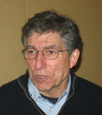
Syndrome induced by adjuvants may be precursor to autoimmune disease
DESTIN, FLA. – Various environmental factors are known to play a role in immune-mediated disease, and the extent to which certain adjuvants contribute to both defined and nondefined immune-mediated diseases warrants a new classification for such diseases, according to Dr. Yehuda Shoenfeld.
He proposes that these adjuvant-induced conditions be classified under a common syndrome entitled Autoimmune Syndrome Induced by Adjuvants (ASIA).

In a 2011 article in the Journal of Autoimmunity, he and coauthor, Dr. Nancy Agmon-Levin of B’Sheba Medical Center, Tel-Hashomer, Israel, reviewed the data demonstrating links between adjuvants and autoimmune disease (J. Autoimmun. 2011;36:4-8).
"ASIA is not necessarily a full-blown, fully characterized autoimmune disease that we are aware of like systemic lupus erythematosus, systemic sclerosis, or rheumatoid arthritis. However, it might be that this syndrome will evolve over the years into a full-blown autoimmune disease," Dr. Shoenfeld of Tel Aviv University, Israel, said at the annual Congress of Clinical Rheumatology.
ASIA is a "special complex of clinical manifestations," he said, noting that chronic fatigue is a particularly important manifestation, as are numerous nonspecific symptoms.
Incubation time after exposure to the offending adjuvant may range from 3 weeks to years and may be associated with chronic stimulation of the immune system by the adjuvants. Multiple vaccines or silicone leakage or rupture – from breast implants, for example – represent such chronic exposure.
As with all autoimmune diseases, there is likely a genetic component that factors into the development of ASIA, he said.
"One of the most important adjuvants is aluminum ... and there is no question that there is involvement of the central nervous system," he said, noting that many affected patients thus experience memory loss and cognitive impairment.
Adjuvants were formerly thought to pose little or no independent threat, but animal models, and some human studies, have suggested otherwise, he said.
In his article and during his presentation, Dr. Shoenfeld reviewed the data on the role of infections, silicone, and aluminum salts commonly found in vaccines as they relate to the development of autoimmunity.
Activation of the autoimmune mechanisms by infectious agents is common, he explained in the article.
"Yet the appearance of an autoimmune disease is not as widespread and apparently not always agent specific. The adjuvant effect of microbial particles, namely the nonantigenic activation of the innate and regulatory immunity as well as the expression of various regulatory cytokines, may determine if an autoimmune response remains limited and harmless or evolves into a full blown disease," he continued.
As for silicone, he discussed multiple studies demonstrating various links with autoimmune disease. In one study, 100 women with silicone breast implants all reported symptoms after about 6 years, including weakness, fatigability, myalgia, morning stiffness, arthralgia, memory loss, sensory loss, headache, and dry mouth.
Abnormal levels of serum immunoglobulin or complement were found in 57% of patients and antibodies were found in 78%.
Other studies have suggested a link between silicone breast implants and lymphoma, polyarthritis, and connective tissue disease. Furthermore, numerous reports have shown that explantation is associated with symptom improvement, he noted.
As for vaccines, Dr. Shoenfeld stressed that he is not "antivaccine," and in his article, he described vaccines as "one of the greatest achievements of modern medicine," but he is concerned about the links between certain vaccines and ASIA.
He recently published a report on three cases of primary ovarian failure in association with the quadrivalent human papillomavirus vaccine (Am. J. Reprod. Immunol. 2013 [doi:10.1111/aji.12151]), and he noted that other studies have suggested an association between the vaccine and SLE, various hypersensitivity reactions, and demyelination syndromes, for example.
"My aim is to draw your attention to the association," he explained.
Reports of "an avalanche" of cases of narcolepsy in Finland following vaccination with the adjuvanted AH1N1 vaccine have also raised concerns – and raised the possibility that narcolepsy is an autoimmune disease, he said, noting that multiple studies have explored and provided support for this possibility.
Dr. Shoenfeld proposes that a diagnosis of ASIA should be based on the presence of either two major criteria or one major and two minor criteria.
Major criteria as defined in his article are:
• Exposure to an external stimulus prior to clinical manifestations.
• The appearance of typical clinical manifestations, including myalgia, myositis, or muscle weakness; arthralgia and/or arthritis; chronic fatigue, unrefreshing sleep or sleep disturbance; neurologic manifestations, cognitive impairment/memory loss; pyrexia and dry mouth.
• Removal of inciting agent induces improvement.
• Typical biopsy of involved organs.
Minor criteria are:
• The appearance of autoantibodies or antibodies directed at the suspected adjuvants.
• Other clinical manifestations such as irritable bowel syndrome.
• Specific human leukocyte antigen (HLA).
• Evolvement of an autoimmune disease.
These criteria are highly specific and sensitive, but could change based on further analysis, he said, noting that the "amassed data" regarding conditions that can be considered part of ASIA "may enable us a spacious view of the immune responses to environmental adjuvants, as well as better definition and diagnosis of these conditions. Moreover, unraveling the pathogenesis of this newly defined syndrome may facilitate the search for preventive and therapeutic inventions," he wrote.
Dr. Shoenfeld reported having no disclosures.
DESTIN, FLA. – Various environmental factors are known to play a role in immune-mediated disease, and the extent to which certain adjuvants contribute to both defined and nondefined immune-mediated diseases warrants a new classification for such diseases, according to Dr. Yehuda Shoenfeld.
He proposes that these adjuvant-induced conditions be classified under a common syndrome entitled Autoimmune Syndrome Induced by Adjuvants (ASIA).
In a 2011 article in the Journal of Autoimmunity, he and coauthor, Dr. Nancy Agmon-Levin of B’Sheba Medical Center, Tel-Hashomer, Israel, reviewed the data demonstrating links between adjuvants and autoimmune disease (J. Autoimmun. 2011;36:4-8).
"ASIA is not necessarily a full-blown, fully characterized autoimmune disease that we are aware of like systemic lupus erythematosus, systemic sclerosis, or rheumatoid arthritis. However, it might be that this syndrome will evolve over the years into a full-blown autoimmune disease," Dr. Shoenfeld of Tel Aviv University, Israel, said at the annual Congress of Clinical Rheumatology.
ASIA is a "special complex of clinical manifestations," he said, noting that chronic fatigue is a particularly important manifestation, as are numerous nonspecific symptoms.
Incubation time after exposure to the offending adjuvant may range from 3 weeks to years and may be associated with chronic stimulation of the immune system by the adjuvants. Multiple vaccines or silicone leakage or rupture – from breast implants, for example – represent such chronic exposure.
As with all autoimmune diseases, there is likely a genetic component that factors into the development of ASIA, he said.
"One of the most important adjuvants is aluminum ... and there is no question that there is involvement of the central nervous system," he said, noting that many affected patients thus experience memory loss and cognitive impairment.
Adjuvants were formerly thought to pose little or no independent threat, but animal models, and some human studies, have suggested otherwise, he said.
In his article and during his presentation, Dr. Shoenfeld reviewed the data on the role of infections, silicone, and aluminum salts commonly found in vaccines as they relate to the development of autoimmunity.
Activation of the autoimmune mechanisms by infectious agents is common, he explained in the article.
"Yet the appearance of an autoimmune disease is not as widespread and apparently not always agent specific. The adjuvant effect of microbial particles, namely the nonantigenic activation of the innate and regulatory immunity as well as the expression of various regulatory cytokines, may determine if an autoimmune response remains limited and harmless or evolves into a full blown disease," he continued.
As for silicone, he discussed multiple studies demonstrating various links with autoimmune disease. In one study, 100 women with silicone breast implants all reported symptoms after about 6 years, including weakness, fatigability, myalgia, morning stiffness, arthralgia, memory loss, sensory loss, headache, and dry mouth.
Abnormal levels of serum immunoglobulin or complement were found in 57% of patients and antibodies were found in 78%.
Other studies have suggested a link between silicone breast implants and lymphoma, polyarthritis, and connective tissue disease. Furthermore, numerous reports have shown that explantation is associated with symptom improvement, he noted.
As for vaccines, Dr. Shoenfeld stressed that he is not "antivaccine," and in his article, he described vaccines as "one of the greatest achievements of modern medicine," but he is concerned about the links between certain vaccines and ASIA.
He recently published a report on three cases of primary ovarian failure in association with the quadrivalent human papillomavirus vaccine (Am. J. Reprod. Immunol. 2013 [doi:10.1111/aji.12151]), and he noted that other studies have suggested an association between the vaccine and SLE, various hypersensitivity reactions, and demyelination syndromes, for example.
"My aim is to draw your attention to the association," he explained.
Reports of "an avalanche" of cases of narcolepsy in Finland following vaccination with the adjuvanted AH1N1 vaccine have also raised concerns – and raised the possibility that narcolepsy is an autoimmune disease, he said, noting that multiple studies have explored and provided support for this possibility.
Dr. Shoenfeld proposes that a diagnosis of ASIA should be based on the presence of either two major criteria or one major and two minor criteria.
Major criteria as defined in his article are:
• Exposure to an external stimulus prior to clinical manifestations.
• The appearance of typical clinical manifestations, including myalgia, myositis, or muscle weakness; arthralgia and/or arthritis; chronic fatigue, unrefreshing sleep or sleep disturbance; neurologic manifestations, cognitive impairment/memory loss; pyrexia and dry mouth.
• Removal of inciting agent induces improvement.
• Typical biopsy of involved organs.
Minor criteria are:
• The appearance of autoantibodies or antibodies directed at the suspected adjuvants.
• Other clinical manifestations such as irritable bowel syndrome.
• Specific human leukocyte antigen (HLA).
• Evolvement of an autoimmune disease.
These criteria are highly specific and sensitive, but could change based on further analysis, he said, noting that the "amassed data" regarding conditions that can be considered part of ASIA "may enable us a spacious view of the immune responses to environmental adjuvants, as well as better definition and diagnosis of these conditions. Moreover, unraveling the pathogenesis of this newly defined syndrome may facilitate the search for preventive and therapeutic inventions," he wrote.
Dr. Shoenfeld reported having no disclosures.
EXPERT OPINION FROM CCR 14

Malar rash flagged by crusty lips, nasolabial sparing
DESTIN, FLA. – A number of conditions are often misdiagnosed as malar rash, but certain clues can help in making the correct diagnosis, according to Dr. Ruth Ann Vleugels.
"One of the most helpful [clues] is this really prominent nasolabial sparing," Dr. Vleugels, assistant professor of dermatology at Harvard Medical School, and director of the autoimmune skin disease program at Brigham and Women’s Hospital, both in Boston, said at the annual Congress of Clinical Rheumatology.

Prominent hemorrhagic crusting on the lips in conjunction with the rash is another telltale sign that the condition is a malar rash, she said.
Often, other skin conditions such as erythematotelangiectatic rosacea are diagnosed as malar rash. Erythematotelangiectatic rosacea is common, affecting about a third of all patients with light skin, and many patients with the condition report having a photosensitive rash on the cheeks.
"So, already you have two criteria for systemic lupus," she said, stressing that self-reported malar erythema is "honestly, not a great criteria" for a malar rash diagnosis.
These patients will have transient flushing on the face or may have papulopustular rosacea.
The nasolabial sparing is thus a "really critical clue to look for," she noted.
Also, a malar rash is not going to go away after a few hours like the flush of erythematotelangiectatic rosacea, but will last for several days, at least.
The presence of alopecia also can help distinguish malar rash from other conditions, as it is a common feature in SLE (systemic lupus erythematosus), Dr. Vleugels noted.
Perhaps the most important differential diagnosis in the dermatology-rheumatology clinic is chronic mid-facial erythema, she said.
When such erythema "hugs or involves the nasolabial fold," think dermatomyositis, Dr. Vleugels advised.
She presented a case in which a patient had a subtle heliotrope along with this type of chronic mid-facial erythema. The patient had been referred for lupus but had amyopathic dermatomyositis, and the subtle edema and erythema of the upper eyelids provided a clue that led to a diagnosis of gastric cancer in the patient.
"When it’s subtle, it’s often missed," she said of the heliotrope a violaceous to dusky erythematous rash, with or without edema, which occurs in a symmetric distribution involving periorbital skin.
Another clue that a rash may indeed be malar rash is the presence of nonblanching erythematous macules. Dr. Vleugels described a patient with this presentation on her chest, in addition to other signs of malar rash, including the nasolabial sparing and hemorrhagic crusting on the lips.
"This is essentially petechiae. This is a sign on her skin of active systemic involvement," she said of the rash on the patient’s chest.
Patients with true malar rash by definition have active SLE, she said.
"So we don’t let them leave even the dermatology clinic without checking their kidneys, blood pressure, renal function, etc.," she said.
Dr. Vleugels also noted that there is a generalized form of acute cutaneous lupus to keep in mind.
"Unfortunately, this is very difficult to distinguish clinically from the morbilliform drug eruption or a viral exanthema. It essentially blanches pink to red," she said, adding that macules and papules may be present.
A high level of suspicion for this condition is needed, even though it is quite rare. A biopsy can confirm the diagnosis, as classic changes associated with lupus will be present on biopsy, she said.
Dr. Vleugels reported having no disclosures.
DESTIN, FLA. – A number of conditions are often misdiagnosed as malar rash, but certain clues can help in making the correct diagnosis, according to Dr. Ruth Ann Vleugels.
"One of the most helpful [clues] is this really prominent nasolabial sparing," Dr. Vleugels, assistant professor of dermatology at Harvard Medical School, and director of the autoimmune skin disease program at Brigham and Women’s Hospital, both in Boston, said at the annual Congress of Clinical Rheumatology.
Prominent hemorrhagic crusting on the lips in conjunction with the rash is another telltale sign that the condition is a malar rash, she said.
Often, other skin conditions such as erythematotelangiectatic rosacea are diagnosed as malar rash. Erythematotelangiectatic rosacea is common, affecting about a third of all patients with light skin, and many patients with the condition report having a photosensitive rash on the cheeks.
"So, already you have two criteria for systemic lupus," she said, stressing that self-reported malar erythema is "honestly, not a great criteria" for a malar rash diagnosis.
These patients will have transient flushing on the face or may have papulopustular rosacea.
The nasolabial sparing is thus a "really critical clue to look for," she noted.
Also, a malar rash is not going to go away after a few hours like the flush of erythematotelangiectatic rosacea, but will last for several days, at least.
The presence of alopecia also can help distinguish malar rash from other conditions, as it is a common feature in SLE (systemic lupus erythematosus), Dr. Vleugels noted.
Perhaps the most important differential diagnosis in the dermatology-rheumatology clinic is chronic mid-facial erythema, she said.
When such erythema "hugs or involves the nasolabial fold," think dermatomyositis, Dr. Vleugels advised.
She presented a case in which a patient had a subtle heliotrope along with this type of chronic mid-facial erythema. The patient had been referred for lupus but had amyopathic dermatomyositis, and the subtle edema and erythema of the upper eyelids provided a clue that led to a diagnosis of gastric cancer in the patient.
"When it’s subtle, it’s often missed," she said of the heliotrope a violaceous to dusky erythematous rash, with or without edema, which occurs in a symmetric distribution involving periorbital skin.
Another clue that a rash may indeed be malar rash is the presence of nonblanching erythematous macules. Dr. Vleugels described a patient with this presentation on her chest, in addition to other signs of malar rash, including the nasolabial sparing and hemorrhagic crusting on the lips.
"This is essentially petechiae. This is a sign on her skin of active systemic involvement," she said of the rash on the patient’s chest.
Patients with true malar rash by definition have active SLE, she said.
"So we don’t let them leave even the dermatology clinic without checking their kidneys, blood pressure, renal function, etc.," she said.
Dr. Vleugels also noted that there is a generalized form of acute cutaneous lupus to keep in mind.
"Unfortunately, this is very difficult to distinguish clinically from the morbilliform drug eruption or a viral exanthema. It essentially blanches pink to red," she said, adding that macules and papules may be present.
A high level of suspicion for this condition is needed, even though it is quite rare. A biopsy can confirm the diagnosis, as classic changes associated with lupus will be present on biopsy, she said.
Dr. Vleugels reported having no disclosures.
EXPERT ANALYSIS FROM CCR 14

Don’t miss these features of dermatomyositis
DESTIN, FLA. – Dermatomyositis doesn’t always present with the violaceous upper eyelid erythema and associated edema seen in textbook cases.
Rather, it often presents without muscle disease and with pink eyelid erythema, mid-facial erythema that hugs the nasolabial folds and extends onto the forehead, and subtle scaly pink plaques on the knuckles, Dr. Ruth Ann Vleugels said at the annual Congress of Clinical Rheumatology.
Dr. Vleugels described an unfortunate case involving a patient with this type of amyopathic presentation who presented to the emergency department with shortness of breath and what was thought to be a pulmonary infection. The patient was admitted to the intensive care unit, and dermatology was not consulted until the third day.
Unfortunately, the patient died in the ICU, said Dr. Vleugels of the department of dermatology at Harvard Medical School and director of the Autoimmune Skin Diseases Program at Brigham & Women’s Hospital, Boston.

The patient had skin-limited dermatomyositis; his family reported that his rash had been present for more than a year, she said.
Even Gottron’s papules, which are pathognomonic for dermatomyositis, don’t always present as pink to violaceous papules on the knuckles as expected. In some cases they can be very subtle – especially in darker skin types – and sometimes there is involvement between the knuckles as well, Dr. Vleugels said.
She described another patient who had been misdiagnosed with flat warts on the back of her hands before being diagnosed with dermatomyositis. In some cases, the papules are very scaly and can be mistaken for psoriasis, she noted.
"We have to recognize these clinical features," she said.
One helpful tip: Examine the nail folds. Nearly all dermatomyositis patients with active disease "have nice-looking nail folds," explained Dr. Vleugels.
"So the feature we will be looking for is cuticular hypertrophy," she said, noting that women tend to be acutely aware of this symptom and may want to push their cuticles back.
Dilated capillary loops, dropout capillaries, and hemosiderin deposition of the nail fold are also common.
"Really look at those nail folds. Lupus patients do not have nail folds like this," Dr. Vleugels emphasized.
Another clue to diagnosing dermatomyositis is poikiloderma in sun-exposed areas, characterized by redness, dispigmentation, and atrophy or thinning of the skin.
Poikiloderma becomes permanent, which is one reason why photoprotection is critical in these patients, said Dr. Vleugels. It can also involve the scalp (sometimes in addition to scaling), and in fact is "probably an underrecognized feature in our dermatomyositis patients," she said.
The condition is very pruritic and detrimental to quality of life, and hair loss can occur in association with the disease activity on the scalp.
"We treat this as a component of the rash, so if we don’t get their scalp better we increase or change their immunosuppressive therapy," Dr. Vleugels noted.
Keep in mind that poikiloderma can also occur on the lateral thighs (the "holster sign"), even though this is not typically a sun-exposed area, she added.
A recent study found that the MDA5 autoantibody, in particular, is associated with certain cutaneous findings in dermatomyositis. The most common was ulcerations in Gottron’s papules, and this is representative of vasculopathy (J. Am. Acad. Dermatol. 2011; 25-34).
Such ulcerations are not a sign of infection, but a sign that the disease is active and required more aggressive treatment.
Calcinosis is another concern, and occurs more often in children than adults.
However, "good data from the pediatric literature say if we treat these patients early and aggressively, they get less calcinosis," Dr. Vleugels said.
In an adult who is developing calcinosis, consider changing their overall treatment regimen, as this is a sign of active disease, she added.
"Mechanic’s hands" can be another sign of dermatomyositis, particularly if there is scaling and fissures on the lateral and palmar aspects of the fingers (as opposed to scaly Gottron’s papules elsewhere on the hands); this cutaneous phenotype is associated with high risk of antisynthetase syndrome or lung involvement, she said.
Recognizing these signs and symptoms of dermatomyositis can help prevent outcomes such as the death of the patient in the ICU after presenting with unrecognized fulminant lung disease associated with dermatomyositis, Dr. Vleugels emphasized.
"Our skin-limited patients are the ones who are often missed, and these are the ones who have approximately equal risk of lung disease and malignancy," she said.
Data suggest these conditions occur in about 25% of patients with skin-limited dermatomyositis
"So even the patients with amyopathic disease need to have regular screening with pulmonary function tests with diffusion capacity," she said.
Data from the Mayo Clinic, Rochester, Minn., suggest that about 20% of dermatomyositis patients have clinically amyopathic disease, and most (76%) of these cases are in women (Arch. Dermatol. 2010;146:26-30).
"The take-home point here is that these skin-limited patients are much harder to diagnose than the ones who come in with the classic muscle weakness, because the rash can be subtle," Dr. Vleugels said, noting that the diagnosis must be made clinically, because there is no confirmatory test.
She described a dermatomyositis patient who had experienced a rash for more than 2 years, and who was referred for lupus – even a biopsy was read as lupus.
"We need to say, ‘Oh, that could also be dermatomyositis,’" she said, adding: "If you see a patient with skin-limited disease, you’re still going to screen them for pulmonary disease, and you’re still going to screen them for cancer."
DESTIN, FLA. – Dermatomyositis doesn’t always present with the violaceous upper eyelid erythema and associated edema seen in textbook cases.
Rather, it often presents without muscle disease and with pink eyelid erythema, mid-facial erythema that hugs the nasolabial folds and extends onto the forehead, and subtle scaly pink plaques on the knuckles, Dr. Ruth Ann Vleugels said at the annual Congress of Clinical Rheumatology.
Dr. Vleugels described an unfortunate case involving a patient with this type of amyopathic presentation who presented to the emergency department with shortness of breath and what was thought to be a pulmonary infection. The patient was admitted to the intensive care unit, and dermatology was not consulted until the third day.
Unfortunately, the patient died in the ICU, said Dr. Vleugels of the department of dermatology at Harvard Medical School and director of the Autoimmune Skin Diseases Program at Brigham & Women’s Hospital, Boston.
The patient had skin-limited dermatomyositis; his family reported that his rash had been present for more than a year, she said.
Even Gottron’s papules, which are pathognomonic for dermatomyositis, don’t always present as pink to violaceous papules on the knuckles as expected. In some cases they can be very subtle – especially in darker skin types – and sometimes there is involvement between the knuckles as well, Dr. Vleugels said.
She described another patient who had been misdiagnosed with flat warts on the back of her hands before being diagnosed with dermatomyositis. In some cases, the papules are very scaly and can be mistaken for psoriasis, she noted.
"We have to recognize these clinical features," she said.
One helpful tip: Examine the nail folds. Nearly all dermatomyositis patients with active disease "have nice-looking nail folds," explained Dr. Vleugels.
"So the feature we will be looking for is cuticular hypertrophy," she said, noting that women tend to be acutely aware of this symptom and may want to push their cuticles back.
Dilated capillary loops, dropout capillaries, and hemosiderin deposition of the nail fold are also common.
"Really look at those nail folds. Lupus patients do not have nail folds like this," Dr. Vleugels emphasized.
Another clue to diagnosing dermatomyositis is poikiloderma in sun-exposed areas, characterized by redness, dispigmentation, and atrophy or thinning of the skin.
Poikiloderma becomes permanent, which is one reason why photoprotection is critical in these patients, said Dr. Vleugels. It can also involve the scalp (sometimes in addition to scaling), and in fact is "probably an underrecognized feature in our dermatomyositis patients," she said.
The condition is very pruritic and detrimental to quality of life, and hair loss can occur in association with the disease activity on the scalp.
"We treat this as a component of the rash, so if we don’t get their scalp better we increase or change their immunosuppressive therapy," Dr. Vleugels noted.
Keep in mind that poikiloderma can also occur on the lateral thighs (the "holster sign"), even though this is not typically a sun-exposed area, she added.
A recent study found that the MDA5 autoantibody, in particular, is associated with certain cutaneous findings in dermatomyositis. The most common was ulcerations in Gottron’s papules, and this is representative of vasculopathy (J. Am. Acad. Dermatol. 2011; 25-34).
Such ulcerations are not a sign of infection, but a sign that the disease is active and required more aggressive treatment.
Calcinosis is another concern, and occurs more often in children than adults.
However, "good data from the pediatric literature say if we treat these patients early and aggressively, they get less calcinosis," Dr. Vleugels said.
In an adult who is developing calcinosis, consider changing their overall treatment regimen, as this is a sign of active disease, she added.
"Mechanic’s hands" can be another sign of dermatomyositis, particularly if there is scaling and fissures on the lateral and palmar aspects of the fingers (as opposed to scaly Gottron’s papules elsewhere on the hands); this cutaneous phenotype is associated with high risk of antisynthetase syndrome or lung involvement, she said.
Recognizing these signs and symptoms of dermatomyositis can help prevent outcomes such as the death of the patient in the ICU after presenting with unrecognized fulminant lung disease associated with dermatomyositis, Dr. Vleugels emphasized.
"Our skin-limited patients are the ones who are often missed, and these are the ones who have approximately equal risk of lung disease and malignancy," she said.
Data suggest these conditions occur in about 25% of patients with skin-limited dermatomyositis
"So even the patients with amyopathic disease need to have regular screening with pulmonary function tests with diffusion capacity," she said.
Data from the Mayo Clinic, Rochester, Minn., suggest that about 20% of dermatomyositis patients have clinically amyopathic disease, and most (76%) of these cases are in women (Arch. Dermatol. 2010;146:26-30).
"The take-home point here is that these skin-limited patients are much harder to diagnose than the ones who come in with the classic muscle weakness, because the rash can be subtle," Dr. Vleugels said, noting that the diagnosis must be made clinically, because there is no confirmatory test.
She described a dermatomyositis patient who had experienced a rash for more than 2 years, and who was referred for lupus – even a biopsy was read as lupus.
"We need to say, ‘Oh, that could also be dermatomyositis,’" she said, adding: "If you see a patient with skin-limited disease, you’re still going to screen them for pulmonary disease, and you’re still going to screen them for cancer."
AT CCR 14

Recognize and treat discoid lupus early to prevent scarring, expert says
DESTIN, FLA. – Recognizing and treating discoid lupus early is imperative for preventing permanent scarring, according to Dr. Ruth Ann Vleugels.
When a patient presents with a "textbook case" of discoid lupus with the classic hyperpigmentation around the border with central atrophy and scarring that is characteristic of unchecked disease, it’s too late, she said at the annual Congress of Clinical Rheumatology.

"When I see a case like this, I unfortunately can do nothing for them. There is no cream I can put on this, there is no laser I can give them ... all of this is permanent damage and scarring," she said, noting that even hair transplants in those with scalp scarring won’t work, because of the scar tissue.
For this reason, it is important to look for signs and symptoms – typically erythema and/or scaling, said Dr. Vleugels of Harvard Medical School, Boston.
Remember that erythema can look very different in different skin types, she advised, explaining that in light skin, the erythema will be red and obvious, but in darker skin it often looks violaceous and can be subtle.
"So we really, in our cutaneous lupus patients, need to pick up that activity in our darker skin patients to prevent scarring," added Dr. Vleugels, director of the autoimmune skin disease program at Brigham and Women’s Hospital, Boston.
Scaling is also an important feature that can help in diagnosing discoid lupus, and it may present with or without erythema.
Dr. Vleugels described one patient with scarring who clearly had existing disease, but who also had plaques with some erythema and scaling at the borders. While it was too late to do anything about the existing scars, the erythema and scaling represented disease activity that would lead to additional scarring without rapid treatment.
On the dorsal hands, discoid lupus tends to spare the knuckles, and on the scalp, the extensive scaling associated with discoid lupus can look like tinea capitis. Conchal bowl involvement is common, as is follicular plugging with excess keratin.
Another patient – a 19-year-old male with severe systemic lupus erythematosus who had been treated with cyclophosphamide and rituximab for renal disease, had impressive skin involvement, and at first glance, his skin disease may have appeared "a lost cause" under the circumstances. However, faint erythema was present throughout his scalp, and with aggressive treatment it was possible to bring back a lot of his hair.
"So in addition to maximizing the systemic regimen, something we do right away in these patients is give intralesional steroid injections to the scalp," Dr. Vleugels said, noting that the injections are very simple and easily learned.
She said she uses 10 mg/cc of intralesional triamcinolone diluted to 5 mg/cc with normal saline for scalp disease (and to a more cautious 3 mg/cc for facial disease), giving 0.1 cc per injection to avoid atrophy. The injections are given in the dermis, about 1 cm apart. The procedure can be completed in about 3 minutes in the office setting, and can be repeated every 4-5 weeks as needed.
Dr. Vleugels noted that she often gets referrals involving alopecia areata that has been confused with discoid lupus.
"Any patient with a circular area of hair loss, erythema, dyspigmentation, or scaling has discoid lupus," she said.
Both conditions can be treated with the intralesional injections, but the patient with signs and symptoms of discoid lupus requires a work-up for cutaneous lupus.
Scalp psoriasis can also be mistaken as discoid lupus because of the prominent scaling, but other characteristics of discoid lupus can help in making the diagnosis, she said, describing one such patient who also had the erythema and conchal bowl involvement that helped distinguish the two conditions.
Knowing the signs and symptoms of discoid lupus is important, because recent research suggests that a higher percentage of patients with cutaneous disease will go on to develop systemic disease than was previously believed, Dr. Vleugels said.
It was long held and taught that about 5%-10% of adult patients would go on to develop systemic disease, but 18% of more than 1,000 patients in a Swedish epidemiologic study who were followed for 3 years developed systemic disease (Br. J. Dermatol. 2011;164:1335-41), and 20% of those in a Mayo Clinic study who were followed for a mean of 8 years developed systemic disease.
"So this changes how we think about skin-only patients," she said, explaining that she follows adult patients a little longer than she used to, and screens them annually for systemic involvement.
Children, however, are more likely to develop systemic disease. "About half will develop systemic involvement, so we comanage them with rheumatology," she said.
Dr. Vleugels reported having no disclosures.
DESTIN, FLA. – Recognizing and treating discoid lupus early is imperative for preventing permanent scarring, according to Dr. Ruth Ann Vleugels.
When a patient presents with a "textbook case" of discoid lupus with the classic hyperpigmentation around the border with central atrophy and scarring that is characteristic of unchecked disease, it’s too late, she said at the annual Congress of Clinical Rheumatology.
"When I see a case like this, I unfortunately can do nothing for them. There is no cream I can put on this, there is no laser I can give them ... all of this is permanent damage and scarring," she said, noting that even hair transplants in those with scalp scarring won’t work, because of the scar tissue.
For this reason, it is important to look for signs and symptoms – typically erythema and/or scaling, said Dr. Vleugels of Harvard Medical School, Boston.
Remember that erythema can look very different in different skin types, she advised, explaining that in light skin, the erythema will be red and obvious, but in darker skin it often looks violaceous and can be subtle.
"So we really, in our cutaneous lupus patients, need to pick up that activity in our darker skin patients to prevent scarring," added Dr. Vleugels, director of the autoimmune skin disease program at Brigham and Women’s Hospital, Boston.
Scaling is also an important feature that can help in diagnosing discoid lupus, and it may present with or without erythema.
Dr. Vleugels described one patient with scarring who clearly had existing disease, but who also had plaques with some erythema and scaling at the borders. While it was too late to do anything about the existing scars, the erythema and scaling represented disease activity that would lead to additional scarring without rapid treatment.
On the dorsal hands, discoid lupus tends to spare the knuckles, and on the scalp, the extensive scaling associated with discoid lupus can look like tinea capitis. Conchal bowl involvement is common, as is follicular plugging with excess keratin.
Another patient – a 19-year-old male with severe systemic lupus erythematosus who had been treated with cyclophosphamide and rituximab for renal disease, had impressive skin involvement, and at first glance, his skin disease may have appeared "a lost cause" under the circumstances. However, faint erythema was present throughout his scalp, and with aggressive treatment it was possible to bring back a lot of his hair.
"So in addition to maximizing the systemic regimen, something we do right away in these patients is give intralesional steroid injections to the scalp," Dr. Vleugels said, noting that the injections are very simple and easily learned.
She said she uses 10 mg/cc of intralesional triamcinolone diluted to 5 mg/cc with normal saline for scalp disease (and to a more cautious 3 mg/cc for facial disease), giving 0.1 cc per injection to avoid atrophy. The injections are given in the dermis, about 1 cm apart. The procedure can be completed in about 3 minutes in the office setting, and can be repeated every 4-5 weeks as needed.
Dr. Vleugels noted that she often gets referrals involving alopecia areata that has been confused with discoid lupus.
"Any patient with a circular area of hair loss, erythema, dyspigmentation, or scaling has discoid lupus," she said.
Both conditions can be treated with the intralesional injections, but the patient with signs and symptoms of discoid lupus requires a work-up for cutaneous lupus.
Scalp psoriasis can also be mistaken as discoid lupus because of the prominent scaling, but other characteristics of discoid lupus can help in making the diagnosis, she said, describing one such patient who also had the erythema and conchal bowl involvement that helped distinguish the two conditions.
Knowing the signs and symptoms of discoid lupus is important, because recent research suggests that a higher percentage of patients with cutaneous disease will go on to develop systemic disease than was previously believed, Dr. Vleugels said.
It was long held and taught that about 5%-10% of adult patients would go on to develop systemic disease, but 18% of more than 1,000 patients in a Swedish epidemiologic study who were followed for 3 years developed systemic disease (Br. J. Dermatol. 2011;164:1335-41), and 20% of those in a Mayo Clinic study who were followed for a mean of 8 years developed systemic disease.
"So this changes how we think about skin-only patients," she said, explaining that she follows adult patients a little longer than she used to, and screens them annually for systemic involvement.
Children, however, are more likely to develop systemic disease. "About half will develop systemic involvement, so we comanage them with rheumatology," she said.
Dr. Vleugels reported having no disclosures.
AT CCR 14
