User login
Investigational Cushing's Drug Eases Symptoms, Ups Hyperglycemia
BOSTON – In the largest clinical trial ever conducted in Cushing’s disease, pasireotide, an investigational somatostatin analogue, was shown to result in a rapid and sustained reduction in urinary free cortisol in the majority of patients, and significantly improved signs and symptoms of the disease.
One troubling finding was that three-quarters of patients experienced at least one hyperglycemic episode, a side effect not found with other somatostatin analogues, according to Dr. Annamaria Colao, who presented the findings at the annual meeting of the Endocrine Society.
Pasireotide presents a new therapeutic possibility for Cushing’s disease, since there is no medical treatment currently approved for the condition, said Dr. Colao, professor of endocrinology and chief of the neuroendocrine unit at the University of Naples (Italy) "Federico II." In Cushing’s disease, a benign pituitary tumor stimulates the adrenal glands to synthesize excess cortisol, leading to weight gain, a moon-shaped face, severe fatigue and weakness, hypertension, and hyperglycemia. Pasireotide is also being studied in patients with carcinoid syndrome, acromegaly, and neuroendocrine tumors, and is not approved for use anywhere in the world.
In this double-blind, phase III study known as the PASPORT (Pasireotide Clinical Trial Portfolio) trial, 162 patients who had confirmed persistent, recurrent, or de novo Cushing’s disease and who were not considered to be surgical candidates were enrolled. Patients came from 18 countries and 62 clinical sites. Inclusion criteria included baseline mean urinary free cortisol (UFC) levels falling between 1.5 and 5 times the upper limit of normal (ULN, defined as 145 nmol/24 hours) and confirmation of pituitary adenoma. Any patient with poorly controlled diabetes was excluded from the trial.
Patients were randomized to receive either pasireotide 600 mcg (n = 82) or 900 mcg (n = 80) subcutaneously twice a day for 3 months. After 3 months, patients with UFC greater than twice the ULN or with UFC levels greater than baseline were unblinded, and the dose was increased by 300 mcg b.i.d. All others remained on the same dosage regimen until 6 months. Between 6 and 12 months, the trial was open label, with dose titration adjusted as needed. The primary end point was UFC levels below or at the ULN at 6 months without dose up-titration from the randomized dose.
Pooled results from both groups showed that within 2 months, there was a 48% mean reduction from baseline in UFC levels, and a drop in UFC levels was seen for a majority of patients. After 6 months of treatment, 15% of those treated with 600 mcg pasireotide and 26% of those treated with 900 mcg reached the primary end point. The effect was sustained, with 13% of the 600-mcg group and 25% of the 900-mcg group meeting the primary end point at 12 months.
"Notice how quickly the effect happens: Most of the decrease is seen within the first month of treatment," noted Dr. Colao. Similar trends were seen for serum and salivary cortisol as well as plasma adrenocorticotropic hormone.
Pasireotide treatment also appeared to normalize UFC levels in a subset of patients, especially those with low levels of elevated baseline UFC.
Because most patients responded quickly to treatment, lack of response within the first weeks of treatment can signal those who will be nonresponders. "Within 1-2 months, patients who will not have an adequate biochemical response can be identified with a high degree of accuracy. This might prove clinically useful," says Dr. Colao.
As UFC levels decreased, significant improvements in signs and symptoms of Cushing’s disease became apparent, including reductions in systolic and diastolic blood pressure, weight, and LDL cholesterol levels. Gains were also noted on a Cushing’s disease quality of life assessment, although Dr. Colao noted that interpretation of such findings was limited by lack of a placebo control group. Although Dr. Colao did not show the results of radiologic assessment of tumors, she said that an overall decrease in tumor size was seen after 12 months of pasireotide treatment.
Most adverse events were gastrointestinal in nature, including nausea (52%) and diarrhea (58%). "Adverse events were generally similar to those seen with other agents in this class of somatostatin analogues, with the exception of hyperglycemia," said Dr. Colao. She noted that 75% of patients treated with pasireotide had at least one hyperglycemic adverse event, which resolved by discontinuing the medication. Of 67 patients who were normoglycemic at baseline, 34% became diabetic during treatment, 43% became prediabetic, and 21% remained normal. Dr. Colao also noted that 8% of treated patients experienced hypocortisolism, a finding she felt would be expected with any effective treatment for Cushing’s disease.
Dr. Colao had nothing to disclose. The study was supported by Novartis Pharma AG.
Two posters at meeting shed light on pasireotide’s biochemical mechanisms of action. Dr. Rob van der Pas of Erasmus Medical Center in Rotterdam, the Netherlands, suggested that the reason the somatostatin analogue octreotide is not effective in Cushing’s disease is because it prefers the somatostatin receptor subtype 2 (sst2), whereas it is the somatostatin receptor 5 (sst5) that plays a key role in sst targeted therapy for Cushing’s disease (Endocr. Rev. 2011;32:P3-520). Dr. Robert R. Henry of the University of California, San Diego, found that the hyperglycemia associated with pasireotide is related to decreases in insulin secretion, with no change in insulin sensitivity, and this effect is most likely mediated by the sst5 receptor, which may also play an important role in glucose metabolism (Endocr. Rev. 2011;32:P3-274).
FROM THE ANNUAL MEETING OF THE ENDOCRINE SOCIETY
Major Finding: Pasireotide reduced urine free cortisol levels by an average of 48% in patients with Cushing’s disease; 73% experienced a hyperglycemic adverse event.
Data Source: A randomized, prospective study of 162 patients with Cushing’s disease.
Disclosures: Dr. Colao had nothing to disclose. The study was supported by Novartis Pharma AG.
Obesity, Estrogen Status Influence Breast Cancer Outcomes
BOSTON – Obesity increases the odds of a woman with breast cancer dying from her disease, according to a retrospective data analysis.
Further, this link between obesity and breast cancer death is strongest among those obese women who have estrogen receptor (ER) positive cancer, according to the analysis of data on nearly 4,000 participants in the California Teachers Study.
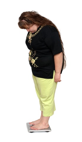
"This is another reason why the public should be aware of the importance of maintaining a healthy body weight, particularly how it pertains to cancer and especially breast cancer," according to Christina M. Dieli-Conwright Ph.D., an assistant research professor at City of Hope National Medical Center, Duarte, Calif. She presented the findings at the annual meeting of The Endocrine Society.
The study looked at a cohort of women who had taken a self-administered baseline questionnaire in 1995-1996 and who were diagnosed with their first primary invasive breast cancer between 1995 and 2006. Of the 3,995 women studied through 2007, 262 died of breast cancer and 321 died of nonbreast cancer causes.
There was a significant association between breast cancer mortality and body mass index (BMI) (P = .03). Women who were obese (defined as BMI greater than or equal to 30 kg/m2) had a 69% increased risk of dying of their breast cancer than women with a BMI less than 25. A similar increased risk in breast cancer mortality was seen in women who were overweight at age 18 (defined as BMI 25-29) compared to those with BMI less than 25. No significant associations were noted between weight and deaths due to nonbreast cancer causes or deaths due to all causes.
When the data were stratified according to estrogen receptor status, weight and breast cancer mortality were significantly related among women who were ER positive (P = 0.04), but not for those who were ER negative (P = .41). Obese women who were ER positive had a 64% increased risk of death due to breast cancer compared to those who had BMI less than 25. No significant associations were seen between weight and death due to all causes according to ER receptor status. Interestingly, there was more than a threefold increase in death from breast cancer in women who were ER negative if they were overweight at age 18 years.
Obesity at baseline almost doubled the breast cancer mortality risk (relative risk 1.91, 95% confidence interval 1.27-2.87) in women diagnosed with regional or distant disease, while there was no such association in obese women with localized disease. Higher risk of death from breast cancer and from all causes were also documented for women who had been overweight at age 18 for both localized and regional/distant disease.
Dr. Dieli-Conwright said that the findings add to a growing body of evidence linking obesity with the risk of developing breast cancer and with the risk of dying from it.
Dr. Dieli-Conwright reported having no conflicts of interest.
BOSTON – Obesity increases the odds of a woman with breast cancer dying from her disease, according to a retrospective data analysis.
Further, this link between obesity and breast cancer death is strongest among those obese women who have estrogen receptor (ER) positive cancer, according to the analysis of data on nearly 4,000 participants in the California Teachers Study.
"This is another reason why the public should be aware of the importance of maintaining a healthy body weight, particularly how it pertains to cancer and especially breast cancer," according to Christina M. Dieli-Conwright Ph.D., an assistant research professor at City of Hope National Medical Center, Duarte, Calif. She presented the findings at the annual meeting of The Endocrine Society.
The study looked at a cohort of women who had taken a self-administered baseline questionnaire in 1995-1996 and who were diagnosed with their first primary invasive breast cancer between 1995 and 2006. Of the 3,995 women studied through 2007, 262 died of breast cancer and 321 died of nonbreast cancer causes.
There was a significant association between breast cancer mortality and body mass index (BMI) (P = .03). Women who were obese (defined as BMI greater than or equal to 30 kg/m2) had a 69% increased risk of dying of their breast cancer than women with a BMI less than 25. A similar increased risk in breast cancer mortality was seen in women who were overweight at age 18 (defined as BMI 25-29) compared to those with BMI less than 25. No significant associations were noted between weight and deaths due to nonbreast cancer causes or deaths due to all causes.
When the data were stratified according to estrogen receptor status, weight and breast cancer mortality were significantly related among women who were ER positive (P = 0.04), but not for those who were ER negative (P = .41). Obese women who were ER positive had a 64% increased risk of death due to breast cancer compared to those who had BMI less than 25. No significant associations were seen between weight and death due to all causes according to ER receptor status. Interestingly, there was more than a threefold increase in death from breast cancer in women who were ER negative if they were overweight at age 18 years.
Obesity at baseline almost doubled the breast cancer mortality risk (relative risk 1.91, 95% confidence interval 1.27-2.87) in women diagnosed with regional or distant disease, while there was no such association in obese women with localized disease. Higher risk of death from breast cancer and from all causes were also documented for women who had been overweight at age 18 for both localized and regional/distant disease.
Dr. Dieli-Conwright said that the findings add to a growing body of evidence linking obesity with the risk of developing breast cancer and with the risk of dying from it.
Dr. Dieli-Conwright reported having no conflicts of interest.
FROM THE ANNUAL MEETING OF THE ENDOCRINE SOCIETY
Major Finding: The risk of death from breast cancer is increased by 69% in obese women, and the risk of death from breast cancer is also increased in women who are estrogen receptor positive.
Data Source: A retrospective analysis of 3,995 participants in the California Teachers Study who were diagnosed with their first primary invasive breast cancer.
Disclosures: Dr. Dieli-Conwright reported having no conflicts of interest.
Less Zoledronate May Prevent Osteoporotic Fractures
BOSTON – An annual dose of zoledronate that has been reduced by 50% or even 80% appears to be as effective as the recommended 5-mg dose in increasing bone mineral density and decreasing markers of bone turnover in osteopenic postmenopausal women, according to Dr. Andrew Grey, who presented the findings at the annual meeting of the Endocrine Society.
"We know that annual administration of intravenous zoledronate (also known as zoldedronic acid) at the 5-mg dose is an effective treatment for preventing fractures and decreasing fractures in osteoporotic, postmenopausal women by 25%-70%, depending on the skeletal site. Such treatment also reduces the risk of dying after hip fracture by 30%. Yet we don’t know the optimum dosing schedule," said Dr. Grey of the University of Auckland (New Zealand). The results of this study are important because reducing the dose can result in significant cost savings as well as decrease the risk of dose-dependent side effects.
In this prospective, controlled trial, 180 osteopenic postmenopausal women were randomized to receive a single dose of placebo or 1 mg, 2.5 mg, or 5 mg of zoledronate. The women had never been treated with zoledronate or any other bisphosphonate medication. The women underwent bone mineral density (BMD) evaluations at baseline and then 12 months after treatment.
For the primary end point (change in lumbar spine BMD at 12 months), the BMD increased significantly in each of the zoledronate groups, compared with placebo (P less than .001 for each dose). Notably, the magnitude of change was comparable for each dose: 3.5% for the 1-mg group, 4% for the 2.5-mg group, and 3.6% for the standard 5-mg group.
Similar results were observed for increases in total hip BMD (2.7% for the 1-mg group, 3.6% for the 2.5-mg group, and 3.6% for the 5-mg group). All results were significantly different from those with placebo (P less than .001).
Dr. Gray also looked at treatment effects on two blood markers of bone resorption, including beta-CTx (beta C-terminal telopeptide) and P1NP (procollagen type 1 N-terminal peptide). At all doses for both bone markers, zoledronate led to significant decreases, compared with placebo (P less than .001). For beta-CTx, the results were –44% for the 1-mg dose, –68% for the 2.5-mg dose and –73% for the 5-mg dose. For P1NP, the reductions were 41% for the 1-mg dose, 58% for the 2.5-mg dose, and 64% for the 5-mg dose.
An acute phase reaction is a side effect of intravenous bisphosphonate use. Fever is the most commonly associated symptom, but occasionally patients experience a flulike syndrome consisting of fever, chills, flushing, myalgias, and bone pain and/or arthralgias. In this study, acute phase reaction occurred less frequently in the zoledronate low-dose 1-mg group (odds ratio, 2.8) compared with either of the higher-dose groups (OR, 8.7 for the 2.5-mg group and 7.4 for the 5-mg group).
"These results show that lower doses than are currently prescribed have substantial antiresorptive effects that approximate those of the 5-mg dose, and suggest that these lower doses will be effective in preventing fractures in people with osteoporosis. Clinical trials should be undertaken to confirm whether annual low doses of zoledronate prevent fractures," according to Dr. Gray.
Dr. Grey had nothing to disclose; he said that the study was funded by public funds.
FROM THE ANNUAL MEETING OF THE ENDOCRINE SOCIETY
Major Finding: Low doses of zoledronate (1 mg or 2.5 mg) given as an annual infusion produce comparable changes on spine and hip bone mineral density and markers of bone resorption to those of the recommended higher dose (5 mg).
Data Source: A prospective, randomized, controlled trial of 180 postmenopausal women with osteopenia.
Disclosures: Dr. Grey had nothing to disclose; he said that the study was funded by public funds.
Many Hospitals Fail to Achieve Routine Inpatient Glycemic Control
BOSTON – Hyperglycemia was found in nearly one-third of patients at some time during their hospital stay, and another 6% of inpatients met criteria for hypoglycemia, an analysis of glucose data from the largest number of U.S. hospitals to date has shown.
In addition, the prevalence of hyperglycemia varied significantly according to hospital size, type, and region. These findings were detected in both intensive care unit and non-ICU patients, Dr. Christine Swanson said at the annual meeting of the Endocrine Society.
"This information is important for hospital administrators as well as providers of care, both hospitalists and endocrinologists, who need to know what blood sugars to target, why, and how their hospitals’ glucose control compares to other hospitals nationwide. Higher glucose levels can complicate hospital course, cause longer hospital stay, more complications, and slower recovery from surgeries or infections. Hypoglycemia can also be related to greater morbidity and mortality," Dr. Swanson said in an interview.
For the analysis, she used an automated laboratory data management system (the Remote Automated Laboratory System–Plus) to extract results of point-of-care bedside glucose (POC-BG) tests during January to December 2009. The identity of the hospitals was kept confidential, and no patient identifying information or characteristics were made available.
From the 575 participating hospitals, more than 49 million measurements were analyzed for nearly 3.5 million patients. The study was a joint venture by the Mayo Clinic in Arizona and the Epsilon Group, a research organization.
Hospital data were obtained from across the United States. Hospitals varied by the number of hospital beds, by type (academic vs. rural community vs. urban community), and by region (Northeast, Midwest, South, and West).
The analysis revealed that about one-third of both ICU and non-ICU patient-days were characterized by hyperglycemia (more than 180 mg/dL), thus exceeding the glucose threshold currently recommended. In addition 6% of patient-days in each group met criteria for hypoglycemia (less than 70 mg/dL).
For both ICU and non-ICU patients, there was a significant relationship between patient-day weighted mean POC-BG levels and hospital characteristics. Larger (more than 400 beds), academic, and Western hospitals were able to achieve the lowest glycemic levels. "This analysis confirms the previous results ... [showing] that hyperglycemia is common in U.S. hospitals" (J. Hosp. Med. 2009;4:E7-14), said Dr. Swanson, an internist at the Mayo Clinic in Scottsdale, Ariz.
Among ICU patients, low blood sugar rates were lowest among hospitals in the Northeast and were significantly higher among larger and academic hospitals.
"Inpatient glycemic control will be important to track because it can be used as a marker to see how hospitals are doing with their inpatient diabetes quality improvement programs," said Dr. Swanson. No hospital received feedback about its performance in this study, since the source of the data was kept blinded.
Dr. Swanson said she hopes that increased hospital participation in data warehousing may support a national benchmarking process for the development of best practices to manage inpatient hyperglycemia.
In an interview, Dr. Thomas W. Barrett said that tight glycemic control in the ICU has become the poster child of quality improvement gone wrong. The clinical question was logical and ambitious: Would tight glycemic control using targets of normoglycemia reduce complications and mortality in the sickest patients in the hospital?
But we know where the path paved with good intentions leads, said Dr. Barrett of the Portland (Ore.) VA Medical Center and the division of hospital medicine at the Oregon Health and Science University, Portland.
After initial small single institutions demonstrated decreased mortality in ICU patients with insulin infusions using targets of normoglycemia in 2001, many centers began doing the same. However, 8 years later, the appropriately powered randomized controlled trial clearly demonstrated that efforts at normo-glycemia produced increased hypoglycemia, which was associated with increased mortality, compared with a looser target of less than 180 mg/dL.
These data come from the critical care world, not the everyday floor-patient world, he said. However, these data remain the focus as there have been very few published studies on clinical outcomes with glycemic control in floor patients. Consensus opinion recommendations do exist for floor patients from the American Diabetes Association and the American College of Endocrinology that endorse the goal of 140-180 mg/dL in hospitalized patients. Clinical inertia–the lack of knowledge or lack of effort at reaching these glucometrics–has been well documented. But the one thing that overcomes inertia is data. We are in need of similar quality research on the topic of glycemic control in floor patients that is correlated to hard clinical outcomes.
To succeed at glycemic control at the whole-hospital level, a multidisciplinary team of dedicated clinical nurse specialists, floor nurses, diabetes educators, information technology staff, pharmacists, and physician champions from hospital medicine and endocrinology is needed. This kind of work is time consuming, moves at its own pace, and generally involves all aspects of the hospital. For example, the nutrition staff that brings the patients’ food tray are crucial members of the glycemic team because the patient’s nurse needs to know when to give the patient insulin to cover that meal. Designing studies in this complex environment will require collaborations between implementation and health services researchers, Dr. Barrett said.
Hospitalist physicians are best positioned to lead these multidisciplinary glycemic groups using hospital level data, such as the aggregate RALS [Remote Automated Laboratory System] glucose results, to minimize hypoglycemia and meet consensus glucometric targets. However, tying glucometrics to patient outcomes instead of consensus opinions is the answer that our patients deserve, he said.
No relevant financial disclosures were reported by Dr. Swanson or Dr. Barrett.
FROM THE ANNUAL MEETING OF THE ENDOCRINE SOCIETY
Major Finding: Thirty-two percent of inpatients had documented hyperglycemia, and 6% had hypoglycemia during their hospital stay.
Data Source: An analysis of glucose data for nearly 3.5 million inpatients at 575 U.S. hospitals.
Disclosures: No disclosures were reported.
Physiologic Estrogen Replacement Improves BMD in Anorexic Girls
BOSTON – Significant increases in bone density at the spine and hip were seen in adolescent girls with anorexia nervosa treated with physiological doses of estrogen, as compared with placebo, according to Dr. Madhusmita Misra, who presented the findings at the annual meeting of the Endocrine Society.
"We know that low bone mineral density is common in adolescent girls with anorexia nervosa. On dual-energy X-ray absorptiometry (DXA), about 50% have low z scores at one or more sites and they also have low bone accrual rates. This is a special concern during adolescence, when peak bone mass accrual normally occurs," said Dr. Misra, a pediatrician at Harvard Medical School and Massachusetts General Hospital in Boston.

While previous studies have shown that giving oral estrogen combined with progesterone as birth control pills is not effective at increasing bone density in these girls, the effect of giving estrogen in a more natural or physiologic form to mimic puberty or as a transdermal patch had not been studied before.
In a prospective, randomized study, 110 girls with anorexia nervosa and 40 normal-weight girls of similar pubertal stage and bone age between the ages of 12 and 19 years were enrolled. The girls with anorexia nervosa were randomized to receive either placebo or estrogen, but which type of estrogen they received depended upon their bone maturity based on wrist and hand x-rays. In order to avoid adversely affecting growth with estrogen, those with immature bone age (n = 14) received low-dose oral ethinyl estradiol in gradually increasing doses (3.75 mcg daily from 0-6 months, 7.5 mcg daily from 6-12 months, and 11.25 mcg daily from 12-18 months) while those with mature bone age (n = 96) received transdermal estradiol at full replacement doses (100 mcg 17-beta estradiol). All received calcium and vitamin D supplementation and were followed for 18 months (J. Bone Miner. Res. 2011 June 22 [doi.10.1002/jbmr.447]).
The results showed that after 18 months, spine and hip bone mineral density z scores increased significantly in girls with anorexia nervosa who received estrogen replacement, compared with those who received placebo, even after controlling for baseline age and weight.
Changes in lumbar bone mineral density were significantly lower at 6, 12, and 18 months in anorexic girls who did not receive estrogen, compared with either anorexic girls who received estrogen or healthy controls. No significant differences were seen in these measures between anorexic girls who received estrogen and healthy controls. Similar trends were seen for hip bone mineral density, with significant differences at all time points between non–estrogen-treated anorexic girls and healthy controls; differences between treated and untreated anorexic girls were significant only at 18 months.
The two anorexic groups did not differ in body mass index, fat mass, or lean mass after estrogen treatment, indicating that estrogen replacement does not cause weight gain or changes in body composition.
"This is very encouraging and exciting data," said Dr. Misra. "I would strongly emphasize that the first step in treating anorexia nervosa has to be to encourage weight gain and menses resumption. As we know, there may be many girls who are recalcitrant about formal treatment, and the adolescent years are a difficult time to accrue bone mass. This is a strategy to optimize bone accrual in girls with anorexia nervosa, even if they are not recovering otherwise." She added that it was important to note that estrogen replacement often falls short of complete restoration of bone density to normal levels.
Dr. Misra received support from Genentech.
BOSTON – Significant increases in bone density at the spine and hip were seen in adolescent girls with anorexia nervosa treated with physiological doses of estrogen, as compared with placebo, according to Dr. Madhusmita Misra, who presented the findings at the annual meeting of the Endocrine Society.
"We know that low bone mineral density is common in adolescent girls with anorexia nervosa. On dual-energy X-ray absorptiometry (DXA), about 50% have low z scores at one or more sites and they also have low bone accrual rates. This is a special concern during adolescence, when peak bone mass accrual normally occurs," said Dr. Misra, a pediatrician at Harvard Medical School and Massachusetts General Hospital in Boston.
While previous studies have shown that giving oral estrogen combined with progesterone as birth control pills is not effective at increasing bone density in these girls, the effect of giving estrogen in a more natural or physiologic form to mimic puberty or as a transdermal patch had not been studied before.
In a prospective, randomized study, 110 girls with anorexia nervosa and 40 normal-weight girls of similar pubertal stage and bone age between the ages of 12 and 19 years were enrolled. The girls with anorexia nervosa were randomized to receive either placebo or estrogen, but which type of estrogen they received depended upon their bone maturity based on wrist and hand x-rays. In order to avoid adversely affecting growth with estrogen, those with immature bone age (n = 14) received low-dose oral ethinyl estradiol in gradually increasing doses (3.75 mcg daily from 0-6 months, 7.5 mcg daily from 6-12 months, and 11.25 mcg daily from 12-18 months) while those with mature bone age (n = 96) received transdermal estradiol at full replacement doses (100 mcg 17-beta estradiol). All received calcium and vitamin D supplementation and were followed for 18 months (J. Bone Miner. Res. 2011 June 22 [doi.10.1002/jbmr.447]).
The results showed that after 18 months, spine and hip bone mineral density z scores increased significantly in girls with anorexia nervosa who received estrogen replacement, compared with those who received placebo, even after controlling for baseline age and weight.
Changes in lumbar bone mineral density were significantly lower at 6, 12, and 18 months in anorexic girls who did not receive estrogen, compared with either anorexic girls who received estrogen or healthy controls. No significant differences were seen in these measures between anorexic girls who received estrogen and healthy controls. Similar trends were seen for hip bone mineral density, with significant differences at all time points between non–estrogen-treated anorexic girls and healthy controls; differences between treated and untreated anorexic girls were significant only at 18 months.
The two anorexic groups did not differ in body mass index, fat mass, or lean mass after estrogen treatment, indicating that estrogen replacement does not cause weight gain or changes in body composition.
"This is very encouraging and exciting data," said Dr. Misra. "I would strongly emphasize that the first step in treating anorexia nervosa has to be to encourage weight gain and menses resumption. As we know, there may be many girls who are recalcitrant about formal treatment, and the adolescent years are a difficult time to accrue bone mass. This is a strategy to optimize bone accrual in girls with anorexia nervosa, even if they are not recovering otherwise." She added that it was important to note that estrogen replacement often falls short of complete restoration of bone density to normal levels.
Dr. Misra received support from Genentech.
FROM THE ANNUAL MEETING OF THE ENDOCRINE SOCIETY
Major Finding: Physiologic doses of estrogen significantly increased bone mineral density z scores in the lumbar spine and hip in adolescent girls with anorexia nervosa.
Data Source: A prospective trial of 110 girls with anorexia nervosa randomized to receive estrogen replacement or placebo, and 40 healthy controls who were followed for 18 months.
Disclosures: Dr. Misra received support from Genentech.
Wrinkles May Help Predict Bone Mineral Density
BOSTON – Skin wrinkling and rigidity could give physicians a clue to bone mineral density, at least among early postmenopausal women, based on a study presented as a poster at the annual meeting of the Endocrine Society.
"In the women that we're talking about, skin wrinkling and skin rigidity – features that are easily appreciable across the table when you are looking at the patient – tie in with bone mineral density as assessed by clinical gold standards, such a as dual x-ray absorptiometry," Dr. Lubna Pal said during a press conference at the meeting.
The researchers explored possible relationships between skin wrinkling/rigidity and bone mineral density (BMD) in a cohort of early menopausal women who were enrolled in the Kronos Early Estrogen Prevention Study, a longitudinal trial of menopausal hormone therapy.
The skin ancillary study to the ongoing Kronos clinical trial included 114 women who had their last menstrual period within the past 3 years. Most of the participants were white, although 30% were not. Cross-sectional baseline data were used, said Dr. Pal, who is a reproductive endocrinologist at Yale University, New Haven, Conn.
Skin wrinkles were assessed at 11 sites on the face and neck using the validated Lemperle wrinkle scale. Skin rigidity was assessed at the forehead and cheek using a durometer. Participants also underwent BMD assessment by dual energy x-ray absorptiometry at the lumbar spine, left hip, and total body. The patients also underwent quantitative heel ultrasound.
Stepwise multivariable linear regression analyses explored the relationship between skin parameters and BMD. Covariates included age, body mass, race/ethnicity, age at menopause, history of smoking, multivitamins intake, and enrollment site.
The researchers found that skin wrinkle severity correlates with BMD. In particular, when wrinkles are severe, BMD is low.
"Our hypothesis, I'm very pleased to say, was substantiated by these findings," said Dr. Pal. "But we are really seeing the tip of the iceberg here. This is a tantalizing association.
"The quest for all of us really is, can we pick out markers in a cost-effective manner that may translate into overall risk detection that would prevent [fractures]?" she added.
Why look at skin wrinkles and bone density? "Well, when you look at the architecture of the skeleton and the architecture of the skin, about 90% of shared properties within tissues exist, which are the protein building blocks," Dr. Pal said.
In the skeleton, bone mineral must be deposited on some struts, and proteins provide that infrastructure, she explained. So, loss of protein in the skeleton translates into increased skeletal fragility. That structural deterioration is also seen in the skin. "As we age, the protein texture in our dermis and the deeper layers of our skin also deteriorate," Dr. Pal noted.
The women in the study are being followed longitudinally. Thus, the researchers may be able to answer questions such as whether more wrinkles are associated with a faster rate of bone loss and whether estrogen therapy has an effect on such a relationship, Dr. Pal said.
Dr. Pal and her coinvestigators reported that they have no relevant financial relationships.
FROM THE ANNUAL MEETING OF THE ENDOCRINE SOCIETY
Major Finding: The worse the skin wrinkles were in terms of depth and number, the lower BMD was in that individual.
Data Source: A skin ancillary study of 114 women also enrolled in the Kronos Early Estrogen Prevention Study, a longitudinal trial of menopausal hormone therapy.
Disclosures: Dr. Pal and her coinvestigators reported that they have no relevant financial relationships.
Severity of Facial Wrinkles May Help Predict Bone Mineral Status
BOSTON – Skin wrinkling and rigidity could give physicians a clue to bone mineral density, at least among early postmenopausal women, based on a study presented as a poster at the annual meeting of the Endocrine Society.
"In the women that we’re talking about, skin wrinkling and skin rigidity – features that are easily appreciable across the table when you are looking at the patient – tie in with bone mineral density as assessed by clinical gold standards, such a as dual x-ray absorptiometry," Dr. Lubna Pal said during a press conference at the meeting.
The researchers explored possible relationships between skin wrinkling/rigidity and bone mineral density (BMD) in a cohort of early menopausal women who were enrolled in the Kronos Early Estrogen Prevention Study, a longitudinal trial of menopausal hormone therapy.
The skin ancillary study to the ongoing Kronos clinical trial included 114 women who had their last menstrual period within the past 3 years. Most of the participants were white, although 30% were not. Cross-sectional baseline data were used, said Dr. Pal, who is a reproductive endocrinologist at Yale University, New Haven, Conn.
Skin wrinkles were assessed at 11 sites on the face and neck using the validated Lemperle wrinkle scale. Skin rigidity was assessed at the forehead and cheek using a durometer. Participants also underwent BMD assessment by dual energy x-ray absorptiometry at the lumbar spine, left hip, and total body. The patients also underwent quantitative heel ultrasound.
Stepwise multivariable linear regression analyses explored the relationship between skin parameters and BMD. Covariates included age, body mass, race/ethnicity, age at menopause, history of smoking, multivitamins intake, and enrollment site.
The researchers found that skin wrinkle severity correlates with BMD. In particular, when wrinkles are severe, BMD is low.
"Our hypothesis, I’m very pleased to say, was substantiated by these findings," said Dr. Pal. "But we are really seeing the tip of the iceberg here. This is a tantalizing association.
"The quest for all of us really is, can we pick out markers in a cost-effective manner that may translate into overall risk detection that would prevent [fractures]?" she added.
Why look at skin wrinkles and bone density? "Well, when you look at the architecture of the skeleton and the architecture of the skin, about 90% of shared properties within tissues exist, which are the protein building blocks," Dr. Pal said.
In the skeleton, bone mineral must be deposited on some struts, and proteins provide that infrastructure, she explained. So, loss of protein in the skeleton translates into increased skeletal fragility. That structural deterioration is also seen in the skin. "As we age, the protein texture in our dermis and the deeper layers of our skin also deteriorate," Dr. Pal noted.
The women in the study are being followed longitudinally. Thus, the researchers may be able to answer questions such as whether more wrinkles are associated with a faster rate of bone loss and whether estrogen therapy has an effect on such a relationship, Dr. Pal said.
Dr. Pal and her coinvestigators reported that they have no relevant financial relationships.
THE ANNUAL MEETING OF THE ENDOCRINE SOCIETY
Major Finding: The worse the skin wrinkles were in terms of depth and number, the lower BMD was in that individual.
Data Source: A skin ancillary study of 114 women also enrolled in the Kronos Early Estrogen Prevention Study, a longitudinal trial of menopausal hormone therapy.
Disclosures: Dr. Pal and her coinvestigators reported that they have no relevant financial relationships.