User login
Prolactin measure didn’t help localize pituitary adenoma
SAN FRANCISCO – Measurements of prolactin levels during inferior petrosal sinus sampling did not help localize pituitary adenomas in patients with Cushing’s disease in a study of 28 patients, contradicting findings from a previous study of 28 patients.
The value of prolactin measurements in tumor localization using inferior petrosal sinus sampling (IPSS) remains unclear and needs further study in a larger, prospective study, Dr. Susmeeta T. Sharma said at the Endocrine Society’s Annual Meeting. The current and previous studies were retrospective analyses.

Although IPSS has been considered the standard test in patients with ACTH-dependent Cushing’s syndrome to differentiate between ectopic ACTH secretion and Cushing’s disease, there has been controversy about its value in localizing adenomas within the pituitary gland once a biochemical diagnosis of Cushing’s disease has been made. Various studies that used an intersinus ACTH ratio of 1.4 or greater before or after corticotropin-releasing hormone (CRH) stimulation have reported success rates as low as 50% and as high as 100% for tumor location.
A previous retrospective study of 28 patients with Cushing’s disease reported that adjusting the ACTH intersinus gradient by levels of prolactin before or after CRH stimulation, and combining the prolactin-adjusted ACTH intersinus ratio, improved pituitary adenoma localization. Magnetic resonance imaging (MRI) alone correctly localized the pituitary adenoma in 17 patients (61%), a prolactin-adjusted ACTH intersinus ratio of at least 1.4 improved the localization rate to 21 patients (75%), and combining MRI and the prolactin-adjusted ACTH intersinus ratio improved localization further to 23 patients, or 82% (Clin. Endocrinol. 2012;77:268-74).
The findings inspired the current retrospective study. The investigators looked at prolactin levels measured in stored petrosal and peripheral venous samples at baseline and at the time of peak ACTH levels after CRH stimulation for 28 patients with Cushing’s disease and ACTH-positive pituitary adenomas who underwent IPSS in 2007-2013. The investigators calculated prolactin-adjusted values by dividing each ACTH value by the concomitant ipsilateral prolactin value. They used an intersinus ACTH ratio of 1.4 or greater to predict tumor location.
At surgery, 26 patients had a single lateral tumor (meaning its epicenter was not in the midline), 1 patient had a central microadenoma, and 1 patient had a macroadenoma, reported Dr. Sharma of the National Institute of Child Health and Human Development, Bethesda, Md.
MRI findings accurately identified the location of 21 of the 26 lateral tumors (81%), compared with accurate localization in 18 patients using either the unadjusted ACTH intersinus ratio or the prolactin-adjusted ACTH intersinus ratio (69% for each), she said.
Incorrect tumor localization occurred with one patient using MRI alone and seven patients using either ratio. In four patients whose tumors could not be localized by MRI, the uncorrected and prolactin-adjusted ratios localized one tumor correctly and three tumors incorrectly. Only MRI correctly localized the one central microadenoma.
"We did not find any difference in localization rates by measurement of prolactin during IPSS," she said. The small size of the study and its retrospective design invite further research in a more robust study.
Dr. Sharma reported having no financial disclosures.
On Twitter @sherryboschert
SAN FRANCISCO – Measurements of prolactin levels during inferior petrosal sinus sampling did not help localize pituitary adenomas in patients with Cushing’s disease in a study of 28 patients, contradicting findings from a previous study of 28 patients.
The value of prolactin measurements in tumor localization using inferior petrosal sinus sampling (IPSS) remains unclear and needs further study in a larger, prospective study, Dr. Susmeeta T. Sharma said at the Endocrine Society’s Annual Meeting. The current and previous studies were retrospective analyses.
Although IPSS has been considered the standard test in patients with ACTH-dependent Cushing’s syndrome to differentiate between ectopic ACTH secretion and Cushing’s disease, there has been controversy about its value in localizing adenomas within the pituitary gland once a biochemical diagnosis of Cushing’s disease has been made. Various studies that used an intersinus ACTH ratio of 1.4 or greater before or after corticotropin-releasing hormone (CRH) stimulation have reported success rates as low as 50% and as high as 100% for tumor location.
A previous retrospective study of 28 patients with Cushing’s disease reported that adjusting the ACTH intersinus gradient by levels of prolactin before or after CRH stimulation, and combining the prolactin-adjusted ACTH intersinus ratio, improved pituitary adenoma localization. Magnetic resonance imaging (MRI) alone correctly localized the pituitary adenoma in 17 patients (61%), a prolactin-adjusted ACTH intersinus ratio of at least 1.4 improved the localization rate to 21 patients (75%), and combining MRI and the prolactin-adjusted ACTH intersinus ratio improved localization further to 23 patients, or 82% (Clin. Endocrinol. 2012;77:268-74).
The findings inspired the current retrospective study. The investigators looked at prolactin levels measured in stored petrosal and peripheral venous samples at baseline and at the time of peak ACTH levels after CRH stimulation for 28 patients with Cushing’s disease and ACTH-positive pituitary adenomas who underwent IPSS in 2007-2013. The investigators calculated prolactin-adjusted values by dividing each ACTH value by the concomitant ipsilateral prolactin value. They used an intersinus ACTH ratio of 1.4 or greater to predict tumor location.
At surgery, 26 patients had a single lateral tumor (meaning its epicenter was not in the midline), 1 patient had a central microadenoma, and 1 patient had a macroadenoma, reported Dr. Sharma of the National Institute of Child Health and Human Development, Bethesda, Md.
MRI findings accurately identified the location of 21 of the 26 lateral tumors (81%), compared with accurate localization in 18 patients using either the unadjusted ACTH intersinus ratio or the prolactin-adjusted ACTH intersinus ratio (69% for each), she said.
Incorrect tumor localization occurred with one patient using MRI alone and seven patients using either ratio. In four patients whose tumors could not be localized by MRI, the uncorrected and prolactin-adjusted ratios localized one tumor correctly and three tumors incorrectly. Only MRI correctly localized the one central microadenoma.
"We did not find any difference in localization rates by measurement of prolactin during IPSS," she said. The small size of the study and its retrospective design invite further research in a more robust study.
Dr. Sharma reported having no financial disclosures.
On Twitter @sherryboschert
AT ENDO 2013
Major finding: The unadjusted and prolactin-adjusted ACTH intersinus ratios correctly localized 18 of 26 lateral pituitary adenomas (69%), compared with 21 localized by MRI (81%).
Data source: Retrospective study of 28 patients with Cushing’s disease and ACTH-positive pituitary adenomas who underwent IPSS in 2007-2013.
Disclosures: Dr. Sharma reported having no financial disclosures.

Long-term mifepristone associated with endometrial thickening, bleeding
SAN FRANCISCO – Long-term mifepristone therapy for Cushing’s syndrome was associated with endometrial thickening, with some women showing histologic changes consistent with progesterone receptor modulator–associated endometrial changes, in two studies of a total of 35 patients.
The women received 300-1,200 mg/day of mifepristone in the 24-week open-label Study of the Efficacy and Safety of Mifepristone in the Treatment of Endogenous Cushing’s Syndrome (SEISMIC) and in an extension of the study in 18 patients who continued for a median of 27 months (range, 14-43 months).

The women received 300-1,200
mg/day of mifepristone (Korlym) in the 24-
week open-label Study of the Effica
cy and Safety of Mifepristone in the
Treatment of Endogenous Cushing’s
Syndrome ( SEISMIC) and in an ex
tension of the study in 18 patients
who continued for a median of 27
months (range, 14-43 months). Korlym is indicated to control hyperglycemia secondary to hypercortisolism in adults with endogenous Cushing's syndrome who have type 2 diabetes mellitus or glucose intolerance and have failed surgery or are not candidates for surgery, according to the product's labeling information.*
Transvaginal ultrasounds in 26 women at baseline and 27 women after the start of the study showed that mifepristone use was associated with endometrial thickening, especially in premenopausal women. The endometrium thickened by more than 5 mm in 8 of 26 premenopausal women (31%) and in two of nine postmenopausal women (22%), with thickening of more than 10 mm in 4 premenopausal women (15%) and one postmenopausal woman (11%), Dr. Ty Carroll reported.
Four (22%) of the 18 women in the extension study who’d been on mifepristone for 14 or more weeks and who had endometrial thickening greater than 20 mm developed clinically relevant endometrial bleeding and underwent hysterectomy (three patients) or dilation and curettage. The endometrial thickening ranged from 25 to 55 mm in these women, who remained on mifepristone therapy throughout the studies, Dr. Carroll and his associates reported in a featured poster presentation at the annual meeting of the Endocrine Society.
"Gynecologic consultation may be required in patients with persistent endometrial bleeding," said Dr. Carroll of the Medical College of Wisconsin, Milwaukee.
Vaginal bleeding of any kind occurred in 10 premenopausal women (38.5%) and two postmenopausal patients (7%). Three premenopausal women reported minor bleeding upon starting mifepristone, and one premenopausal woman reported minor bleeding after stopping mifepristone. Two premenopausal and two postmenopausal women reported intermittent, self-limited spotting or bleeding during treatment.
Bleeding did not always occur following endometrial thickening. Three patients with endometrial thickening greater than 20 mm after 6 months reported no bleeding.
Mifepristone use was not associated with precancerous endometrial lesions. In 33 endometrial biopsies obtained from 15 patients (11 premenopausal and 4 postmenopausal women), 31 biopsies (94%) had benign histology with variable findings of inactive, atrophic, disordered, or mixed-pattern endometrium, and 18 biopsies (56%) showed findings of progesterone receptor modulator–associated endometrial changes. Simple hyperplasia in one patient could not be confirmed on a repeat biopsy, and complex atypical endometrial hyperplasia in a second patient was thought to have existed prior to the study, Dr. Carroll reported. No patients showed evidence of endometrial carcinoma.
Previous studies have reported progesterone receptor modulator–associated endometrial changes from the use of mifepristone, a competitive progesterone receptor antagonist, in doses of 5-200 mg/day that are not associated with glucocorticoid receptor antagonism.
In the current study, median endometrial thickness for the premenopausal women was 5 mm at baseline and 11 mm at 6 months, and for postmenopausal women, was 3 mm at baseline and 6.4 mm at 6 months. The gain was statistically significant for premenopausal but not postmenopausal women. Endometrial thickness continued to increase in the extension study.
The SEISMIC study included adult females with endogenous Cushing’s syndrome and type 2 diabetes or impaired glucose function and/or hypertension. It excluded premenopausal women with an endometrial thickness greater than 20 mm, postmenopausal women with an endometrial thickness greater than 5 mm, patients with ovarian cysts with diameters measuring greater than 5 cm (premenopausal) or 2 cm (postmenopausal), or women with free fluid pockets greater than 4 cm. Premenopausal participants had a mean age of 39 years, and postmenopausal participants had a mean age of 57 years.
Dr. Carroll has been a speaker and researcher for Corcept Therapeutics, which markets mifepristone. His coinvestigators were employees, contractors, or consultants for Corcept, which provided some funding for the study.
*Clarification, 8/22/2013: An earlier version of this story did not state that the brand of mifepristone used in this study was Korlym, which is indicated for treating Cushing's syndrome.
On Twitter @sherryboschert
SAN FRANCISCO – Long-term mifepristone therapy for Cushing’s syndrome was associated with endometrial thickening, with some women showing histologic changes consistent with progesterone receptor modulator–associated endometrial changes, in two studies of a total of 35 patients.
The women received 300-1,200 mg/day of mifepristone in the 24-week open-label Study of the Efficacy and Safety of Mifepristone in the Treatment of Endogenous Cushing’s Syndrome (SEISMIC) and in an extension of the study in 18 patients who continued for a median of 27 months (range, 14-43 months).
The women received 300-1,200
mg/day of mifepristone (Korlym) in the 24-
week open-label Study of the Effica
cy and Safety of Mifepristone in the
Treatment of Endogenous Cushing’s
Syndrome ( SEISMIC) and in an ex
tension of the study in 18 patients
who continued for a median of 27
months (range, 14-43 months). Korlym is indicated to control hyperglycemia secondary to hypercortisolism in adults with endogenous Cushing's syndrome who have type 2 diabetes mellitus or glucose intolerance and have failed surgery or are not candidates for surgery, according to the product's labeling information.*
Transvaginal ultrasounds in 26 women at baseline and 27 women after the start of the study showed that mifepristone use was associated with endometrial thickening, especially in premenopausal women. The endometrium thickened by more than 5 mm in 8 of 26 premenopausal women (31%) and in two of nine postmenopausal women (22%), with thickening of more than 10 mm in 4 premenopausal women (15%) and one postmenopausal woman (11%), Dr. Ty Carroll reported.
Four (22%) of the 18 women in the extension study who’d been on mifepristone for 14 or more weeks and who had endometrial thickening greater than 20 mm developed clinically relevant endometrial bleeding and underwent hysterectomy (three patients) or dilation and curettage. The endometrial thickening ranged from 25 to 55 mm in these women, who remained on mifepristone therapy throughout the studies, Dr. Carroll and his associates reported in a featured poster presentation at the annual meeting of the Endocrine Society.
"Gynecologic consultation may be required in patients with persistent endometrial bleeding," said Dr. Carroll of the Medical College of Wisconsin, Milwaukee.
Vaginal bleeding of any kind occurred in 10 premenopausal women (38.5%) and two postmenopausal patients (7%). Three premenopausal women reported minor bleeding upon starting mifepristone, and one premenopausal woman reported minor bleeding after stopping mifepristone. Two premenopausal and two postmenopausal women reported intermittent, self-limited spotting or bleeding during treatment.
Bleeding did not always occur following endometrial thickening. Three patients with endometrial thickening greater than 20 mm after 6 months reported no bleeding.
Mifepristone use was not associated with precancerous endometrial lesions. In 33 endometrial biopsies obtained from 15 patients (11 premenopausal and 4 postmenopausal women), 31 biopsies (94%) had benign histology with variable findings of inactive, atrophic, disordered, or mixed-pattern endometrium, and 18 biopsies (56%) showed findings of progesterone receptor modulator–associated endometrial changes. Simple hyperplasia in one patient could not be confirmed on a repeat biopsy, and complex atypical endometrial hyperplasia in a second patient was thought to have existed prior to the study, Dr. Carroll reported. No patients showed evidence of endometrial carcinoma.
Previous studies have reported progesterone receptor modulator–associated endometrial changes from the use of mifepristone, a competitive progesterone receptor antagonist, in doses of 5-200 mg/day that are not associated with glucocorticoid receptor antagonism.
In the current study, median endometrial thickness for the premenopausal women was 5 mm at baseline and 11 mm at 6 months, and for postmenopausal women, was 3 mm at baseline and 6.4 mm at 6 months. The gain was statistically significant for premenopausal but not postmenopausal women. Endometrial thickness continued to increase in the extension study.
The SEISMIC study included adult females with endogenous Cushing’s syndrome and type 2 diabetes or impaired glucose function and/or hypertension. It excluded premenopausal women with an endometrial thickness greater than 20 mm, postmenopausal women with an endometrial thickness greater than 5 mm, patients with ovarian cysts with diameters measuring greater than 5 cm (premenopausal) or 2 cm (postmenopausal), or women with free fluid pockets greater than 4 cm. Premenopausal participants had a mean age of 39 years, and postmenopausal participants had a mean age of 57 years.
Dr. Carroll has been a speaker and researcher for Corcept Therapeutics, which markets mifepristone. His coinvestigators were employees, contractors, or consultants for Corcept, which provided some funding for the study.
*Clarification, 8/22/2013: An earlier version of this story did not state that the brand of mifepristone used in this study was Korlym, which is indicated for treating Cushing's syndrome.
On Twitter @sherryboschert
AT ENDO 2013
Major finding: Clinically relevant bleeding developed in four women with greater than 20 mm of endometrial thickening after 14 or more weeks of mifepristone 300-1,200 mg/day for Cushing’s disease.
Data source: An open-label 24-week study and a median 27-month extension study of 35 adult women with Cushing’s syndrome, type 2 diabetes or impaired glucose function, and/or hypertension.
Disclosures: Dr. Carroll has been a speaker and researcher for Corcept Therapeutics, which markets mifepristone. His coinvestigators were employees, contractors, or consultants for Corcept, which provided some funding for the study.

Glycemic Variation Increased in Nondiabetic Obese People
HOUSTON – Data from 42 morbidly obese people who applied to be on the television show "The Biggest Loser" showed significantly greater glycemic variability even without diabetes, compared with previous data on nondiabetic, normal-weight adults.
Oxidative stress from the glycemic variability may contribute to microvascular and macrovascular changes in obese people and help explain why some obese people develop advanced cardiovascular disease and increased risk for cardiovascular events, investigators said at the annual meeting of the Endocrine Society.
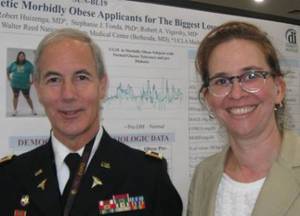
"The Biggest Loser" applicants wore a masked continuous glucose monitor for 3-8 days as part of the application process. Four patients with type 1 or type 2 diabetes were excluded from the analysis, leaving 23 people with normal glucose tolerance and 15 who were classified as having prediabetes. The investigators compared the swings between high and low glucose readings in these 38 patients with historic data on 37 age-matched, nondiabetic, normal-weight people from the Juvenile Diabetes Research Foundation Continuous Glucose Monitoring Study Group (Diabetes Care 33;6:1297-9).
The average hemoglobin A1c was 5.3% in the normoglycemic but obese applicants and 5.4% in the nonobese control group, compared with 5.8% in the obese applicants.
The standard deviation in glucose measures was 24 mg/dL in both the normoglycemic applicants and the prediabetic applicants, compared with 12 mg/dL in the nonobese control group, Sara J. Salkind and her associates reported. Other standard measures of glycemic variability showed similar trends. The mean amplitude of glycemic excursion was 48 mg/dL in the normoglycemic applicants and 50 mg/dL in the prediabetic group, compared with 27 mg/dL the control group.
The percentage of glucose values that fell within the range of 81-139 mg/dL was 77% in the normoglycemic applicants and 78% in the prediabetic group, compared with 99% in the control group. Glucose values below 80 mg/dL comprised 11% of values in the "normal" obese group and 5% of values in the prediabetes group, compared with 0.3% of values in the control group. Glucose values higher than 140 mg/dL comprised 12% of measures in the "normal" obese group, 18% of measures in the prediabetes group, and 0.3% of values in the control group.
If "normal" glycemic status is defined as a HbA1c less than 5.7%, a fasting glucose level less than 100 mg/dL, and a 2-hour postprandial glucose load of less than 140 mg/dL, then "these obese people, who by almost all the traditional measures have normal glycemia, have increased variability and much greater variability than normals of similar age" who are not obese, coinvestigator Robert A. Vigersky, Col., MC, USA, said in an interview.
Both Ms. Salkind and Dr. Vigersky are with the endocrinology service at Walter Reed National Military Medical Center, Bethesda, Md. The investigators’ report reflects the personal views of the authors and not the official views of the United States Army or the Department of Defense.
People with diabetes are known to have glycemic variability, but there are few previous data on glycemic variability in people without diabetes. The current findings have implications for the pathophysiology of microvascular disease and, combined with previous studies suggesting that obese people have increased carotid intima-media thickness, compared with normal-weight people, may increase understanding of cardiovascular risk from obesity.
Measures of glycemic variability also may have a more practical application as a behavior-modification tool to help obese people understand the effects of their eating habits on their bodies. The investigators said they will pursue studies to see if continuous glucose monitoring might help obese patients lose weight and avoid converting to prediabetes or diabetes.
The average age in the current study was 32 years in the "normal" group and 34 years in the prediabetes group. The average body mass index was 50 and 51 kg/m2, respectively. The 2-hour post-glucose load averaged 107 mg/dL for normoglycemic applicants and 145 mg/dL in the prediabetes group. The fasting plasma glucose averaged 89 mg/dL in the "normal" group and 97 mg/dL in the prediabetes group. Hypertension was present in 76% and 93%, respectively.
Dexcom, which makes continuous glucose meters, funded the study. The investigators reported having no other financial disclosures.
HOUSTON – Data from 42 morbidly obese people who applied to be on the television show "The Biggest Loser" showed significantly greater glycemic variability even without diabetes, compared with previous data on nondiabetic, normal-weight adults.
Oxidative stress from the glycemic variability may contribute to microvascular and macrovascular changes in obese people and help explain why some obese people develop advanced cardiovascular disease and increased risk for cardiovascular events, investigators said at the annual meeting of the Endocrine Society.
"The Biggest Loser" applicants wore a masked continuous glucose monitor for 3-8 days as part of the application process. Four patients with type 1 or type 2 diabetes were excluded from the analysis, leaving 23 people with normal glucose tolerance and 15 who were classified as having prediabetes. The investigators compared the swings between high and low glucose readings in these 38 patients with historic data on 37 age-matched, nondiabetic, normal-weight people from the Juvenile Diabetes Research Foundation Continuous Glucose Monitoring Study Group (Diabetes Care 33;6:1297-9).
The average hemoglobin A1c was 5.3% in the normoglycemic but obese applicants and 5.4% in the nonobese control group, compared with 5.8% in the obese applicants.
The standard deviation in glucose measures was 24 mg/dL in both the normoglycemic applicants and the prediabetic applicants, compared with 12 mg/dL in the nonobese control group, Sara J. Salkind and her associates reported. Other standard measures of glycemic variability showed similar trends. The mean amplitude of glycemic excursion was 48 mg/dL in the normoglycemic applicants and 50 mg/dL in the prediabetic group, compared with 27 mg/dL the control group.
The percentage of glucose values that fell within the range of 81-139 mg/dL was 77% in the normoglycemic applicants and 78% in the prediabetic group, compared with 99% in the control group. Glucose values below 80 mg/dL comprised 11% of values in the "normal" obese group and 5% of values in the prediabetes group, compared with 0.3% of values in the control group. Glucose values higher than 140 mg/dL comprised 12% of measures in the "normal" obese group, 18% of measures in the prediabetes group, and 0.3% of values in the control group.
If "normal" glycemic status is defined as a HbA1c less than 5.7%, a fasting glucose level less than 100 mg/dL, and a 2-hour postprandial glucose load of less than 140 mg/dL, then "these obese people, who by almost all the traditional measures have normal glycemia, have increased variability and much greater variability than normals of similar age" who are not obese, coinvestigator Robert A. Vigersky, Col., MC, USA, said in an interview.
Both Ms. Salkind and Dr. Vigersky are with the endocrinology service at Walter Reed National Military Medical Center, Bethesda, Md. The investigators’ report reflects the personal views of the authors and not the official views of the United States Army or the Department of Defense.
People with diabetes are known to have glycemic variability, but there are few previous data on glycemic variability in people without diabetes. The current findings have implications for the pathophysiology of microvascular disease and, combined with previous studies suggesting that obese people have increased carotid intima-media thickness, compared with normal-weight people, may increase understanding of cardiovascular risk from obesity.
Measures of glycemic variability also may have a more practical application as a behavior-modification tool to help obese people understand the effects of their eating habits on their bodies. The investigators said they will pursue studies to see if continuous glucose monitoring might help obese patients lose weight and avoid converting to prediabetes or diabetes.
The average age in the current study was 32 years in the "normal" group and 34 years in the prediabetes group. The average body mass index was 50 and 51 kg/m2, respectively. The 2-hour post-glucose load averaged 107 mg/dL for normoglycemic applicants and 145 mg/dL in the prediabetes group. The fasting plasma glucose averaged 89 mg/dL in the "normal" group and 97 mg/dL in the prediabetes group. Hypertension was present in 76% and 93%, respectively.
Dexcom, which makes continuous glucose meters, funded the study. The investigators reported having no other financial disclosures.
AT THE ANNUAL MEETING OF THE ENDOCRINE SOCIETY
Major Finding: Continuous glucose meter readings showed a standard deviation of 24 mg/dL in obese people with normal glycemic status or prediabetes, compared with 12 mg/dL in normal-weight healthy controls.
Data Source: Prospective continuous glucose monitoring in 38 nondiabetic obese people, compared with historical data on 37 nonobese, nondiabetic people of similar age.
Disclosures: Dexcom, which makes continuous glucose meters, funded the study. The investigators reported having no other financial disclosures.

Pamidronate Deemed Kidney-Safe in Chronic Critical Illness
HOUSTON – Intravenous pamidronate for management of bone hyperresorption in ventilator-dependent patients doesn’t adversely affect renal function, even in those with chronic kidney disease, according to a single-center retrospective study.
"This stands in contrast to prevailing concerns about bisphosphonate therapy to manage bone hyperresorption in chronically critically ill patients, many of whom have chronic kidney disease," Dr. Rifka C. Schulman reported at the annual meeting of the Endocrine Society.
"It is hoped that these results will remove barriers to more aggressive management of metabolic bone disease in chronic critical illness, which may have salutary downstream effects on morbidity and mortality," declared Dr. Schulman of the Mount Sinai School of Medicine in New York.
She presented a retrospective observational study of 315 patients admitted to the Mount Sinai Hospital respiratory care unit with ventilator-dependent chronic critical illness after surviving a bout of acute critical illness. In all, 115 received 30-90 mg of intravenous pamidronate (Aredia) infused over 4 hours, with dosing based on body weight. The other 200 patients did not receive pamidronate. All participants got calcitriol, calcium carbonate, and ergocalciferol to help protect their bones.
The study population consisted of 204 patients with either no or stage 1-2 chronic kidney disease, 41 with stage 3 CKD, 33 with stage 4 disease, and 37 with stage 5 CKD who were on hemodialysis.
The primary study end points were change in glomerular filtration rate and creatinine level following pamidronate administration. Importantly, none of the pamidronate-treated patients showed a 25% or greater reduction in GFR immediately after or at 7 or 14 days post infusion, regardless of their CKD status or dose received.
The group with no or only mild CKD showed no change in median GFR between baseline and day 7 post infusion. Those with stage 3 CKD had a median 4% drop in GFR. So did those with stage 4 disease. Patients with stage 5 CKD had a median 9% reduction in GFR on day 7. Among controls, those with stage 0-3 CKD had no change in median GFR, while those with stage 4 or stage 5 disease averaged a 6% increase in GFR during 7 days. These small fluctuations in renal function aren’t clinically meaningful, according to Dr. Schulman.
Creatinine levels rose between baseline and day 7 in lockstep with CKD status, but to the same extent in pamidronate-treated patients and controls. For example, creatinine climbed by 6.7% and 18.2%, respectively, in pamidronate-treated patients with stage 4 and stage 5 CKD, and by 8.8% and 20.8% in stage 4 and 5 controls.
She observed that metabolic bone disease involving bone hyperresorption with elevated levels of the bone turnover biomarker N-telopeptide is present in more than 90% of patients with chronic critical illness. Contributing factors include immobilization, inflammation, neuroendocrine abnormalities, low vitamin D levels and secondary hyperparathyroidism, and the use of high-dose corticosteroids and other medications with an adverse impact on bone.
Bone loss during critical illness is challenging to reverse. It predisposes to osteoporosis, fractures, and poor quality of life in patients who recover from chronic critical illness. That’s why Dr. Schulman and her coinvestigators favor turning to pamidronate when patients have a 24-hour urine N-telopeptide of 70 nmol BCE/mmol creatinine or a serum level in excess of 40 nmol BCE/L.
The study was supported by a grant from Select Medical. Dr. Schulman reported having no financial conflicts.
AT THE ANNUAL MEETING OF THE ENDOCRINE SOCIETY
Major Finding: In pamidronate-treated patients with chronic critical illness and stage 4 and stage 5 chronic kidney disease, creatinine climbed by 6.7% and 18.2%, respectively. In matched controls who didn’t receive pamidronate, creatinine rose by 8.8% and 20.8%.
Data Source: This was a retrospective, observational, single-center study of 315 patients with ventilator-dependent chronic critical illness, 200 of whom received intravenous pamidronate.
Disclosures: The study was supported by a grant from Select Medical. Dr. Schulman reported having no financial conflicts.
Traumatic Brain Injury Linked to GH Deficiency
HOUSTON – Growth hormone deficiency appears to be common among military veterans with mild traumatic brain injury sustained in combat, according to the first study to look at the issue.
Future studies will attempt to confirm the new finding that GH deficiency in the setting of traumatic brain injury (TBI) appears to be associated with specific neuropsychologic abnormalities, and, further, whether GH replacement therapy in affected veterans enhances TBI rehabilitation efforts, according to Dr. Adriana G. Ioachimescu of Emory University, Atlanta.
At the annual meeting of the Endocrine Society, she presented the results of a pilot study of 20 men (mean age, 34 years) with mild TBI resulting from military combat. The last injury occurred an average of 44 months earlier.
Five subjects, or 25%, were GH deficient on the basis of a peak value of less than 3 ng/mL in response to a glucagon stimulation test. One GH-deficient veteran also had an abnormally low insulin-like growth factor-1 level. But all subjects had normal thyroid status and cortisol function.
Four GH-deficient men and 12 GH-sufficient men were able to put enough effort into their neuropsychologic testing for the results to be valid. The two groups performed similarly on measures of memory, learning, and simple and complex attention.
In contrast, GH deficiency appeared to be associated with executive dysfunction, as manifest in worse performance on measures of inhibitory control and self-monitoring. Depression also was more severe in the GH-deficient men, although they did not experience greater levels of fatigue or posttraumatic stress disorder. The GH-deficient group also scored significantly lower on a validated quality-of-life measure.
This study was supported by Novo Nordisk. The presenter reported having no financial conflicts.
AT THE ANNUAL MEETING OF THE ENDOCRINE SOCIETY
Major Finding: Twenty-five percent of a group of military veterans with mild traumatic brain injury had growth hormone deficiency, which was associated with specific neuropsychologic abnormalities.
Data Source: This was a pilot study of 20 men with combat-induced mild traumatic brain injury.
Disclosures: The study was supported by Novo Nordisk. The presenter reported having no financial conflicts.
Oral Relaxin Improves Poststroke Recovery
HOUSTON – Oral relaxin therapy resulted in significantly improved cognitive and functional recovery in poststroke patients participating in a randomized controlled trial.
"We speculate that in the near future, relaxin hormone could represent a new therapeutic and preventative tool to afford reduction of both incidence and disability of vascular ischemic diseases, not only of the brain but also of other organs and apparatuses," Dr. Paolo Milia declared in presenting the study findings at the annual meeting of the Endocrine Society.
The trial involved 36 poststroke patients admitted to a rehabilitation unit, where they were randomized to rehabilitation therapy plus 40 mg/day of oral porcine relaxin or to rehab alone. Investigators administered validated tests of cognitive function, global impairment, and daily activity at admission and again on days 20 and 40.
Cognitive function as assessed using the Trail Making Test was significantly better in the relaxin group on day 20, when their mean score was 3.5, compared with 2.0 in controls. The difference in cognitive outcome broadened by day 40, when the average score in the relaxin group was 4.0, compared with 2.0 in controls on rehabilitation therapy only, reported Dr. Milia of the Prosperius Institute in Umbertide, Italy.
Daily activity as measured using the Functional Independence Measure improved in the relaxin group from a baseline score of 53 to 78 at day 20 and 96 on day 40. In the control group, the average score was 59 at baseline, 69 at day 20, and 75 at day 40, indicating significantly greater functional recovery in the relaxin group at the 40-day mark.
Global function as assessed using the Modified Rankin Scale was higher in the relaxin group than in controls on days 20 and 40, with the difference between groups significant at both times.
The hormone treatment was safe as well as efficacious. No side effects occurred, he said.
Relaxin, discovered in 1926, belongs to the same peptide hormone superfamily as the insulin-like peptides. Relaxin’s known function revolves around pregnancy; however, the hormone also promotes angiogenesis, inhibits collagen synthesis, enhances nitric oxide synthesis, and boosts production of matrix metalloproteinases, a spectrum of effects leaving the door open to possible broader functions not well characterized as yet. In animal models, relaxin protects against ischemic injury to the myocardium and brain (J. Chem. Neuroanat. 2011;42:262-75), Dr. Milia noted.
The porcine relaxin utilized in this study, known as Vitalaxin Plus, is marketed by Sky BioHealth. The investigators reported having no financial disclosures.
AT THE ANNUAL MEETING OF THE ENDOCRINE SOCIETY
Major Finding: Treatment with oral porcine relaxin at 40 mg/day plus rehabilitation therapy resulted in greater psychoneurologic and functional recovery than rehabilitation therapy alone in a group of poststroke patients.
Data Source: A prospective, randomized, controlled trial of 40 poststroke patients admitted to a rehabilitation center.
Disclosures: The porcine relaxin utilized in this study, known as Vitalaxin Plus, is marketed by Sky BioHealth. The investigators reported having no financial disclosures.
Tomatoes Boosted Low HDL in Small Study
HOUSTON – Consuming two midsize uncooked tomatoes daily for a month resulted in a mean 5 mg/dL gain in HDL cholesterol level in a randomized trial in patients with low HDL.
A control group assigned to eat an equal quantity of cucumber – 300 g/day – saw no change in HDL, according to Dr. Daniel Cuevas Ramos of the National Institute of Medical Sciences and Nutrition in Mexico City.
He reported on 41 women and 11 men with low HDL but normal triglyceride levels who participated in the month-long randomized trial, during which they consumed an isocaloric diet.
Over the course of 1 month of follow-up, mean HDL levels in the tomato eaters climbed from 36.5 mg/dL at baseline to 41.6 mg/dL, while the cucumber-eating controls saw no significant change over time.
A linear regression analysis adjusted for age, sex, waist-to-hip ratio, physical activity, body mass index, and intake of alcohol, simple sugars, and omega-3 fatty acids showed that tomato consumption was independently associated with the increase in HDL.
Two medium tomatoes contain about 30 mg of lycopene, the nutrient thought responsible for the HDL boost. Prior cross-sectional studies had linked lycopene intake to higher HDL, he noted.
Dr. Cuevas Ramos reported having no financial conflicts.
AT THE ANNUAL MEETING OF THE ENDOCRINE SOCIETY
Major Finding: Eating two midsize tomatoes per day for 1 month led to a mean 5 mg/dL increase in HDL in a randomized trial involving men and women with low HDL.
Data Source: A month-long open-label randomized trial in 52 low-HDL participants.
Disclosures: The presenter reported having no financial conflicts.
Computer Algorithm Improved ICU Glucose Control
HOUSTON – Nurses using a computerized algorithm maintained better glycemic control, compared with expert nursing care without the algorithm, in a prospective, randomized study of 300 patients in one institution’s ICU.
Baseline characteristics did not differ between the 151 patients randomized to nursing care and the 149 patients cared for by nurses using the LOGIC-Insulin computerized algorithm developed by Dr. Dieter Mesotten and his associates at Catholic University of Leuven, Belgium. Mean blood glucose levels during ICU care also were similar between groups: 106 mg/dL in both groups.

On other measures of glycemic control, however, the LOGIC group scored significantly better than did the nurses group, he reported in a poster presentation at the annual meeting of the Endocrine Society. The LOGIC group had a significantly lower mean score on the Glycemic Penalty Index, a marker of efficacy of blood glucose control that was the primary outcome measure of efficacy in the study: 9.8 vs. 12.4 in the nurses group.
Patients in the LOGIC group were less likely to develop critically low glucose levels, they spent more time in the study’s target range of 80-110mg/dL, and they had narrower swings between minimum and maximum blood-sugar measurements, indicating less blood glucose variability.
Mean scores on the Hyperglycemic Index were significantly lower in the LOGIC group (2.5 mg/dL), compared with the nurses group (4.2 mg/dL). Patients in the LOGIC group reached the target range of 80-110 mg/dL faster (in 2 hours on average instead of 3 hours in the nurses group) and were within the target range 69% of the time while in the ICU, compared with 60% of the time for patients in the nurses group. The mean daily difference between minimum and maximum blood glucose levels was 31 mg/dL in the LOGIC group and 37 mg/dL in the nurses group, Dr. Mesotten reported.
The patients had been admitted to the ICU after cardiac surgery (49% in the nurses group and 51% in the LOGIC group), following transplantation (17% and 13%, respectively), because of medical problems (15% and 17%), respectively, or for other reasons.
In general, maintaining tight blood control in critically ill patients is labor intensive and difficult. The LOGIC algorithm may have made this task more successful but also appeared to add a bit to the nursing team’s workload by increasing the frequency of glucose measurements. Nurses in the LOGIC group measured blood glucose levels every 2.2 hours on average, compared with every 2.5 hours in the nursing-only group, a statistically significant difference.
Patients averaged 64 years in age, 60% were male, and 21% had diabetes when admitted to the ICU.
Dr. Mesotten reported having no financial disclosures. Belgian public agencies funded the study.
nursing care, LOGIC-Insulin computerized algorithm, Dr. Dieter Mesotten, Glycemic Penalty Index, Hyperglycemic Index,
HOUSTON – Nurses using a computerized algorithm maintained better glycemic control, compared with expert nursing care without the algorithm, in a prospective, randomized study of 300 patients in one institution’s ICU.
Baseline characteristics did not differ between the 151 patients randomized to nursing care and the 149 patients cared for by nurses using the LOGIC-Insulin computerized algorithm developed by Dr. Dieter Mesotten and his associates at Catholic University of Leuven, Belgium. Mean blood glucose levels during ICU care also were similar between groups: 106 mg/dL in both groups.
On other measures of glycemic control, however, the LOGIC group scored significantly better than did the nurses group, he reported in a poster presentation at the annual meeting of the Endocrine Society. The LOGIC group had a significantly lower mean score on the Glycemic Penalty Index, a marker of efficacy of blood glucose control that was the primary outcome measure of efficacy in the study: 9.8 vs. 12.4 in the nurses group.
Patients in the LOGIC group were less likely to develop critically low glucose levels, they spent more time in the study’s target range of 80-110mg/dL, and they had narrower swings between minimum and maximum blood-sugar measurements, indicating less blood glucose variability.
Mean scores on the Hyperglycemic Index were significantly lower in the LOGIC group (2.5 mg/dL), compared with the nurses group (4.2 mg/dL). Patients in the LOGIC group reached the target range of 80-110 mg/dL faster (in 2 hours on average instead of 3 hours in the nurses group) and were within the target range 69% of the time while in the ICU, compared with 60% of the time for patients in the nurses group. The mean daily difference between minimum and maximum blood glucose levels was 31 mg/dL in the LOGIC group and 37 mg/dL in the nurses group, Dr. Mesotten reported.
The patients had been admitted to the ICU after cardiac surgery (49% in the nurses group and 51% in the LOGIC group), following transplantation (17% and 13%, respectively), because of medical problems (15% and 17%), respectively, or for other reasons.
In general, maintaining tight blood control in critically ill patients is labor intensive and difficult. The LOGIC algorithm may have made this task more successful but also appeared to add a bit to the nursing team’s workload by increasing the frequency of glucose measurements. Nurses in the LOGIC group measured blood glucose levels every 2.2 hours on average, compared with every 2.5 hours in the nursing-only group, a statistically significant difference.
Patients averaged 64 years in age, 60% were male, and 21% had diabetes when admitted to the ICU.
Dr. Mesotten reported having no financial disclosures. Belgian public agencies funded the study.
nursing care, LOGIC-Insulin computerized algorithm, Dr. Dieter Mesotten, Glycemic Penalty Index, Hyperglycemic Index,
nursing care, LOGIC-Insulin computerized algorithm, Dr. Dieter Mesotten, Glycemic Penalty Index, Hyperglycemic Index,
FROM THE ANNUAL MEETING OF THE ENDOCRINE SOCIETY
Major Finding: Use of the LOGIC computerized algorithm improved scores on the Glycemic Penalty Index to a mean of 9.8 during ICU care, compared with a mean score of 12.4 under expert nursing care alone.
Data Source: Prospective, randomized, single-blind study in 300 patients managed in one institution’s ICU.
Disclosures: Dr. Mesotten reported having no financial disclosures. Belgian public agencies funded the study.

Novel Estrogen Combination Shows Favorable Coagulation Profile
HOUSTON – The novel investigational tissue selective estrogen complex comprising bazedoxifene plus conjugated estrogens displayed an overall favorable coagulation profile in a pooled analysis of three randomized, placebo-controlled, double-blind, phase III clinical trials.
Moreover, the risk of venous thromboembolism or cerebrovascular events was low, at 0.2% or less during up to 2 years of treatment, a rate not significantly different from that of control patients, Dr. Sven O. Skouby of Herlev (Denmark) Hospital said at the annual meeting of the Endocrine Society.
He presented an analysis of serial coagulation parameters obtained in 1,978 postmenopausal women with intact uteri who were randomized to bazedoxifene at 20 mg per day plus either 0.45 or 0.625 mg of conjugated estrogens or to placebo in the first, fourth, and fifth SMART (Selective Estrogens, Menopause, and Response to Therapy) trials. SMART-1 assessed changes in coagulation variables at 12 and 24 months, while SMART-4 and -5 did so at 12 months.
Changes from baseline in protein C activity, d-dimer level, and prothrombin and partial thromboplastin times were similar in the two bazedoxifene/conjugated estrogens study arms and in placebo-treated controls.
The two active treatment arms showed modest but not clinically meaningful decreases in plasminogen activator-1, antithrombin III, and fibrinogen, compared with placebo. Similarly, plasminogen activity in the two active treatment groups increased by a mean of 0.1 L/L over the course of 24 months, a statistically greater increase than the 0.06 L/L seen in the control group, but not a clinically significant difference, Dr. Skouby observed.
SMART-1, -4, and -5 included 3,549 randomized patients with follow-up data on hard clinical thromboembolic events. Three patients on bazedoxifene/conjugated estrogens experienced deep vein thrombosis during 2 years of therapy, as did one on placebo. There were no cases of pulmonary embolism. Two of the treated patients and no controls had a cerebrovascular event.
In a separate presentation, Dr. Sebastian Mirkin reported on a pooled analysis of lipid changes in 4,409 postmenopausal women in SMART-1 through -5.
At 24 months, LDL cholesterol fell by a mean 7.4% from baseline in the bazedoxifene 20 mg/conjugated estrogens 0.45 mg group, and by 8.4% in those on bazedoxifene 20 mg/conjugated estrogens 0.625 mg, compared with a 2% increase in controls.
HDL levels rose by 7.3% in the lower-dose and 7.9% in the higher-dose groups, a significantly greater benefit than the 2.8% increase with placebo. Apolipoprotein B declined by a mean of 0.4% in the lower- and 0.5% in the higher-dose bazedoxifene/conjugated estrogens group, compared with a 4.3% increase in controls.
The one negative was that triglyceride levels increased by 20.3% in the lower- and 18.7% in the higher-dose groups, significantly greater than the 7.1% rise with placebo, reported Dr. Mirkin.
Bazedoxifene is not approved in the United States. Pfizer submitted a new drug application to the Food and Drug Administration in 2006, and in 2007 got an "approvable letter" that asked for more information. There is no public record of Pfizer’s response. Pfizer has announced that it will submit an application to the FDA for the bazedoxifene/conjugated estrogen combination this year.
The SMART trials were sponsored by Pfizer. Dr. Skouby serves as a consultant to the company. Dr. Mirkin is a Pfizer employee.
AT THE ANNUAL MEETING OF THE ENDOCRINE SOCIETY
Major Finding: An overall favorable coagulation profile was documented in women on an investigational tissue-selective estrogen complex for up to 2 years.
Data Source: This was a pooled analysis of several thousand postmenopausal women with intact uteri who were randomized to bazedoxifene 20 mg/conjugated estrogens 0.45 mg, bazedoxifene 20 mg/conjugated estrogens 0.625 mg, or placebo for up to 2 years in three phase III clinical trials.
Disclosures: The SMART trials were sponsored by Pfizer. Dr. Skouby serves as a consultant to the company. Dr. Mirkin is a Pfizer employee.
Experimental Drug Improves Muscle Strength in Cancer
HOUSTON – An experimental drug significantly improved physical function in men with cancer-related muscle wasting, compared with placebo – and more so in the men with low testosterone levels – in a secondary analysis of a randomized, double-blind, phase II clinical trial.
In all, 60% of the 93 men for whom baseline testosterone levels were available had hypogonadism when the patients were randomized to treatment with daily placebo or 1 mg or 3 mg of enobosarm for 16 weeks. Before treatment, the eugonadal males showed significantly better physical function, as measured by stair-climb power, compared with the hypogonadal patients (174 W vs. 147 W). Lower testosterone levels correlated with lower physical function.
Enobosarm significantly improved physical function (a secondary end point in the trial) with an average 17% increase in stair-climb power in hypogonadal men, and a 12% increase in power in eugonadal men, compared with baseline, Dr. Adrian Dobs and her associates reported at the annual meeting of the Endocrine Society.
Among men on placebo, stair-climb power increased by about 10% in hypogonadal men and by roughly 1% in eugonadal men, compared with baseline, but the changes were not statistically significant, said Dr. Dobs, professor of medicine and oncology at Johns Hopkins University, Baltimore.
Adverse events included fatigue in 21% of both treatment groups, anemia in 19% of those on enobosarm and 15% in those on placebo, nausea in 18% on enobosarm and 14% on placebo, diarrhea in 16% on enobosarm and 14% on placebo, vomiting in 12% of each treatment group, weight decrease in 12% on enobosarm and 10% on placebo, and constipation in 14% on enobosarm and 4% on placebo.
The investigators defined hypogonadism as a testosterone level below 300 ng/dL.
Enobosarm is a nonsteroidal SARM (selective androgen receptor modulator) that produces anabolic effects in bone and muscle without causing the prostate effects in men or hair growth in women that is seen with steroids.
The analysis used data from a larger trial in 159 people with cancer (65% of whom were men) who had lost at least 2% (and an average of 8%) of their weight in the previous 6 months. The men were older than 45 years of age, the women were postmenopausal, and each had a body mass index less than 35 mg/kg2.
The multicenter study as a whole met its primary end point of increasing lean body mass (muscle) and its secondary end point of improving physical function, Dr. Dobs said in an interview. Those results were reported on the website of the company that is developing the drug, GTx Inc., and at previous medical meetings, according to an interview in the Los Angeles Times.
Dr. Dobs did not report results for lean body mass in the current analysis based on gonadal status.
Cancer diagnoses included colorectal cancer, non–small cell lung cancer, non-Hodgkin’s lymphoma, chronic lymphocytic leukemia, and breast cancer. Patients participated in the study prior to or between courses of chemotherapy. Rates of hypogonadism were similar for each type of cancer. Patients with hypogonadism were less likely to complete the study than were eugonadal men.
Patients with cancer develop cachexia (muscle wasting) because of reduced anabolic activity, increased catabolic activity, or both, accounting for a progressive catabolic state. Up to 50% of patients with lung cancer show severe cachexia at the time of diagnosis, and the muscle wasting increases throughout the course of the malignancy, Dr. Dobs said.
Two phase III clinical trials are underway to study effects of 3 mg/day of enobosarm for the prevention and treatment of muscle wasting in patients with non–small cell lung cancer.
GTx Inc., the company that is developing enobosarm, funded the current study. Dr. Dobs reported having no other financial disclosures.
AT THE ANNUAL MEETING OF THE ENDOCRINE SOCIETY
Major Finding: The experimental drug enobosarm significantly improved physical function on a stair-climb test by 17% in hypogonadal men with cancer and by 12% in eugonadal men with cancer, compared with nonsignificant improvements in men on placebo.
Data Source: Data are from an analysis of data on a secondary end point (physical function) in 93 men from a randomized, double-blind, multicenter trial in 159 cancer patients.
Disclosures: GTx, the company that is developing enobosarm, funded the study. Dr. Dobs reported having no other financial disclosures.