User login
Laser Therapy of Nonmelanoma Cancers Continues to Grow
BOCA RATON, FLA. – Experience with various lasers for the treatment and prophylaxis of nonmelanoma skin cancers is increasing, and the outcomes are promising.
One setting where lasers have particular potential is in patients with multiple basal cell carcinomas, Dr. Keyvan Nouri said at the annual meeting of the Florida Society of Dermatology and Dermatologic Surgery.
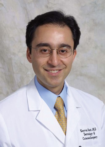
BCCs tend to have telangiectasias, and by specifically targeting the tumor vasculature, the BCC burden can be decreased or eliminated by laser treatment with little damage to surrounding skin. An ablative approach can also eliminate premalignant cells, said Dr. Nouri, professor of dermatology and otolaryngology and director of Mohs, dermatologic, and laser surgery at the University of Miami.
Studies using this approach have been conducted over the past several years using CO2, Er:YAG, pulsed-dye, and alexandrite lasers.
The 10,600-nm CO2 laser achieves a depth of 15-200 mcm in a single pass, which is adequate for coagulating the epidermis and superficial dermis, and for eliminating squamous cells at risk for evolution to actinic keratosis or squamous cell carcinoma. It also is partially effective for eliminating cells en route to basal cell carcinoma, he said, noting that deeper treatment can be achieved with multiple passes or protocol modifications.
In one study using this CO2 laser, 14 patients with diffuse facial AKs underwent resurfacing and experienced significant long-term reduction in AK burden (Dermatol. Surg. 1997;23:885-9). In another report of two patients with a history of multiple nonmelanoma skin cancer who were treated with two-pass CO2 resurfacing, no new lesions developed in the treated area at 33 and 52 months (Dermatol. Surg. 1999;25:513-16).
In a randomized controlled study of 34 patients, no differences in outcomes were seen at 5 years regardless of whether patients were treated with the CO2 laser, a 30% trichloroacetic acid peel, or 5% fluorouracil cream twice daily for 3 weeks. All of the treatments reduced AK counts by 83%-92%, and reduced the incidence of nonmelanoma skin cancers, compared with control groups (Dermatol. Surg. 1999;25:729-32), Dr. Nouri said.
In three of his patients with basal cell nevus syndrome, the Ultrapulse CO2 laser also proved effective for treatment. Postoperative Mohs micrographic surgical sections verified complete histologic clearance, and the patients had minimal scarring at follow-up (Dermatol. Surg. 2002;28:287-90).
A high-energy pulsed CO2 laser has also been used to treat a squamous cell carcinoma in situ on the nose with one pass (at 250 mJ, 8 W). In that case, no recurrence had occurred at 5 months’ follow-up, and the cosmetic result was excellent, Dr. Nouri said.
In a series of patients with nodular and superficial BCCs, a combination of curettage and one or two passes with the high-energy pulsed CO2 laser was also effective, with no histologic evidence or residual tumor and no known recurrences.
In another study, the pulsed CO2 laser was effective for treating 30 neoplasms, including 17 BCCs and 13 SCCs (Arch. Dermatol. 1998;134:1247-52). Two or three passes were used (at 500 mJ and 2-4 W). Treatment with a 4-mm margin and three passes is recommended for complete vaporization of neoplasms using this laser, Dr. Nouri said. Also, one SCC in situ in that study was incompletely ablated after three passes, so this modality alone is not recommended for thick or keratotic lesions, he said.
More recently, fractional laser resurfacing and pulsed-dye laser treatment have also shown promise for nonmelanoma skin cancers. In a pilot study of 28 patients with mild to moderate actinic damage who were treated with a fractional Er:YAG laser (2,940 nm) with one to four treatments at 4-week intervals, 75% had excellent results and the other 25% had good results. The effects were maintained at up to 9 months (Dermatol. Surg. 2008;34:1048-53).
And the 595-nm pulsed-dye laser has been used successfully to target tumor vasculature in glottal dysplasia and SCCs. Depending on fluence and spot size, the 595-nm pulsed-dye laser can penetrate skin to thicknesses ranging from 0.75 to 1.25 mm, which encompasses most BCCs, Dr. Nouri said.
In one study, a single treatment on seven BCCs resulted in histologic clearance of only one tumor (Lasers Med. Sci. 2003;18:125-6), but in two other studies of patients with superficial BCCs, this laser was associated with no clinical recurrences at a minimum follow-up of 1 year in 16 of 20 BCCs (Lasers Med. Sci. 2005;20:147-8; Dermatol. Ther. 2008; 21:402-5).
In another study, 20 BCCs treated with four 595-nm pulsed-dye laser treatments at 2-week intervals showed that nearly all BCCs less than 1.5 cm responded completely, compared with about 17% of controls. Larger BCCs had a complete response rate of 25% vs. 0% in controls.
Tumors with an incomplete response showed a significant reduction in tumor burden following treatment, with residual histologic tumor burden ranging from less than 1% to 29% of the original clinical tumor diameter, compared with 13%-68% residual tumor burden for the corresponding controls.
The investigators took the study one step further, treating 14 patients with a total of 20 BCCs on the trunk or extremities with four treatments at 3- to 4-week intervals; 19 of 20 BCCs had a complete clinical response, regardless of size and histological subtype (Lasers Surg. Med. 2009; 41:417-22). The investigators concluded that the 595-nm pulsed-dye laser is a "novel, quick, and relatively nonpainful treatment" for BCCs when used in the appropriate clinical setting, Dr. Nouri said.
As for the long-pulse alexandrite laser, it is selective like the pulsed-dye laser, but has deeper penetration, and may be helpful in significantly reducing tumor burden with a single treatment, he said.
In 15 of 18 patients with basal cell nevus syndrome who were treated in one study, a complete clinical response was seen at follow-ups of 2 and 7 months (Lasers Surg. Med. 2010;42:68-71).
No particular laser has emerged as the ideal tool for the treatment or prevention of skin cancer, but work is ongoing. More research is needed to confirm the observations made thus far, as well as to optimize treatment parameters, but the findings to date are encouraging, Dr. Nouri said, noting that he is confident that laser and light sources will eventually be used more for both medical and oncologic purposes.
Dr. Nouri disclosed that he has received grants or research support from Aesthera, CureLight, and Omnilux.
BOCA RATON, FLA. – Experience with various lasers for the treatment and prophylaxis of nonmelanoma skin cancers is increasing, and the outcomes are promising.
One setting where lasers have particular potential is in patients with multiple basal cell carcinomas, Dr. Keyvan Nouri said at the annual meeting of the Florida Society of Dermatology and Dermatologic Surgery.
BCCs tend to have telangiectasias, and by specifically targeting the tumor vasculature, the BCC burden can be decreased or eliminated by laser treatment with little damage to surrounding skin. An ablative approach can also eliminate premalignant cells, said Dr. Nouri, professor of dermatology and otolaryngology and director of Mohs, dermatologic, and laser surgery at the University of Miami.
Studies using this approach have been conducted over the past several years using CO2, Er:YAG, pulsed-dye, and alexandrite lasers.
The 10,600-nm CO2 laser achieves a depth of 15-200 mcm in a single pass, which is adequate for coagulating the epidermis and superficial dermis, and for eliminating squamous cells at risk for evolution to actinic keratosis or squamous cell carcinoma. It also is partially effective for eliminating cells en route to basal cell carcinoma, he said, noting that deeper treatment can be achieved with multiple passes or protocol modifications.
In one study using this CO2 laser, 14 patients with diffuse facial AKs underwent resurfacing and experienced significant long-term reduction in AK burden (Dermatol. Surg. 1997;23:885-9). In another report of two patients with a history of multiple nonmelanoma skin cancer who were treated with two-pass CO2 resurfacing, no new lesions developed in the treated area at 33 and 52 months (Dermatol. Surg. 1999;25:513-16).
In a randomized controlled study of 34 patients, no differences in outcomes were seen at 5 years regardless of whether patients were treated with the CO2 laser, a 30% trichloroacetic acid peel, or 5% fluorouracil cream twice daily for 3 weeks. All of the treatments reduced AK counts by 83%-92%, and reduced the incidence of nonmelanoma skin cancers, compared with control groups (Dermatol. Surg. 1999;25:729-32), Dr. Nouri said.
In three of his patients with basal cell nevus syndrome, the Ultrapulse CO2 laser also proved effective for treatment. Postoperative Mohs micrographic surgical sections verified complete histologic clearance, and the patients had minimal scarring at follow-up (Dermatol. Surg. 2002;28:287-90).
A high-energy pulsed CO2 laser has also been used to treat a squamous cell carcinoma in situ on the nose with one pass (at 250 mJ, 8 W). In that case, no recurrence had occurred at 5 months’ follow-up, and the cosmetic result was excellent, Dr. Nouri said.
In a series of patients with nodular and superficial BCCs, a combination of curettage and one or two passes with the high-energy pulsed CO2 laser was also effective, with no histologic evidence or residual tumor and no known recurrences.
In another study, the pulsed CO2 laser was effective for treating 30 neoplasms, including 17 BCCs and 13 SCCs (Arch. Dermatol. 1998;134:1247-52). Two or three passes were used (at 500 mJ and 2-4 W). Treatment with a 4-mm margin and three passes is recommended for complete vaporization of neoplasms using this laser, Dr. Nouri said. Also, one SCC in situ in that study was incompletely ablated after three passes, so this modality alone is not recommended for thick or keratotic lesions, he said.
More recently, fractional laser resurfacing and pulsed-dye laser treatment have also shown promise for nonmelanoma skin cancers. In a pilot study of 28 patients with mild to moderate actinic damage who were treated with a fractional Er:YAG laser (2,940 nm) with one to four treatments at 4-week intervals, 75% had excellent results and the other 25% had good results. The effects were maintained at up to 9 months (Dermatol. Surg. 2008;34:1048-53).
And the 595-nm pulsed-dye laser has been used successfully to target tumor vasculature in glottal dysplasia and SCCs. Depending on fluence and spot size, the 595-nm pulsed-dye laser can penetrate skin to thicknesses ranging from 0.75 to 1.25 mm, which encompasses most BCCs, Dr. Nouri said.
In one study, a single treatment on seven BCCs resulted in histologic clearance of only one tumor (Lasers Med. Sci. 2003;18:125-6), but in two other studies of patients with superficial BCCs, this laser was associated with no clinical recurrences at a minimum follow-up of 1 year in 16 of 20 BCCs (Lasers Med. Sci. 2005;20:147-8; Dermatol. Ther. 2008; 21:402-5).
In another study, 20 BCCs treated with four 595-nm pulsed-dye laser treatments at 2-week intervals showed that nearly all BCCs less than 1.5 cm responded completely, compared with about 17% of controls. Larger BCCs had a complete response rate of 25% vs. 0% in controls.
Tumors with an incomplete response showed a significant reduction in tumor burden following treatment, with residual histologic tumor burden ranging from less than 1% to 29% of the original clinical tumor diameter, compared with 13%-68% residual tumor burden for the corresponding controls.
The investigators took the study one step further, treating 14 patients with a total of 20 BCCs on the trunk or extremities with four treatments at 3- to 4-week intervals; 19 of 20 BCCs had a complete clinical response, regardless of size and histological subtype (Lasers Surg. Med. 2009; 41:417-22). The investigators concluded that the 595-nm pulsed-dye laser is a "novel, quick, and relatively nonpainful treatment" for BCCs when used in the appropriate clinical setting, Dr. Nouri said.
As for the long-pulse alexandrite laser, it is selective like the pulsed-dye laser, but has deeper penetration, and may be helpful in significantly reducing tumor burden with a single treatment, he said.
In 15 of 18 patients with basal cell nevus syndrome who were treated in one study, a complete clinical response was seen at follow-ups of 2 and 7 months (Lasers Surg. Med. 2010;42:68-71).
No particular laser has emerged as the ideal tool for the treatment or prevention of skin cancer, but work is ongoing. More research is needed to confirm the observations made thus far, as well as to optimize treatment parameters, but the findings to date are encouraging, Dr. Nouri said, noting that he is confident that laser and light sources will eventually be used more for both medical and oncologic purposes.
Dr. Nouri disclosed that he has received grants or research support from Aesthera, CureLight, and Omnilux.
EXPERT ANALYSIS FROM THE ANNUAL MEETING OF THE FLORIDA SOCIETY OF DERMATOLOGY AND DERMATOLOGIC SURGERY
Giant Congenital Melanocytic Nevi May Signal Melanoma in Kids
BOCA RATON, FLA. – Pediatric melanoma can be difficult to predict, as the known risk factors in adults typically don't apply in children, but one exception is in the setting of giant congenital melanocytic nevi.
Giant congenital melanocytic nevi (CMN) are rare, occurring in fewer than 1 in 100,000 live births, but the estimated incidence of soft tissue melanoma in association with such nevi, based on a number of retrospective studies, is about 4%, said Dr. Seth J. Orlow at the annual meeting of the Florida Society of Dermatology and Dermatologic Surgery.

Interestingly, the risk of melanoma of the central nervous system or other internal site is slightly higher at about 5% (J. Am. Acad. Dermatol. 1979;1:123-300) , said Dr. Orlow, chair of dermatology and a professor of pediatric dermatology at New York University.
The risk of melanoma in association with giant CMN across the lifetime is up to 10%. The risk appears based on findings from several prospective trials that half of that risk is concentrated in the first 5 years of life, he said.
The greatest risk, in his experience, is in children with giant CMN involving the scalp or posterior axial location, and in particular in those with satellite congenital nevi.
"The child who is born with multiple small congenital nevi – in fact, even if they don't have a giant congenital nevus with multiple small congenital nevi – has a risk factor," he said. Giant CMN by itself doesn't seem to be much of an issue, he noted.
To date there have been no reports of melanoma developing within a satellite nevus, despite some of these patients having up to 100 congenital nevi for years, he said.
When melanomas do arise in patients with giant CMN, they can be very difficult to recognize early, he added. In addition to those that arise in the central nervous system, some develop in the dermis or below, and in some cases no primary site can be found.
The risk of melanoma in children who have medium-sized CMN is much lower than with giant CMN and is estimated at less than 0.1% over the lifetime, he noted.
In one study of 227 patients with medium CMN followed for nearly 7 years, no melanomas developed (J. Am. Acad. Dermatol. 1998;39:428-33). Findings from another study involving information from the National Cancer Institute’s Surveillance, Epidemiology, and End Results (SEER) Program database showed that the risk is about 1 in 10,000 patients before puberty, and 1 in 4,000 patients over the lifetime (Pediatr. Dermatol. 1994;11:204-8).
Thus it appears that most of the medium CMN occur after puberty, and they tend to arise at the dermal-epidermal junction, Dr. Orlow said.
The risk with small CMN can't be quantified, but is "much, much, much lower," he said.
Dr. Orlow had no disclosures relevant to his presentation.
BOCA RATON, FLA. – Pediatric melanoma can be difficult to predict, as the known risk factors in adults typically don't apply in children, but one exception is in the setting of giant congenital melanocytic nevi.
Giant congenital melanocytic nevi (CMN) are rare, occurring in fewer than 1 in 100,000 live births, but the estimated incidence of soft tissue melanoma in association with such nevi, based on a number of retrospective studies, is about 4%, said Dr. Seth J. Orlow at the annual meeting of the Florida Society of Dermatology and Dermatologic Surgery.
Interestingly, the risk of melanoma of the central nervous system or other internal site is slightly higher at about 5% (J. Am. Acad. Dermatol. 1979;1:123-300) , said Dr. Orlow, chair of dermatology and a professor of pediatric dermatology at New York University.
The risk of melanoma in association with giant CMN across the lifetime is up to 10%. The risk appears based on findings from several prospective trials that half of that risk is concentrated in the first 5 years of life, he said.
The greatest risk, in his experience, is in children with giant CMN involving the scalp or posterior axial location, and in particular in those with satellite congenital nevi.
"The child who is born with multiple small congenital nevi – in fact, even if they don't have a giant congenital nevus with multiple small congenital nevi – has a risk factor," he said. Giant CMN by itself doesn't seem to be much of an issue, he noted.
To date there have been no reports of melanoma developing within a satellite nevus, despite some of these patients having up to 100 congenital nevi for years, he said.
When melanomas do arise in patients with giant CMN, they can be very difficult to recognize early, he added. In addition to those that arise in the central nervous system, some develop in the dermis or below, and in some cases no primary site can be found.
The risk of melanoma in children who have medium-sized CMN is much lower than with giant CMN and is estimated at less than 0.1% over the lifetime, he noted.
In one study of 227 patients with medium CMN followed for nearly 7 years, no melanomas developed (J. Am. Acad. Dermatol. 1998;39:428-33). Findings from another study involving information from the National Cancer Institute’s Surveillance, Epidemiology, and End Results (SEER) Program database showed that the risk is about 1 in 10,000 patients before puberty, and 1 in 4,000 patients over the lifetime (Pediatr. Dermatol. 1994;11:204-8).
Thus it appears that most of the medium CMN occur after puberty, and they tend to arise at the dermal-epidermal junction, Dr. Orlow said.
The risk with small CMN can't be quantified, but is "much, much, much lower," he said.
Dr. Orlow had no disclosures relevant to his presentation.
EXPERT ANALYSIS FROM THE ANNUAL MEETING OF THE FLORIDA SOCIETY OF DERMATOLOGY AND DERMATOLOGIC SURGERY
Spitz Nevi Do Not Predict Pediatric Melanoma
BOCA RATON, FLA. – Pigmented Spitz nevi, and even atypical Spitzoid melanocytic tumors, are unlikely to be melanoma in children.
"Boy, does this create controversy," noted Dr. Seth J. Orlow at the meeting of the Florida Society of Dermatology and Dermatologic Surgery.

Pigmented Spitz nevi and atypical Spitzoid melanocytic tumors (ASMT) can appear suddenly and grow rapidly in children. Part of the problem is that these lesions have been shown to be associated with microscopic foci in draining lymph nodes in children who have been subjected to sentinel lymph node biopsy.
In some cases, this has been used as a basis for a diagnosis of melanoma.
"It turns logic upside down, but it's no proof of anything – only that the child has Spitz nevus," said Dr. Orlow, chair of dermatology and the Samuel Weinberg professor of pediatric dermatology at New York University.
Spitz nevi are common, and typically appear as pink-red papules on the face that are less than 1 cm in diameter. However, they can also develop as pigmented lesions, particularly on the limbs.
Although a pigmented lesion in a child is likely a Spitz nevus, the dermatoscope is particularly helpful for confirming the diagnosis, as Spitz nevi have a very characteristic starburst or peripheral globule appearance.
"This is one of those examples where having a dermatoscope really changes things I do," Dr. Orlow said, explaining that although he tends to assume that a pigmented lesion in a child is a Spitz nevus, seeing the typical pattern that is diagnostic of a Spitz nevus is reassuring for both him and the patient’s parents.
Even with ASMT (a subset of Spitzlike growths with features such as mitoses that are associated with melanoma), the risk of melanoma is minimal, he noted.
Although interobserver agreement on diagnosis and prognosis is poor with these lesions, data show that the prognosis is favorable in patients both with and without positive lymph nodes. In one recent study of 11 children with ASMT and positive lymph nodes, all remained disease free at an average of 47 months’ follow-up. By comparison, two of five children younger than age 18 years with histologically unambiguous melanoma and positive sentinel lymph nodes died from metastatic melanoma (Am. J. Surg. Pathol. 2009;33:1386-95).
This finding is not surprising, because these are not melanomas, Dr. Orlow said.
What are pediatric dermatologists' attitudes about Spitz nevi and ASMTs?
Findings from a recent Web-based survey of 350 pediatric dermatologists conducted by Dr. Orlow and his associates showed that academic pediatric dermatologists are most comfortable following Spitz nevi clinically, and are more likely to do so than are those in private practice. It also showed that both never seeing an unambiguous melanoma in a child and believing that Spitz nevi are not precursors to melanoma are – not unexpectedly – predictors of following patients clinically, Dr. Orlow said.
Also, clinicians observe both involution and evolution of Spitz nevi to more banal nevus subtypes as evidence of their benign nature, he said.
Of all those surveyed, only two (who were in private practice) had reported that a death resulting from melanoma had occurred in a patient of theirs.
"Not a single pediatric dermatologist at any academic medical center had ever seen a death from an atypical Spitz nevus in their entire career" he concluded.
Dr. Orlow had no disclosures relevant to his presentation.
BOCA RATON, FLA. – Pigmented Spitz nevi, and even atypical Spitzoid melanocytic tumors, are unlikely to be melanoma in children.
"Boy, does this create controversy," noted Dr. Seth J. Orlow at the meeting of the Florida Society of Dermatology and Dermatologic Surgery.
Pigmented Spitz nevi and atypical Spitzoid melanocytic tumors (ASMT) can appear suddenly and grow rapidly in children. Part of the problem is that these lesions have been shown to be associated with microscopic foci in draining lymph nodes in children who have been subjected to sentinel lymph node biopsy.
In some cases, this has been used as a basis for a diagnosis of melanoma.
"It turns logic upside down, but it's no proof of anything – only that the child has Spitz nevus," said Dr. Orlow, chair of dermatology and the Samuel Weinberg professor of pediatric dermatology at New York University.
Spitz nevi are common, and typically appear as pink-red papules on the face that are less than 1 cm in diameter. However, they can also develop as pigmented lesions, particularly on the limbs.
Although a pigmented lesion in a child is likely a Spitz nevus, the dermatoscope is particularly helpful for confirming the diagnosis, as Spitz nevi have a very characteristic starburst or peripheral globule appearance.
"This is one of those examples where having a dermatoscope really changes things I do," Dr. Orlow said, explaining that although he tends to assume that a pigmented lesion in a child is a Spitz nevus, seeing the typical pattern that is diagnostic of a Spitz nevus is reassuring for both him and the patient’s parents.
Even with ASMT (a subset of Spitzlike growths with features such as mitoses that are associated with melanoma), the risk of melanoma is minimal, he noted.
Although interobserver agreement on diagnosis and prognosis is poor with these lesions, data show that the prognosis is favorable in patients both with and without positive lymph nodes. In one recent study of 11 children with ASMT and positive lymph nodes, all remained disease free at an average of 47 months’ follow-up. By comparison, two of five children younger than age 18 years with histologically unambiguous melanoma and positive sentinel lymph nodes died from metastatic melanoma (Am. J. Surg. Pathol. 2009;33:1386-95).
This finding is not surprising, because these are not melanomas, Dr. Orlow said.
What are pediatric dermatologists' attitudes about Spitz nevi and ASMTs?
Findings from a recent Web-based survey of 350 pediatric dermatologists conducted by Dr. Orlow and his associates showed that academic pediatric dermatologists are most comfortable following Spitz nevi clinically, and are more likely to do so than are those in private practice. It also showed that both never seeing an unambiguous melanoma in a child and believing that Spitz nevi are not precursors to melanoma are – not unexpectedly – predictors of following patients clinically, Dr. Orlow said.
Also, clinicians observe both involution and evolution of Spitz nevi to more banal nevus subtypes as evidence of their benign nature, he said.
Of all those surveyed, only two (who were in private practice) had reported that a death resulting from melanoma had occurred in a patient of theirs.
"Not a single pediatric dermatologist at any academic medical center had ever seen a death from an atypical Spitz nevus in their entire career" he concluded.
Dr. Orlow had no disclosures relevant to his presentation.
EXPERT ANALYSIS FROM THE ANNUAL MEETING OF THE FLORIDA SOCIETY OF DERMATOLOGY AND DERMATOLOGIC SURGERY
Risk for Melanoma in Kids Not Clear Cut
BOCA RATON, FLA. – The risk factors for melanoma are well established in adults, as is the well-known ABCD method for self-evaluation for the disease, but assessing risk and recognizing melanoma in children are far less clear cut.
The typical risk factors, such as a history of changing moles, white race, and sun sensitivity, don't seem to apply in children to the degree they do in adults, and although the ABCDs (asymmetry, border irregularity, color variations, diameter over 6 mm) have withstood the test of time in adults, for whom they were developed, they don’t necessarily apply in children, Dr. Seth J. Orlow said at the meeting of the Florida Society of Dermatology and Dermatologic Surgery.
The relatively recent addition of an E to the ABCD method adds an element that is applicable to assessment of suspected melanoma in children, noted Dr. Orlow, chair of dermatology and the Samuel Weinberg professor of pediatric dermatology at New York University.
The E stands for evolving, and it refers to any unexpected change in a mole. It is the word "unexpected" that is important when it comes to moles in children, because in many cases – such as with Spitz nevi – some change is normal, he noted.
Adding to the challenge of predicting and diagnosing melanoma in children is the fact that the disease is very rare in this population, he said.
Based on information from the National Cancer Institute's Surveillance, Epidemiology and End Results (SEER) Program database from 1973 to 2001, only 95 of 140,206 cases of melanoma were in children under age 10 years. Compared with older children with melanoma, those in this age group who had melanoma were more likely to be nonwhite; have metastatic disease; show nodular histology; have primary tumors on the head, face, and neck; and have a history of other cancer (J. Clin. Oncol. 2005;23:4735-41).
Among the melanoma patients younger than 10 years old, survival was about 90%, which was comparable to that in individuals aged 10-19 years and 20-24 years. Among those under age 20, survival was 100% if the melanoma was in situ, and 96%, 77%, and 57% if it was localized, regional, or distant, respectively.

Dr. Orlow noted that findings from other series over the years have also underscored the rarity of pediatric melanomas: Children's Hospital of Boston reported only 23 cases over 36 years, a Montreal hospital reported only 13 cases over 22 years, and Istituto Nazionale Tumor reported only 33 cases over 25 years. Many of the cases included nodular or amelanotic disease, reinforcing other data suggesting that children tend to have a different presentation than adults. For example, studies have shown that about 20% of melanomas are nodular in adults, compared with 30%-40% in children, and about 10%-20% of melanomas in adults are amelanotic, compared with about 30% in children.
Dr. Orlow noted that his own experience over 21 years underscores the rarity and differences of melanoma in children, compared with adults.
He has encountered only five authentic cases (excluding giant congenital melanocytic nevi), including one that he said "made the most sense" because it involved a 12-year-old boy from Chernobyl, the site of a 1986 large-scale explosion at a nuclear power plant in Ukraine. Radiation levels at that site remain high to this day. The child had a lesion on his lower back and innumerable dysplastic nevi. The boy's mother also had a history of melanoma as a child.
The other cases included one involving a 12-year old Ashkenazic Jewish girl with a less than 1-mm-thick lesion on her shoulder, one involving a 14-year-old Peruvian Indian girl with deeply pigmented skin and a 1-mm-thick lesion on her fingertip, one involving a 15-year old Ashkenazic/Sephardic boy with xeroderma pigmentosum C and two melanomas in situ, and one involving a 16-year-old Jamaican girl with very deeply pigmented skin who had an 8-mm-deep lesion on her posterior thigh and who died of her disease within 3 months.
"I think this list is typical in that it shows you that these are not people you would necessarily predict to have a problem," he said.
Children seem to differ from adults not only in terms of who gets melanoma, but also in terms of how they develop the disease.
Molecular alterations appear to differ in children, Dr. Orlow noted. While BRAF/NRAS pathway genetic mutations are present in 50%-70% of adults with superficial spreading melanomas, and mutations in the c-KIT gene are present in acral lentiginous and mucosal melanomas in adults, in children melanomas appear to have an increased incidence of deletion of c-KIT as well as mutations in the CDKN2A gene, according to one study (J. Invest. Dermatol. 2009;129:1759-68).
The workup and treatment of children with pediatric melanoma are well established. The prognostic value of sentinel lymph node biopsy – which is well established in adults – is also believed to apply in children. There is no evidence regarding lymph node dissection in either adults or children, although it is used frequently. Risks of lymph node dissection include infection and lymphedema, he noted.
As for treatment, adjuvant therapy includes high-dose interferon-alpha, which is approved for use in adults but not children. It has been used in several case series in children. Risks include fever, malaise, neutropenia, and abnormal liver function tests.
While there remains a great deal to learn about melanoma in children, there are several fallacies about the disease that dermatologists should know. The top five falacies, according to Dr. Orlow, are:
• Any new mole that appears suddenly in a child, or a mole that has grown over the past 6 months, should prompt concern about melanoma just as it does in adults. Reality: The appearance of new nevi in children is normal, and it is also normal for nevi to grow and evolve until they reach their zenith.
• Nevi on the scalp are particularly worrisome and should be removed because they are difficult to follow. Reality: The scalp is a common site for nevi to arise in white children. Such nevi will often have a targetlike appearance or resemble a fried egg. Many resolve by adulthood.
• All atypical/dysplastic nevi must be removed. Reality: Even in patients with the highest risk, such as those with familial atypical mole melanoma syndrome, more than 50% of melanomas will not have any evidence of a preexisting nevus.
• If melanocytic cells are found in a sentinel lymph node biopsy, it must be melanoma. Reality: Such cells can be found in the lymph nodes of children with Spitz nevi, atypical spitzoid melanocytic tumors, and blue nevi.
• We can prevent most prepubertal melanomas by applying our experience with melanoma and melanoma risk in adults. Reality: Such melanomas are unusual both clinically and with respect to the patients in whom they arise.
Dr. Orlow had no disclosures relevant to his presentation.
BOCA RATON, FLA. – The risk factors for melanoma are well established in adults, as is the well-known ABCD method for self-evaluation for the disease, but assessing risk and recognizing melanoma in children are far less clear cut.
The typical risk factors, such as a history of changing moles, white race, and sun sensitivity, don't seem to apply in children to the degree they do in adults, and although the ABCDs (asymmetry, border irregularity, color variations, diameter over 6 mm) have withstood the test of time in adults, for whom they were developed, they don’t necessarily apply in children, Dr. Seth J. Orlow said at the meeting of the Florida Society of Dermatology and Dermatologic Surgery.
The relatively recent addition of an E to the ABCD method adds an element that is applicable to assessment of suspected melanoma in children, noted Dr. Orlow, chair of dermatology and the Samuel Weinberg professor of pediatric dermatology at New York University.
The E stands for evolving, and it refers to any unexpected change in a mole. It is the word "unexpected" that is important when it comes to moles in children, because in many cases – such as with Spitz nevi – some change is normal, he noted.
Adding to the challenge of predicting and diagnosing melanoma in children is the fact that the disease is very rare in this population, he said.
Based on information from the National Cancer Institute's Surveillance, Epidemiology and End Results (SEER) Program database from 1973 to 2001, only 95 of 140,206 cases of melanoma were in children under age 10 years. Compared with older children with melanoma, those in this age group who had melanoma were more likely to be nonwhite; have metastatic disease; show nodular histology; have primary tumors on the head, face, and neck; and have a history of other cancer (J. Clin. Oncol. 2005;23:4735-41).
Among the melanoma patients younger than 10 years old, survival was about 90%, which was comparable to that in individuals aged 10-19 years and 20-24 years. Among those under age 20, survival was 100% if the melanoma was in situ, and 96%, 77%, and 57% if it was localized, regional, or distant, respectively.
Dr. Orlow noted that findings from other series over the years have also underscored the rarity of pediatric melanomas: Children's Hospital of Boston reported only 23 cases over 36 years, a Montreal hospital reported only 13 cases over 22 years, and Istituto Nazionale Tumor reported only 33 cases over 25 years. Many of the cases included nodular or amelanotic disease, reinforcing other data suggesting that children tend to have a different presentation than adults. For example, studies have shown that about 20% of melanomas are nodular in adults, compared with 30%-40% in children, and about 10%-20% of melanomas in adults are amelanotic, compared with about 30% in children.
Dr. Orlow noted that his own experience over 21 years underscores the rarity and differences of melanoma in children, compared with adults.
He has encountered only five authentic cases (excluding giant congenital melanocytic nevi), including one that he said "made the most sense" because it involved a 12-year-old boy from Chernobyl, the site of a 1986 large-scale explosion at a nuclear power plant in Ukraine. Radiation levels at that site remain high to this day. The child had a lesion on his lower back and innumerable dysplastic nevi. The boy's mother also had a history of melanoma as a child.
The other cases included one involving a 12-year old Ashkenazic Jewish girl with a less than 1-mm-thick lesion on her shoulder, one involving a 14-year-old Peruvian Indian girl with deeply pigmented skin and a 1-mm-thick lesion on her fingertip, one involving a 15-year old Ashkenazic/Sephardic boy with xeroderma pigmentosum C and two melanomas in situ, and one involving a 16-year-old Jamaican girl with very deeply pigmented skin who had an 8-mm-deep lesion on her posterior thigh and who died of her disease within 3 months.
"I think this list is typical in that it shows you that these are not people you would necessarily predict to have a problem," he said.
Children seem to differ from adults not only in terms of who gets melanoma, but also in terms of how they develop the disease.
Molecular alterations appear to differ in children, Dr. Orlow noted. While BRAF/NRAS pathway genetic mutations are present in 50%-70% of adults with superficial spreading melanomas, and mutations in the c-KIT gene are present in acral lentiginous and mucosal melanomas in adults, in children melanomas appear to have an increased incidence of deletion of c-KIT as well as mutations in the CDKN2A gene, according to one study (J. Invest. Dermatol. 2009;129:1759-68).
The workup and treatment of children with pediatric melanoma are well established. The prognostic value of sentinel lymph node biopsy – which is well established in adults – is also believed to apply in children. There is no evidence regarding lymph node dissection in either adults or children, although it is used frequently. Risks of lymph node dissection include infection and lymphedema, he noted.
As for treatment, adjuvant therapy includes high-dose interferon-alpha, which is approved for use in adults but not children. It has been used in several case series in children. Risks include fever, malaise, neutropenia, and abnormal liver function tests.
While there remains a great deal to learn about melanoma in children, there are several fallacies about the disease that dermatologists should know. The top five falacies, according to Dr. Orlow, are:
• Any new mole that appears suddenly in a child, or a mole that has grown over the past 6 months, should prompt concern about melanoma just as it does in adults. Reality: The appearance of new nevi in children is normal, and it is also normal for nevi to grow and evolve until they reach their zenith.
• Nevi on the scalp are particularly worrisome and should be removed because they are difficult to follow. Reality: The scalp is a common site for nevi to arise in white children. Such nevi will often have a targetlike appearance or resemble a fried egg. Many resolve by adulthood.
• All atypical/dysplastic nevi must be removed. Reality: Even in patients with the highest risk, such as those with familial atypical mole melanoma syndrome, more than 50% of melanomas will not have any evidence of a preexisting nevus.
• If melanocytic cells are found in a sentinel lymph node biopsy, it must be melanoma. Reality: Such cells can be found in the lymph nodes of children with Spitz nevi, atypical spitzoid melanocytic tumors, and blue nevi.
• We can prevent most prepubertal melanomas by applying our experience with melanoma and melanoma risk in adults. Reality: Such melanomas are unusual both clinically and with respect to the patients in whom they arise.
Dr. Orlow had no disclosures relevant to his presentation.
EXPERT ANALYSIS FROM THE ANNUAL MEETING OF THE FLORIDA SOCIETY OF DERMATOLOGY AND DERMATOLOGIC SURGERY
Online Doctor Bashers Losing Ground
BOCA RATON, FLA. – There’s good news and bad news when it comes to doctor-bashing websites that can destroy a reputation that has taken years to build, according to Dr. Kevin C. Smith.
The bad news is that the sites aren’t going away. The good news is that they don’t appear to be lucrative. Site views are declining, "rate the rating sites" are emerging, and laws that may help protect victims of anonymous doctor-bashing posts are evolving, Dr. Smith, a dermatologist in private practice in Niagara Falls, Ontario, said at the Annual meeting of the Florida Society of Dermatology and Dermatologic Surgery.
One site, www.ratemds.com, had a value estimation of nearly $75,000 in February 2010 but was valued at under $65,500 in February 2011 by an online resource, which showed that page views over that time period declined from about 150,000 page views/month to about 90,000/month. Three other well-known sites have current estimated values of only $10,820 (www.doctorscorecard.com), $8,447 (www.healthcarereviews.com), and $2,992 (www.mydochub.com), based on their number of page views, he said.
"These things have very little value," he said, noting that the decline in page views is likely a result of consumers becoming more savvy and knowing that in many cases "something just isn’t right" about a lot of the sites.
Sites that rate rating sites could also be useful for patients. One such website, Informed Patient Institute (www.informedpatientinstitute.org), grades sites and provides a "What we like" and "What we don’t like" section that spells out the pros and cons of a given site.
Current Law
Under current U.S. law, website owners cannot be held liable for comments posted on their sites; the right to speak anonymously both in print and online is constitutionally protected free speech, Dr. Smith said.

In Canada, the rules governing online defamation differ from U.S. laws because they make no distinction between libel published in a newspaper or online. Canadian laws hold both the individual who authors the statements and the individual who arranged for its publication or republication responsible. Doctors in Canada have "unmasked people making false statements ... and caused them enormous amounts of trouble," he noted.
Furthermore, according to at least one Canadian legal expert, courts there would have jurisdiction in cases in which a libelous statement about a Canadian citizen was made on a U.S. website, he said.
He predicted that U.S. and Canadian laws will "eventually converge."
"Patients and others who post ill-considered or defamatory comments about physicians on the Internet may feel like they are ‘getting away with it’ ... but they might feel differently and use better judgment if they were aware that the laws regarding anonymous online defamation are evolving; and they may, in the not-too-distant future, be stripped of their anonymity and dragged into court to defend themselves and their online statements," he said.
Protecting Your Online Reputation
In the meantime, there are steps that can be taken to protect one’s reputation and perform some damage control in the face of damaging statements made online.
In some cases, a site will remove a post upon request, particularly if there is no evidence the person who posted the comment is a patient. Dr. Smith gave an example of someone who did not like his billboard advertisement and posted a negative comment.
Dr. Clifford W. Lober of the dermatology and cutaneous surgery department at the University of South Florida, Tampa, added that in almost a third of cases, a site will agree to remove posts upon request, particularly if the person who posts a comment violates a site rule (such as using obscenities).
Using a notification system, such as Google Alerts, can also help physicians stay abreast of negative information. Steps can be taken – by using search engine optimization techniques, for example – to suppress such negative information by "burying" it under positive posts, Dr. Smith said.
By developing a deep social network, issuing press releases, and asking satisfied patients to post their own reviews, negative information can be buried; studies show that most Web users don’t look past the first few pages of an Internet search, according to Dr. Lober, who is also an attorney.
Using services such as www.drscore.com can also help. DrScore.com, a service run by a North Carolina dermatologist, does not allow anonymous posting. Instead, patients are invited to offer comments, which are sent only to the doctor, not posted online. Unhappy patients may provide useful feedback, and since they are given the opportunity to do so, they may be less likely to go to the trouble of posting comments or ratings elsewhere online, Dr. Smith said.
Seeking help through one of the emerging reputation-management sites or seeking legal remedies are also options, as is posting responses on the offending sites – some of which allow doctors to create a free account to reply or request that a post be removed. However, it is important to consider the additional attention that can be brought to negative reviews by taking such actions, he said.
Dr. Lober had no disclosures. Dr. Smith disclosed having no relevant disclosures.
BOCA RATON, FLA. – There’s good news and bad news when it comes to doctor-bashing websites that can destroy a reputation that has taken years to build, according to Dr. Kevin C. Smith.
The bad news is that the sites aren’t going away. The good news is that they don’t appear to be lucrative. Site views are declining, "rate the rating sites" are emerging, and laws that may help protect victims of anonymous doctor-bashing posts are evolving, Dr. Smith, a dermatologist in private practice in Niagara Falls, Ontario, said at the Annual meeting of the Florida Society of Dermatology and Dermatologic Surgery.
One site, www.ratemds.com, had a value estimation of nearly $75,000 in February 2010 but was valued at under $65,500 in February 2011 by an online resource, which showed that page views over that time period declined from about 150,000 page views/month to about 90,000/month. Three other well-known sites have current estimated values of only $10,820 (www.doctorscorecard.com), $8,447 (www.healthcarereviews.com), and $2,992 (www.mydochub.com), based on their number of page views, he said.
"These things have very little value," he said, noting that the decline in page views is likely a result of consumers becoming more savvy and knowing that in many cases "something just isn’t right" about a lot of the sites.
Sites that rate rating sites could also be useful for patients. One such website, Informed Patient Institute (www.informedpatientinstitute.org), grades sites and provides a "What we like" and "What we don’t like" section that spells out the pros and cons of a given site.
Current Law
Under current U.S. law, website owners cannot be held liable for comments posted on their sites; the right to speak anonymously both in print and online is constitutionally protected free speech, Dr. Smith said.
In Canada, the rules governing online defamation differ from U.S. laws because they make no distinction between libel published in a newspaper or online. Canadian laws hold both the individual who authors the statements and the individual who arranged for its publication or republication responsible. Doctors in Canada have "unmasked people making false statements ... and caused them enormous amounts of trouble," he noted.
Furthermore, according to at least one Canadian legal expert, courts there would have jurisdiction in cases in which a libelous statement about a Canadian citizen was made on a U.S. website, he said.
He predicted that U.S. and Canadian laws will "eventually converge."
"Patients and others who post ill-considered or defamatory comments about physicians on the Internet may feel like they are ‘getting away with it’ ... but they might feel differently and use better judgment if they were aware that the laws regarding anonymous online defamation are evolving; and they may, in the not-too-distant future, be stripped of their anonymity and dragged into court to defend themselves and their online statements," he said.
Protecting Your Online Reputation
In the meantime, there are steps that can be taken to protect one’s reputation and perform some damage control in the face of damaging statements made online.
In some cases, a site will remove a post upon request, particularly if there is no evidence the person who posted the comment is a patient. Dr. Smith gave an example of someone who did not like his billboard advertisement and posted a negative comment.
Dr. Clifford W. Lober of the dermatology and cutaneous surgery department at the University of South Florida, Tampa, added that in almost a third of cases, a site will agree to remove posts upon request, particularly if the person who posts a comment violates a site rule (such as using obscenities).
Using a notification system, such as Google Alerts, can also help physicians stay abreast of negative information. Steps can be taken – by using search engine optimization techniques, for example – to suppress such negative information by "burying" it under positive posts, Dr. Smith said.
By developing a deep social network, issuing press releases, and asking satisfied patients to post their own reviews, negative information can be buried; studies show that most Web users don’t look past the first few pages of an Internet search, according to Dr. Lober, who is also an attorney.
Using services such as www.drscore.com can also help. DrScore.com, a service run by a North Carolina dermatologist, does not allow anonymous posting. Instead, patients are invited to offer comments, which are sent only to the doctor, not posted online. Unhappy patients may provide useful feedback, and since they are given the opportunity to do so, they may be less likely to go to the trouble of posting comments or ratings elsewhere online, Dr. Smith said.
Seeking help through one of the emerging reputation-management sites or seeking legal remedies are also options, as is posting responses on the offending sites – some of which allow doctors to create a free account to reply or request that a post be removed. However, it is important to consider the additional attention that can be brought to negative reviews by taking such actions, he said.
Dr. Lober had no disclosures. Dr. Smith disclosed having no relevant disclosures.
EXPERT ANALYSIS FROM THE ANNUAL MEETING OF THE FLORIDA SOCIETY OF DERMATOLOGY AND DERMATOLOGIC SURGERY
Online Doctor Bashers Losing Ground
BOCA RATON, FLA. – There’s good news and bad news when it comes to doctor-bashing websites that can destroy a reputation that has taken years to build, according to Dr. Kevin C. Smith.
The bad news is that the sites aren’t going away. The good news is that they don’t appear to be lucrative. Site views are declining, "rate the rating sites" are emerging, and laws that may help protect victims of anonymous doctor-bashing posts are evolving, Dr. Smith, a dermatologist in private practice in Niagara Falls, Ontario, said at the Annual meeting of the Florida Society of Dermatology and Dermatologic Surgery.
One site, www.ratemds.com, had a value estimation of nearly $75,000 in February 2010 but was valued at under $65,500 in February 2011 by an online resource, which showed that page views over that time period declined from about 150,000 page views/month to about 90,000/month. Three other well-known sites have current estimated values of only $10,820 (www.doctorscorecard.com), $8,447 (www.healthcarereviews.com), and $2,992 (www.mydochub.com), based on their number of page views, he said.
"These things have very little value," he said, noting that the decline in page views is likely a result of consumers becoming more savvy and knowing that in many cases "something just isn’t right" about a lot of the sites.
Sites that rate rating sites could also be useful for patients. One such website, Informed Patient Institute (www.informedpatientinstitute.org), grades sites and provides a "What we like" and "What we don’t like" section that spells out the pros and cons of a given site.
Current Law
Under current U.S. law, website owners cannot be held liable for comments posted on their sites; the right to speak anonymously both in print and online is constitutionally protected free speech, Dr. Smith said.
In Canada, the rules governing online defamation differ from U.S. laws because they make no distinction between libel published in a newspaper or online. Canadian laws hold both the individual who authors the statements and the individual who arranged for its publication or republication responsible. Doctors in Canada have "unmasked people making false statements ... and caused them enormous amounts of trouble," he noted.
Furthermore, according to at least one Canadian legal expert, courts there would have jurisdiction in cases in which a libelous statement about a Canadian citizen was made on a U.S. website, he said.
He predicted that U.S. and Canadian laws will "eventually converge."
"Patients and others who post ill-considered or defamatory comments about physicians on the Internet may feel like they are ‘getting away with it’ ... but they might feel differently and use better judgment if they were aware that the laws regarding anonymous online defamation are evolving; and they may, in the not-too-distant future, be stripped of their anonymity and dragged into court to defend themselves and their online statements," he said.
Protecting Your Online Reputation
In the meantime, there are steps that can be taken to protect one’s reputation and perform some damage control in the face of damaging statements made online.
In some cases, a site will remove a post upon request, particularly if there is no evidence the person who posted the comment is a patient. Dr. Smith gave an example of someone who did not like his billboard advertisement and posted a negative comment.
Dr. Clifford W. Lober of the dermatology and cutaneous surgery department at the University of South Florida, Tampa, added that in almost a third of cases, a site will agree to remove posts upon request, particularly if the person who posts a comment violates a site rule (such as using obscenities).
Using a notification system, such as Google Alerts, can also help physicians stay abreast of negative information. Steps can be taken – by using search engine optimization techniques, for example – to suppress such negative information by "burying" it under positive posts, Dr. Smith said.
By developing a deep social network, issuing press releases, and asking satisfied patients to post their own reviews, negative information can be buried; studies show that most Web users don’t look past the first few pages of an Internet search, according to Dr. Lober, who is also an attorney.
Using services such as www.drscore.com can also help. DrScore.com, a service run by a North Carolina dermatologist, does not allow anonymous posting. Instead, patients are invited to offer comments, which are sent only to the doctor, not posted online. Unhappy patients may provide useful feedback, and since they are given the opportunity to do so, they may be less likely to go to the trouble of posting comments or ratings elsewhere online, Dr. Smith said.
Seeking help through one of the emerging reputation-management sites or seeking legal remedies are also options, as is posting responses on the offending sites – some of which allow doctors to create a free account to reply or request that a post be removed. However, it is important to consider the additional attention that can be brought to negative reviews by taking such actions, he said.
Dr. Lober had no disclosures. Dr. Smith disclosed having no relevant disclosures.
EXPERT ANALYSIS FROM THE ANNUAL MEETING OF THE FLORIDA SOCIETY OF DERMATOLOGY AND DERMATOLOGIC SURGERY
Androgenetic Alopecia Not Uncommon in Children, Teens
BOCA RATON, FLA.– Androgenetic alopecia is fairly common in the pediatric population, and in adolescent girls it should prompt an evaluation for hyperandrogenism, according to Dr. Seth J. Orlow.
Androgenetic alopecia is a presenting symptom of polycystic ovary syndrome (PCOS) in a considerable number of cases, he said at the annual meeting of the Florida Society for Dermatology and Dermatologic Surgery. "I think this is a place where we can really make a difference as dermatologists," said Dr. Orlow, chair of dermatology and professor of pediatric dermatology, cell biology, and pediatrics at New York University.
One of the most useful laboratory tests in adolescent girls presenting with early androgenetic alopecia is free and total testosterone, which at elevated levels can serve as a marker for PCOS.
"In a girl who presents with early-onset androgenetic alopecia, think about early presentation of PCOS. It’s definitely worth it to test them," he said.
A chart review of 438 consecutive pediatric patients with alopecia seen by Dr. Orlow and his colleagues over a 12-year period underscored the importance of looking for this diagnosis, and illustrated other characteristics of androgenetic alopecia in both girls and boys. The study showed that androgenetic alopecia was the second most common type of alopecia (after alopecia areata, which accounted for 55% of cases), involving 13% of the cases overall.
Among the 123 adolescent patients, however, 42% (52 patients: 36 boys and 16 girls) had androgenetic alopecia, for a total of 38 boys and 19 girls with androgenetic alopecia among the 438 studied.
Female Findings
Of the 19 girls, 9 had hyperandrogenism. Three had clinical signs and 6 had biochemical signs of hyperandrogenism. Of the six with biochemical signs, three had elevated free and total testosterone levels, one had elevated free and total testosterone and elevated dihydroepiandrosterone sulfate, one had an elevated free testosterone level only, and one had an elevated total testosterone level only
Seven of the girls were oligomenorrheic, and two were premenarchal. Clinical signs other than the androgenetic alopecia included hirsutism in four girls, acne in six, and seborrheic dermatitis in two.
Other laboratory findings in the 19 girls with androgenetic alopecia included antithyroid antibodies in 1of 5 tested and low serum iron in 3 of 14 tested. None of the girls tested had abnormal thyroid function, iron deficiency anemia, or low testosterone levels, Dr. Orlow said.
The most common presentations in girls were diffuse scalp thinning and thinning at the crown, each occurring in 8 of the 19 patients. The remaining three girls presented with frontal thinning only.
Male Findings
Findings in the boys presenting with androgenetic alopecia included antithyroid antibodies in 1 of 7 tested, hyperandrogenemia in 2 of 14 tested, and low testosterone levels in 3 of 14 tested. None of the boys had abnormal thyroid function, low serum iron, or iron deficiency anemia.
A disproportionate number of boys (13 of the 38) presented with classic female pattern androgenetic alopecia with diffuse thinning of the crown. The remaining boys presented with bitemporal vertex thinning (18 boys), vertex only thinning (4 boys), or frontal and vertex thinning (3 boys), Dr. Orlow said.
Concomitant findings included acne in 32% of the girls and 50% of the boys, and seborrheic dermatitis in 37% of the girls and 16% of the boys. A family history of androgenetic alopecia was present in both, with 82% of the boys and 87% of the girls having an affected first- or second-degree relative.
Differential Diagnoses
It is important to consider possible differential diagnoses in patients presenting with what appears to be androgenetic alopecia, Dr. Orlow said.
These include acute telogen effluvium, chronic telogen effluvium (particularly in girls), and diffuse alopecia areata.
If the clinical diagnosis is unclear – in boys with female pattern hair loss, in girls with very young onset, or if the patient or parents have a great deal of anxiety about the diagnosis – a biopsy may be helpful, he said.
Of the 57 patients with androgenetic alopecia included in his chart review, 14 (5 girls and 9 boys) underwent biopsy; all of the biopsies showed typical features of androgenetic alopecia, including increased vellus/telogen hairs and connective tissue streamers/follicular stelae below small vellus follicles.
Eight of the 14 also had varying degrees of peri-infundibular lymphocytic inflammatory infiltrate and fibrosis.
Treatment
Treatment options for patients with androgenetic alopecia include minoxidil, finasteride (in boys), and spironolactone.
Minoxidil was used in 16 of the 19 girls; 4 of 6 with greater than 6 months of follow-up had stabilized at 1 year. One developed increased facial hair on treatment, which resolved with a switch from a 5% to a 2% formulation, Dr. Orlow said.
Two patients discontinued treatment because of a lack of efficacy and/or headache and nausea.
In the boys, 36 of the 38 were treated with minoxidil, and 18 of 23 with at least 6 months of follow-up were stabilized. Two patients never started treatment and two discontinued for lack of efficacy and acne.
Finasteride was used in nine boys, including seven who also received minoxidil. In six boys, with at least 6 months of follow-up, all had better hair density (including four on concomitant minoxidil). One experienced sexual dysfunction, which resolved spontaneously, Dr. Orlow said.
"I did not treat – and would not treat girls [with finasteride], nor did I find any case reports of finasteride use in girls," he said.
There are a few case reports, however, of spironolactone being used in girls with some success, he noted.
The findings of the chart review (Br. J. Dermatol. 2010;163:378-85) underscore the importance of understanding that alopecia is a common complaint in the pediatric population, that androgenetic alopecia is the most common form of hair loss in adolescents, and that androgenetic alopecia can be a presenting sign of an underlying endocrine disorder, Dr. Orlow said.
He reported having no relevant financial disclosures.
EXPERT ANALYSIS FROM THE ANNUAL MEETING OF THE FLORIDA SOCIETY OF DERMATOLOGY AND DERMATOLOGIC SURGERY