User login
Patient selection for acute stroke thrombectomy stirs controversy
HONOLULU – A little more than a year ago, results from the DAWN and DEFUSE 3 trials substantially broadened the time window for endovascular thrombectomy of acute ischemic stroke by selecting patients using brain imaging. Stroke clinicians are now trying to reconcile widespread, routine use of this life-changing treatment against an uncertain need to replicate the higher-end perfusion CT and analytical software imaging that these landmark trials used for patient selection. This has produced a schism in what experts advise for using endovascular thrombectomy on acute ischemic stroke patients.

“Go open the artery, people!” Michael D. Hill, MD, exhorted during a talk at the International Stroke Conference, sponsored by the American Heart Association. “Don’t get over-selective; more people will benefit than you think,” said Dr. Hill, a professor of clinical neurosciences at the University of Calgary (Alta.).
“We are over-selecting, and depriving patients,” commented Raul C. Nogueira, MD, professor of neurology at Emory University in Atlanta and a lead investigator of the DAWN trial, speaking from the audience during a discussion at the session where Dr. Hill spoke.
“The prevalence of treatable [acute ischemic stroke] patients is far higher than the prevalence of patients who are not good candidates, so you just want to exclude the ‘wipe-outs’; that’s what we do,” Dr. Hill explained. “Fortunately, endovascular therapy is very safe, and you’re not going to harm many patients. With other treatments [used routinely in medicine] some patients don’t benefit, but when you have a large effect size we use the treatment on almost everyone. The effect size from thrombectomy is so large it’s an argument to treat almost everyone, although the patients in the trials were selected by imaging.”
Dr. Hill repeatedly stressed that for most patients a non-contrast CT image is usually adequate to identify patients with salvageable brain tissue and a low risk for hemorrhage from intervention, and he endorsed also doing CT angiography to further inform the diagnosis. But he dismissed CT perfusion imaging as unnecessary. “Noncontrast CT and CT angiography are more than adequate to make treatment decisions,” he said. “The prevalence of poor collaterals is quite low, about 10%,” which means that about 90% of acute ischemic stroke patients will have more slowly progressing infarction,” making them amenable to treatment in an expanded time window and boosting the volume of salvageable tissue.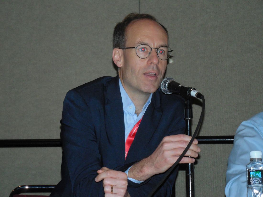
But these appeals for more liberal use of thrombectomy without the perfusion CT imaging used in DAWN (N Engl J Med. 2018 Jan 4;378[1]:11-21)and DEFUSE-3 (N Engl J Med. 2018 Feb 22;378[8]:708-18) received push back. Maarten G. Lansberg, MD, a co-investigator on the DEFUSE 3 trial, highlighted the speed and simplicity of CT perfusion imaging, and its utility in helping to better target thrombectomy to the right patients. It’s “speedy, simple, and safe,” it “excludes patients who will not benefit” from thrombectomy, and it helps when the patient’s history and noncontrast CT images are inconclusive, said Dr. Lansberg, a neurologist at Stanford (Calif.) University.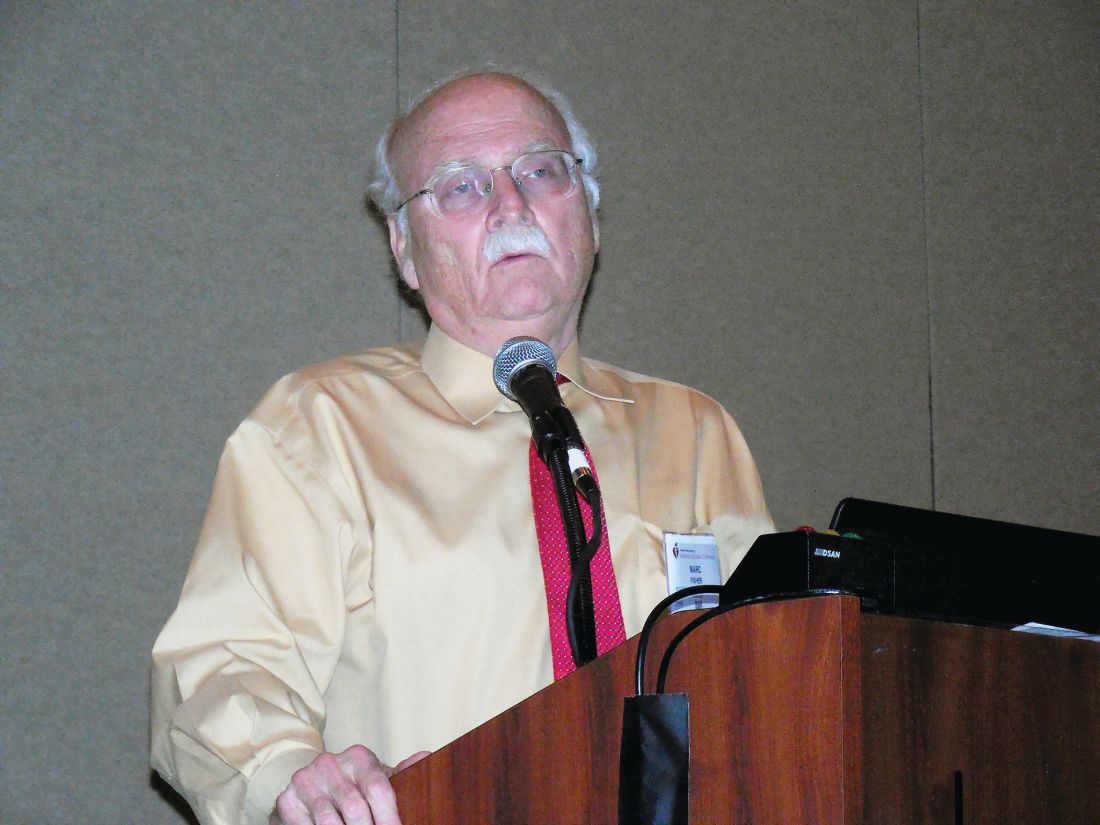
“Clinical presentation will only tell you so much.” With imaging that includes CT perfusion, “you can find out, in 5, 10 minutes, whether there is an occlusion, its location, the extent of dead tissue – that’s all really helpful,” said Marc Fisher, MD, professor of neurology at Harvard Medical School in Boston. “There is a tension now between doing treatment really fast and the concept of slow and fast evolvers. For slow evolvers, the concern about speed is irrelevant because it can take days” for their brains to have substantial damage. “For the fast evolvers, time matters, but they could also possibly be harmed; that’s why we need more data.”
A pitch for more data also came from Pooja Khatri, MD, who also spoke at the session. “There is a real tension now between personalizing the imaging and figuring out exactly the right patients against the time trade off for doing that. Some argue to keep it simple and move fast, and by doing that you’ll wash out any difference from doing more fancy stuff. Plus some places, even in developed countries, can’t afford the image-processing software” used in the DAWN and DEFUSE 3 trials. The correct approach remains unclear and has created “an area ripe for a trial,” declared Dr. Khatri, professor of neurology at director of acute stroke at the University of Cincinnati.
Dr. Hill has received honoraria from Merck and received research funding from Boehringer Ingelheim, Covidien, Medtronic, and Stryker. He has an ownership interest in Calgary Scientific and holds a patent on acute stroke triage methods. Dr. Nogueira has financial relationships with many companies. Dr. Lansberg and Dr. Fisher had no disclosures. Dr. Khatri has been a consultant to Lumosa and has received research funding from Cerenovus/Johnson & Johnson, Genentech, and Nervive.
HONOLULU – A little more than a year ago, results from the DAWN and DEFUSE 3 trials substantially broadened the time window for endovascular thrombectomy of acute ischemic stroke by selecting patients using brain imaging. Stroke clinicians are now trying to reconcile widespread, routine use of this life-changing treatment against an uncertain need to replicate the higher-end perfusion CT and analytical software imaging that these landmark trials used for patient selection. This has produced a schism in what experts advise for using endovascular thrombectomy on acute ischemic stroke patients.
“Go open the artery, people!” Michael D. Hill, MD, exhorted during a talk at the International Stroke Conference, sponsored by the American Heart Association. “Don’t get over-selective; more people will benefit than you think,” said Dr. Hill, a professor of clinical neurosciences at the University of Calgary (Alta.).
“We are over-selecting, and depriving patients,” commented Raul C. Nogueira, MD, professor of neurology at Emory University in Atlanta and a lead investigator of the DAWN trial, speaking from the audience during a discussion at the session where Dr. Hill spoke.
“The prevalence of treatable [acute ischemic stroke] patients is far higher than the prevalence of patients who are not good candidates, so you just want to exclude the ‘wipe-outs’; that’s what we do,” Dr. Hill explained. “Fortunately, endovascular therapy is very safe, and you’re not going to harm many patients. With other treatments [used routinely in medicine] some patients don’t benefit, but when you have a large effect size we use the treatment on almost everyone. The effect size from thrombectomy is so large it’s an argument to treat almost everyone, although the patients in the trials were selected by imaging.”
Dr. Hill repeatedly stressed that for most patients a non-contrast CT image is usually adequate to identify patients with salvageable brain tissue and a low risk for hemorrhage from intervention, and he endorsed also doing CT angiography to further inform the diagnosis. But he dismissed CT perfusion imaging as unnecessary. “Noncontrast CT and CT angiography are more than adequate to make treatment decisions,” he said. “The prevalence of poor collaterals is quite low, about 10%,” which means that about 90% of acute ischemic stroke patients will have more slowly progressing infarction,” making them amenable to treatment in an expanded time window and boosting the volume of salvageable tissue.
But these appeals for more liberal use of thrombectomy without the perfusion CT imaging used in DAWN (N Engl J Med. 2018 Jan 4;378[1]:11-21)and DEFUSE-3 (N Engl J Med. 2018 Feb 22;378[8]:708-18) received push back. Maarten G. Lansberg, MD, a co-investigator on the DEFUSE 3 trial, highlighted the speed and simplicity of CT perfusion imaging, and its utility in helping to better target thrombectomy to the right patients. It’s “speedy, simple, and safe,” it “excludes patients who will not benefit” from thrombectomy, and it helps when the patient’s history and noncontrast CT images are inconclusive, said Dr. Lansberg, a neurologist at Stanford (Calif.) University.
“Clinical presentation will only tell you so much.” With imaging that includes CT perfusion, “you can find out, in 5, 10 minutes, whether there is an occlusion, its location, the extent of dead tissue – that’s all really helpful,” said Marc Fisher, MD, professor of neurology at Harvard Medical School in Boston. “There is a tension now between doing treatment really fast and the concept of slow and fast evolvers. For slow evolvers, the concern about speed is irrelevant because it can take days” for their brains to have substantial damage. “For the fast evolvers, time matters, but they could also possibly be harmed; that’s why we need more data.”
A pitch for more data also came from Pooja Khatri, MD, who also spoke at the session. “There is a real tension now between personalizing the imaging and figuring out exactly the right patients against the time trade off for doing that. Some argue to keep it simple and move fast, and by doing that you’ll wash out any difference from doing more fancy stuff. Plus some places, even in developed countries, can’t afford the image-processing software” used in the DAWN and DEFUSE 3 trials. The correct approach remains unclear and has created “an area ripe for a trial,” declared Dr. Khatri, professor of neurology at director of acute stroke at the University of Cincinnati.
Dr. Hill has received honoraria from Merck and received research funding from Boehringer Ingelheim, Covidien, Medtronic, and Stryker. He has an ownership interest in Calgary Scientific and holds a patent on acute stroke triage methods. Dr. Nogueira has financial relationships with many companies. Dr. Lansberg and Dr. Fisher had no disclosures. Dr. Khatri has been a consultant to Lumosa and has received research funding from Cerenovus/Johnson & Johnson, Genentech, and Nervive.
REPORTING FROM ISC 2019
Telerehabilitation is noninferior to in-clinic rehabilitation for poststroke arm function
HONOLULU – according to research presented at the International Stroke Conference sponsored by the American Heart Association. Telerehabilitation also provides patient education as effectively as in-clinic rehabilitation, said Steven C. Cramer, MD, professor of neurology at the University of California, Irvine.
Stroke is a leading cause of disability, and more than 80% of patients with stroke have motor deficits when they present to the ED. Research indicates that high doses of rehabilitation therapy improve brain and motor function. However, many patients get low amounts of rehabilitation because of obstacles such as travel difficulties and shortages of therapy providers. “We reasoned that telerehabilitation is ideally suited to efficiently provide a large dose of useful, high-quality rehab therapy after stroke,” Dr. Cramer said.
Participants received supervised and unsupervised therapy
He and his colleagues enrolled patients who had experienced a stroke during the previous 4-36 weeks and who had arm motor deficits into their study. Eligible participants were adults, had experienced ischemic stroke or intracerebral hemorrhage, and had an arm Fugl-Meyer score between 22 and 56 out of 66.
Dr. Cramer’s group randomized 124 participants at 11 National Institutes of Health StrokeNet sites to 6 weeks of intensive arm rehabilitation therapy, plus stroke education, delivered in clinic or at home by a telehealth system. For both groups, treatment included 36 sessions that each lasted for 70 minutes. Half of the sessions were supervised and half were not. All sessions included at least 15 minutes of arm exercises and at least 15 minutes of functional training. Unsupervised sessions also included at least 5 minutes of stroke education on topics such as prevention, risk factors, recognition, and treatment. Participants in the in-clinic group worked with therapists in the clinic on supervised days and at home with a personalized booklet on unsupervised days. Participants in the telerehabilitation group played specially designed and individually tailored computer games at home on all days and had video conferences with therapists on supervised days. The treatment groups included approximately equal numbers of patients; treatment duration, intensity, and frequency were matched between groups.
The investigators hypothesized that telerehabilitation was not inferior to in-clinic rehabilitation. The study’s primary endpoint was change in Fugl-Meyer score from baseline to 30 days after the end of therapy. Secondary end points included Box and Blocks score (that is, a measure of arm function), Stroke Impact Scale–hand, and gains in stroke knowledge. The researchers defined the noninferiority margin as 30% of the gains of the in-clinic group. End points were evaluated by blinded assessors.
Patients had clinically meaningful gains
Participants’ average age was 61 years; the mean baseline arm Fugl-Meyer score was 42. Stroke onset had occurred at a mean of 4.5 months previously, and most strokes were ischemic. In all, 10 participants dropped out of the study. The rate of compliance was 98.3% in the telerehabilitation group and 93.0% in the in-clinic group.
The change in Fugl-Meyer score from baseline to 30 days post therapy was 8.36 points in the in-clinic group and 7.86 points in the telerehabilitation group. The changes in this score were higher than the minimal clinically important difference. The difference between groups, adjusted for covariance, was approximately 0. In addition, the 95% confidence interval for the change in score in the telerehabilitation group was within the noninferiority margin. “We can say that telerehabilitation is not inferior” to in-clinic therapy, said Dr. Cramer.
Telerehabilitation also was noninferior to in-clinic rehabilitation on the Box and Blocks score, and gains in stroke knowledge were significant and comparable in both groups. “Interestingly, the arm motor gains did not differ whether the subjects had aphasia or not,” said Dr. Cramer.
The investigators measured activity-inherent motivation (that is, how much a patient likes rehabilitation) using the Physical Activity Enjoyment Scale. Scores were higher in the in-clinic group, compared with the telerehabilitation group. “People like going to sit with a live human, and they like the longer time with the live human. This is something for us to study further and understand,” said Dr. Cramer.
Dr. Cramer and colleagues observed six serious adverse events in the in-clinic group and one in the telerehabilitation group, such as pneumonia or palpitations, all of which were deemed unrelated to therapy. Adverse events related to therapy (for example, shoulder pain and fatigue) were equally distributed between the two groups.
“What we were trying to do with home-based telehealth does not compete with or replace traditional rehab medicine. It is expanding tools for occupational and physical therapists, for nurses and physicians,” said Dr. Cramer.
Future studies could examine the efficacy of telerehabilitation in the treatment of language deficits, leg weakness, micturition, and dysphagia. “We might also study telehealth such as this to see how we can improve access and lower the cost of poststroke rehab care,” he concluded.
The study was funded by the Eunice Kennedy Shriver National Institute Of Child Health & Human Development and several grants from the National Institute of Neurological Disorders and Stroke. Dr. Cramer has an ownership interest in TRCare, a company that plans to market a telerehabilitation system and was not involved in the study. In addition, he is a consultant or advisor for MicroTransponder, Dart Neuroscience, Neurolutions, Regenera, Abbvie, SanBio, and TRCare.
SOURCE: Cramer SC et al. ISC 2019, Abstract LB23.
REPORTING FROM ISC 2019
Intensive blood pressure lowering may not reduce risk of recurrent stroke
HONOLULU – according to research presented at the International Stroke Conference sponsored by the American Heart Association.
Combined with data from previous trials, these results support a target systolic blood pressure of less than 130 mm Hg and a diastolic blood pressure of less than 80 mm Hg for secondary stroke prevention, said Kazuo Kitagawa, MD, PhD.
Lowering blood pressure reduces the risk of recurrent stroke, but investigators have not identified the best target blood pressure for this indication. The Secondary Prevention of Small Subcortical Strokes Trial (SPS3) examined the efficacy of intensive blood pressure treatment for secondary stroke prevention. The investigators randomized more than 3,000 patients with recent lacunar stroke to intensive or standard blood pressure treatment. Intensive treatment (a target systolic blood pressure of less than 130 mm Hg) conferred a nonsignificant reduction of the risk of recurrent stroke. A 2018 meta-analysis of SPS3 and two smaller randomized controlled trials also showed that intensive treatment did not significantly reduce the risk of recurrent stroke.
A new multicenter trial
Dr. Kitagawa, of Tokyo Women’s Medical University, and colleagues conducted a new trial to evaluate whether intensive blood pressure reduction significantly reduced the risk of recurrent stroke, compared with standard treatment (a systolic target of less than 140 mm Hg and a diastolic target of less than 90 mm Hg). Between 2010 and 2016, they enrolled patients with a history of stroke within the previous 3 years at 140 hospitals in Japan. Participants were randomized to standard blood pressure treatment or intensive blood pressure treatment (defined in this study as a systolic target of less than 120 mm Hg and a diastolic target of less than 80 mm Hg). The primary end point was recurrent stroke.
Both treatment regimens were based on stepwise multidrug rationing. Step 1 was an angiotensin II receptor blockade (ARB), step 2 was the addition of diuretics, step 3 was the addition of calcium channel blockers, step 4 was an increase of the ARB, step 5 was increase of the calcium channel blocker, and step 6 was the addition of spironolactone.
This trial was stopped at the end of 2016 because of slow recruitment and funding cessation. Investigators randomized 1,280 patients out of a planned 2,000. Seventeen patients were excluded from analysis. At baseline, participants’ mean age was 67 years, and mean systolic blood pressure was 145 mm Hg. The qualifying event was ischemic stroke for 85% of patients and intracerebral hemorrhage for 15%. Mean follow-up duration was 3.9 years.
Intensive treatment reduced blood pressure
At 1 year, the mean systolic blood pressure was 132.0 mm Hg in the standard-treatment group and 123.7 mm Hg in the intensive-treatment group. Mean diastolic blood pressure was 77.5 mm Hg in the standard-treatment group and 72.8 mm Hg in the intensive-treatment group. The investigators observed a significant difference in blood pressure between the groups throughout the study period.
The annual rate of stroke recurrence was 2.26% in the standard-treatment group and 1.65% in the intensive-treatment group. Intensive treatment tended to reduce stroke recurrence (hazard ratio, 0.73), but the result was not statistically significant. “The nonsignificant finding might be due to early termination or the modest difference in blood pressure level [between groups],” said Dr. Kitagawa.
Subgroup analyses did not indicate any interaction between treatment group and age, sex, qualifying event, mean systolic blood pressure at baseline, or diabetes. The rate of ischemic stroke was similar between the two groups, but the rate of intracerebral hemorrhage was lower in the intensive treatment group than in the standard treatment group. The rate of serious adverse events was similar between treatment groups.
When Dr. Kitagawa and colleagues pooled their data with those examined in the 2018 meta-analysis, they found that intensive treatment significantly reduced the risk of recurrent stroke (hazard ratio, 0.68), compared with standard treatment.
This study was sponsored by Biomedis International.
SOURCE: Kitagawa K et al. ISC 2019, Abstract LB10.
REPORTING FROM ISC 2019
Andexanet alfa effectively reverses factor Xa inhibition
HONOLULU – according to a study presented at the International Stroke Conference sponsored by the American Heart Association. The medication is associated with a low rate of mortality resulting from intracerebral hemorrhage (ICH), compared with the general population of patients with ICH receiving anticoagulation.

Factor Xa inhibitors such as apixaban and rivaroxaban effectively prevent thromboembolic events but may cause or exacerbate acute major bleeding. Andexanet alfa, a modified, recombinant, inactive form of human factor Xa, was developed and approved as a reversal agent for factor Xa inhibitors. In a 2015 study, andexanet rapidly and safely reversed anti–factor Xa activity in large cohorts of patients without bleeding.
A single-cohort study
Truman John Milling Jr., MD, an emergency medicine physician at Dell Seton Medical Center at the University of Texas in Austin, and his colleagues conducted the Andexanet Alfa, a Novel Antidote to the Anticoagulation Effects of Factor Xa Inhibitors (ANNEXA-4) study to evaluate the drug’s safety and efficacy in patients with acute major bleeding associated with treatment with a factor Xa inhibitor. For participants to be eligible, their bleeding had to be life threatening with signs of hemodynamic compromise, be associated with a decrease in hemoglobin level of at least 2 g/dL, or occur in a critical organ such as the brain. An independent academic committee determined whether patients met these criteria.
The trial’s primary efficacy outcomes were change from baseline in anti–factor Xa activity and the percentage of patients with excellent or good hemostatic efficacy at 12 hours. The primary safety endpoints were death, thrombotic events, and the development of neutralizing antibodies to andexanet or to native factor X and factor Xa. The efficacy population included patients with major bleeding and baseline anti–factor Xa activity of at least 75 ng/mL. The safety population included all patients who received a dose of andexanet. The independent committee adjudicated the efficacy and safety outcomes.
Hemostasis was sustained for 12 hours
The investigators enrolled 352 participants into the study, all of whom received andexanet and were followed for at least 30 days or until death. The population’s mean age was 77 years. “These were older and sicker patients with a significant amount of comorbid disease,” said Dr. Milling. The primary indication for anticoagulation was atrial fibrillation in 80% of patients. The primary site of bleeding was intracranial in 64% of patients and gastrointestinal in 26% of patients. The remaining 10% of patients had bleeding affecting other areas (such as pericardial or intramuscular bleeding).
The investigators included 254 patients in the efficacy population. At the end of the administration of the andexanet bolus, the median value for anti–factor Xa activity decreased by 92% among participants receiving apixaban, 92% among participants receiving rivaroxaban, and 75% among patients receiving enoxaparin. Among patients receiving apixaban, the median value for anti–factor Xa activity was decreased by 32% at 4 hours, 34% at 8 hours, and 38% at 12 hours. Among patients receiving rivaroxaban, the median value for anti–factor Xa activity was decreased by 42% at 4 hours, 48% at 8 hours, and 62% at 12 hours.
Dr. Milling and his colleagues assessed hemostatic efficacy in 249 patients. Of this group, 82% achieved good or excellent hemostasis. Among participants with good or excellent hemostasis, 84% had excellent results, and 16% had good results. Subanalysis by factor Xa inhibitor, type of bleed, age, and dose of andexanet did not alter the findings significantly.
To determine whether hemostasis had been sustained sufficiently to prevent clinical deterioration, the investigators examined 71 patients with ICH and a single-compartment bleed. From 1 hour to 12 hours, one patient’s outcome changed from excellent/good to poor/none, and one patient’s outcome changed from excellent to good. For the majority of these patients, however, good hemostasis was sustained from 1 to 12 hours.
The rate of thromboembolic events was 9.7%, which is in the expected range for this population, said Dr. Milling. These events were distributed evenly among the 4 weeks of the study. Stroke and deep vein thrombosis accounted for most of these events, and pulmonary emboli and heart attacks occurred as well. “Once we restarted oral anticoagulation ... there were no more thrombotic events,” said Dr. Milling. No patient developed neutralizing antibodies to factor X or factor Xa, nor did any patient develop neutralizing antibodies to andexanet.
The overall mortality rate was 13.9%. The rate of mortality resulting from ICH was 15%, and the rate of mortality resulting from gastrointestinal bleeding was 11%. These results are impressive, considering that patients had received anticoagulants, said Dr. Milling.
Portola Pharmaceuticals, the maker of andexanet alfa, funded the study. Dr. Milling reported receiving funding and honoraria from the Population Health Research Institute at McMasters University, Janssen, CSL Behring, and Octapharma. He also received a small research payment from Portola Pharmaceuticals. Several of the investigators reported receiving funding from Portola Pharmaceuticals.
SOURCE: Milling TJ et al. ISC 2019, Abstract LB7.
HONOLULU – according to a study presented at the International Stroke Conference sponsored by the American Heart Association. The medication is associated with a low rate of mortality resulting from intracerebral hemorrhage (ICH), compared with the general population of patients with ICH receiving anticoagulation.
Factor Xa inhibitors such as apixaban and rivaroxaban effectively prevent thromboembolic events but may cause or exacerbate acute major bleeding. Andexanet alfa, a modified, recombinant, inactive form of human factor Xa, was developed and approved as a reversal agent for factor Xa inhibitors. In a 2015 study, andexanet rapidly and safely reversed anti–factor Xa activity in large cohorts of patients without bleeding.
A single-cohort study
Truman John Milling Jr., MD, an emergency medicine physician at Dell Seton Medical Center at the University of Texas in Austin, and his colleagues conducted the Andexanet Alfa, a Novel Antidote to the Anticoagulation Effects of Factor Xa Inhibitors (ANNEXA-4) study to evaluate the drug’s safety and efficacy in patients with acute major bleeding associated with treatment with a factor Xa inhibitor. For participants to be eligible, their bleeding had to be life threatening with signs of hemodynamic compromise, be associated with a decrease in hemoglobin level of at least 2 g/dL, or occur in a critical organ such as the brain. An independent academic committee determined whether patients met these criteria.
The trial’s primary efficacy outcomes were change from baseline in anti–factor Xa activity and the percentage of patients with excellent or good hemostatic efficacy at 12 hours. The primary safety endpoints were death, thrombotic events, and the development of neutralizing antibodies to andexanet or to native factor X and factor Xa. The efficacy population included patients with major bleeding and baseline anti–factor Xa activity of at least 75 ng/mL. The safety population included all patients who received a dose of andexanet. The independent committee adjudicated the efficacy and safety outcomes.
Hemostasis was sustained for 12 hours
The investigators enrolled 352 participants into the study, all of whom received andexanet and were followed for at least 30 days or until death. The population’s mean age was 77 years. “These were older and sicker patients with a significant amount of comorbid disease,” said Dr. Milling. The primary indication for anticoagulation was atrial fibrillation in 80% of patients. The primary site of bleeding was intracranial in 64% of patients and gastrointestinal in 26% of patients. The remaining 10% of patients had bleeding affecting other areas (such as pericardial or intramuscular bleeding).
The investigators included 254 patients in the efficacy population. At the end of the administration of the andexanet bolus, the median value for anti–factor Xa activity decreased by 92% among participants receiving apixaban, 92% among participants receiving rivaroxaban, and 75% among patients receiving enoxaparin. Among patients receiving apixaban, the median value for anti–factor Xa activity was decreased by 32% at 4 hours, 34% at 8 hours, and 38% at 12 hours. Among patients receiving rivaroxaban, the median value for anti–factor Xa activity was decreased by 42% at 4 hours, 48% at 8 hours, and 62% at 12 hours.
Dr. Milling and his colleagues assessed hemostatic efficacy in 249 patients. Of this group, 82% achieved good or excellent hemostasis. Among participants with good or excellent hemostasis, 84% had excellent results, and 16% had good results. Subanalysis by factor Xa inhibitor, type of bleed, age, and dose of andexanet did not alter the findings significantly.
To determine whether hemostasis had been sustained sufficiently to prevent clinical deterioration, the investigators examined 71 patients with ICH and a single-compartment bleed. From 1 hour to 12 hours, one patient’s outcome changed from excellent/good to poor/none, and one patient’s outcome changed from excellent to good. For the majority of these patients, however, good hemostasis was sustained from 1 to 12 hours.
The rate of thromboembolic events was 9.7%, which is in the expected range for this population, said Dr. Milling. These events were distributed evenly among the 4 weeks of the study. Stroke and deep vein thrombosis accounted for most of these events, and pulmonary emboli and heart attacks occurred as well. “Once we restarted oral anticoagulation ... there were no more thrombotic events,” said Dr. Milling. No patient developed neutralizing antibodies to factor X or factor Xa, nor did any patient develop neutralizing antibodies to andexanet.
The overall mortality rate was 13.9%. The rate of mortality resulting from ICH was 15%, and the rate of mortality resulting from gastrointestinal bleeding was 11%. These results are impressive, considering that patients had received anticoagulants, said Dr. Milling.
Portola Pharmaceuticals, the maker of andexanet alfa, funded the study. Dr. Milling reported receiving funding and honoraria from the Population Health Research Institute at McMasters University, Janssen, CSL Behring, and Octapharma. He also received a small research payment from Portola Pharmaceuticals. Several of the investigators reported receiving funding from Portola Pharmaceuticals.
SOURCE: Milling TJ et al. ISC 2019, Abstract LB7.
REPORTING FROM ISC 2019
Endarterectomy and stenting have similar efficacy in carotid stenosis
HONOLULU – , according to a pooled analysis presented at the International Stroke Conference sponsored by the American Heart Association. The treatments have similar rates of procedural complications and 4-year ipsilateral stroke, said Jon S. Matsumura, MD, chairman of the division of vascular surgery at the University of Wisconsin in Madison.
Asymptomatic severe carotid stenosis is the most common indication for carotid operations in the United States. Data support carotid endarterectomy in selected asymptomatic patients. Carotid artery stenting with embolic protection is a newer treatment option. Two of the five most recent large, randomized trials – CREST and ACT I – compared carotid stenting with endarterectomy in asymptomatic patients. Dr. Matsumura and his colleagues conducted a pooled analysis of these two trials to help inform the choice of treatment.
The investigators analyzed data from the CREST and ACT I studies, which had many similarities. The researchers in these trials carefully selected the surgeons and the interventionalists who participated in them. Each trial used single carotid stent systems, and both trials used routine, distally placed embolic protection. The trials had independent neurologic assessment, routine cardiac enzyme screening, and central clinical and adjudication committees.
Dr. Matsumura and his colleagues decided to conduct a patient-level pooled analysis and defined the primary endpoint as a composite of death, stroke, and myocardial infarction in the periprocedural period and any ipsilateral stroke within 4 years of randomization. They included in their analysis all randomized, asymptomatic patients who were younger than 80 years.
The analysis comprised 2,544 patients, 1,637 of whom were randomized to stenting, and 907 of whom were randomized to endarterectomy. The population included more than 1,000 patients with 3-year follow-up and more than 500 with 4-year follow-up. Patients randomized to stenting were slightly younger, but the percentage of patients older than age 65 was similar between groups. Current cigarette smoking was slightly more common among patients randomized to stenting. The groups were well balanced by sex, race, and risk factors such as hypertension, hyperlipidemia, and diabetes.
The rate of primary endpoint events was 5.3% in the stenting arm and 5.1% in the endarterectomy arm (hazard ratio with stenting, 1.02; 95% confidence interval, 0.7-1.5; P = .91). The rate of periprocedural stroke was 2.7% in the stenting arm and 1.5% in the endarterectomy arm (P = .07). The rate of periprocedural myocardial infarction was 0.6% in the stenting arm and 1.7% in the endarterectomy arm (P = .01). The rate of periprocedural stroke and death was 2.7% in the stenting arm and 1.6% in the endarterectomy arm (P = .07). The rate of 4-year ipsilateral stroke was 2.3% in the stenting arm and 2.2% in the endarterectomy arm (P = .97).
A secondary analysis indicated that the cumulative, 4-year rate of stroke-free survival was 93.2% in the stenting arm and 95.1% in the endarterectomy arm (P = .10). “Almost all this difference is the initial periprocedural hazard difference,” said Dr. Matsumura. The rate of cumulative 4-year survival was 91% in the stenting arm and 90.2% in the endarterectomy arm.
The results of the pooled analysis do not support the perception that stenting entails an increased risk of periprocedural stroke. “The majority of trials have been in symptomatic patients,” said Dr. Matsumura. “We’re studying asymptomatic patients. We’re also studying them in the context of second-generation devices.” The results may reflect the amount of device-related training that the researchers undertook, as well as the decision to use single-stent dedicated carotid systems, he added.
Sponsors of the analysis included the University of Wisconsin, Massachusetts General Hospital, the Medical University of South Carolina, the University of Alabama at Birmingham, Cardiovascular Associates, and Mayo Clinic Jacksonville.
REPORTING FROM ISC 2019
Injectable nimodipine does not improve outcomes of subarachnoid hemorrhage
HONOLULU – according to a study presented at the International Stroke Conference sponsored by the American Heart Association. However, the treatment does reduce the rate of angiographic vasospasm significantly and does not raise significant safety concerns.

Approximately 70% of patients with subarachnoid hemorrhage develop vasospasm, which can in turn cause delayed cerebral ischemia. The only evidence-based treatment for ischemia of the brain resulting from vasospasm is nimodipine, which has been the standard of care for more than a decade.
EG-1962 was developed to deliver higher amounts of nimodipine to the CNS and spastic vessels than oral nimodipine. The formulation contains 50-mcm particles of nimodipine combined with a biodegradable polymer. Pharmacokinetic studies indicate that EG-1962 successfully delivers higher amounts of nimodipine to the CNS than the oral formulation does.
A multisite, phase 3 trial
Stephan A. Mayer, MD, William T. Gossett Endowed Chair of Neurology at the Henry Ford Health System in Detroit, presented the trial results. He and his colleagues conducted NEWTON2, a phase 3 trial to evaluate the safety and efficacy of a single 600-mg intraventricular dose of EG-1962 in patients with aneurysmal subarachnoid hemorrhage, compared with those of oral nimodipine. Eligible participants had a World Federation of Neurosurgeons Score (WFNS) of 2-4, Glasgow Coma Scale scores of 7-14, and an indication for placement of an external ventricular drain (such as hydrocephalus). All patients had their aneurysms clipped or coiled.
On the first day after the operation, the investigators randomized patients in equal groups to EG-1962 plus oral placebo or oral nimodipine plus placebo injection. Patients in the EG-1962 arm received a 600-mg injection of nimodipine into the ventricular system. The primary endpoint was the proportion of subjects with an extended Glasgow Outcome Scale (eGOS) score of 6-8 (that is, minimal or mild disability) at day 90. The main secondary endpoint was the proportion of subjects with a Montreal Cognitive Assessment (MOCA) score of 26 or greater (indicating no important cognitive disability) at day 90. Safety outcomes included cerebral infarction, hypotension, and ventriculitis.
Dr. Mayer and his colleagues conducted the study at 65 centers in 11 countries. They planned to enroll 374 patients and conduct an interim analysis after the first 210 participants had their 90-day follow-up. An independent data monitoring committee reviewed the safety data as it was collected. The investigators stopped the study for futility after the preplanned interim analysis was completed.
The challenge of identifying responders
The two study arms were well matched on age and gender. The proportion of patients with poor WFNS was 45% in the EG-1962 arm and 53% in the oral nimodipine arm.
The rate of any vasospasm (such as symptomatic or by imaging) was 56% in the intervention group and 70% in the oral nimodipine group, a statistically significant difference. Follow-up angiography showed vasospasm in 50% of the intervention group, compared with 63% of the oral nimodipine group, which was also statistically significant.
When the investigators examined the primary endpoint, they found that 46% of patients who received EG-1962 had a favorable outcome, compared with 43% of patients who received oral nimodipine, a nonsignificant difference. If the investigators had defined a favorable outcome as an eGOS score of 4 or greater, “there would have been a more favorable effect,” said Dr. Mayer. The investigators found no difference between groups in MOCA score at day 90.
Subgroup analyses produced “puzzling” results, said Dr. Mayer. For example, more patients with poor-grade WFNS had a favorable outcome at 3 months in the EG-1962 group than in the nimodipine group, but fewer patients with good-grade WFNS had a favorable outcome at 3 months in the EG-1962 group than in the nimodipine group. In addition, the treatment effect was more evident in the United States than abroad.
The safety analysis indicated a trend toward less rescue therapy – defined as hypertensive therapy or any interventional approach – in the EG-1962 group (27%), compared with the oral nimodipine group (35%). The rate of hypotension was slightly lower in the EG-1962 group. The rates of treatment-emergent serious adverse events and hydrocephalus were not significantly different between groups.
The results may encourage neurologists to treat “extremely sick patients with highly refractory vasospasms,” said Dr. Mayer. “We have a biologically active agent. The problem is, though, that it’s very hard ... to prove efficacy in trials, because we do not fully understand who the responders were going to be.”
Dr. Mayer reported receiving consulting fees from Edge Therapeutics, the company that developed EG-1962, in the past for activities unrelated to this study. Other investigators are employees of Edge Therapeutics.
SOURCE: Mayer SA et al. ISC 2019, Abstract LB15.
HONOLULU – according to a study presented at the International Stroke Conference sponsored by the American Heart Association. However, the treatment does reduce the rate of angiographic vasospasm significantly and does not raise significant safety concerns.
Approximately 70% of patients with subarachnoid hemorrhage develop vasospasm, which can in turn cause delayed cerebral ischemia. The only evidence-based treatment for ischemia of the brain resulting from vasospasm is nimodipine, which has been the standard of care for more than a decade.
EG-1962 was developed to deliver higher amounts of nimodipine to the CNS and spastic vessels than oral nimodipine. The formulation contains 50-mcm particles of nimodipine combined with a biodegradable polymer. Pharmacokinetic studies indicate that EG-1962 successfully delivers higher amounts of nimodipine to the CNS than the oral formulation does.
A multisite, phase 3 trial
Stephan A. Mayer, MD, William T. Gossett Endowed Chair of Neurology at the Henry Ford Health System in Detroit, presented the trial results. He and his colleagues conducted NEWTON2, a phase 3 trial to evaluate the safety and efficacy of a single 600-mg intraventricular dose of EG-1962 in patients with aneurysmal subarachnoid hemorrhage, compared with those of oral nimodipine. Eligible participants had a World Federation of Neurosurgeons Score (WFNS) of 2-4, Glasgow Coma Scale scores of 7-14, and an indication for placement of an external ventricular drain (such as hydrocephalus). All patients had their aneurysms clipped or coiled.
On the first day after the operation, the investigators randomized patients in equal groups to EG-1962 plus oral placebo or oral nimodipine plus placebo injection. Patients in the EG-1962 arm received a 600-mg injection of nimodipine into the ventricular system. The primary endpoint was the proportion of subjects with an extended Glasgow Outcome Scale (eGOS) score of 6-8 (that is, minimal or mild disability) at day 90. The main secondary endpoint was the proportion of subjects with a Montreal Cognitive Assessment (MOCA) score of 26 or greater (indicating no important cognitive disability) at day 90. Safety outcomes included cerebral infarction, hypotension, and ventriculitis.
Dr. Mayer and his colleagues conducted the study at 65 centers in 11 countries. They planned to enroll 374 patients and conduct an interim analysis after the first 210 participants had their 90-day follow-up. An independent data monitoring committee reviewed the safety data as it was collected. The investigators stopped the study for futility after the preplanned interim analysis was completed.
The challenge of identifying responders
The two study arms were well matched on age and gender. The proportion of patients with poor WFNS was 45% in the EG-1962 arm and 53% in the oral nimodipine arm.
The rate of any vasospasm (such as symptomatic or by imaging) was 56% in the intervention group and 70% in the oral nimodipine group, a statistically significant difference. Follow-up angiography showed vasospasm in 50% of the intervention group, compared with 63% of the oral nimodipine group, which was also statistically significant.
When the investigators examined the primary endpoint, they found that 46% of patients who received EG-1962 had a favorable outcome, compared with 43% of patients who received oral nimodipine, a nonsignificant difference. If the investigators had defined a favorable outcome as an eGOS score of 4 or greater, “there would have been a more favorable effect,” said Dr. Mayer. The investigators found no difference between groups in MOCA score at day 90.
Subgroup analyses produced “puzzling” results, said Dr. Mayer. For example, more patients with poor-grade WFNS had a favorable outcome at 3 months in the EG-1962 group than in the nimodipine group, but fewer patients with good-grade WFNS had a favorable outcome at 3 months in the EG-1962 group than in the nimodipine group. In addition, the treatment effect was more evident in the United States than abroad.
The safety analysis indicated a trend toward less rescue therapy – defined as hypertensive therapy or any interventional approach – in the EG-1962 group (27%), compared with the oral nimodipine group (35%). The rate of hypotension was slightly lower in the EG-1962 group. The rates of treatment-emergent serious adverse events and hydrocephalus were not significantly different between groups.
The results may encourage neurologists to treat “extremely sick patients with highly refractory vasospasms,” said Dr. Mayer. “We have a biologically active agent. The problem is, though, that it’s very hard ... to prove efficacy in trials, because we do not fully understand who the responders were going to be.”
Dr. Mayer reported receiving consulting fees from Edge Therapeutics, the company that developed EG-1962, in the past for activities unrelated to this study. Other investigators are employees of Edge Therapeutics.
SOURCE: Mayer SA et al. ISC 2019, Abstract LB15.
REPORTING FROM ISC 2019
Second-generation hydrogel surpasses platinum for brain aneurysm closure
HONOLULU – A second-generation hydrogel coil that’s already approved for U.S. marketing proved superior to bare platinum wire for durably embolizing brain aneurysms in a multicenter, randomized trial with 600 patients.

For the study’s primary endpoint of recurrent intracranial aneurysm during follow-up as long as 18-24 months, the 297 patients treated with the hydrogel coil had a recurrence rate of 4%, compared with a 15% rate among patients treated with platinum wire, Bernard R. Bendok, MD, said at the International Stroke Conference sponsored by the American Heart Association. The hydrogel coil materials were also relatively easy to use – much easier than the first-generation hydrogel materials, according to Dr. Bendok – and the adverse event rate using the hydrogel coils was similar to the rate using platinum wire, the standard material for brain aneurysm embolization.
Given the similar safety and ease of use and superior efficacy, clinicians treating brain aneurysms may now increasingly use hydrogel coil. “I think these results will change practice. It’s compelling data,” Dr. Bendok predicted in an interview. “The problem with platinum is it is not conducive to healing.” When an aneurysm is “closed” by insertion of platinum wire, in reality most of the space within the aneurysm remains unfilled by metal. In contrast, animal models suggest that when hydrogel fills an aneurysm it forms a matrix for collagen deposition that further fills the aneurysm space in a wound-healing process, thereby making recurrence less likely, said Dr. Bendok, professor and chair of neurosurgery at the Mayo Clinic in Phoenix.
The HEAT (Hydrogel Endovascular Aneurysm Treatment) trial enrolled patients at 46 centers in the United States and Canada during June 2012–January 2016. Enrolled patients had a ruptured or unruptured brain aneurysm 3-14 mm in diameter, and averaged 57 years old. A blinded core laboratory reviewed brain scans to identify recurrent aneurysms. Of the 297 patients randomized to hydrogel coil treatment, 254 underwent follow-up at 3-12 months and 222 had additional follow-up at 18-24 months, the study’s prespecified time for the primary endpoint. Among 303 patients treated with platinum, 258 had follow-up after 3-12 months and 231 had follow-up at 18-24 months.
Among patients who entered the study with an unruptured aneurysm (nearly three-quarters of enrolled patients), the recurrence rate was 12% with platinum and 3% with hydrogel. Among the patients with a ruptured aneurysm, the recurrence rate was 24% with platinum and 8% with hydrogel. Adverse events occurred in 25% of those treated with platinum and 22% of those treated with hydrogel coil. Mortality during follow-up was 3% in the platinum arm and 2% in the hydrogel coil arm. The two groups also had similar outcomes as measured by the modified Rankin Scale and by quality-of-life measures.
HEAT was sponsored by MicroVention, the company that markets the tested hydrogel coil. Dr. Bendok had no personal disclosures.
mzoler@mdedge.com
SOURCE: Abi-Aad KR et al. ISC 2019, Abstract LB20.
HONOLULU – A second-generation hydrogel coil that’s already approved for U.S. marketing proved superior to bare platinum wire for durably embolizing brain aneurysms in a multicenter, randomized trial with 600 patients.
For the study’s primary endpoint of recurrent intracranial aneurysm during follow-up as long as 18-24 months, the 297 patients treated with the hydrogel coil had a recurrence rate of 4%, compared with a 15% rate among patients treated with platinum wire, Bernard R. Bendok, MD, said at the International Stroke Conference sponsored by the American Heart Association. The hydrogel coil materials were also relatively easy to use – much easier than the first-generation hydrogel materials, according to Dr. Bendok – and the adverse event rate using the hydrogel coils was similar to the rate using platinum wire, the standard material for brain aneurysm embolization.
Given the similar safety and ease of use and superior efficacy, clinicians treating brain aneurysms may now increasingly use hydrogel coil. “I think these results will change practice. It’s compelling data,” Dr. Bendok predicted in an interview. “The problem with platinum is it is not conducive to healing.” When an aneurysm is “closed” by insertion of platinum wire, in reality most of the space within the aneurysm remains unfilled by metal. In contrast, animal models suggest that when hydrogel fills an aneurysm it forms a matrix for collagen deposition that further fills the aneurysm space in a wound-healing process, thereby making recurrence less likely, said Dr. Bendok, professor and chair of neurosurgery at the Mayo Clinic in Phoenix.
The HEAT (Hydrogel Endovascular Aneurysm Treatment) trial enrolled patients at 46 centers in the United States and Canada during June 2012–January 2016. Enrolled patients had a ruptured or unruptured brain aneurysm 3-14 mm in diameter, and averaged 57 years old. A blinded core laboratory reviewed brain scans to identify recurrent aneurysms. Of the 297 patients randomized to hydrogel coil treatment, 254 underwent follow-up at 3-12 months and 222 had additional follow-up at 18-24 months, the study’s prespecified time for the primary endpoint. Among 303 patients treated with platinum, 258 had follow-up after 3-12 months and 231 had follow-up at 18-24 months.
Among patients who entered the study with an unruptured aneurysm (nearly three-quarters of enrolled patients), the recurrence rate was 12% with platinum and 3% with hydrogel. Among the patients with a ruptured aneurysm, the recurrence rate was 24% with platinum and 8% with hydrogel. Adverse events occurred in 25% of those treated with platinum and 22% of those treated with hydrogel coil. Mortality during follow-up was 3% in the platinum arm and 2% in the hydrogel coil arm. The two groups also had similar outcomes as measured by the modified Rankin Scale and by quality-of-life measures.
HEAT was sponsored by MicroVention, the company that markets the tested hydrogel coil. Dr. Bendok had no personal disclosures.
mzoler@mdedge.com
SOURCE: Abi-Aad KR et al. ISC 2019, Abstract LB20.
REPORTING FROM ISC 2019
MI, strokes spike during 30 days after cancer diagnosis
HONOLULU – During the first month after a new cancer diagnosis, patients face a substantially elevated risk for an arterial thromboembolic event – an MI or stroke – consistent with the well-described increased risk newly diagnosed cancer patients face from venous thromboembolism, based on findings from a prospective study of more than 4,000 people.
In the new study, 836 patients newly diagnosed with cancer had a 480% increased rate of a fatal or nonfatal MI or stroke during the 30 days following their diagnosis, compared with 3,339 matched people without cancer and after adjustment for baseline differences in demographics and cardiovascular risk factors, Babak B. Navi, MD, said while presenting a poster at the International Stroke Conference sponsored by the American Heart Association.
An additional analysis that focused on 210 of the 836 patients with incident cancer who had any of seven of the cancers known to pose the highest venous thromboembolism risk (lymphoma; gynecologic cancer; or cancer of the pancreas, stomach, lung, bladder, or testes) showed an 1,750% greater rate of incident MI or stroke during the first 30 days after diagnosis, compared with matched people without cancer, reported Dr. Navi, chief of the stroke and hospital neurology at Weill Cornell Medicine, New York.
In contrast, during both the period 1-3 months after the cancer diagnosis and more than 3 months after, the rate of MI or stroke among recently diagnosed cancer patients was not significantly different from the rate in comparator individuals, although the data showed modest trends toward more arterial thromboembolic events after a month, and the lack of statistically significant differences may have been a power issue, Dr. Navi suggested.
The reasons for this acutely increased risk for arterial thromboembolic events, as well as the early spike in venous thromboembolic events, are not completely clear, but they likely result from factors released by tumors, effects from the drugs that patients receive for cancer treatment, stress, and interruption of antithrombotic treatment. Dr. Navi fingered cancer-induced hypercoagulability as likely the biggest culprit. It may now be reasonable to test the idea of treating newly diagnosed cancer patients with agents that could reduce their risk for MI or stroke, such as aspirin or a statin, he said in an interview.
The new analysis used data collected in the REGARDS (Reasons for Geographic and Racial Differences in Stroke) study, which during 2003-2007 enrolled more than 30,000 U.S. residents who were at least 45 years old. Dr. Navi and his associates used data collected from all REGARDS participants who developed incident cancer and had continuous Medicare coverage for at least 1 year before entering REGARDS, excluding those with cancer before enrollment. They used the Medicare records to identify the cancer diagnoses, and matched each of these people with four similar but cancer-free people enrolled in the study. Follow-up continued through September 2015. The average age of this REGARDS subgroup at enrollment was 72 years old, and nearly half were women. The incident cancers included 640 patients with a solid tumor, 71 with hematologic cancers, 13 with brain tumors, and 112 with an unknown primary cancer site.
Dr. Navi and his associates designed this study to validate previously reported findings of increased arterial thromboembolic events in newly diagnosed cancer patients from studies of insurance claims databases. Although the increased risk for venous thromboembolism in cancer patients is already well established, documenting a similar risk for arterial events is important because they are “generally more impactful for patients than venous thromboembolism,” Dr. Navi said.
REGARDS has received no commercial funding. Dr. Navi reported no disclosures.
SOURCE: Navi BB et al. Stroke. 2019 Feb;50(Suppl_1): Abstract WMP53.
REPORTING FROM ISC 2019
Higher blood pressure after thrombectomy links with bad stroke outcomes
HONOLULU – Acute ischemic stroke patients who underwent endovascular thrombectomy and then had a peak systolic blood pressure of greater than 158 mm Hg during the next 24 hours had worse 90-day outcomes than did patients whose peak systolic pressure remained at or below 158 mm Hg in a prospective, multicenter, observational study with 485 patients.

The results hint that maintaining a lower systolic blood pressure after thrombectomy in acute ischemic stroke patients may improve outcomes, but because the current study was observational, the hypothesis that patients benefit when treatment keeps their systolic pressure at or below 158 mm Hg must undergo testing in a prospective, randomized trial, Eva A. Mistry, MBBS, said at the International Stroke Conference, sponsored by the American Heart Association.
The finding from this study that 158 mm Hg provided the best dichotomous division between systolic blood pressures linked with good or bad outcomes is a first step toward trying to devise a more systematic and evidence-based approach to blood pressure management in acute ischemic stroke patients following endovascular thrombectomy, said Dr. Mistry, a neurologist at Vanderbilt University in Nashville, Tenn.
Neither Vanderbilt nor any of the other 11 major U.S. stroke centers that participated in the current study currently have an established protocol for blood pressure management after thrombectomy, Dr. Mistry said in an interview.
“We usually treat to reduce blood pressure, but we don’t have a [broadly agreed on] threshold” to trigger treatment. “It depends on a collective decision” by the various medical specialists who care for an individual acute stroke patient. In addition, no consensus yet exists for the best treatment strategy for blood pressure lowering in acute ischemic stroke patients. Intravenous nicardipine is often the top choice because it is fast-acting and easy to administer and control as an intravenous agent. Those same properties make the beta blocker labetalol a frequently used second drug, she said.
The BEST (Blood Pressure After Endovascular Stroke Therapy) study ran at 12 U.S. centers and enrolled 485 patients who underwent endovascular thrombectomy to treat an acute ischemic stroke. The patients averaged 69 years old, and 48% also underwent thrombolytic treatment. The study’s primary outcome was the percentage of patients with a modified Rankin Scale score of 0-2 at 90 days after their stroke, an outcome reached by 39% of all patients in the study.
Statistical analysis of the collected data showed that a peak systolic blood pressure of 158 mm Hg reached during the 24 hours following thrombectomy best divided patients with good 90-day outcomes from those with worse outcomes. Patients with a postthrombectomy peak systolic pressure above 158 mm Hg had a 2.2-fold increased rate of having a modified Rankin Scale score of 3 or higher after 90 days, a statistically significant relationship, Dr. Mistry reported. However, in an analysis that also adjusted for age, baseline stroke severity, glucose level, time to reperfusion, ASPECTS score, history of hypertension, and recanalization status, the elevated risk for a bad outcome linked with higher systolic pressure dropped to 39% greater than that for patients with systolic pressures that did not rise above 158 mm Hg, a difference that was not statistically significant. This suggests that these adjustments were unable to account for all confounders and further highlighted the need for a prospective, randomized trial to test the value of controlling blood pressure following thrombectomy, Dr. Mistry said. The unadjusted results confirmed a prior report from Dr. Mistry and her associates that found a link between higher blood pressure after stroke thrombectomy and worse outcomes (J Am Heart Assoc. 2017 May 18. doi: 10.1161/JAHA.117.006167).
The analysis also showed that patients who were successfully recanalized by thrombectomy, achieving a thrombolysis in cerebral infarction (TICI) score of 2b or 3, had lower peak systolic blood pressures than did patients who failed to get this level of restored cerebral blood flow from thrombectomy.
BEST received no commercial funding. Dr. Mistry had no disclosures.
SOURCE: Mistry EA et al. Stroke. 2019 Feb;50(Suppl_1): Abstract 94.
HONOLULU – Acute ischemic stroke patients who underwent endovascular thrombectomy and then had a peak systolic blood pressure of greater than 158 mm Hg during the next 24 hours had worse 90-day outcomes than did patients whose peak systolic pressure remained at or below 158 mm Hg in a prospective, multicenter, observational study with 485 patients.
The results hint that maintaining a lower systolic blood pressure after thrombectomy in acute ischemic stroke patients may improve outcomes, but because the current study was observational, the hypothesis that patients benefit when treatment keeps their systolic pressure at or below 158 mm Hg must undergo testing in a prospective, randomized trial, Eva A. Mistry, MBBS, said at the International Stroke Conference, sponsored by the American Heart Association.
The finding from this study that 158 mm Hg provided the best dichotomous division between systolic blood pressures linked with good or bad outcomes is a first step toward trying to devise a more systematic and evidence-based approach to blood pressure management in acute ischemic stroke patients following endovascular thrombectomy, said Dr. Mistry, a neurologist at Vanderbilt University in Nashville, Tenn.
Neither Vanderbilt nor any of the other 11 major U.S. stroke centers that participated in the current study currently have an established protocol for blood pressure management after thrombectomy, Dr. Mistry said in an interview.
“We usually treat to reduce blood pressure, but we don’t have a [broadly agreed on] threshold” to trigger treatment. “It depends on a collective decision” by the various medical specialists who care for an individual acute stroke patient. In addition, no consensus yet exists for the best treatment strategy for blood pressure lowering in acute ischemic stroke patients. Intravenous nicardipine is often the top choice because it is fast-acting and easy to administer and control as an intravenous agent. Those same properties make the beta blocker labetalol a frequently used second drug, she said.
The BEST (Blood Pressure After Endovascular Stroke Therapy) study ran at 12 U.S. centers and enrolled 485 patients who underwent endovascular thrombectomy to treat an acute ischemic stroke. The patients averaged 69 years old, and 48% also underwent thrombolytic treatment. The study’s primary outcome was the percentage of patients with a modified Rankin Scale score of 0-2 at 90 days after their stroke, an outcome reached by 39% of all patients in the study.
Statistical analysis of the collected data showed that a peak systolic blood pressure of 158 mm Hg reached during the 24 hours following thrombectomy best divided patients with good 90-day outcomes from those with worse outcomes. Patients with a postthrombectomy peak systolic pressure above 158 mm Hg had a 2.2-fold increased rate of having a modified Rankin Scale score of 3 or higher after 90 days, a statistically significant relationship, Dr. Mistry reported. However, in an analysis that also adjusted for age, baseline stroke severity, glucose level, time to reperfusion, ASPECTS score, history of hypertension, and recanalization status, the elevated risk for a bad outcome linked with higher systolic pressure dropped to 39% greater than that for patients with systolic pressures that did not rise above 158 mm Hg, a difference that was not statistically significant. This suggests that these adjustments were unable to account for all confounders and further highlighted the need for a prospective, randomized trial to test the value of controlling blood pressure following thrombectomy, Dr. Mistry said. The unadjusted results confirmed a prior report from Dr. Mistry and her associates that found a link between higher blood pressure after stroke thrombectomy and worse outcomes (J Am Heart Assoc. 2017 May 18. doi: 10.1161/JAHA.117.006167).
The analysis also showed that patients who were successfully recanalized by thrombectomy, achieving a thrombolysis in cerebral infarction (TICI) score of 2b or 3, had lower peak systolic blood pressures than did patients who failed to get this level of restored cerebral blood flow from thrombectomy.
BEST received no commercial funding. Dr. Mistry had no disclosures.
SOURCE: Mistry EA et al. Stroke. 2019 Feb;50(Suppl_1): Abstract 94.
REPORTING FROM ISC 2019
Glyceryl trinitrate does not improve outcomes of ischemic stroke
HONOLULU – , according to data presented at the International Stroke Conference sponsored by the American Heart Association. Results suggest that GTN causes adverse effects in patients with intracerebral hemorrhage (ICH), but this observation is not definitive, according to the researchers. Study results were published online ahead of print Feb. 6 in the Lancet.
Nitric oxide is a regulatory molecule that has vasoactive effects and promotes blood pressure reduction. Vascular levels of nitric oxide are low in stroke, which suggests that the molecule may be a target for stroke treatment. GTN, a nitric oxide donor, lowered blood pressure and improved functional outcome among patients with acute stroke in the phase 2 Rapid Intervention with GTN in Hypertensive Stroke Trial (RIGHT).
Philip Bath, MD, Stroke Association Professor of Stroke Medicine at the University of Nottingham (England), and colleagues conducted the RIGHT-2 study to evaluate the safety and efficacy of GTN when administered early after onset of suspected stroke. Paramedics randomized patients in equal groups to a GTN patch or a sham patch in the ambulance. Three more patches were administered in the hospital on the following days. Active and sham patches looked similar and had no writing on them, thus ensuring effective blinding upon administration. Investigators followed up patients by telephone at 90 days to assess the modified Rankin Scale score and markers of disability, mood, cognition, and quality of life.
Eligible participants were adults who had dialed emergency services, independently or with assistance, because of a possible stroke. They had a Face, Arm, Speech, Time (FAST) score of 2 or 3, were within 4 hours of onset, and had a systolic blood pressure greater than 120 mm Hg. Patients from nursing homes, those with hypoglycemia, those who were unconscious, and those with a witnessed seizure were excluded.
Dr. Bath and colleagues planned to enroll 850 patients from five ambulance services in 30 hospitals across the United Kingdom. Data were to be examined through an intention-to-treat analysis. During the trial, however, the investigators observed that the rate of stroke mimics was 26%, rather than the 12% that they had anticipated. To ensure the proper power for the study, the investigators increased the sample size to 1,149 patients. They also changed the planned data analysis from intention-to-treat to hierarchical analysis. Specifically, the researchers planned to perform the primary analysis in patients with stroke or TIA. If the results were positive, then they would perform a standard intention-to-treat analysis.
More than 99% of patients received the first patch. Approximately 57% of the population received the first two patches. One reason for this decrease in adherence was that many patients were discharged from the hospital with a TIA or a stroke mimic. Participants’ average age was 72. The median time from onset to randomization was 71 minutes, and the median time to treatment was 73 minutes. Participants’ mean systolic blood pressure was 162 mm Hg. Approximately 60% of the patients had a FAST score of 3. About 50% of participants had ischemic stroke, 13% had ICH, 10% had TIA, and 26% had stroke mimics.
At 1 hour after treatment initiation, systolic blood pressure decreased by 6.2 mm Hg and diastolic blood pressure decreased by 2.7 mm Hg among patients who received GTN, compared with controls. At one day, the differences were 5.2 mm Hg and 2.5 mm Hg, respectively, in treated patients, compared with controls. Blood pressure became similar between groups thereafter, “in part because of the tachyphylaxis that we know happens with GTN,” said Dr. Bath.
The researchers found no evidence of an effect of GTN on functional outcome at 90 days in participants with stroke or transient ischemic attack. The adjusted common odds ratio of poor outcome was 1.25 in the GTN group, compared with the control group (95 % confidence interval, 0.97-1.60; P = .083). “We were close to getting a negative trial,” said Dr. Bath.
Subgroup analyses revealed differences in outcome according to the time to randomization. GTN had a negative effect in patients treated within 1 hour of onset. Results were neutral, but tended to be negative, in patients treated between 1 and 2 hours of onset. Results were neutral, but tended to be positive, among patients treated at more than 2 hours after onset. There was no difference between groups in the rate of mortality.
One of the study’s limitations was its single-blind design. In addition, the trial was conducted in a single country, and the investigators changed the protocol after it was initiated. “We had a higher-than-expected [stroke] mimic rate, although I’m reassured by most experts that ... this is probably about right,” said Dr. Bath.
A potential reason for the neutral results is the negative effect that GTN had among patients with ICH, said Dr. Bath. “In that very early first hour, we are of course breaking a law that we learned in medical school, which is that the first part of hemostasis is spasm. We gave an antispasmodic: a vasodilator,” he added. “That is speculation.”
The trial was funded by the British Heart Foundation. Dr. Bath declared a modest ownership interest in Platelet Solutions and consultant or advisory board positions with Moleac, DiaMedica, Phagenesis, Nestle, and ReNeuron. The other investigators declared no conflicts of interest.
The RIGHT-2 trial shows the limitations of a prehospital enrollment model, wrote Karen C. Johnston, MD, professor of neurology at the University of Virginia in Charlottesville, and Valerie L. Durkalski-Mauldin, PhD, professor of medicine at Medical University of South Carolina in Charleston, in an editorial accompanying the RIGHT-2 trial results. The rate of nonstroke diagnoses was so high that it would have reduced the study’s power to assess the efficacy of glyceryl trinitrate (GTN), had the investigators not increased the sample size and changed the statistical analysis plan.
“Future prehospital trials need to consider the implications of enrolling, yet excluding, stroke mimics in the primary analysis,” said Dr. Johnston and Dr. Durkalski-Mauldin. Using telemedicine in the ambulance to facilitate direct contact between the stroke provider and the patient and emergency medical services provider could reduce the enrollment of patients with stroke mimics in clinical trials, they added. “Improved tools to exclude stroke mimics in the field have been difficult to develop and validate. The absence of imaging in most ambulances will continue to limit field personnel from definitively determining ischemic stroke from intracerebral hemorrhage, which will limit hyperacute trials to interventions presumed safe in both populations.”
In addition, the blood pressure reduction that GTN provided might not be clinically relevant, said Dr. Johnston and Dr. Durkalski-Mauldin. “The RIGHT-2 investigators report no difference in blood pressure at day 3 or day 4 of treatment, which might have been related to the very low adherence to study protocol by day 4.
“Regardless of these limitations, RIGHT-2 has provided high-level evidence that GTN given within 4 hours of onset does not significantly improve outcome in hyperacute patients presenting with possible stroke,” the authors concluded (Lancet. 2019 Feb 6. doi: 10.1016/
S0140-6736(19)30276-4). Dr. Johnston and Dr. Durkalski-Mauldin declared no conflicts of interest.
HONOLULU – , according to data presented at the International Stroke Conference sponsored by the American Heart Association. Results suggest that GTN causes adverse effects in patients with intracerebral hemorrhage (ICH), but this observation is not definitive, according to the researchers. Study results were published online ahead of print Feb. 6 in the Lancet.
Nitric oxide is a regulatory molecule that has vasoactive effects and promotes blood pressure reduction. Vascular levels of nitric oxide are low in stroke, which suggests that the molecule may be a target for stroke treatment. GTN, a nitric oxide donor, lowered blood pressure and improved functional outcome among patients with acute stroke in the phase 2 Rapid Intervention with GTN in Hypertensive Stroke Trial (RIGHT).
Philip Bath, MD, Stroke Association Professor of Stroke Medicine at the University of Nottingham (England), and colleagues conducted the RIGHT-2 study to evaluate the safety and efficacy of GTN when administered early after onset of suspected stroke. Paramedics randomized patients in equal groups to a GTN patch or a sham patch in the ambulance. Three more patches were administered in the hospital on the following days. Active and sham patches looked similar and had no writing on them, thus ensuring effective blinding upon administration. Investigators followed up patients by telephone at 90 days to assess the modified Rankin Scale score and markers of disability, mood, cognition, and quality of life.
Eligible participants were adults who had dialed emergency services, independently or with assistance, because of a possible stroke. They had a Face, Arm, Speech, Time (FAST) score of 2 or 3, were within 4 hours of onset, and had a systolic blood pressure greater than 120 mm Hg. Patients from nursing homes, those with hypoglycemia, those who were unconscious, and those with a witnessed seizure were excluded.
Dr. Bath and colleagues planned to enroll 850 patients from five ambulance services in 30 hospitals across the United Kingdom. Data were to be examined through an intention-to-treat analysis. During the trial, however, the investigators observed that the rate of stroke mimics was 26%, rather than the 12% that they had anticipated. To ensure the proper power for the study, the investigators increased the sample size to 1,149 patients. They also changed the planned data analysis from intention-to-treat to hierarchical analysis. Specifically, the researchers planned to perform the primary analysis in patients with stroke or TIA. If the results were positive, then they would perform a standard intention-to-treat analysis.
More than 99% of patients received the first patch. Approximately 57% of the population received the first two patches. One reason for this decrease in adherence was that many patients were discharged from the hospital with a TIA or a stroke mimic. Participants’ average age was 72. The median time from onset to randomization was 71 minutes, and the median time to treatment was 73 minutes. Participants’ mean systolic blood pressure was 162 mm Hg. Approximately 60% of the patients had a FAST score of 3. About 50% of participants had ischemic stroke, 13% had ICH, 10% had TIA, and 26% had stroke mimics.
At 1 hour after treatment initiation, systolic blood pressure decreased by 6.2 mm Hg and diastolic blood pressure decreased by 2.7 mm Hg among patients who received GTN, compared with controls. At one day, the differences were 5.2 mm Hg and 2.5 mm Hg, respectively, in treated patients, compared with controls. Blood pressure became similar between groups thereafter, “in part because of the tachyphylaxis that we know happens with GTN,” said Dr. Bath.
The researchers found no evidence of an effect of GTN on functional outcome at 90 days in participants with stroke or transient ischemic attack. The adjusted common odds ratio of poor outcome was 1.25 in the GTN group, compared with the control group (95 % confidence interval, 0.97-1.60; P = .083). “We were close to getting a negative trial,” said Dr. Bath.
Subgroup analyses revealed differences in outcome according to the time to randomization. GTN had a negative effect in patients treated within 1 hour of onset. Results were neutral, but tended to be negative, in patients treated between 1 and 2 hours of onset. Results were neutral, but tended to be positive, among patients treated at more than 2 hours after onset. There was no difference between groups in the rate of mortality.
One of the study’s limitations was its single-blind design. In addition, the trial was conducted in a single country, and the investigators changed the protocol after it was initiated. “We had a higher-than-expected [stroke] mimic rate, although I’m reassured by most experts that ... this is probably about right,” said Dr. Bath.
A potential reason for the neutral results is the negative effect that GTN had among patients with ICH, said Dr. Bath. “In that very early first hour, we are of course breaking a law that we learned in medical school, which is that the first part of hemostasis is spasm. We gave an antispasmodic: a vasodilator,” he added. “That is speculation.”
The trial was funded by the British Heart Foundation. Dr. Bath declared a modest ownership interest in Platelet Solutions and consultant or advisory board positions with Moleac, DiaMedica, Phagenesis, Nestle, and ReNeuron. The other investigators declared no conflicts of interest.
REPORTING FROM ISC 2019