User login
Skin Disease Education Foundation (SDEF): Hawaii Dermatology Seminar
New study supports imiquimod for lentigo maligna
WAIKOLOA, HAWAII – The topical immunomodulator imiquimod appears to be beneficial therapy in selected patients with lentigo maligna or lentigo maligna melanoma, according to the largest case series to date.
"Based upon our experience, imiquimod 5% cream is a viable option as primary or adjunctive therapy of lentigo maligna and the in situ component of lentigo maligna melanoma in patients where surgery is really not feasible, or you’ve optimized surgical margins without achieving a clear histologic margin. I emphasize this should only be considered for in situ melanoma; we’re not using it to treat invasive melanoma," Dr. Susan M. Swetter said at the Hawaii Dermatology Seminar sponsored by the Global Academy for Medical Education/Skin Disease Education Foundation.

A cautionary note: Imiquimod (Aldara) for lentigo maligna (LM) is off-label therapy. It’s crucial to discuss with candidates the limitations of nonsurgical treatment for LM and lentigo maligna melanoma (LMM), which includes increased risk of local recurrence because of the lack of margin control, the possibility of a missed invasive melanoma, and the lack of supporting evidence from randomized trials with long-term follow-up.
"When we’re talking about imiquimod as potential treatment for lentigo maligna – and I can’t emphasize this enough – it requires close clinical follow-up, [carefully] documented discussion with the patient, and strong patient compliance. We have had cases of lentigo maligna melanoma where there have been metastases, not because of the in situ component, but because of the initial invasive tumor. You’ve got to follow these patients long term like a hawk. I follow them with Wood’s lamp and dermoscopy, and I biopsy any pigmentation," said Dr. Swetter, professor of dermatology and director of the pigmented lesion and cutaneous melanoma clinic at Stanford (Calif.) University Medical Center.
That being said, there are many scenarios in which surgical excision – the recommended first-line treatment for LM and LMM – might not be feasible. These include the sizable numbers of patients of advanced age who have limiting comorbid medical conditions or cognitive impairment. Also, LM has a strong predilection for the head and neck, and some affected patients avidly wish to avoid potentially disfiguring surgery.
Dr. Swetter presented a retrospective study of 60 patients with 62 LM or LMM lesions treated with imiquimod 5% cream at Stanford or the Veterans Affairs Palo Alto Health Care System. Most she treated personally. Imiquimod was primary therapy in 20 cases and adjuvant therapy following failed surgical attempts to achieve histologic clearance in the rest.
Pathologically, 45% of the lesions were LM, 29% were atypical intraepidermal melanocytic proliferations or poorly evolving LM, and 26% were LMM with histologic transection of LM and excision of the invasive melanoma component. Three-quarters of treated lesions were on the head or neck.
The lesions had a mean Breslow depth of 1.17 mm. Prior to imiquimod, half of the patients had one excision and another 19% had two or more. The mean duration of imiquimod therapy was 10.9 weeks, with a mean post treatment follow-up of 38 months.
"I typically treat to a 2-cm margin around the lesion," the dermatologist noted.
The overall clinical or histologic clearance rate was 86%. This is strikingly similar to the collective 82% rate in 46 reports involving 264 treated patients described in a published review by dermatologists at the University of Colorado, Denver (Dermatol. Surg. 2012;38:937-46).
An inflammatory response to imiquimod was predictive of favorable treatment outcome, especially in patients using the topical agent as primary therapy. Of the 20 patients who used imiquimod as primary therapy, all 16 who experienced clinical and/or histologic clearance had an initial inflammatory response. In contrast, none of the four clinical nonresponders showed an inflammatory response.
Outcomes weren’t quite as good in patients using imiquimod as adjuvant therapy.
"This is likely related to more challenging cases in the adjuvant setting, where the patients may have failed multiple attempts at wide local excision for histologic clearance," Dr. Swetter surmised.
Although the use of imiquimod as treatment for LM remains off label, it’s worth noting that it receives support in major practice guidelines. National Comprehensive Cancer Network guidelines recommend consideration of topical imiquimod or radiotherapy as adjunctive treatment for LM in selected patients after optimal surgery, with a level 2B recommendation. And the American Academy of Dermatology lists imiquimod as among the adjunctive options (J. Am. Acad. Dermatol. 2009; 61:865-7). What’s really needed now, according to Dr. Swetter, is a randomized controlled trial to firmly demonstrate benefit.

Session chair Dr. Allan C. Halpern said he and his colleagues have also been using topical imiquimod to treat LM with impressive results.
"What we’ve been doing, without literature to support it, is a broad shave biopsy, essentially removing the lesion, and then going right in with imiquimod before the patient heals. We’ve gotten some really wonderful clinical results," said Dr. Halpern, chief of the dermatology service at Memorial Sloan-Kettering Cancer Center in New York.
"That said, I think it’s important to remind people that – even with surgery – we do see occasional recurrences of desmoplastic melanomas in association with prior lentigo maligna. There’s no reason to think that’s going to be any less common in the imiquimod era. It’s going to happen to us," he cautioned.
Dr. Swetter reported having no financial conflicts. Dr. Halpern has received research grants from SciBase, DermTech, Caliber, and Canfield.
SDEF and this news organization are owned by the same parent company.
WAIKOLOA, HAWAII – The topical immunomodulator imiquimod appears to be beneficial therapy in selected patients with lentigo maligna or lentigo maligna melanoma, according to the largest case series to date.
"Based upon our experience, imiquimod 5% cream is a viable option as primary or adjunctive therapy of lentigo maligna and the in situ component of lentigo maligna melanoma in patients where surgery is really not feasible, or you’ve optimized surgical margins without achieving a clear histologic margin. I emphasize this should only be considered for in situ melanoma; we’re not using it to treat invasive melanoma," Dr. Susan M. Swetter said at the Hawaii Dermatology Seminar sponsored by the Global Academy for Medical Education/Skin Disease Education Foundation.
A cautionary note: Imiquimod (Aldara) for lentigo maligna (LM) is off-label therapy. It’s crucial to discuss with candidates the limitations of nonsurgical treatment for LM and lentigo maligna melanoma (LMM), which includes increased risk of local recurrence because of the lack of margin control, the possibility of a missed invasive melanoma, and the lack of supporting evidence from randomized trials with long-term follow-up.
"When we’re talking about imiquimod as potential treatment for lentigo maligna – and I can’t emphasize this enough – it requires close clinical follow-up, [carefully] documented discussion with the patient, and strong patient compliance. We have had cases of lentigo maligna melanoma where there have been metastases, not because of the in situ component, but because of the initial invasive tumor. You’ve got to follow these patients long term like a hawk. I follow them with Wood’s lamp and dermoscopy, and I biopsy any pigmentation," said Dr. Swetter, professor of dermatology and director of the pigmented lesion and cutaneous melanoma clinic at Stanford (Calif.) University Medical Center.
That being said, there are many scenarios in which surgical excision – the recommended first-line treatment for LM and LMM – might not be feasible. These include the sizable numbers of patients of advanced age who have limiting comorbid medical conditions or cognitive impairment. Also, LM has a strong predilection for the head and neck, and some affected patients avidly wish to avoid potentially disfiguring surgery.
Dr. Swetter presented a retrospective study of 60 patients with 62 LM or LMM lesions treated with imiquimod 5% cream at Stanford or the Veterans Affairs Palo Alto Health Care System. Most she treated personally. Imiquimod was primary therapy in 20 cases and adjuvant therapy following failed surgical attempts to achieve histologic clearance in the rest.
Pathologically, 45% of the lesions were LM, 29% were atypical intraepidermal melanocytic proliferations or poorly evolving LM, and 26% were LMM with histologic transection of LM and excision of the invasive melanoma component. Three-quarters of treated lesions were on the head or neck.
The lesions had a mean Breslow depth of 1.17 mm. Prior to imiquimod, half of the patients had one excision and another 19% had two or more. The mean duration of imiquimod therapy was 10.9 weeks, with a mean post treatment follow-up of 38 months.
"I typically treat to a 2-cm margin around the lesion," the dermatologist noted.
The overall clinical or histologic clearance rate was 86%. This is strikingly similar to the collective 82% rate in 46 reports involving 264 treated patients described in a published review by dermatologists at the University of Colorado, Denver (Dermatol. Surg. 2012;38:937-46).
An inflammatory response to imiquimod was predictive of favorable treatment outcome, especially in patients using the topical agent as primary therapy. Of the 20 patients who used imiquimod as primary therapy, all 16 who experienced clinical and/or histologic clearance had an initial inflammatory response. In contrast, none of the four clinical nonresponders showed an inflammatory response.
Outcomes weren’t quite as good in patients using imiquimod as adjuvant therapy.
"This is likely related to more challenging cases in the adjuvant setting, where the patients may have failed multiple attempts at wide local excision for histologic clearance," Dr. Swetter surmised.
Although the use of imiquimod as treatment for LM remains off label, it’s worth noting that it receives support in major practice guidelines. National Comprehensive Cancer Network guidelines recommend consideration of topical imiquimod or radiotherapy as adjunctive treatment for LM in selected patients after optimal surgery, with a level 2B recommendation. And the American Academy of Dermatology lists imiquimod as among the adjunctive options (J. Am. Acad. Dermatol. 2009; 61:865-7). What’s really needed now, according to Dr. Swetter, is a randomized controlled trial to firmly demonstrate benefit.
Session chair Dr. Allan C. Halpern said he and his colleagues have also been using topical imiquimod to treat LM with impressive results.
"What we’ve been doing, without literature to support it, is a broad shave biopsy, essentially removing the lesion, and then going right in with imiquimod before the patient heals. We’ve gotten some really wonderful clinical results," said Dr. Halpern, chief of the dermatology service at Memorial Sloan-Kettering Cancer Center in New York.
"That said, I think it’s important to remind people that – even with surgery – we do see occasional recurrences of desmoplastic melanomas in association with prior lentigo maligna. There’s no reason to think that’s going to be any less common in the imiquimod era. It’s going to happen to us," he cautioned.
Dr. Swetter reported having no financial conflicts. Dr. Halpern has received research grants from SciBase, DermTech, Caliber, and Canfield.
SDEF and this news organization are owned by the same parent company.
EXPERT ANALYSIS FROM SDEF HAWAII DERMATOLOGY SEMINAR

Childhood Rosacea May Wear Acne Mask
WAIKOLOA, HAWAII – Rosacea is generally thought of as a common adult disorder with onset typically at age 30-50 years. But recent evidence indicates it can occur during early childhood, too.
Idiopathic facial aseptic granuloma – an uncommon condition sometimes mistaken for unusually early acne – is actually often an expression of childhood rosacea.

"If you see these atypical skin lesions that look like acne cysts in young children who also have facial flushing, erythema, and perhaps pustules without comedones, be sure to look carefully at the eyes. Many times, they will have ocular rosacea with recurrent chalazions, conjunctival hyperemia, or keratitis. And if you think it’s ocular rosacea, you may want to make a referral to ophthalmology for assistance with ocular rosacea management," Dr. Lawrence F. Eichenfield said at the Hawaii Dermatology Seminar sponsored by Global Academy for Medical Education/Skin Disease Education Foundation.
"I find that many times oral antibiotics are highly useful in this subset," added Dr. Eichenfield, chief of pediatric and adolescent dermatology at Rady Children’s Hospital and professor of clinical pediatrics and medicine at the University of California, San Diego.
The pathogenesis of idiopathic facial aseptic granuloma (IFAG) is poorly understood. French dermatologists were first to identify the link between IFAG and rosacea. In a multicenter study involving 20 girls and 18 boys with IFAG, the investigators determined that 16 of the children, or 42%, met two or more criteria for rosacea (Pediatr. Dermatol. 2013;30:429-32).
Median age at diagnosis of IFAG was 43 months, and the children were subsequently followed for a median of 3.9 years before their evaluation for possible rosacea. Eleven of the 32 (34%) children with a single IFAG lesion met criteria for childhood rosacea, as did 5 of the 6 (83%) children with multiple skin lesions.
"So be aware: Although rosacea is very uncommon in our preteens and teens, it can occur," Dr. Eichenfield observed.
He reported having served as a clinical investigator and/or consultant to Allergan, Galderma, GSK-Stiefel, and Medicis/Valeant.
The SDEF and this news organization are owned by the same parent company.
WAIKOLOA, HAWAII – Rosacea is generally thought of as a common adult disorder with onset typically at age 30-50 years. But recent evidence indicates it can occur during early childhood, too.
Idiopathic facial aseptic granuloma – an uncommon condition sometimes mistaken for unusually early acne – is actually often an expression of childhood rosacea.
"If you see these atypical skin lesions that look like acne cysts in young children who also have facial flushing, erythema, and perhaps pustules without comedones, be sure to look carefully at the eyes. Many times, they will have ocular rosacea with recurrent chalazions, conjunctival hyperemia, or keratitis. And if you think it’s ocular rosacea, you may want to make a referral to ophthalmology for assistance with ocular rosacea management," Dr. Lawrence F. Eichenfield said at the Hawaii Dermatology Seminar sponsored by Global Academy for Medical Education/Skin Disease Education Foundation.
"I find that many times oral antibiotics are highly useful in this subset," added Dr. Eichenfield, chief of pediatric and adolescent dermatology at Rady Children’s Hospital and professor of clinical pediatrics and medicine at the University of California, San Diego.
The pathogenesis of idiopathic facial aseptic granuloma (IFAG) is poorly understood. French dermatologists were first to identify the link between IFAG and rosacea. In a multicenter study involving 20 girls and 18 boys with IFAG, the investigators determined that 16 of the children, or 42%, met two or more criteria for rosacea (Pediatr. Dermatol. 2013;30:429-32).
Median age at diagnosis of IFAG was 43 months, and the children were subsequently followed for a median of 3.9 years before their evaluation for possible rosacea. Eleven of the 32 (34%) children with a single IFAG lesion met criteria for childhood rosacea, as did 5 of the 6 (83%) children with multiple skin lesions.
"So be aware: Although rosacea is very uncommon in our preteens and teens, it can occur," Dr. Eichenfield observed.
He reported having served as a clinical investigator and/or consultant to Allergan, Galderma, GSK-Stiefel, and Medicis/Valeant.
The SDEF and this news organization are owned by the same parent company.
EXPERT ANALYSIS FROM SDEF HAWAII DERMATOLOGY SEMINAR

Childhood rosacea may wear acne mask
WAIKOLOA, HAWAII – Rosacea is generally thought of as a common adult disorder with onset typically at age 30-50 years. But recent evidence indicates it can occur during early childhood, too.
Idiopathic facial aseptic granuloma – an uncommon condition sometimes mistaken for unusually early acne – is actually often an expression of childhood rosacea.

"If you see these atypical skin lesions that look like acne cysts in young children who also have facial flushing, erythema, and perhaps pustules without comedones, be sure to look carefully at the eyes. Many times, they will have ocular rosacea with recurrent chalazions, conjunctival hyperemia, or keratitis. And if you think it’s ocular rosacea, you may want to make a referral to ophthalmology for assistance with ocular rosacea management," Dr. Lawrence F. Eichenfield said at the Hawaii Dermatology Seminar sponsored by Global Academy for Medical Education/Skin Disease Education Foundation.
"I find that many times oral antibiotics are highly useful in this subset," added Dr. Eichenfield, chief of pediatric and adolescent dermatology at Rady Children’s Hospital and professor of clinical pediatrics and medicine at the University of California, San Diego.
The pathogenesis of idiopathic facial aseptic granuloma (IFAG) is poorly understood. French dermatologists were first to identify the link between IFAG and rosacea. In a multicenter study involving 20 girls and 18 boys with IFAG, the investigators determined that 16 of the children, or 42%, met two or more criteria for rosacea (Pediatr. Dermatol. 2013;30:429-32).
Median age at diagnosis of IFAG was 43 months, and the children were subsequently followed for a median of 3.9 years before their evaluation for possible rosacea. Eleven of the 32 (34%) children with a single IFAG lesion met criteria for childhood rosacea, as did 5 of the 6 (83%) children with multiple skin lesions.
"So be aware: Although rosacea is very uncommon in our preteens and teens, it can occur," Dr. Eichenfield observed.
He reported having served as a clinical investigator and/or consultant to Allergan, Galderma, GSK-Stiefel, and Medicis/Valeant.
The SDEF and this news organization are owned by the same parent company.
WAIKOLOA, HAWAII – Rosacea is generally thought of as a common adult disorder with onset typically at age 30-50 years. But recent evidence indicates it can occur during early childhood, too.
Idiopathic facial aseptic granuloma – an uncommon condition sometimes mistaken for unusually early acne – is actually often an expression of childhood rosacea.
"If you see these atypical skin lesions that look like acne cysts in young children who also have facial flushing, erythema, and perhaps pustules without comedones, be sure to look carefully at the eyes. Many times, they will have ocular rosacea with recurrent chalazions, conjunctival hyperemia, or keratitis. And if you think it’s ocular rosacea, you may want to make a referral to ophthalmology for assistance with ocular rosacea management," Dr. Lawrence F. Eichenfield said at the Hawaii Dermatology Seminar sponsored by Global Academy for Medical Education/Skin Disease Education Foundation.
"I find that many times oral antibiotics are highly useful in this subset," added Dr. Eichenfield, chief of pediatric and adolescent dermatology at Rady Children’s Hospital and professor of clinical pediatrics and medicine at the University of California, San Diego.
The pathogenesis of idiopathic facial aseptic granuloma (IFAG) is poorly understood. French dermatologists were first to identify the link between IFAG and rosacea. In a multicenter study involving 20 girls and 18 boys with IFAG, the investigators determined that 16 of the children, or 42%, met two or more criteria for rosacea (Pediatr. Dermatol. 2013;30:429-32).
Median age at diagnosis of IFAG was 43 months, and the children were subsequently followed for a median of 3.9 years before their evaluation for possible rosacea. Eleven of the 32 (34%) children with a single IFAG lesion met criteria for childhood rosacea, as did 5 of the 6 (83%) children with multiple skin lesions.
"So be aware: Although rosacea is very uncommon in our preteens and teens, it can occur," Dr. Eichenfield observed.
He reported having served as a clinical investigator and/or consultant to Allergan, Galderma, GSK-Stiefel, and Medicis/Valeant.
The SDEF and this news organization are owned by the same parent company.
EXPERT ANALYSIS FROM SDEF HAWAII DERMATOLOGY SEMINAR

Ustekinumab again linked to cardiovascular events
WAIKOLOA, HAWAII – The anti-interleukin-12/23 biologic agents ustekinumab and briakinumab were associated with a statistically significant 4.23-fold increased risk of major adverse cardiac events in the latest meta-analysis of placebo-controlled clinical trials conducted in patients with chronic plaque psoriasis.
"What are the implications? It’s probably a class effect. That’s the way I practice. I think there is a slightly increased risk of myocardial infarction in our anti-IL-12/23-treated patients. So I think you should consider all of your options when selecting a biologic therapy. We know that our psoriasis patients typically have multiple cardiac risk factors, and that TNF antagonists are cardioprotective," Dr. Craig L. Leonardi said at the Hawaii Dermatology Seminar sponsored by Global Academy for Medical Education/Skin Disease Education Foundation.

The latest industry-independent meta-analysis (J. Eur. Acad. Dermatol. Venereol. 2013;27:622-7), led by dermatologists at Dessau (Germany) Medical Center, examined the same nine phase-II and phase-III placebo-controlled randomized trials scrutinized in an earlier meta-analysis, also industry independent, carried out by Dr. Leonardi and coinvestigators (JAMA 2011;306:864-71).
The earlier meta-analysis identified 10 major adverse cardiovascular events (MACE) in 3,179 IL-12/23-treated subjects and none among 1,474 placebo-treated controls. This signal was deemed not statistically significant, although Dr. Leonardi and coworkers noted that their meta-analysis may have been underpowered to detect a small increase in risk.
Given that the two meta-analyses relied upon essentially the same data, how did they reach such different conclusions? It’s all in the statistical methods. Without getting geeky about the statistical fine points, Dr. Leonardi noted that his group used the Mantel-Haenszel fixed-effects model, while the Dessau group employed the Peto method, which their statisticians deemed more appropriate on the basis of its previously established superior performance in detecting rare events.
The Peto method is named for Sir Richard Peto, a renowned University of Oxford epidemiologist knighted for his statistical contributions.
Dr. Leonardi, a dermatologist in private practice in St. Louis and a clinical professor of dermatology at Saint Louis University, wasn’t about to quibble about statistics. Taken together, he said, the story conveyed by the two meta-analyses is one of a small but real increase in the absolute risk of MACE in psoriasis patients exposed to anti-IL-12/23 biologics. Based upon this evidence, when he starts a psoriasis patient on ustekinumab (Stelara), he now generally does so at the lower 45-mg dose regardless of the patient’s weight, even though the 90-mg dose is approved for use in patients weighing more than 100 kg.
"And I’m placing my patients on aspirin at 81 mg/day while we await further data," he added.
He anticipates that clinically meaningful data will eventually come from the Psoriasis Longitudinal Assessment and Registry (PSOLAR) study, an ongoing observational registry that has enrolled nearly 12,000 psoriasis patients for a planned follow-up period of at least 8 years at 266 investigative sites in 15 countries. The Janssen-funded registry includes roughly 3,800 patients on ustekinumab, lesser numbers on the other biologics, as well as a large group on phototherapy and no biologics.
Dr. Leonardi presented an early interim analysis in which the rate of MACE in the ustekinumab group was 0.28 per 100 patient-years. This was numerically slightly lower than but still comparable to the rates observed with infliximab and other biologics, and half the rate in patients on no biologics. He stressed, however, that these are unadjusted rates. Planned formal comparisons will require longer follow-up periods, more MACE, and multivariate analysis to control for baseline differences in comorbid conditions and cardiovascular risk factors. The no-biologics group, for example, was significantly older than patients on biologic agents, because once patients become Medicare eligible it is quite difficult to get a prescription for a biologic. And of course cardiovascular risk climbs with advancing age.
Dr. Leonardi is a PSOLAR coinvestigator, and a big fan of the project’s potential.
"Don’t wait for an NIH-sponsored disease-specific registry. The fact is the government is not going to fund it, so we have to do the best we can with the tools we have. PSOLAR is the largest psoriasis registry in the world. It will allow us to answer questions about the emergence of comorbid diseases like psoriatic arthritis and cardiovascular disease, and treatment-specific questions like rates of infection, cancer, and MACE, as well as unanticipated issues nobody knew about," he said.
Dr. Leonardi disclosed ties with Janssen and nearly two dozen other pharmaceutical companies.
SDEF and this news organization are owned by the same parent company.
WAIKOLOA, HAWAII – The anti-interleukin-12/23 biologic agents ustekinumab and briakinumab were associated with a statistically significant 4.23-fold increased risk of major adverse cardiac events in the latest meta-analysis of placebo-controlled clinical trials conducted in patients with chronic plaque psoriasis.
"What are the implications? It’s probably a class effect. That’s the way I practice. I think there is a slightly increased risk of myocardial infarction in our anti-IL-12/23-treated patients. So I think you should consider all of your options when selecting a biologic therapy. We know that our psoriasis patients typically have multiple cardiac risk factors, and that TNF antagonists are cardioprotective," Dr. Craig L. Leonardi said at the Hawaii Dermatology Seminar sponsored by Global Academy for Medical Education/Skin Disease Education Foundation.
The latest industry-independent meta-analysis (J. Eur. Acad. Dermatol. Venereol. 2013;27:622-7), led by dermatologists at Dessau (Germany) Medical Center, examined the same nine phase-II and phase-III placebo-controlled randomized trials scrutinized in an earlier meta-analysis, also industry independent, carried out by Dr. Leonardi and coinvestigators (JAMA 2011;306:864-71).
The earlier meta-analysis identified 10 major adverse cardiovascular events (MACE) in 3,179 IL-12/23-treated subjects and none among 1,474 placebo-treated controls. This signal was deemed not statistically significant, although Dr. Leonardi and coworkers noted that their meta-analysis may have been underpowered to detect a small increase in risk.
Given that the two meta-analyses relied upon essentially the same data, how did they reach such different conclusions? It’s all in the statistical methods. Without getting geeky about the statistical fine points, Dr. Leonardi noted that his group used the Mantel-Haenszel fixed-effects model, while the Dessau group employed the Peto method, which their statisticians deemed more appropriate on the basis of its previously established superior performance in detecting rare events.
The Peto method is named for Sir Richard Peto, a renowned University of Oxford epidemiologist knighted for his statistical contributions.
Dr. Leonardi, a dermatologist in private practice in St. Louis and a clinical professor of dermatology at Saint Louis University, wasn’t about to quibble about statistics. Taken together, he said, the story conveyed by the two meta-analyses is one of a small but real increase in the absolute risk of MACE in psoriasis patients exposed to anti-IL-12/23 biologics. Based upon this evidence, when he starts a psoriasis patient on ustekinumab (Stelara), he now generally does so at the lower 45-mg dose regardless of the patient’s weight, even though the 90-mg dose is approved for use in patients weighing more than 100 kg.
"And I’m placing my patients on aspirin at 81 mg/day while we await further data," he added.
He anticipates that clinically meaningful data will eventually come from the Psoriasis Longitudinal Assessment and Registry (PSOLAR) study, an ongoing observational registry that has enrolled nearly 12,000 psoriasis patients for a planned follow-up period of at least 8 years at 266 investigative sites in 15 countries. The Janssen-funded registry includes roughly 3,800 patients on ustekinumab, lesser numbers on the other biologics, as well as a large group on phototherapy and no biologics.
Dr. Leonardi presented an early interim analysis in which the rate of MACE in the ustekinumab group was 0.28 per 100 patient-years. This was numerically slightly lower than but still comparable to the rates observed with infliximab and other biologics, and half the rate in patients on no biologics. He stressed, however, that these are unadjusted rates. Planned formal comparisons will require longer follow-up periods, more MACE, and multivariate analysis to control for baseline differences in comorbid conditions and cardiovascular risk factors. The no-biologics group, for example, was significantly older than patients on biologic agents, because once patients become Medicare eligible it is quite difficult to get a prescription for a biologic. And of course cardiovascular risk climbs with advancing age.
Dr. Leonardi is a PSOLAR coinvestigator, and a big fan of the project’s potential.
"Don’t wait for an NIH-sponsored disease-specific registry. The fact is the government is not going to fund it, so we have to do the best we can with the tools we have. PSOLAR is the largest psoriasis registry in the world. It will allow us to answer questions about the emergence of comorbid diseases like psoriatic arthritis and cardiovascular disease, and treatment-specific questions like rates of infection, cancer, and MACE, as well as unanticipated issues nobody knew about," he said.
Dr. Leonardi disclosed ties with Janssen and nearly two dozen other pharmaceutical companies.
SDEF and this news organization are owned by the same parent company.
EXPERT ANALYSIS FROM SDEF HAWAII DERMATOLOGY SEMINAR

How an expert uses Voluma
WAIKOLOA, HAWAII – A key point to understand about Juvederm Voluma XC, the recently approved filler for age-related midface volume deficit, is that it’s a pillar or lift product, according to Dr. Sue Ellen Cox.
"Voluma loves to lift. It works great when placed on bone, such as the malar bone. With a supraperiosteal vertical puncture, you’ll see the skin lift right in front of your eyes," said Dr. Cox, a dermatologist at the University of North Carolina at Chapel Hill and principal investigator in the pivotal clinical trial that led to Food and Drug Administration approval of Voluma.

This characteristic of the highly cohesive 20-mg/mL hyaluronic acid filler has important implications for the product’s optimal use and achieving maximal patient satisfaction. For one, Voluma absolutely should not be used for patients with thin skin. For these patients, a more effective option is a product containing monophasic monodensified hyaluronic acids, such as Juvederm Ultra or Ultra Plus, Dr. Cox said at the Hawaii Dermatology Seminar sponsored by Global Academy for Medical Education/Skin Disease Education Foundation.
As a rule of thumb, approximately 40% of Voluma is needed compared with the amount of monophasic monodensified hyaluronic acid fillers dermatologists are accustomed to working with, she said.
It’s crucial to inject Voluma extremely slowly, Dr. Cox emphasized. She advised scheduling 30 minutes for a patient’s first volumizing session. It’s also important to avoid using a large bolus, and be sure not to overcorrect. Voluma loves water and will draw it from tissue, Dr. Cox noted. Therefore it’s important to use the exact correction. Remember that at 9 months post treatment, 50% or more of the original correction will remain, so the 9-month mark is a good time to schedule a touch-up, she added.
Another pearl: Inject struts or pillars from the periostium; then blend and mold them, Dr. Cox continued.
She urged her colleagues to be conservative in using Voluma around the eyes. In her experience, too much Voluma in this area causes the product to migrate anteriorly on the cheek, which could result in an unwelcome doughy appearance.
To achieve improvement in the submalar area, it’s best to utilize tangential microdroplets of Voluma after reconstitution with saline so the filler doesn’t affect the nerve and cause a lip drop, according to Dr. Cox.
Should it become necessary to dissolve Voluma, use twice as much hyaluronidase (Hylenex).
Dr. Cox reported serving as a consultant to Allergan and Medicis and serving as principal investigator in trials funded by those companies, as well as in studies funded by Revance and Kythera.
SDEF and this news organization are owned by the same parent company.
WAIKOLOA, HAWAII – A key point to understand about Juvederm Voluma XC, the recently approved filler for age-related midface volume deficit, is that it’s a pillar or lift product, according to Dr. Sue Ellen Cox.
"Voluma loves to lift. It works great when placed on bone, such as the malar bone. With a supraperiosteal vertical puncture, you’ll see the skin lift right in front of your eyes," said Dr. Cox, a dermatologist at the University of North Carolina at Chapel Hill and principal investigator in the pivotal clinical trial that led to Food and Drug Administration approval of Voluma.
This characteristic of the highly cohesive 20-mg/mL hyaluronic acid filler has important implications for the product’s optimal use and achieving maximal patient satisfaction. For one, Voluma absolutely should not be used for patients with thin skin. For these patients, a more effective option is a product containing monophasic monodensified hyaluronic acids, such as Juvederm Ultra or Ultra Plus, Dr. Cox said at the Hawaii Dermatology Seminar sponsored by Global Academy for Medical Education/Skin Disease Education Foundation.
As a rule of thumb, approximately 40% of Voluma is needed compared with the amount of monophasic monodensified hyaluronic acid fillers dermatologists are accustomed to working with, she said.
It’s crucial to inject Voluma extremely slowly, Dr. Cox emphasized. She advised scheduling 30 minutes for a patient’s first volumizing session. It’s also important to avoid using a large bolus, and be sure not to overcorrect. Voluma loves water and will draw it from tissue, Dr. Cox noted. Therefore it’s important to use the exact correction. Remember that at 9 months post treatment, 50% or more of the original correction will remain, so the 9-month mark is a good time to schedule a touch-up, she added.
Another pearl: Inject struts or pillars from the periostium; then blend and mold them, Dr. Cox continued.
She urged her colleagues to be conservative in using Voluma around the eyes. In her experience, too much Voluma in this area causes the product to migrate anteriorly on the cheek, which could result in an unwelcome doughy appearance.
To achieve improvement in the submalar area, it’s best to utilize tangential microdroplets of Voluma after reconstitution with saline so the filler doesn’t affect the nerve and cause a lip drop, according to Dr. Cox.
Should it become necessary to dissolve Voluma, use twice as much hyaluronidase (Hylenex).
Dr. Cox reported serving as a consultant to Allergan and Medicis and serving as principal investigator in trials funded by those companies, as well as in studies funded by Revance and Kythera.
SDEF and this news organization are owned by the same parent company.
EXPERT ANALYSIS FROM SDEF HAWAII DERMATOLOGY SEMINAR

Midface filler Voluma provides long-lasting patient satisfaction
WAIKOLOA, HAWAII – Juvederm Voluma XC continues to show significant results in extended follow-up data from the pivotal phase III trial that earned the product marketing approval from the Food and Drug Administration late last year as the first filler indicated specifically for treating age-related midface volume deficit.
One of the notable new findings: At 6 months post treatment, 73% of Voluma-treated study participants rated themselves as looking younger than at baseline – and by an average of 5 years less than their mean baseline chronologic age of 56 years. Moreover, at 24 months, 55% of patients said they felt that they still looked younger by an average of 3 years, Dr. Sue Ellen Cox reported at the Hawaii Dermatology Seminar sponsored by Global Academy for Medical Education/Skin Disease Education Foundation.

"The improvement was really profound. What was also profound was how long it lasted. I’m now 3 years out seeing these patients and they still have retention of their product. So I am a believer," said Dr. Cox, a dermatologist at the University of North Carolina at Chapel Hill, who was principal investigator in the pivotal phase III trial.
Dr. Cox shared highlights of the extended follow-up data, along with her personal observations regarding how to use Voluma most effectively.
Voluma XC is a highly cohesive volumizing hyaluronic acid filler formulated at a concentration of 20 mg/mL. It fills what has been widely regarded as a major unmet need in aesthetic dermatology, said Dr. Cox
"It’s a wonderful filler we’re all really going to enjoy using. I think it’s going to prove to be everything we want it to be," she said.
The pivotal data reviewed by the FDA came from a 15-center, randomized, single-blind clinical trial including 235 Voluma-treated patients and 47 no-treatment controls. All patients had moderate or severe baseline midface volume deficits as reflected by scores of 3-5 on a standardized 0-5 scoring system. The active treatment group received one treatment with the option of a touch-up session a month later.
The primary study endpoint prespecified by the FDA was an improvement of at least 1 point between baseline and 6 months on the Mid-Facial Volume Deficit Scale (MFVDS). This endpoint was achieved in 86% of the Voluma group and 39% of controls. Moreover, 51% of the active treatment group had an improvement of 2 points or greater compared with 11% of controls. And 26% of the Voluma group, but none of the controls, showed a 2.5-point improvement or better.
The durability of the treatment response was noteworthy, Dr. Cox added. At 2 years, 67% of patients in the Voluma group maintained a clinically significant improvement based upon MFVDS scores.
Every 3 months for the 2 years of follow-up, patients were asked how they felt about their appearance. As Dr. Cox noted, this is the true litmus test for any aesthetic dermatology procedure. At 6 months, 90% of patients pronounced themselves satisfied with the improvement in their facial appearance. At 12 months, 82% said they were satisfied; and at 2 years post treatment, 76% of patients remained satisfied with their facial appearance.
At baseline, 67% of patients rated their midface appearance as making them look "very much" older; at 6 months post treatment, only 12% of patients felt that way. Similarly, at baseline 55%-66% of patients characterized their midface appearance as variously "very much" unattractive, sad, and/or tired; at 6 months post treatment, only 9%-11% of subjects did so.
Treatment of the nasolabial folds and tear ducts was not permitted in the pivotal trial. Yet by investigator assessment at 6 months’ follow-up 32% of the active treatment group had a clinically meaningful improvement of at least 1 point on the 5-point Nasolabial Fold Photo Severity Scale, compared with 8% of controls, said Dr. Cox. Moreover, 54% of Voluma-treated patients rated themselves as moderately, very much, or completely satisfied with the appearance of their tear trough area, a marked improvement over the 17% rate at baseline. These findings underscore the point that effectively reinflating the midface and reestablishing optimal proportion provides ancillary benefits that may render treatment of the tear troughs and nasolabial folds unnecessary, she said.
Common treatment side effects consisted of mild to moderate injection site tenderness, swelling, bruising, lumps and bumps, and pain. All cases of side effects resolved within 30 days, and most resolved within 2 weeks.
Dr. Cox reported acting as a consultant to Allergan and Medicis and serving as principal investigator in trials funded by those companies, as well as in studies funded by Revance and Kythera.
SDEF and this news organization are owned by the same parent company.
WAIKOLOA, HAWAII – Juvederm Voluma XC continues to show significant results in extended follow-up data from the pivotal phase III trial that earned the product marketing approval from the Food and Drug Administration late last year as the first filler indicated specifically for treating age-related midface volume deficit.
One of the notable new findings: At 6 months post treatment, 73% of Voluma-treated study participants rated themselves as looking younger than at baseline – and by an average of 5 years less than their mean baseline chronologic age of 56 years. Moreover, at 24 months, 55% of patients said they felt that they still looked younger by an average of 3 years, Dr. Sue Ellen Cox reported at the Hawaii Dermatology Seminar sponsored by Global Academy for Medical Education/Skin Disease Education Foundation.
"The improvement was really profound. What was also profound was how long it lasted. I’m now 3 years out seeing these patients and they still have retention of their product. So I am a believer," said Dr. Cox, a dermatologist at the University of North Carolina at Chapel Hill, who was principal investigator in the pivotal phase III trial.
Dr. Cox shared highlights of the extended follow-up data, along with her personal observations regarding how to use Voluma most effectively.
Voluma XC is a highly cohesive volumizing hyaluronic acid filler formulated at a concentration of 20 mg/mL. It fills what has been widely regarded as a major unmet need in aesthetic dermatology, said Dr. Cox
"It’s a wonderful filler we’re all really going to enjoy using. I think it’s going to prove to be everything we want it to be," she said.
The pivotal data reviewed by the FDA came from a 15-center, randomized, single-blind clinical trial including 235 Voluma-treated patients and 47 no-treatment controls. All patients had moderate or severe baseline midface volume deficits as reflected by scores of 3-5 on a standardized 0-5 scoring system. The active treatment group received one treatment with the option of a touch-up session a month later.
The primary study endpoint prespecified by the FDA was an improvement of at least 1 point between baseline and 6 months on the Mid-Facial Volume Deficit Scale (MFVDS). This endpoint was achieved in 86% of the Voluma group and 39% of controls. Moreover, 51% of the active treatment group had an improvement of 2 points or greater compared with 11% of controls. And 26% of the Voluma group, but none of the controls, showed a 2.5-point improvement or better.
The durability of the treatment response was noteworthy, Dr. Cox added. At 2 years, 67% of patients in the Voluma group maintained a clinically significant improvement based upon MFVDS scores.
Every 3 months for the 2 years of follow-up, patients were asked how they felt about their appearance. As Dr. Cox noted, this is the true litmus test for any aesthetic dermatology procedure. At 6 months, 90% of patients pronounced themselves satisfied with the improvement in their facial appearance. At 12 months, 82% said they were satisfied; and at 2 years post treatment, 76% of patients remained satisfied with their facial appearance.
At baseline, 67% of patients rated their midface appearance as making them look "very much" older; at 6 months post treatment, only 12% of patients felt that way. Similarly, at baseline 55%-66% of patients characterized their midface appearance as variously "very much" unattractive, sad, and/or tired; at 6 months post treatment, only 9%-11% of subjects did so.
Treatment of the nasolabial folds and tear ducts was not permitted in the pivotal trial. Yet by investigator assessment at 6 months’ follow-up 32% of the active treatment group had a clinically meaningful improvement of at least 1 point on the 5-point Nasolabial Fold Photo Severity Scale, compared with 8% of controls, said Dr. Cox. Moreover, 54% of Voluma-treated patients rated themselves as moderately, very much, or completely satisfied with the appearance of their tear trough area, a marked improvement over the 17% rate at baseline. These findings underscore the point that effectively reinflating the midface and reestablishing optimal proportion provides ancillary benefits that may render treatment of the tear troughs and nasolabial folds unnecessary, she said.
Common treatment side effects consisted of mild to moderate injection site tenderness, swelling, bruising, lumps and bumps, and pain. All cases of side effects resolved within 30 days, and most resolved within 2 weeks.
Dr. Cox reported acting as a consultant to Allergan and Medicis and serving as principal investigator in trials funded by those companies, as well as in studies funded by Revance and Kythera.
SDEF and this news organization are owned by the same parent company.
EXPERT ANALYSIS FROM SDEF HAWAII DERMATOLOGY SEMINAR

VIDEO: Bright future ahead for U.S. sunscreen market
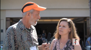
WAIKOLOA, HAWAII – A new wave of better sunscreens may soon wash into the U.S. marketplace, predicted Dr. Susan Swetter of Stanford (Calif.) University. She spoke with us at the Hawaii Dermatology Seminar about what might be in the pipeline for U.S. patients, and she outlined her own choices for sun protection in patients with a history of melanoma or who have very fair skin.
The video associated with this article is no longer available on this site. Please view all of our videos on the MDedge YouTube channel
EXPERT ANALYSIS FROM SDEF HAWAII DERMATOLOGY SEMINAR
VIDEO: Coffee Break 4: What did you learn at the meeting?
WAIKOLOA, HAWAII – Our editor, Heidi Splete, catches up with attendees at the Hawaii Dermatology Seminar to find out what they learned at the meeting that they will take back to their practices.
During a coffee break video interview, doctors discussed the use of topical vitamin D in psoriasis treatment, proper use of dermal fillers, and the latest techniques for the use of dermoscopy in melanoma.
The video associated with this article is no longer available on this site. Please view all of our videos on the MDedge YouTube channel
FROM SDEF HAWAII DERMATOLOGY SEMINAR

VIDEO: Treatment tips for pediatric molluscum contagiosum
WAIKOLOA, HAWAII – Dr. Sheila F. Friedlander of the University of California, San Diego, speaks about reassuring parents of children with molluscum contagiosum, the concept of "BOTE" – the beginning of the end –and the safe, effective use of topical cantharidin. She made her comments in a video interview during the SDEF Hawaii Dermatology Seminar.
WAIKOLOA, HAWAII – Dr. Sheila F. Friedlander of the University of California, San Diego, speaks about reassuring parents of children with molluscum contagiosum, the concept of "BOTE" – the beginning of the end –and the safe, effective use of topical cantharidin. She made her comments in a video interview during the SDEF Hawaii Dermatology Seminar.
WAIKOLOA, HAWAII – Dr. Sheila F. Friedlander of the University of California, San Diego, speaks about reassuring parents of children with molluscum contagiosum, the concept of "BOTE" – the beginning of the end –and the safe, effective use of topical cantharidin. She made her comments in a video interview during the SDEF Hawaii Dermatology Seminar.
EXPERT ANALYSIS FROM SDEF HAWAII DERMATOLOGY SEMINAR

VIDEO: A closer look at new pediatric acne guidelines
WAIKOLOA, HAWAII – Dr. Lawrence Eichenfield of the University of California, San Diego, speaks about the recent publication of comprehensive guidelines for the management of pediatric acne and outlines the treatment gaps that persist.
Dr. Eichenfield made his remarks during a video interview at the Hawaii Dermatology Seminar.
The video associated with this article is no longer available on this site. Please view all of our videos on the MDedge YouTube channel
EXPERT ANALYSIS FROM SDEF HAWAII DERMATOLOGY SEMINAR