User login
Skin Disease Education Foundation (SDEF): Hawaii Dermatology Seminar
Microwave therapy zaps hyperhidrosis long-term
MAUI, HAWAII – The future of hyperhidrosis therapy may be microwave ablation, a Food and Drug Administration–cleared method of destroying sweat glands noninvasively.
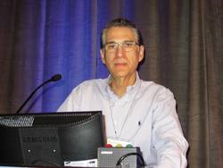
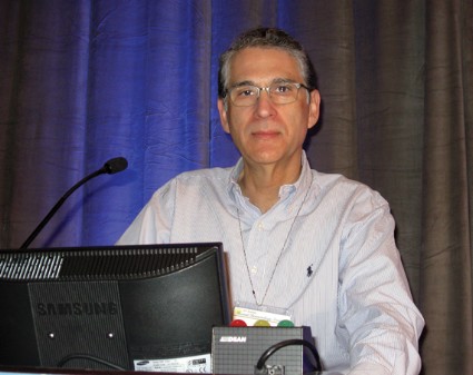
"We’ve seen that it’s effective in about 90% of patients, the patients are really satisfied, and the side effects are transient," Dr. Nazanin Saedi said at the Hawaii Dermatology Seminar, sponsored by Global Academy for Medical Education/Skin Disease Education Foundation.
Larger patient numbers and more long-term data are needed before the therapy becomes common in clinical practice, but it shouldn’t take long to accumulate such data, because the proprietary miraDry microwave treatment system (marketed by Miramar Labs) is much in demand by patients, noted Dr. Saedi of Thomas Jefferson University, Philadelphia.
An estimated 4.3 million Americans have axillary hyperhidrosis, Dr. Saedi said. Numerous treatment options are available, but most have significant drawbacks, she noted. First-line therapy is topical aluminum chloride, with or without salicylic acid; irritant dermatitis is a common complaint.
Second-line options include iontophoresis, which requires treatments two or three times per week and can be quite painful, Dr. Saedi said. Botulinum toxin A injections are highly effective, but they must be repeated every 4-6 months, and can cause weakness of the underlying muscles. Oral medications include anticholinergic agents and alpha2-adrenergic drugs, all with side effects many patients find problematic.
Before microwave therapy, surgical procedures were the only means of destroying sweat glands, and surgery is considered third-line therapy, Dr. Saedi said.
"Now that we have the newer technologies, I think surgery will become even more rare," she predicted.
The study that earned the miraDry device its Food and Drug Administration marketing clearance was a blinded trial in which 120 patients were randomized 2:1 to microwave therapy or a sham procedure. At baseline, all participants were rated 3 or 4 on the 1-4 Hyperhidrosis Disease Severity Scale (HDSS). At a follow-up point of 30 days, 89% of patients in the treatment group met the primary endpoint – an HDSS score of 1 or 2 – compared with 54% of controls. Roughly 70% of patients in the microwave treatment group still had an HDSS score of 1-2 at 12 months (Dermatol. Surg. 2012;38:185-91).
However, that study involved an earlier-generation, less efficient device, Dr. Saedi noted. In an open-label study of 31 patients treated with the current-generation, commercially available miraDry device, all subjects had a baseline HDSS score of 3 or 4. Twelve months after patients received 1-3 treatment sessions, 90% had an HDSS score of 1 or 2. In addition, 90% of patients had at least a 50% reduction in gravimetrically measured axillary sweat, and 85% had at least a 5-point improvement on the Dermatologic Life Quality Index (Dermatol. Surg. 2012;38:728-35).
In the microwave treatment procedure, an applicator is placed on the skin surface to deliver focused energy to the dermal-fat interface, where sweat glands are located. Microwaves are preferentially absorbed by tissues with high water content, such as sweat glands, and poorly absorbed by fat. A cooling system keeps the heat at the lower skin layer. A heat dome at about 60° C is created along the dermal-fat junction, resulting in sweat gland thermolysis, Dr. Saedi explained.
Two hours prior to treatment, the patient takes 800 mg of ibuprofen. A grid of temporary markings is placed over the area to be treated, and the dermis is anesthetized with injections of a 1:1 mix of 1% plain lidocaine and 1% lidocaine plus epinephrine at 1-cm intervals.
Microwave therapy entails two treatment sessions roughly 3 months apart. The first typically takes 60-90 minutes, the second 30-60 minutes.
Dr. Saedi said swelling, bruising, discomfort, and altered sensation in and around the treatment area are all common side effects of microwave therapy, but all are short-term. Less common side effects include swelling extending beyond the treatment area, a tight banding sensation in the axilla, and arm and hand numbness lasting less than 24 hours, she said.
One audience member who has treated 130 patients via microwave therapy reported that one of them developed a significant brachial nerve palsy that has lasted for 5 months. As a consequence, the commenter said he now routinely employs tumescent anesthesia in an effort to steer clear of the axillary nerve bundles. But Dr. Suzanne L. Kilmer said she doubts that will prevent the occasional case of brachial nerve palsy, which she believes is the result of variant anatomy.
"We’ve done the procedure on many thin people, and not had brachial nerve palsy be an issue, so I’m not sure that tumescent anesthesia will make a difference," said Dr. Kilmer of the University of California, Davis.
Pulsed ultrasound is currently under investigation as a means of treating hyperhidrosis through destruction of sweat glands, session moderator Dr. Christopher B. Zachary said in response to an audience question. "This would be an off-label indication right now, but you can absolutely hit that region with an ultrasound device and cause heating. It’s quite likely that ultrasound will be used extensively in exactly the same way as microwave, and probably with the same efficacy," predicted Dr. Zachary of the University of California, Irvine.
Dr. Zachary is a consultant to and has received research funding from numerous laser and other medical device companies, as has Dr. Kilmer. Dr. Saedi had no financial conflicts to disclose. SDEF and this news organization are owned by the same parent company.
MAUI, HAWAII – The future of hyperhidrosis therapy may be microwave ablation, a Food and Drug Administration–cleared method of destroying sweat glands noninvasively.
"We’ve seen that it’s effective in about 90% of patients, the patients are really satisfied, and the side effects are transient," Dr. Nazanin Saedi said at the Hawaii Dermatology Seminar, sponsored by Global Academy for Medical Education/Skin Disease Education Foundation.
Larger patient numbers and more long-term data are needed before the therapy becomes common in clinical practice, but it shouldn’t take long to accumulate such data, because the proprietary miraDry microwave treatment system (marketed by Miramar Labs) is much in demand by patients, noted Dr. Saedi of Thomas Jefferson University, Philadelphia.
An estimated 4.3 million Americans have axillary hyperhidrosis, Dr. Saedi said. Numerous treatment options are available, but most have significant drawbacks, she noted. First-line therapy is topical aluminum chloride, with or without salicylic acid; irritant dermatitis is a common complaint.
Second-line options include iontophoresis, which requires treatments two or three times per week and can be quite painful, Dr. Saedi said. Botulinum toxin A injections are highly effective, but they must be repeated every 4-6 months, and can cause weakness of the underlying muscles. Oral medications include anticholinergic agents and alpha2-adrenergic drugs, all with side effects many patients find problematic.
Before microwave therapy, surgical procedures were the only means of destroying sweat glands, and surgery is considered third-line therapy, Dr. Saedi said.
"Now that we have the newer technologies, I think surgery will become even more rare," she predicted.
The study that earned the miraDry device its Food and Drug Administration marketing clearance was a blinded trial in which 120 patients were randomized 2:1 to microwave therapy or a sham procedure. At baseline, all participants were rated 3 or 4 on the 1-4 Hyperhidrosis Disease Severity Scale (HDSS). At a follow-up point of 30 days, 89% of patients in the treatment group met the primary endpoint – an HDSS score of 1 or 2 – compared with 54% of controls. Roughly 70% of patients in the microwave treatment group still had an HDSS score of 1-2 at 12 months (Dermatol. Surg. 2012;38:185-91).
However, that study involved an earlier-generation, less efficient device, Dr. Saedi noted. In an open-label study of 31 patients treated with the current-generation, commercially available miraDry device, all subjects had a baseline HDSS score of 3 or 4. Twelve months after patients received 1-3 treatment sessions, 90% had an HDSS score of 1 or 2. In addition, 90% of patients had at least a 50% reduction in gravimetrically measured axillary sweat, and 85% had at least a 5-point improvement on the Dermatologic Life Quality Index (Dermatol. Surg. 2012;38:728-35).
In the microwave treatment procedure, an applicator is placed on the skin surface to deliver focused energy to the dermal-fat interface, where sweat glands are located. Microwaves are preferentially absorbed by tissues with high water content, such as sweat glands, and poorly absorbed by fat. A cooling system keeps the heat at the lower skin layer. A heat dome at about 60° C is created along the dermal-fat junction, resulting in sweat gland thermolysis, Dr. Saedi explained.
Two hours prior to treatment, the patient takes 800 mg of ibuprofen. A grid of temporary markings is placed over the area to be treated, and the dermis is anesthetized with injections of a 1:1 mix of 1% plain lidocaine and 1% lidocaine plus epinephrine at 1-cm intervals.
Microwave therapy entails two treatment sessions roughly 3 months apart. The first typically takes 60-90 minutes, the second 30-60 minutes.
Dr. Saedi said swelling, bruising, discomfort, and altered sensation in and around the treatment area are all common side effects of microwave therapy, but all are short-term. Less common side effects include swelling extending beyond the treatment area, a tight banding sensation in the axilla, and arm and hand numbness lasting less than 24 hours, she said.
One audience member who has treated 130 patients via microwave therapy reported that one of them developed a significant brachial nerve palsy that has lasted for 5 months. As a consequence, the commenter said he now routinely employs tumescent anesthesia in an effort to steer clear of the axillary nerve bundles. But Dr. Suzanne L. Kilmer said she doubts that will prevent the occasional case of brachial nerve palsy, which she believes is the result of variant anatomy.
"We’ve done the procedure on many thin people, and not had brachial nerve palsy be an issue, so I’m not sure that tumescent anesthesia will make a difference," said Dr. Kilmer of the University of California, Davis.
Pulsed ultrasound is currently under investigation as a means of treating hyperhidrosis through destruction of sweat glands, session moderator Dr. Christopher B. Zachary said in response to an audience question. "This would be an off-label indication right now, but you can absolutely hit that region with an ultrasound device and cause heating. It’s quite likely that ultrasound will be used extensively in exactly the same way as microwave, and probably with the same efficacy," predicted Dr. Zachary of the University of California, Irvine.
Dr. Zachary is a consultant to and has received research funding from numerous laser and other medical device companies, as has Dr. Kilmer. Dr. Saedi had no financial conflicts to disclose. SDEF and this news organization are owned by the same parent company.
EXPERT ANALYSIS FROM SDEF HAWAII DERMATOLOGY SEMINAR

'Slow Mohs' advised for lentigo maligna
MAUI, HAWAII – "Slow Mohs" has gained near-universal acceptance among skin cancer specialists as a definitive surgical technique for complete removal of lentigo maligna melanoma while simultaneously sparing normal tissue, according to Dr. Ellen Marmur of Mount Sinai School of Medicine, New York.
The big advantage that slow Mohs has over standard wide local excision with 0.5- to 1-cm margins is a 5-year cure rate approaching 100%. In contrast, standard excision has a recurrence rate of up to 20%, she said at the Hawaii Dermatology Seminar, sponsored by Global Academy for Medical Education/Skin Disease Education Foundation.

Slow Mohs is a modified form of Mohs micrographic surgery. The surgery compares with conventional Mohs: It is staged, margin-controlled excision. But in slow Mohs, rush permanent sections are sent off to the pathologist rather than the frozen sections integral to conventional Mohs.
Dr. Marmur relies upon slow Mohs, with "bread-loafing" of the central tumor by the pathologist, because the permanent sections better preserve the tumor’s microscopic features. Interpreting atypical melanocytes in frozen sections can be quite a challenge. However, she added, some Mohs surgeons have found that using rapid immunostains also markedly improves the sensitivity and specificity of frozen sections in lentigo maligna surgery.
Slow Mohs takes place over the course of days, she said. "Basically, you do your Wood’s lamp to define the lesion diameter, you measure out your margins, you excise the tumor, pack the area with a bandage, and send the patient home. You rush your pathology, and you don’t do any reconstruction until you get the margins clear."
A pathology report that comes back stating narrow margins are present is "a heart stopper," she added.
"You have the option of observing the area if the margin is clear but the tumor was close to the margin. That’s a good approach for an elderly patient or when the lentigo maligna was in a cosmetically important area."
Lentigo maligna melanoma accounts for 4% of all cases of melanoma. It typically arises on sun-damaged skin in individuals in their 70s or older. Common sites include the malar area, forehead, nose, and temple. The differential diagnosis includes seborrheic keratosis, pigmented actinic keratosis, and pigmented nevus.
Lentigo maligna becomes lentigo maligna melanoma when malignant melanoma cells invade the dermis and deeper appendages. Roughly 5% of lentigo malignas eventually progress to invasive melanoma, according to Dr. Marmur. Typically, a lentigo maligna undergoes extended gradual horizontal growth before beginning a vertical growth phase.
"It spreads like an oil slick for many years," Dr. Marmur said at the seminar.
Established treatment modalities for patients who aren’t surgical candidates include cryotherapy, radiotherapy, and topical imiquimod 5%. All have disadvantages, including high 5-year recurrence rates.
Dr. Marmur noted that a newer nonsurgical therapy drawing considerable interest involves off-label use of topical combination therapy with imiquimod and tazarotene gel. The concept is to use the topical retinoid to disrupt the stratum corneum in order to enhance imiquimod penetration, thereby achieving a greater inflammatory response than possible with imiquimod alone.
Initial data have been published by researchers at the University of Utah, Salt Lake City. They randomized 90 patients with 91 lentigo malignas to imiquimod 5% cream applied 5 days per week for 3 months or to the imiquimod regimen plus tazarotene 0.1% gel on the other 2 days per week. After 3 months of topical therapy, patients underwent conservative staged excision with frozen section analysis with Melan A immunostaining to confirm negative margins.
Of those treated with dual topical therapy for 3 months, 29 of 37 lesions (78%) had complete responses with no residual lentigo maligna at the time of staged excision. So did 27 of 42 (64%) treated with imiquimod alone (Arch. Dermatol. 2012;148:592-6).
The modest difference in outcome was not significant (P = .17). Nevertheless, the Utah investigators wrote that topical pretreatment appears to reduce surgical defect sizes, an important consideration in lentigo maligna because the lesions are often large and located on cosmetically sensitive facial sites. At the patient’s first visit, the researchers saucerize the entire tumor to remove all visible evidence of lentigo maligna and nip in the bud any invasive element that might be present. One month later, after the wound has healed by secondary intention, the patient begins topical imiquimod therapy 5 days per week. If no inflammation is observed, tazarotene gel is added on the other 2 days per week. After 3 months of topical therapy, the patient goes off treatment for 2 months so the inflammatory response can subside. Then a staged excision is performed with 2-mm margins around the perimeter of the original tumor outline.
"This is an interesting thought, but it’s still not considered standard of care," Dr. Marmur commented in describing the Utah research.
Session chair Dr. Allan C. Halpern, chief of the dermatology service at Memorial Sloan-Kettering Cancer Center in New York, said he and his colleagues, like the Utah group, are using a lot of topical imiquimod as adjunctive therapy for lentigo maligna.
"Because we’re a referral center, we see all those patients whose melanomas unfortunately do recur at the edge of large surgeries. We see a lot of patients whose local surgeons have chased around their faces for a certain amount of time. So I think the general concept of the cure rates of surgery is a bit overstated. I’m not trying to sell an off-label use of a drug, but I think sometimes imiquimod can be very helpful," Dr. Halpern said.
He offered a clinical pearl in diagnosing lentigo maligna: Consider a broad shave biopsy of a suspicious lesion rather than a deeper, smaller-diameter biopsy.
"Sampling errors are a real problem with lentigo maligna. We learned early on that if you take a large lentigo maligna and do multiple biopsies of the same lesion and send them to what we think are our very expert pathologists, on average, we get two or three back that were melanoma and two or three back that were pigmented actinic keratoses. The sampling error tends to be more in the horizontal than in the depth for these very early lesions," according to Dr. Halpern.
Dr. Marmur and Dr. Halpern reported having no relevant financial interests. SDEF and this news organization are owned by the same parent company.
MAUI, HAWAII – "Slow Mohs" has gained near-universal acceptance among skin cancer specialists as a definitive surgical technique for complete removal of lentigo maligna melanoma while simultaneously sparing normal tissue, according to Dr. Ellen Marmur of Mount Sinai School of Medicine, New York.
The big advantage that slow Mohs has over standard wide local excision with 0.5- to 1-cm margins is a 5-year cure rate approaching 100%. In contrast, standard excision has a recurrence rate of up to 20%, she said at the Hawaii Dermatology Seminar, sponsored by Global Academy for Medical Education/Skin Disease Education Foundation.
Slow Mohs is a modified form of Mohs micrographic surgery. The surgery compares with conventional Mohs: It is staged, margin-controlled excision. But in slow Mohs, rush permanent sections are sent off to the pathologist rather than the frozen sections integral to conventional Mohs.
Dr. Marmur relies upon slow Mohs, with "bread-loafing" of the central tumor by the pathologist, because the permanent sections better preserve the tumor’s microscopic features. Interpreting atypical melanocytes in frozen sections can be quite a challenge. However, she added, some Mohs surgeons have found that using rapid immunostains also markedly improves the sensitivity and specificity of frozen sections in lentigo maligna surgery.
Slow Mohs takes place over the course of days, she said. "Basically, you do your Wood’s lamp to define the lesion diameter, you measure out your margins, you excise the tumor, pack the area with a bandage, and send the patient home. You rush your pathology, and you don’t do any reconstruction until you get the margins clear."
A pathology report that comes back stating narrow margins are present is "a heart stopper," she added.
"You have the option of observing the area if the margin is clear but the tumor was close to the margin. That’s a good approach for an elderly patient or when the lentigo maligna was in a cosmetically important area."
Lentigo maligna melanoma accounts for 4% of all cases of melanoma. It typically arises on sun-damaged skin in individuals in their 70s or older. Common sites include the malar area, forehead, nose, and temple. The differential diagnosis includes seborrheic keratosis, pigmented actinic keratosis, and pigmented nevus.
Lentigo maligna becomes lentigo maligna melanoma when malignant melanoma cells invade the dermis and deeper appendages. Roughly 5% of lentigo malignas eventually progress to invasive melanoma, according to Dr. Marmur. Typically, a lentigo maligna undergoes extended gradual horizontal growth before beginning a vertical growth phase.
"It spreads like an oil slick for many years," Dr. Marmur said at the seminar.
Established treatment modalities for patients who aren’t surgical candidates include cryotherapy, radiotherapy, and topical imiquimod 5%. All have disadvantages, including high 5-year recurrence rates.
Dr. Marmur noted that a newer nonsurgical therapy drawing considerable interest involves off-label use of topical combination therapy with imiquimod and tazarotene gel. The concept is to use the topical retinoid to disrupt the stratum corneum in order to enhance imiquimod penetration, thereby achieving a greater inflammatory response than possible with imiquimod alone.
Initial data have been published by researchers at the University of Utah, Salt Lake City. They randomized 90 patients with 91 lentigo malignas to imiquimod 5% cream applied 5 days per week for 3 months or to the imiquimod regimen plus tazarotene 0.1% gel on the other 2 days per week. After 3 months of topical therapy, patients underwent conservative staged excision with frozen section analysis with Melan A immunostaining to confirm negative margins.
Of those treated with dual topical therapy for 3 months, 29 of 37 lesions (78%) had complete responses with no residual lentigo maligna at the time of staged excision. So did 27 of 42 (64%) treated with imiquimod alone (Arch. Dermatol. 2012;148:592-6).
The modest difference in outcome was not significant (P = .17). Nevertheless, the Utah investigators wrote that topical pretreatment appears to reduce surgical defect sizes, an important consideration in lentigo maligna because the lesions are often large and located on cosmetically sensitive facial sites. At the patient’s first visit, the researchers saucerize the entire tumor to remove all visible evidence of lentigo maligna and nip in the bud any invasive element that might be present. One month later, after the wound has healed by secondary intention, the patient begins topical imiquimod therapy 5 days per week. If no inflammation is observed, tazarotene gel is added on the other 2 days per week. After 3 months of topical therapy, the patient goes off treatment for 2 months so the inflammatory response can subside. Then a staged excision is performed with 2-mm margins around the perimeter of the original tumor outline.
"This is an interesting thought, but it’s still not considered standard of care," Dr. Marmur commented in describing the Utah research.
Session chair Dr. Allan C. Halpern, chief of the dermatology service at Memorial Sloan-Kettering Cancer Center in New York, said he and his colleagues, like the Utah group, are using a lot of topical imiquimod as adjunctive therapy for lentigo maligna.
"Because we’re a referral center, we see all those patients whose melanomas unfortunately do recur at the edge of large surgeries. We see a lot of patients whose local surgeons have chased around their faces for a certain amount of time. So I think the general concept of the cure rates of surgery is a bit overstated. I’m not trying to sell an off-label use of a drug, but I think sometimes imiquimod can be very helpful," Dr. Halpern said.
He offered a clinical pearl in diagnosing lentigo maligna: Consider a broad shave biopsy of a suspicious lesion rather than a deeper, smaller-diameter biopsy.
"Sampling errors are a real problem with lentigo maligna. We learned early on that if you take a large lentigo maligna and do multiple biopsies of the same lesion and send them to what we think are our very expert pathologists, on average, we get two or three back that were melanoma and two or three back that were pigmented actinic keratoses. The sampling error tends to be more in the horizontal than in the depth for these very early lesions," according to Dr. Halpern.
Dr. Marmur and Dr. Halpern reported having no relevant financial interests. SDEF and this news organization are owned by the same parent company.
EXPERT ANALYSIS FROM SDEF HAWAII DERMATOLOGY SEMINAR
Polymorphic light eruption: A diagnosis of exclusion
MAUI, HAWAII – Polymorphic light eruption is the most common of all the photodermatoses, yet few physicians will ever actually see the rash in their office, according to Dr. Vincent A. DeLeo.
That’s because polymorphic light eruption (PMLE) typically occurs when someone goes on vacation someplace sunny. By the time the patient returns home, the itchy rash is gone. But the history tells the tale, Dr. DeLeo said at the Hawaii Dermatology Seminar sponsored by Global Academy for Medical Education/Skin Disease Education Foundation.
"PMLE is a clinical diagnosis you make when you’ve ruled out photoallergic contact dermatitis and lupus. It’s a diagnosis of exclusion," explained Dr. DeLeo, chairman of the department of dermatology at St. Luke’s-Roosevelt Hospital Center and Beth Israel Medical Center, both in New York.
The salient features of PMLE are that it’s a recurrent rash, which typically erupts several days after – not within minutes of – sun exposure; it lasts for days; and it spares the face.
The face is spared because patients with PMLE gradually become "hardened" to sunlight. The more sun they get on a body part over time, the less likely they are to experience an eruption at that location. The face gets more sunlight than other body parts during everyday outdoor exposure. The rash of PMLE occurs on skin sites that are usually covered in winter, but exposed to the sun in the spring or while vacationing, such as the upper chest and arms.
A study conducted several decades ago among Harvard Medical School students suggested that PMLE affects about 10% of Americans. European studies indicate 21% of Swedes and 15% of U.K. residents are affected. Women outnumber men with PMLE by at least 2:1. The onset of the condition is typically in the 20s among women and somewhat later in men.
The itchy rash is variable in appearance. It can take the form of papules, papulovesicular lesions, or plaques. In the unlikely event the physician actually sees the rash and takes a biopsy, the histology shows a dense perivascular dermal lymphocytic infiltrate.
The differential diagnosis involves photoallergic contact dermatitis and lupus, both of which, like PMLE, feature recurrent, persistent rashes that erupt after a delay in response to sun exposure. Photoallergic contact dermatitis usually affects the face and almost always is due to sunscreens, with oxybenzone the No. 1 culprit. Photopatch testing will confirm the diagnosis. Titanium- or zinc-based physical sun blockers are protective.
It’s important to recognize that oxybenzone and other offenders are also present in shampoos, rinses, and other personal care products in addition to sunscreens, the dermatologist noted.
The plaque form of PMLE looks morphologically like lupus erythematosus (LE). But LE can be ruled out on the basis of negative results for antinuclear antibody, anti-Ro, and anti-La testing.
Because episodes of PMLE are recurrent and predictable, prophylaxis is the best approach. Dr. DeLeo recommended the use of sunscreens containing avobenzone and ecamsule; the combination works better than either agent alone.
If sunscreens don’t work, his top choice is narrow-band UVB phototherapy. "We use it like you would for psoriasis. We give most patients three treatments per week for 3-4 weeks before they go away on their trip. That usually suppresses the eruption. Essentially we’re hardening them," he said.
A product that physicians can expect to be hearing a lot more about is Polypodium leucotomos, an oral fern extract marketed as a sunscreen in pill form. In one recent open-label study involving 57 patients with PMLE, three-quarters of them benefited (G. Ital. Dermatol. Venereol. 2011;146:85-7). In another open-label study by other investigators, this one involving 35 PMLE patients, daily P. leucotomos also resulted in a significant reduction in sensitivity to sunlight (J. Am. Acad. Dermatol. 2012;66:58-62). The usual dosage is 240 mg twice daily.
P. leucotomos has been a commercially available natural product in Europe for 3 decades, with well-established safety. It has been acquired by Ferndale Pharma, which plans to raise its market profile in the United States.
Dr. DeLeo reported serving as a consultant to numerous pharmaceutical and cosmetics companies. SDEF and this news organization are owned by the same parent company.
EXPERT ANALYSIS FROM SDEF HAWAII DERMATOLOGY SEMINAR

Therapeutic combos make inroads in advanced melanoma
WAILEA, HAWAII – "The past 2 years have been a really exciting time for those of us who have spent the last several decades" in the field of melanoma, said Dr. Allan C. Halpern, chief of the dermatology service at Memorial Sloan Kettering Cancer Center, New York.
"We are in a whole new place with a very promising future for turning stage IV melanoma into maybe a chronic disease for many patients, instead of a death sentence. For some patients, we’re already seeing what may be cures," he said at the Hawaii Dermatology Seminar sponsored by the Global Academy for Medical Education/Skin Disease Education Foundation.

The greatest enthusiasm in the field now involves combining a pathway-targeted agent, such as vemurafenib, with an immunologic checkpoint blocker, such as ipilimumab. The vemurafenib knocks down 60%-70% of metastatic melanomas temporarily and the ipilimumab promotes durable responses.
But there’s a formidable economic obstacle to this approach: The strongest drug combinations often put big pharmaceutical companies in the uncomfortable position of having to cooperate with their competitors in expensive research projects. "A lot of the drug companies, to their credit, are finding ways to make it work," Dr. Halpern said.
Dr. Halpern detailed the therapeutic history that has revolutionized the treatment of metastatic melanoma.
Prior to 2011 there were only two Food and Drug Administration–approved therapies for metastatic melanoma, dacarbazine and high-dose interleukin II. Both were unimpressive. The therapeutic dry spell has ended, he said. "There are for the first time in melanoma, instead of no hopeful drugs, a slew of hopeful drugs."
Targeted therapeutic approaches, the result of laboratory insights into the molecular pathways to melanoma and the key genetic mutations involved, led to the development of vemurafenib, a selective, first-in-class BRAF inhibitor approved in 2011.
"Vemurafenib is an astounding drug. When you give it to a BRAF-mutated cell, it essentially turns off the cell’s metabolic activity." When given to patients whose tumors test positive for the BRAF mutation, "it’s dramatically effective in 60%-70%." But the response does not persist. "After about 6-18 months, the tumor develops resistance to the drug. It’s like somebody hit a switch to turn the tumor back on. The tumor comes roaring back, in the same places for the most part," Dr. Halpern said.
As a result of this limited success, ongoing clinical trials are aimed at determining whether dual pathway blockade using combination therapy will provide more durable responses. Trials are underway with the oral BRAF inhibitor dabrafenib plus the oral MEK 1/2 pathway inhibitor trametinib. Other dual pathway combinations are also under study in melanoma.
The prospects are even more promising, according to Dr. Halpern, for immunologic checkpoint blockade, which is based upon the concept that some cancers progress because the immune system turns off prematurely and stops battling the malignancy. Ipilimumab is one such agent. An anti-CTLA-4 antibody, ipilimumab enhances T-cell activation and proliferation and has earned FDA approval as single-agent therapy in advanced melanoma.
Tumors often don’t begin to shrink until after 3-4 months, but the response is impressively durable in the roughly 30% of patients who respond to immunologic checkpoint blockade.
"These people look like they might be cured," said Dr. Halpern.
Another important immunologic checkpoint molecule is PD-1. The anti-PD-1 agent known as MDX-1106 appears to be nearly as effective as ipilimumab, but with less toxicity. The early impression from ongoing clinical trials is that dual immunologic checkpoint blockade using anti-CTLA-4 therapy along with an anti-PD-1 drug provides synergistic anti-tumor activity.
Dr. Halpern reported serving as a consultant to Canfield Scientific, DermTech, SciBase, Quintiles, and Lucid.
SDEF and this news organization are owned by the same parent company.
WAILEA, HAWAII – "The past 2 years have been a really exciting time for those of us who have spent the last several decades" in the field of melanoma, said Dr. Allan C. Halpern, chief of the dermatology service at Memorial Sloan Kettering Cancer Center, New York.
"We are in a whole new place with a very promising future for turning stage IV melanoma into maybe a chronic disease for many patients, instead of a death sentence. For some patients, we’re already seeing what may be cures," he said at the Hawaii Dermatology Seminar sponsored by the Global Academy for Medical Education/Skin Disease Education Foundation.
The greatest enthusiasm in the field now involves combining a pathway-targeted agent, such as vemurafenib, with an immunologic checkpoint blocker, such as ipilimumab. The vemurafenib knocks down 60%-70% of metastatic melanomas temporarily and the ipilimumab promotes durable responses.
But there’s a formidable economic obstacle to this approach: The strongest drug combinations often put big pharmaceutical companies in the uncomfortable position of having to cooperate with their competitors in expensive research projects. "A lot of the drug companies, to their credit, are finding ways to make it work," Dr. Halpern said.
Dr. Halpern detailed the therapeutic history that has revolutionized the treatment of metastatic melanoma.
Prior to 2011 there were only two Food and Drug Administration–approved therapies for metastatic melanoma, dacarbazine and high-dose interleukin II. Both were unimpressive. The therapeutic dry spell has ended, he said. "There are for the first time in melanoma, instead of no hopeful drugs, a slew of hopeful drugs."
Targeted therapeutic approaches, the result of laboratory insights into the molecular pathways to melanoma and the key genetic mutations involved, led to the development of vemurafenib, a selective, first-in-class BRAF inhibitor approved in 2011.
"Vemurafenib is an astounding drug. When you give it to a BRAF-mutated cell, it essentially turns off the cell’s metabolic activity." When given to patients whose tumors test positive for the BRAF mutation, "it’s dramatically effective in 60%-70%." But the response does not persist. "After about 6-18 months, the tumor develops resistance to the drug. It’s like somebody hit a switch to turn the tumor back on. The tumor comes roaring back, in the same places for the most part," Dr. Halpern said.
As a result of this limited success, ongoing clinical trials are aimed at determining whether dual pathway blockade using combination therapy will provide more durable responses. Trials are underway with the oral BRAF inhibitor dabrafenib plus the oral MEK 1/2 pathway inhibitor trametinib. Other dual pathway combinations are also under study in melanoma.
The prospects are even more promising, according to Dr. Halpern, for immunologic checkpoint blockade, which is based upon the concept that some cancers progress because the immune system turns off prematurely and stops battling the malignancy. Ipilimumab is one such agent. An anti-CTLA-4 antibody, ipilimumab enhances T-cell activation and proliferation and has earned FDA approval as single-agent therapy in advanced melanoma.
Tumors often don’t begin to shrink until after 3-4 months, but the response is impressively durable in the roughly 30% of patients who respond to immunologic checkpoint blockade.
"These people look like they might be cured," said Dr. Halpern.
Another important immunologic checkpoint molecule is PD-1. The anti-PD-1 agent known as MDX-1106 appears to be nearly as effective as ipilimumab, but with less toxicity. The early impression from ongoing clinical trials is that dual immunologic checkpoint blockade using anti-CTLA-4 therapy along with an anti-PD-1 drug provides synergistic anti-tumor activity.
Dr. Halpern reported serving as a consultant to Canfield Scientific, DermTech, SciBase, Quintiles, and Lucid.
SDEF and this news organization are owned by the same parent company.
EXPERT ANALYSIS FROM SDEF HAWAII DERMATOLOGY SEMINAR
Dermoscopy characterized as patient trust builder
MAUI, HAWAII – Dermatologists in the know view dermoscopy as a powerful tool to increase diagnostic accuracy in evaluating pigmented lesions; less well appreciated is dermoscopy’s value in building patient trust in the physician, according to Dr. Steven Q. Wang.
"With the dermoscope, people feel like you’re providing a much more detailed exam," observed Dr. Wang, director of dermatologic surgery and dermatology at Memorial Sloan-Kettering Cancer Center’s Basking Ridge, N.J., campus.

"We are a tertiary referral center. We have lots of patients come in who are high risk, with a personal or family history of melanoma and numerous nevi. I always ask why they have transferred their care. The common answer I hear is they feel their dermatologist was not giving them a detailed examination," he said at the Hawaii Dermatology Seminar sponsored by the Global Academy for Medical Education/Skin Disease Education Foundation.
Dr. Wang said he makes a point of giving every patient a full-body clinical examination, including looking between the toes. And he views every single lesion with his dermoscope, up close and personal, scope to skin.
"The dermoscope makes it easier to spot the outlier lesions, the ugly ducklings. And we’re all so busy in the office, running from room to room – dermoscopy helps me slow down my mind and really look," he explained.
It doesn’t take all that long, either. In a classic multicenter study, 1,328 patients with one or more melanocytic or nonmelanocytic skin lesions were randomized to receive a complete skin examination with or without dermoscopy. The median time for a complete skin examination alone was 70 seconds; with dermoscopy it rose to 142 seconds (Arch. Dermatol. 2008;144:509-13).
"You double the time required, but it’s still only a little over 2 minutes. Yet you’ve changed the patient’s perception," Dr. Wang said.
A digital dermoscope is basically a handheld microscope that permits detailed visualization of structures in the deep epidermis and superficial dermis not visible to the naked eye. There’s a learning curve involved. Dermatologists who pick up a dermoscope and try to use it without formal training have worse diagnostic accuracy than with clinical examination, while experienced dermoscopists have significantly greater diagnostic accuracy than can be achieved with clinical exam alone (Lancet Oncol. 2002;3:159-65).
Dr. Wang said that because patients have more trust in their dermatologist when they feel they are receiving a thorough skin examination including dermoscopy, they are more likely to be adherent to scheduled follow-up evaluations. And that, in turn, spells improved long-term outcomes in patients at elevated risk for melanoma.
This point was already brought home forcefully for him nearly a decade ago, he said, when he and his coinvestigators reported their experience with long-term follow-up of 258 patients at high risk for melanoma. The monitoring strategy consisted of annual total body photography, total skin examination, and dermoscopy. The cumulative 10-year incidence of melanoma was 14% in the 160 patients with classic atypical mole syndrome and 10% in the other 98 high-risk patients. Impressively, all of the melanomas were either in situ or less than 1 mm thick. There were no metastases and no melanoma-related deaths (J. Am. Acad. Dermatol. 2004;50:15-20).
Although dermoscopy is used primarily in examining pigmented skin lesions, it has other applications. For example, in performing Mohs surgery for basal cell carcinomas, Dr. Wang has found dermoscopy to be of assistance in a couple of ways: In patients with ill-defined tumor borders on clinical examination, dermoscopy can define the tumor borders presurgically, thereby reducing the number of Mohs surgical stages required; and when a patient returns for Mohs surgery 6 weeks after skin biopsy and the biopsy site has healed so completely it can’t be found with the naked eye, the dermoscope can identify the site by visualizing subtle scars and telangiectasias.
In general dermatology, Dr. Wang said he turns to dermoscopy as an aid in diagnosing connective tissue diseases, including dermatomyositis, scleroderma, and lupus. He applies ultrasound gel to the proximal nail fold and examines the site using the dermoscope. A finding of dilated blood vessels stands out as a helpful diagnostic clue.
In addition, Dr. Wang said he has utilized the dermoscope in diagnosing scabies by spotting the mites and their trails, in detecting the telltale Wickham striae of lichen planus, and in diagnosing other dermatologic disorders.
Dr. Wang reported having no relevant financial conflicts.
SDEF and this news organization are owned by the same parent company.
MAUI, HAWAII – Dermatologists in the know view dermoscopy as a powerful tool to increase diagnostic accuracy in evaluating pigmented lesions; less well appreciated is dermoscopy’s value in building patient trust in the physician, according to Dr. Steven Q. Wang.
"With the dermoscope, people feel like you’re providing a much more detailed exam," observed Dr. Wang, director of dermatologic surgery and dermatology at Memorial Sloan-Kettering Cancer Center’s Basking Ridge, N.J., campus.
"We are a tertiary referral center. We have lots of patients come in who are high risk, with a personal or family history of melanoma and numerous nevi. I always ask why they have transferred their care. The common answer I hear is they feel their dermatologist was not giving them a detailed examination," he said at the Hawaii Dermatology Seminar sponsored by the Global Academy for Medical Education/Skin Disease Education Foundation.
Dr. Wang said he makes a point of giving every patient a full-body clinical examination, including looking between the toes. And he views every single lesion with his dermoscope, up close and personal, scope to skin.
"The dermoscope makes it easier to spot the outlier lesions, the ugly ducklings. And we’re all so busy in the office, running from room to room – dermoscopy helps me slow down my mind and really look," he explained.
It doesn’t take all that long, either. In a classic multicenter study, 1,328 patients with one or more melanocytic or nonmelanocytic skin lesions were randomized to receive a complete skin examination with or without dermoscopy. The median time for a complete skin examination alone was 70 seconds; with dermoscopy it rose to 142 seconds (Arch. Dermatol. 2008;144:509-13).
"You double the time required, but it’s still only a little over 2 minutes. Yet you’ve changed the patient’s perception," Dr. Wang said.
A digital dermoscope is basically a handheld microscope that permits detailed visualization of structures in the deep epidermis and superficial dermis not visible to the naked eye. There’s a learning curve involved. Dermatologists who pick up a dermoscope and try to use it without formal training have worse diagnostic accuracy than with clinical examination, while experienced dermoscopists have significantly greater diagnostic accuracy than can be achieved with clinical exam alone (Lancet Oncol. 2002;3:159-65).
Dr. Wang said that because patients have more trust in their dermatologist when they feel they are receiving a thorough skin examination including dermoscopy, they are more likely to be adherent to scheduled follow-up evaluations. And that, in turn, spells improved long-term outcomes in patients at elevated risk for melanoma.
This point was already brought home forcefully for him nearly a decade ago, he said, when he and his coinvestigators reported their experience with long-term follow-up of 258 patients at high risk for melanoma. The monitoring strategy consisted of annual total body photography, total skin examination, and dermoscopy. The cumulative 10-year incidence of melanoma was 14% in the 160 patients with classic atypical mole syndrome and 10% in the other 98 high-risk patients. Impressively, all of the melanomas were either in situ or less than 1 mm thick. There were no metastases and no melanoma-related deaths (J. Am. Acad. Dermatol. 2004;50:15-20).
Although dermoscopy is used primarily in examining pigmented skin lesions, it has other applications. For example, in performing Mohs surgery for basal cell carcinomas, Dr. Wang has found dermoscopy to be of assistance in a couple of ways: In patients with ill-defined tumor borders on clinical examination, dermoscopy can define the tumor borders presurgically, thereby reducing the number of Mohs surgical stages required; and when a patient returns for Mohs surgery 6 weeks after skin biopsy and the biopsy site has healed so completely it can’t be found with the naked eye, the dermoscope can identify the site by visualizing subtle scars and telangiectasias.
In general dermatology, Dr. Wang said he turns to dermoscopy as an aid in diagnosing connective tissue diseases, including dermatomyositis, scleroderma, and lupus. He applies ultrasound gel to the proximal nail fold and examines the site using the dermoscope. A finding of dilated blood vessels stands out as a helpful diagnostic clue.
In addition, Dr. Wang said he has utilized the dermoscope in diagnosing scabies by spotting the mites and their trails, in detecting the telltale Wickham striae of lichen planus, and in diagnosing other dermatologic disorders.
Dr. Wang reported having no relevant financial conflicts.
SDEF and this news organization are owned by the same parent company.
EXPERT ANALYSIS FROM SDEF HAWAII DERMATOLOGY SEMINAR

Where Belotero Balance excels in cosmetic dermatology
MAUI, HAWAII – Belotero Balance, the most recent filler to receive Food and Drug Administration approval, offers unique advantages over other hyaluronic acid fillers in treatment of superficial fine lines and wrinkles in the perioral area, according to Dr. Mark G. Rubin, a dermatologist in Beverly Hills, Calif.
"This product has become my collagen replacement. It’s a filler you want to inject into the dermis. It enables dermatologists to go after very superficial thin lines without causing lumpiness, ridges, or the Tyndall effect," he said at the Hawaii Dermatology Seminar sponsored by Global Academy for Medical Education/Skin Disease Education Foundation.

Be aware, though, that this is not a product to use straight out of the box. Unlike the older, widely used hyaluronic acid fillers Restylane and Juvederm, it doesn’t come premixed with anesthetic, so patients will find that Belotero injected without first being diluted with lidocaine hurts more than other fillers.
The other reason Dr. Rubin dilutes Belotero is that even though it is thinner and softer than other hyaluronic acid fillers, it can still result in unsightly ridges lasting for several weeks if injected intradermally in too strong a concentration.
For treatment of thin perioral lines, he typically dilutes 1 cc of Belotero with 0.5 cc of 1% lidocaine with epinephrine. He favors a serial puncture technique, although some other cosmetic dermatologists prefer a linear threading strategy.
"This behaves a lot like Zyderm did. It can be injected superficially and still give people significant improvement in very fine lines," said Dr. Rubin, also of the University of California, San Diego.
Zyderm and other collagen implants were taken off the U.S. market by FDA request. Cosmetic dermatologists have been looking for a suitable replacement thin filler for superficial use ever since – and Belotero is it, he said.
To treat deeper folds and contour irregularities, Dr. Rubin will dilute 1 cc of Belotero with 0.3 cc of 1% lidocaine with epinephrine, which provides more lift. Sometimes he’ll partially fill in a deeper perioral line with the less dilute product before topping it off with the more dilute version in the same treatment session.
Belotero Balance is a non-animal–derived hyaluronic acid–based cohesive gel that is crosslinked twice. Its clinical behavior is significantly different from the other hyaluronic acid products; it’s stretchy, like a rubber band, while the earlier-generation hyaluronic acid fillers tend to break when stretched, the dermatologist explained.
"It’s like an amoeba, working its way between cells and fibers. It really does smooth out better than the hyaluronic acids we’re used to," he continued.
On the other hand, Belotero provides less lift when injected deep than the other hyaluronic acid fillers.
"It’s not a go-to product for volume replacement. It requires more product to achieve a similar result. I still use a lot of Restylane and Juvederm, particularly where I’m looking for volume," Dr. Rubin said.
The FDA approval of Belotero Balance was based upon a phase III study of 118 patients. In contrast, Dr. Rubin has injected more than 1,300 syringes of Belotero into his patients. He offered several tips based upon his experience:
– Don’t overcorrect. "Correct to 100%. Because of the dilution, a week or two later the patient will no longer be filled all the way and may need a second treatment," Dr. Rubin said. "I tell patients who come in for superficial wrinkle therapy that they’re going to be treated twice. They’ll have a good result the first time and they’ll have a great result the second time when they come in a couple weeks later."
– Massage the treated area vigorously right after the injection to help smooth out the product. "I’ll stick my finger inside their cheek and squash them hard for 5 seconds," he said.
– Expect the results to last at least 6 months before beginning to fade.
– Anticipate a day or two of beadiness or ridging until the Belotero settles into the skin. Occasionally this can take up to 5 or 6 days.
– Consider prescribing an antihistamine if the ridging and edema lasts more than about 5 days. "There is some type of a histamine-mediated response in those patients. It almost looks urticarial. It’s a small group, probably no greater than 5%-10% of the people I inject, who have this persistent ridginess and puffiness," Dr. Rubin said.
Dr. Rubin reported serving as a paid researcher and consultant for Medicis, but has no financial relationship with Merz, which markets Belotero Balance. SDEF and this news organization are owned by the same parent company.
MAUI, HAWAII – Belotero Balance, the most recent filler to receive Food and Drug Administration approval, offers unique advantages over other hyaluronic acid fillers in treatment of superficial fine lines and wrinkles in the perioral area, according to Dr. Mark G. Rubin, a dermatologist in Beverly Hills, Calif.
"This product has become my collagen replacement. It’s a filler you want to inject into the dermis. It enables dermatologists to go after very superficial thin lines without causing lumpiness, ridges, or the Tyndall effect," he said at the Hawaii Dermatology Seminar sponsored by Global Academy for Medical Education/Skin Disease Education Foundation.
Be aware, though, that this is not a product to use straight out of the box. Unlike the older, widely used hyaluronic acid fillers Restylane and Juvederm, it doesn’t come premixed with anesthetic, so patients will find that Belotero injected without first being diluted with lidocaine hurts more than other fillers.
The other reason Dr. Rubin dilutes Belotero is that even though it is thinner and softer than other hyaluronic acid fillers, it can still result in unsightly ridges lasting for several weeks if injected intradermally in too strong a concentration.
For treatment of thin perioral lines, he typically dilutes 1 cc of Belotero with 0.5 cc of 1% lidocaine with epinephrine. He favors a serial puncture technique, although some other cosmetic dermatologists prefer a linear threading strategy.
"This behaves a lot like Zyderm did. It can be injected superficially and still give people significant improvement in very fine lines," said Dr. Rubin, also of the University of California, San Diego.
Zyderm and other collagen implants were taken off the U.S. market by FDA request. Cosmetic dermatologists have been looking for a suitable replacement thin filler for superficial use ever since – and Belotero is it, he said.
To treat deeper folds and contour irregularities, Dr. Rubin will dilute 1 cc of Belotero with 0.3 cc of 1% lidocaine with epinephrine, which provides more lift. Sometimes he’ll partially fill in a deeper perioral line with the less dilute product before topping it off with the more dilute version in the same treatment session.
Belotero Balance is a non-animal–derived hyaluronic acid–based cohesive gel that is crosslinked twice. Its clinical behavior is significantly different from the other hyaluronic acid products; it’s stretchy, like a rubber band, while the earlier-generation hyaluronic acid fillers tend to break when stretched, the dermatologist explained.
"It’s like an amoeba, working its way between cells and fibers. It really does smooth out better than the hyaluronic acids we’re used to," he continued.
On the other hand, Belotero provides less lift when injected deep than the other hyaluronic acid fillers.
"It’s not a go-to product for volume replacement. It requires more product to achieve a similar result. I still use a lot of Restylane and Juvederm, particularly where I’m looking for volume," Dr. Rubin said.
The FDA approval of Belotero Balance was based upon a phase III study of 118 patients. In contrast, Dr. Rubin has injected more than 1,300 syringes of Belotero into his patients. He offered several tips based upon his experience:
– Don’t overcorrect. "Correct to 100%. Because of the dilution, a week or two later the patient will no longer be filled all the way and may need a second treatment," Dr. Rubin said. "I tell patients who come in for superficial wrinkle therapy that they’re going to be treated twice. They’ll have a good result the first time and they’ll have a great result the second time when they come in a couple weeks later."
– Massage the treated area vigorously right after the injection to help smooth out the product. "I’ll stick my finger inside their cheek and squash them hard for 5 seconds," he said.
– Expect the results to last at least 6 months before beginning to fade.
– Anticipate a day or two of beadiness or ridging until the Belotero settles into the skin. Occasionally this can take up to 5 or 6 days.
– Consider prescribing an antihistamine if the ridging and edema lasts more than about 5 days. "There is some type of a histamine-mediated response in those patients. It almost looks urticarial. It’s a small group, probably no greater than 5%-10% of the people I inject, who have this persistent ridginess and puffiness," Dr. Rubin said.
Dr. Rubin reported serving as a paid researcher and consultant for Medicis, but has no financial relationship with Merz, which markets Belotero Balance. SDEF and this news organization are owned by the same parent company.
EXPERT ANALYSIS FROM SDEF HAWAII DERMATOLOGY SEMINAR
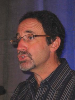
Be mindful of psoriasis/cutaneous T-cell lymphoma link
MAUI, HAWAII – The strong association between psoriasis and cutaneous T-cell lymphoma warrants a low threshold for skin biopsy in psoriasis patients, particularly since the two diseases can be tough to differentiate, experts emphasized at the Hawaii Dermatology Seminar sponsored by Global Academy for Medical Education/Skin Disease Education Foundation.
Indeed, skin biopsy is appropriate in any psoriasis patient with atypical features or psoriasis that isn’t responding to treatment, according to Dr. Joel M. Gelfand of the University of Pennsylvania, Philadelphia.
"I’ve found that my psoriasis patients actually like to be biopsied. They’ve been living with their disease for an average of 20 years, and they’re curious," he said. "And they’re also suspicious. They wonder, ‘How could I have this for 20 years and it’s still here?’ They’re worried that it’s really cancer or something else. So my patients never decline a biopsy of psoriasis," he said.
The literature on psoriasis and solid malignancies is "a mess," with some observational studies showing a positive association and others not, Dr. Gelfand noted.
In contrast, the studies looking at psoriasis and cutaneous T-cell lymphoma (CTCL) have been consistently positive. Case in point: when Dr. Gelfand and colleagues turned to the U.K. General Practice Research Database to study more than 153,000 psoriasis patients and nearly 800,000 controls, they found participants with mild psoriasis had a 4.1-fold increased relative risk of developing CTCL, while those with severe psoriasis had a 10.75-fold increased risk (J. Invest. Dermatol. 2006;126:2194-201).
One limitation of large, observational studies is that it’s hard to know whether some psoriasis patients who developed CTCL were initially misdiagnosed and actually had CTCL all along, Dr. Gelfand said.
"We have a large lymphoma study group at Penn, and we see all kinds: we see some patients who clearly had psoriasis for many years and then developed erythrodermic CTCL, and we have people who clearly were misdiagnosed as having psoriasis," he said.

Whether the increased lymphoma risk is caused by psoriasis itself, as opposed to the chronic use of immunosuppressive drugs employed in treating the skin disease, remains uncertain. To date, the studies are mostly reassuring that the psoriasis medications don’t significantly increase the risk. One thing that is clear, however, is that CTCL can progress very rapidly in a patient who is on potent immunosuppressive therapy for what is mistakenly thought to be psoriasis.
"The patient with tumor-stage mycosis fungoides is not a person you want to hit with a TNF inhibitor," Dr. Gelfand observed.
The distinction between severe psoriasis and CTCL can be tricky, according to veteran psoriasis researcher Dr. Craig L. Leonardi of Saint Louis University.
"In the last 13 years, while we’ve been developing the biologic agents, every now and then we’ll get a patient in a research trial who actually has CTCL. This is in an environment where patients are being examined closely, and yet the clinician is totally fooled by it," Dr. Leonardi said. "These drugs have evolved to the point that we get PASI-75 responses in the 70%-85% range. Most patients will have a rocking experience on these medications. So it’s a good general rule that if you find somebody who doesn’t respond, it’s time to start thinking about other diagnoses."
When Dr. Gelfand’s suspicions are raised, he typically biopsies one or two representative lesions after the patient has been off of topical steroids for at least a week, since the medication can reduce the epidermal infiltrate.

Be cautious if the histologic specimen is going to be read by a general pathologist. The situation is fairly straightforward if the report comes back as unequivocally psoriasis or CTCL. But if the report says, ‘psoriasiform dermatitis NOS’ it’s appropriate to insist that a dermatopathologist takes a look at the slides, Dr. Gelfand said.
"A lot of malpractice cases come across my desk, and ‘failure to diagnose’ is always a big one," said Dr. Guy F. Webster of Thomas Jefferson University, Philadelphia.
"It’s not always melanoma, either," he continued. "The story is always that somebody had a rash for a long time. It looked like eczema, psoriasis, whatever. And the physician treated it without ever questioning the diagnosis when the patient didn’t get better."
Dr. Alan Menter noted convincing evidence that, at pretreatment baseline, psoriasis patients, like those with rheumatoid arthritis, have a slightly increased risk of lymphoma.
"It’s kind of sensitive to talk to patients about the cancer issue. They all ask, ‘Is there a chance you’ll cause cancer by giving me this biologic drug?’ You have to gently tell them they are already at slightly increased risk," Dr. Menter said.

"Is the use of an anti-TNF agent going to increase that risk? The answer is that in all of the more than 2 million patients who’ve been treated with TNF inhibitors, there does not appear to be a statistical increase above the baseline risk," said Dr. Menter of Baylor University Medical Center, Dallas, and chair of the American Academy of Dermatology psoriasis guidelines committee.
SDEF and this news organization are owned by the same parent company. Dr. Menter, Dr. Gelfand, Dr. Webster, and Dr. Leonardi have received research funds and/or served as consultants to numerous pharmaceutical companies.
MAUI, HAWAII – The strong association between psoriasis and cutaneous T-cell lymphoma warrants a low threshold for skin biopsy in psoriasis patients, particularly since the two diseases can be tough to differentiate, experts emphasized at the Hawaii Dermatology Seminar sponsored by Global Academy for Medical Education/Skin Disease Education Foundation.
Indeed, skin biopsy is appropriate in any psoriasis patient with atypical features or psoriasis that isn’t responding to treatment, according to Dr. Joel M. Gelfand of the University of Pennsylvania, Philadelphia.
"I’ve found that my psoriasis patients actually like to be biopsied. They’ve been living with their disease for an average of 20 years, and they’re curious," he said. "And they’re also suspicious. They wonder, ‘How could I have this for 20 years and it’s still here?’ They’re worried that it’s really cancer or something else. So my patients never decline a biopsy of psoriasis," he said.
The literature on psoriasis and solid malignancies is "a mess," with some observational studies showing a positive association and others not, Dr. Gelfand noted.
In contrast, the studies looking at psoriasis and cutaneous T-cell lymphoma (CTCL) have been consistently positive. Case in point: when Dr. Gelfand and colleagues turned to the U.K. General Practice Research Database to study more than 153,000 psoriasis patients and nearly 800,000 controls, they found participants with mild psoriasis had a 4.1-fold increased relative risk of developing CTCL, while those with severe psoriasis had a 10.75-fold increased risk (J. Invest. Dermatol. 2006;126:2194-201).
One limitation of large, observational studies is that it’s hard to know whether some psoriasis patients who developed CTCL were initially misdiagnosed and actually had CTCL all along, Dr. Gelfand said.
"We have a large lymphoma study group at Penn, and we see all kinds: we see some patients who clearly had psoriasis for many years and then developed erythrodermic CTCL, and we have people who clearly were misdiagnosed as having psoriasis," he said.
Whether the increased lymphoma risk is caused by psoriasis itself, as opposed to the chronic use of immunosuppressive drugs employed in treating the skin disease, remains uncertain. To date, the studies are mostly reassuring that the psoriasis medications don’t significantly increase the risk. One thing that is clear, however, is that CTCL can progress very rapidly in a patient who is on potent immunosuppressive therapy for what is mistakenly thought to be psoriasis.
"The patient with tumor-stage mycosis fungoides is not a person you want to hit with a TNF inhibitor," Dr. Gelfand observed.
The distinction between severe psoriasis and CTCL can be tricky, according to veteran psoriasis researcher Dr. Craig L. Leonardi of Saint Louis University.
"In the last 13 years, while we’ve been developing the biologic agents, every now and then we’ll get a patient in a research trial who actually has CTCL. This is in an environment where patients are being examined closely, and yet the clinician is totally fooled by it," Dr. Leonardi said. "These drugs have evolved to the point that we get PASI-75 responses in the 70%-85% range. Most patients will have a rocking experience on these medications. So it’s a good general rule that if you find somebody who doesn’t respond, it’s time to start thinking about other diagnoses."
When Dr. Gelfand’s suspicions are raised, he typically biopsies one or two representative lesions after the patient has been off of topical steroids for at least a week, since the medication can reduce the epidermal infiltrate.
Be cautious if the histologic specimen is going to be read by a general pathologist. The situation is fairly straightforward if the report comes back as unequivocally psoriasis or CTCL. But if the report says, ‘psoriasiform dermatitis NOS’ it’s appropriate to insist that a dermatopathologist takes a look at the slides, Dr. Gelfand said.
"A lot of malpractice cases come across my desk, and ‘failure to diagnose’ is always a big one," said Dr. Guy F. Webster of Thomas Jefferson University, Philadelphia.
"It’s not always melanoma, either," he continued. "The story is always that somebody had a rash for a long time. It looked like eczema, psoriasis, whatever. And the physician treated it without ever questioning the diagnosis when the patient didn’t get better."
Dr. Alan Menter noted convincing evidence that, at pretreatment baseline, psoriasis patients, like those with rheumatoid arthritis, have a slightly increased risk of lymphoma.
"It’s kind of sensitive to talk to patients about the cancer issue. They all ask, ‘Is there a chance you’ll cause cancer by giving me this biologic drug?’ You have to gently tell them they are already at slightly increased risk," Dr. Menter said.
"Is the use of an anti-TNF agent going to increase that risk? The answer is that in all of the more than 2 million patients who’ve been treated with TNF inhibitors, there does not appear to be a statistical increase above the baseline risk," said Dr. Menter of Baylor University Medical Center, Dallas, and chair of the American Academy of Dermatology psoriasis guidelines committee.
SDEF and this news organization are owned by the same parent company. Dr. Menter, Dr. Gelfand, Dr. Webster, and Dr. Leonardi have received research funds and/or served as consultants to numerous pharmaceutical companies.
EXPERT OPINION FROM SDEF HAWAII DERMATOLOGY SEMINAR

Infantile hemangiomas: New insights into pathogenesis
MAUI, HAWAII – Placental anomalies emerged as a major risk factor for infantile hemangiomas in the first large, prospective study examining the incidence and outcomes of these common vascular abnormalities.
The study included 594 babies born in San Diego who were examined by pediatric dermatologists within the first 48 hours of life and followed prospectively through 9 months of age (J. Pediatr. 2012;161:240-5).
Overall, 4.5% of babies developed infantile hemangiomas (IHs), the most common vascular tumors of childhood, by age 3 months. One of the major unexpected findings was that 53% of the lesions were located on the trunk. A mere 12% in this prospective study were on the head and neck, the locations dermatologists have traditionally been taught that most IHs are to be found, pediatric dermatologist Sheila Fallon Friedlander noted.

Another surprise: 91% of the IHs were focal, and 23% were abortive/telangiectasic. In other words, most are "not that big or troublesome," according to Dr. Friedlander, who was senior author of the study.
Indeed, only 1 of 34 lesions – a segmental lesion on the hand – required therapeutic intervention.
"This study tells us there’s a wide spectrum of presentation. The patients you see, the ones who come in with a problem, are just the tip of the iceberg. Many infantile hemangiomas that occur are just not a big issue," stressed Dr. Friedlander, professor of clinical pediatrics and medicine at the University of California, San Diego, and president of the Society for Pediatric Dermatology.
Placenta previa or other placental abnormalities were present in 35% of deliveries of children who subsequently developed IH. The other significant risk factors for IHs in this large study, as in previous studies, were extreme prematurity and white race.
The observation of a high rate of placental abnormalities in kids with IH figured prominently in a recent update by Dr. Friedlander and her coworkers on the pathogenesis and treatment of IH (Pediatrics 2013;131:99-108). One theory as to the origin of IHs holds that the lesions consist of embolized placental tissue sheared off during pregnancy. IHs and placental tissue share in common several surface markers of tissue of chorionic villous mesenchymal core origin: notably, glucose transporter–1 and hypoxia-inducible factor. DNA clustering analyses conducted by other investigators are consistent with the placental embolization theory.
"There’s lots of evidence that if infantile hemangiomas and placenta are not the same tissue, they at least seem to be first cousins," Dr. Friedlander said at the seminar, sponsored by the Global Academy for Medical Education/Skin Disease Education Foundation.
A second theory holds that IHs are the result of a somatic mutation in the gene mediating vascular endothelial growth factor–1, which is responsible for putting the brakes on the proliferation of endothelial cells.
The third theory is that hypoxic cells in the placenta or fetal tissue produce HIF to send a message to the bone marrow that they need more oxygen. The bone marrow responds by generating more endothelial progenitor cells, which then home in and proliferate at the hypoxic site. Support for this theory comes from the observation that babies with IHs have increased numbers of circulating endothelial progenitor cells, the pediatric dermatologist continued.
Although the large San Diego study indicates that the great majority of IHs don’t require treatment, the current thinking is to act quickly in the minority that do, particularly now that oral propranolol is firmly established as a treatment of unprecedented effectiveness and safety when lesions warrant systemic therapy.
"It’s the inclination of many primary care physicians to wait and see what’s going to happen with these lesions. That’s not a good idea because it will often result in permanent cosmetic disfigurement. You want to get in there before the damage is done," Dr. Friedlander emphasized.
Early treatment is especially important in high-risk areas, including the central face and periorbital and oral areas, she added.
Dr. Friedlander reported having no relevant financial conflicts. The SDEF and this news organization are owned by the same parent company.
MAUI, HAWAII – Placental anomalies emerged as a major risk factor for infantile hemangiomas in the first large, prospective study examining the incidence and outcomes of these common vascular abnormalities.
The study included 594 babies born in San Diego who were examined by pediatric dermatologists within the first 48 hours of life and followed prospectively through 9 months of age (J. Pediatr. 2012;161:240-5).
Overall, 4.5% of babies developed infantile hemangiomas (IHs), the most common vascular tumors of childhood, by age 3 months. One of the major unexpected findings was that 53% of the lesions were located on the trunk. A mere 12% in this prospective study were on the head and neck, the locations dermatologists have traditionally been taught that most IHs are to be found, pediatric dermatologist Sheila Fallon Friedlander noted.
Another surprise: 91% of the IHs were focal, and 23% were abortive/telangiectasic. In other words, most are "not that big or troublesome," according to Dr. Friedlander, who was senior author of the study.
Indeed, only 1 of 34 lesions – a segmental lesion on the hand – required therapeutic intervention.
"This study tells us there’s a wide spectrum of presentation. The patients you see, the ones who come in with a problem, are just the tip of the iceberg. Many infantile hemangiomas that occur are just not a big issue," stressed Dr. Friedlander, professor of clinical pediatrics and medicine at the University of California, San Diego, and president of the Society for Pediatric Dermatology.
Placenta previa or other placental abnormalities were present in 35% of deliveries of children who subsequently developed IH. The other significant risk factors for IHs in this large study, as in previous studies, were extreme prematurity and white race.
The observation of a high rate of placental abnormalities in kids with IH figured prominently in a recent update by Dr. Friedlander and her coworkers on the pathogenesis and treatment of IH (Pediatrics 2013;131:99-108). One theory as to the origin of IHs holds that the lesions consist of embolized placental tissue sheared off during pregnancy. IHs and placental tissue share in common several surface markers of tissue of chorionic villous mesenchymal core origin: notably, glucose transporter–1 and hypoxia-inducible factor. DNA clustering analyses conducted by other investigators are consistent with the placental embolization theory.
"There’s lots of evidence that if infantile hemangiomas and placenta are not the same tissue, they at least seem to be first cousins," Dr. Friedlander said at the seminar, sponsored by the Global Academy for Medical Education/Skin Disease Education Foundation.
A second theory holds that IHs are the result of a somatic mutation in the gene mediating vascular endothelial growth factor–1, which is responsible for putting the brakes on the proliferation of endothelial cells.
The third theory is that hypoxic cells in the placenta or fetal tissue produce HIF to send a message to the bone marrow that they need more oxygen. The bone marrow responds by generating more endothelial progenitor cells, which then home in and proliferate at the hypoxic site. Support for this theory comes from the observation that babies with IHs have increased numbers of circulating endothelial progenitor cells, the pediatric dermatologist continued.
Although the large San Diego study indicates that the great majority of IHs don’t require treatment, the current thinking is to act quickly in the minority that do, particularly now that oral propranolol is firmly established as a treatment of unprecedented effectiveness and safety when lesions warrant systemic therapy.
"It’s the inclination of many primary care physicians to wait and see what’s going to happen with these lesions. That’s not a good idea because it will often result in permanent cosmetic disfigurement. You want to get in there before the damage is done," Dr. Friedlander emphasized.
Early treatment is especially important in high-risk areas, including the central face and periorbital and oral areas, she added.
Dr. Friedlander reported having no relevant financial conflicts. The SDEF and this news organization are owned by the same parent company.
EXPERT ANALYSIS FROM SDEF HAWAII DERMATOLOGY SEMINAR

Are dermatologists falling behind the technology curve?
WAILEA, MAUI – Dermatology is arguably the least technologically advanced specialty in medicine, but forces are at work to remedy that situation, according to Dr. Allan C. Halpern.
"We are this astoundingly visual specialty. Everything we do is captured in images. And there has been this astounding imaging revolution occurring all around us," said Dr. Halpern, chief of the dermatology service at Memorial Sloan-Kettering Cancer Center, New York.
"Yet, not only are we not driving the bus, most of us aren’t even sitting in one of the seats on the bus," Dr. Halpern said.

"Everywhere in medicine there has been application of technologies. We [skin cancer specialists] really are the slowest adopters outside of the laser/cosmetic space," Dr. Halpern noted. "If we don’t figure out how to leverage all these technologies in our astounding information age, we will get left behind in the dirt," he cautioned at the Hawaii Dermatology Seminar sponsored by Global Academy for Medical Education/Skin Disease Education Foundation.
Dr. Halpern is a key figure in a major collaborative effort to help bring dermatology into the 21st century by upgrading the state of melanoma detection. The International Skin Imaging Collaboration Melanoma Project is an ambitious new effort coordinated by Memorial Sloan-Kettering Cancer Center. Key collaborators include the National Institutes of Health cancer bioinformatics working group, the International Dermoscopy Society, the International Society for Digital Imaging of the Skin, and International Business Machines (IBM).
For the ISIC Melanoma Project, IBM intends to apply the same effort used in training its computer, Watson, to become the champion of the game show Jeopardy. But this time the challenge will be to improve methods of analyzing clinical and dermoscopic digital skin lesion images to create powerful new decision support and automated diagnostic systems, Dr. Halpern said.
The consortium already has commitments from academic institutions and industry to provide hundreds of thousands of images of melanomas and benign skin lesions accurately annotated for pathology and clinical diagnosis. This archive will be in the public domain, and will be available for teaching purposes, teledermatology, and for commercial development of automated diagnostic systems. An inventor who, for example, comes up with what he thinks is a superior smartphone app for melanoma detection can test its accuracy on 50,000 validated images, Dr. Halpern explained.
Dr. Halpern said he remains bullish about the future for smartphone melanoma apps, despite the much-publicized disappointing diagnostic accuracy of four such apps in a study published earlier this year (JAMA Dermatol. doi:10.1001/jamadermatol.2013.2382).
"That study made it to the front page of the Wall Street Journal. But the fact of the matter is it’s not surprising that the apps now are not all that accurate. Most of them are garage startups where somebody got their hands on images by going to their local dermatologist and getting a convenient sample of images," Dr. Halpern said.
By contrast, the ISIC Melanoma Project collaborative will make it possible for innovators to develop their apps and other diagnostic tools using vast sums of imaging data. Five years from now, phone apps for melanoma detection are going to be enormously improved, Dr. Halpern predicted.
"Why are these apps so important? Well, who finds most melanomas? It’s not us," he said. "If we’re not screening the public for melanoma, then how can we be negative about anything that makes the public better at finding their own melanomas?" he added.
"You hear doctors saying, ‘This is dangerous, people are going to use these things and they’re not going to go see their doctor.’ Well, right now there’s a good chance they’re not seeing their doctor anyway. And if these apps are making [individuals] dramatically more aware and more comfortable in checking their skin and coming in for a visit, that’s a good thing," Dr. Halpern asserted.
The data show that physicians detect about 15% of melanomas, while the other 85% are detected by the patients themselves, and there is strong demand for tools to help them do even better, Dr. Halpern added.
"The melanomas that many of us saw coming into our practices 30 years ago can’t be compared to the overwhelming majority of the melanomas we’re seeing in our practices today brought to our attention by patients. They’re getting much better at early detection," Dr. Halpern said.
Another major goal for the melanoma project is to develop dermatologic digital imaging standards and standardized terminology for industry in terms of camera quality, resolution, techniques, image encryption and compression, and other basic issues.
"Imagine going to get a chest x-ray in different parts of the world if every technician had their own way of doing it and there were no standards in the machines. That’s the current state of dermatologic imaging," Dr. Halpern noted.
One useful technology for melanoma is serial total body photography to detect dynamically changing and therefore suspicious skin lesions.
"You’d want it if you were the patient. But it’s a logistical nightmare to set it up," Dr. Halpern observed.
Conventional total body photography is two-dimensional. Dr. Halpern and his colleagues are working with industry to develop a 3-D total body imaging system. The prototype takes 1 millisecond to acquire a 360-degree color total-body representation. Although such technology is not practical for the typical office practice, it may be a boon for patient care in high-volume skin cancer centers.
American dermatologists as a whole are about a decade behind their European colleagues in embracing dermoscopy as a tool for enhanced assessment of concerning lesions, Dr. Halpern noted.
"We’re still not as good at it as many of them, because they started on the learning curve earlier. But we are rapidly catching up," he added.
The next major advance in melanoma detection that improves upon dermoscopy will require subsurface imaging that provides cellular detail. Optical coherence tomography and high-resolution ultrasound are among the technologies under development, Dr. Halpern said.
He said he believes that reflectance confocal microscopy holds the greatest promise. "Now there’s a handheld device that runs off of a laptop [VivaNet by Lucid Inc.]. There are clinics in Europe using it routinely. It may or may not play out in clinical practice here," he said.
At present, the only Food and Drug Administration–approved device for melanoma detection is MelaFind, the multispectral imaging system marketed by Mela Sciences. Other diagnostic systems the pipeline for melanoma include the use of electrical impedance spectroscopy and the noninvasive genomic detection of melanoma via RNA analysis of a scraping of stratum corneum off of the lesion.
Dr. Halpern reported serving as a consultant to Canfield Scientific, DermTech, SciBase, Quintiles, and Lucid.
SDEF and this news organization are owned by the same parent company.
WAILEA, MAUI – Dermatology is arguably the least technologically advanced specialty in medicine, but forces are at work to remedy that situation, according to Dr. Allan C. Halpern.
"We are this astoundingly visual specialty. Everything we do is captured in images. And there has been this astounding imaging revolution occurring all around us," said Dr. Halpern, chief of the dermatology service at Memorial Sloan-Kettering Cancer Center, New York.
"Yet, not only are we not driving the bus, most of us aren’t even sitting in one of the seats on the bus," Dr. Halpern said.
"Everywhere in medicine there has been application of technologies. We [skin cancer specialists] really are the slowest adopters outside of the laser/cosmetic space," Dr. Halpern noted. "If we don’t figure out how to leverage all these technologies in our astounding information age, we will get left behind in the dirt," he cautioned at the Hawaii Dermatology Seminar sponsored by Global Academy for Medical Education/Skin Disease Education Foundation.
Dr. Halpern is a key figure in a major collaborative effort to help bring dermatology into the 21st century by upgrading the state of melanoma detection. The International Skin Imaging Collaboration Melanoma Project is an ambitious new effort coordinated by Memorial Sloan-Kettering Cancer Center. Key collaborators include the National Institutes of Health cancer bioinformatics working group, the International Dermoscopy Society, the International Society for Digital Imaging of the Skin, and International Business Machines (IBM).
For the ISIC Melanoma Project, IBM intends to apply the same effort used in training its computer, Watson, to become the champion of the game show Jeopardy. But this time the challenge will be to improve methods of analyzing clinical and dermoscopic digital skin lesion images to create powerful new decision support and automated diagnostic systems, Dr. Halpern said.
The consortium already has commitments from academic institutions and industry to provide hundreds of thousands of images of melanomas and benign skin lesions accurately annotated for pathology and clinical diagnosis. This archive will be in the public domain, and will be available for teaching purposes, teledermatology, and for commercial development of automated diagnostic systems. An inventor who, for example, comes up with what he thinks is a superior smartphone app for melanoma detection can test its accuracy on 50,000 validated images, Dr. Halpern explained.
Dr. Halpern said he remains bullish about the future for smartphone melanoma apps, despite the much-publicized disappointing diagnostic accuracy of four such apps in a study published earlier this year (JAMA Dermatol. doi:10.1001/jamadermatol.2013.2382).
"That study made it to the front page of the Wall Street Journal. But the fact of the matter is it’s not surprising that the apps now are not all that accurate. Most of them are garage startups where somebody got their hands on images by going to their local dermatologist and getting a convenient sample of images," Dr. Halpern said.
By contrast, the ISIC Melanoma Project collaborative will make it possible for innovators to develop their apps and other diagnostic tools using vast sums of imaging data. Five years from now, phone apps for melanoma detection are going to be enormously improved, Dr. Halpern predicted.
"Why are these apps so important? Well, who finds most melanomas? It’s not us," he said. "If we’re not screening the public for melanoma, then how can we be negative about anything that makes the public better at finding their own melanomas?" he added.
"You hear doctors saying, ‘This is dangerous, people are going to use these things and they’re not going to go see their doctor.’ Well, right now there’s a good chance they’re not seeing their doctor anyway. And if these apps are making [individuals] dramatically more aware and more comfortable in checking their skin and coming in for a visit, that’s a good thing," Dr. Halpern asserted.
The data show that physicians detect about 15% of melanomas, while the other 85% are detected by the patients themselves, and there is strong demand for tools to help them do even better, Dr. Halpern added.
"The melanomas that many of us saw coming into our practices 30 years ago can’t be compared to the overwhelming majority of the melanomas we’re seeing in our practices today brought to our attention by patients. They’re getting much better at early detection," Dr. Halpern said.
Another major goal for the melanoma project is to develop dermatologic digital imaging standards and standardized terminology for industry in terms of camera quality, resolution, techniques, image encryption and compression, and other basic issues.
"Imagine going to get a chest x-ray in different parts of the world if every technician had their own way of doing it and there were no standards in the machines. That’s the current state of dermatologic imaging," Dr. Halpern noted.
One useful technology for melanoma is serial total body photography to detect dynamically changing and therefore suspicious skin lesions.
"You’d want it if you were the patient. But it’s a logistical nightmare to set it up," Dr. Halpern observed.
Conventional total body photography is two-dimensional. Dr. Halpern and his colleagues are working with industry to develop a 3-D total body imaging system. The prototype takes 1 millisecond to acquire a 360-degree color total-body representation. Although such technology is not practical for the typical office practice, it may be a boon for patient care in high-volume skin cancer centers.
American dermatologists as a whole are about a decade behind their European colleagues in embracing dermoscopy as a tool for enhanced assessment of concerning lesions, Dr. Halpern noted.
"We’re still not as good at it as many of them, because they started on the learning curve earlier. But we are rapidly catching up," he added.
The next major advance in melanoma detection that improves upon dermoscopy will require subsurface imaging that provides cellular detail. Optical coherence tomography and high-resolution ultrasound are among the technologies under development, Dr. Halpern said.
He said he believes that reflectance confocal microscopy holds the greatest promise. "Now there’s a handheld device that runs off of a laptop [VivaNet by Lucid Inc.]. There are clinics in Europe using it routinely. It may or may not play out in clinical practice here," he said.
At present, the only Food and Drug Administration–approved device for melanoma detection is MelaFind, the multispectral imaging system marketed by Mela Sciences. Other diagnostic systems the pipeline for melanoma include the use of electrical impedance spectroscopy and the noninvasive genomic detection of melanoma via RNA analysis of a scraping of stratum corneum off of the lesion.
Dr. Halpern reported serving as a consultant to Canfield Scientific, DermTech, SciBase, Quintiles, and Lucid.
SDEF and this news organization are owned by the same parent company.
EXPERT ANALYSIS FROM SDEF HAWAII DERMATOLOGY SEMINAR
Turn your laser into a Swiss Army knife!
WAILEA, HAWAII – Laser manufacturers want to convince dermatologists to buy a different device for every application. Nothing doing: With an understanding of laser physics and the laser/tissue interaction, a dermatologist can coax a laser to perform a variety of tasks, according to Dr. E. Victor Ross.
"Making the lasers you already have smarter can really save you money. The critical thing, especially if you’re in a private practice, is to take three or four lasers, or maybe just one or two, and make those lasers do as many things as they can. Play to their strengths. Take the tool you have and make it like a Swiss Army knife. You want a multiple-trick pony, not a one-trick pony," he said at the Hawaii Dermatology Seminar sponsored by the Global Academy for Medical Education/Skin Disease Education Foundation.

It’s all about knowing the laser energy’s relative absorption by blood, melanin, and water, according to Dr. Ross, director of the laser and cosmetic dermatology center at the Scripps Clinic, San Diego.
Armed with this understanding, here is some of the multitasking that popular dermatologic lasers are capable of:
• 810-nm diode laser: A popular device for hair removal, it’s also effective in treating deeper venous lakes and reticular facial veins with the handpiece placed against the skin and cooling turned on. With the cooling off, the 810-nm diode laser is also useful in treating various epidermal pigmented lesions.
• Nonfractional CO2 laser: An old dog capable of many new tricks, according to Dr. Ross. Among them are treatment of seborrheic keratoses, skin tags, nevi, hidrocystomas, and lyomas and lasering and curettage of basal cell carcinomas. With several treatment sessions, roughly 90% of the ink in red lip-liner tattoos can be eliminated.
"I use this laser every day. I’m still a big fan," the dermatologist said.
• Pulsed-dye laser: This laser have been typecast for years as a tool for removal of port wine stains, but it can be applied to anything that’s red and inflammatory. It works particularly well for red striae on fair-skinned patients, scars, warts, sebaceous hyperplasia, lentigo, and red fibrous papules. Dr. Ross said he’s had mixed results with pulsed dye laser therapy for granuloma annulare. He has even used the laser to treat superficial basal cell carcinomas.
"You have to hit them hard, with stacked pulses at about 14 Joules/cm2, no cooling, at a duration of 1.5-3.0 millisec. But I have to say, I think excision and curettage works about as well," the dermatologist continued.
• KTP laser: The potassium titanyl phosphate laser is among the devices Dr. Ross turns to most often. It’s the best laser out there for vascular lesions. But it’s well suited for any red or brown lesions, including seborrheic keratoses, red fibrous papules, leg veins, poikiloderma, warts, and dermatosis papulosa nigra.
• Q-switched YAG: Often employed for tattoo removal. But it can also be utilized for treatment of scars, Hori’s nevus, melasma, and for laser skin toning. In addition, it works very well for compound nevi.
"Hit them multiple times with 10 Hz at about 12 Joules/cm2. After two or three treatments, they’re gone," according to Dr. Ross.
• Long-pulsed alexandrite laser: It’s normally used for hair removal. But it’s also excellent for epidermal pigmented lesions. In addition, it can provide good single-treatment results for venous lakes, telangiectasias, vascular lesions, seborrheic keratoses, and port wine stains.
"Put numbing cream on about 45 minutes beforehand, turn the cooling off, and blast away," the dermatologist advised.
• Nonfractional erbium YAG: A good laser in addressing stucco keratoses, flesh-colored fibrous papules on the nose, large sebaceous hyperplasia lesions, and seborrheic keratoses.
• Intense pulsed light devices: Lots of applications for these, including activation of photodynamic therapy, removal of port wine stains, and eradication of warts. Templates can be used to mask the beam, allowing high-energy treatment of focal lesions while sparing normal skin.
The SDEF and this news organization are owned by the same parent company.
Dr. Ross reported financial relationships with Alma, Lumenis, Miramar Laboratories, Palomar, and Synernon.
WAILEA, HAWAII – Laser manufacturers want to convince dermatologists to buy a different device for every application. Nothing doing: With an understanding of laser physics and the laser/tissue interaction, a dermatologist can coax a laser to perform a variety of tasks, according to Dr. E. Victor Ross.
"Making the lasers you already have smarter can really save you money. The critical thing, especially if you’re in a private practice, is to take three or four lasers, or maybe just one or two, and make those lasers do as many things as they can. Play to their strengths. Take the tool you have and make it like a Swiss Army knife. You want a multiple-trick pony, not a one-trick pony," he said at the Hawaii Dermatology Seminar sponsored by the Global Academy for Medical Education/Skin Disease Education Foundation.
It’s all about knowing the laser energy’s relative absorption by blood, melanin, and water, according to Dr. Ross, director of the laser and cosmetic dermatology center at the Scripps Clinic, San Diego.
Armed with this understanding, here is some of the multitasking that popular dermatologic lasers are capable of:
• 810-nm diode laser: A popular device for hair removal, it’s also effective in treating deeper venous lakes and reticular facial veins with the handpiece placed against the skin and cooling turned on. With the cooling off, the 810-nm diode laser is also useful in treating various epidermal pigmented lesions.
• Nonfractional CO2 laser: An old dog capable of many new tricks, according to Dr. Ross. Among them are treatment of seborrheic keratoses, skin tags, nevi, hidrocystomas, and lyomas and lasering and curettage of basal cell carcinomas. With several treatment sessions, roughly 90% of the ink in red lip-liner tattoos can be eliminated.
"I use this laser every day. I’m still a big fan," the dermatologist said.
• Pulsed-dye laser: This laser have been typecast for years as a tool for removal of port wine stains, but it can be applied to anything that’s red and inflammatory. It works particularly well for red striae on fair-skinned patients, scars, warts, sebaceous hyperplasia, lentigo, and red fibrous papules. Dr. Ross said he’s had mixed results with pulsed dye laser therapy for granuloma annulare. He has even used the laser to treat superficial basal cell carcinomas.
"You have to hit them hard, with stacked pulses at about 14 Joules/cm2, no cooling, at a duration of 1.5-3.0 millisec. But I have to say, I think excision and curettage works about as well," the dermatologist continued.
• KTP laser: The potassium titanyl phosphate laser is among the devices Dr. Ross turns to most often. It’s the best laser out there for vascular lesions. But it’s well suited for any red or brown lesions, including seborrheic keratoses, red fibrous papules, leg veins, poikiloderma, warts, and dermatosis papulosa nigra.
• Q-switched YAG: Often employed for tattoo removal. But it can also be utilized for treatment of scars, Hori’s nevus, melasma, and for laser skin toning. In addition, it works very well for compound nevi.
"Hit them multiple times with 10 Hz at about 12 Joules/cm2. After two or three treatments, they’re gone," according to Dr. Ross.
• Long-pulsed alexandrite laser: It’s normally used for hair removal. But it’s also excellent for epidermal pigmented lesions. In addition, it can provide good single-treatment results for venous lakes, telangiectasias, vascular lesions, seborrheic keratoses, and port wine stains.
"Put numbing cream on about 45 minutes beforehand, turn the cooling off, and blast away," the dermatologist advised.
• Nonfractional erbium YAG: A good laser in addressing stucco keratoses, flesh-colored fibrous papules on the nose, large sebaceous hyperplasia lesions, and seborrheic keratoses.
• Intense pulsed light devices: Lots of applications for these, including activation of photodynamic therapy, removal of port wine stains, and eradication of warts. Templates can be used to mask the beam, allowing high-energy treatment of focal lesions while sparing normal skin.
The SDEF and this news organization are owned by the same parent company.
Dr. Ross reported financial relationships with Alma, Lumenis, Miramar Laboratories, Palomar, and Synernon.
EXPERT OPINION FROM SDEF HAWAII DERMATOLOGY SEMINAR