User login
Multiple CALMs in Infancy May Signal NF1
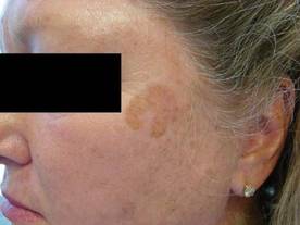
Single café au lait macules are found in about 2.5% of infants and typically are of little concern, but certain syndromes should be suspected in children with multiple lesions or in those who develop new lesions after the first 5-6 years of life, according to Dr. Wynnis L. Tom.
For example, multiple café au lait macules (CALMs) could be a sign of neurofibromatosis type I (NF1), Dr. Tom of the University of California, San Diego, said during a presentation at the annual Hawaii Dermatology Seminar sponsored by Skin Disease Education Foundation (SDEF).

Diagnosis of the autosomal dominant syndrome – which results from a neurofibromin defect and occurs in 1 of 2,000-4,500 individuals – requires the presence of two or more of the following diagnostic criteria: six or more CALMs greater than 0.5 cm in diameter in prepubertal children, or greater than 1.5 cm in those postpubertal; two or more neurofibromas of any type, or one plexiform neurofibroma; axillary and/or inguinal freckling (Crowe’s sign); optic glioma; two or more Lisch nodules; a bony lesion (such as sphenoid dysplasia or long bone dysplasia); and a first-degree relative with NF1.
After CALMs, the most common finding in patients with NF1 is axillary and/or inguinal freckling, which occurs in 75%-90% of cases.
"Recognizing NF1 is important, because the syndrome is associated with multiple complications. For example, ocular disease is common in these patients, and can include eye displacement, glaucoma, and visual impairment if the optical nerve is involved," Dr. Tom said.
Also, about 50% of patients have learning disabilities, and 5% have more severe developmental delay. Other complications include hypertension due to renal artery stenosis or (rarely) pheochromocytoma, and an increased risk of malignancy (particularly astrocytomas and malignant peripheral nerve sheath tumors).
The work-up for patients who meet the criteria for NF1 should include referrals for genetic evaluation and counseling, ophthalmological care, and neurologic and developmental services. Also, watch for scoliosis and consider imaging and performing biopsies for lesions with pain or sudden growth, Dr. Tom advised.

Legius syndrome is a more-recently defined NF1-like syndrome, which also is characterized by multiple CALMs with or without intertriginous freckling, although both are often fewer than in patients with true NF1.
This autosomal dominant, gene-related syndrome can be associated with macrocephaly; attention-deficit/hyperactivity disorder and/or other learning disabilities; lipomas; or vascular anomalies, but they do not have the neurofibromas or tumors of NF1. Thus, they likely have a better prognosis.
Other possible diagnoses in patients with multiple CALMs include familial CALMs (without any other findings) and other neuro-cardio-facial-cutaneous syndromes involving the ras/mitogen-activated protein kinase (ras/MAPK) signaling pathway.
Such syndromes may be associated with pigmentary anomalies, facial dysmorphism, short stature, congenital heart defects, neurocognitive defects, and predisposition to malignancy, Dr. Tom said. "Thus, advances in genetic analysis and diagnosis have expanded the number of disorders in which multiple CALMs can be seen. Children with multiple CALMs should, therefore, have thorough physical exams, regular monitoring for additional skin lesions, and appropriate other specialty evaluation," she concluded.
Dr. Tom reported having no disclosures. SDEF and this news organization are owned by Elsevier.
Single café au lait macules are found in about 2.5% of infants and typically are of little concern, but certain syndromes should be suspected in children with multiple lesions or in those who develop new lesions after the first 5-6 years of life, according to Dr. Wynnis L. Tom.
For example, multiple café au lait macules (CALMs) could be a sign of neurofibromatosis type I (NF1), Dr. Tom of the University of California, San Diego, said during a presentation at the annual Hawaii Dermatology Seminar sponsored by Skin Disease Education Foundation (SDEF).
Diagnosis of the autosomal dominant syndrome – which results from a neurofibromin defect and occurs in 1 of 2,000-4,500 individuals – requires the presence of two or more of the following diagnostic criteria: six or more CALMs greater than 0.5 cm in diameter in prepubertal children, or greater than 1.5 cm in those postpubertal; two or more neurofibromas of any type, or one plexiform neurofibroma; axillary and/or inguinal freckling (Crowe’s sign); optic glioma; two or more Lisch nodules; a bony lesion (such as sphenoid dysplasia or long bone dysplasia); and a first-degree relative with NF1.
After CALMs, the most common finding in patients with NF1 is axillary and/or inguinal freckling, which occurs in 75%-90% of cases.
"Recognizing NF1 is important, because the syndrome is associated with multiple complications. For example, ocular disease is common in these patients, and can include eye displacement, glaucoma, and visual impairment if the optical nerve is involved," Dr. Tom said.
Also, about 50% of patients have learning disabilities, and 5% have more severe developmental delay. Other complications include hypertension due to renal artery stenosis or (rarely) pheochromocytoma, and an increased risk of malignancy (particularly astrocytomas and malignant peripheral nerve sheath tumors).
The work-up for patients who meet the criteria for NF1 should include referrals for genetic evaluation and counseling, ophthalmological care, and neurologic and developmental services. Also, watch for scoliosis and consider imaging and performing biopsies for lesions with pain or sudden growth, Dr. Tom advised.
Legius syndrome is a more-recently defined NF1-like syndrome, which also is characterized by multiple CALMs with or without intertriginous freckling, although both are often fewer than in patients with true NF1.
This autosomal dominant, gene-related syndrome can be associated with macrocephaly; attention-deficit/hyperactivity disorder and/or other learning disabilities; lipomas; or vascular anomalies, but they do not have the neurofibromas or tumors of NF1. Thus, they likely have a better prognosis.
Other possible diagnoses in patients with multiple CALMs include familial CALMs (without any other findings) and other neuro-cardio-facial-cutaneous syndromes involving the ras/mitogen-activated protein kinase (ras/MAPK) signaling pathway.
Such syndromes may be associated with pigmentary anomalies, facial dysmorphism, short stature, congenital heart defects, neurocognitive defects, and predisposition to malignancy, Dr. Tom said. "Thus, advances in genetic analysis and diagnosis have expanded the number of disorders in which multiple CALMs can be seen. Children with multiple CALMs should, therefore, have thorough physical exams, regular monitoring for additional skin lesions, and appropriate other specialty evaluation," she concluded.
Dr. Tom reported having no disclosures. SDEF and this news organization are owned by Elsevier.
EXPERT ANALYSIS FROM THE SDEF HAWAII DERMATOLOGY SEMINAR
Heel Pain May Presage Psoriatic Arthritis
WAIKOLOA, HAWAII – Joint pain arising in a patient with psoriasis is by no means necessarily due to psoriatic arthritis.
Psoriasis patients can get osteoarthritis or rheumatoid arthritis, just like individuals without the skin disease. The distribution pattern of affected joints provides a useful guide as to which arthropathy is involved.

"When a patient who’s got psoriasis comes to me and says, ‘I have a tremendous amount of joint pain,’ the first place I look is the heel. Heel involvement doesn’t happen very often in rheumatoid arthritis. If the heel hurts, it’s probable that the patient has dactylitis and some form of psoriatic arthritis," according to Dr. Daniel E. Furst, the Carl M. Pearson professor of rheumatology at the University of California, Los Angeles.
Another useful clue: Psoriatic arthritis often affects the distal phalangeal joints of the fingers and toes, with accompanying characteristic psoriatic nail changes, but it spares the metacarpophalangeal (MCP) joints. In contrast, MCP involvement is a typical feature of rheumatoid arthritis, he explained at the Hawaii Dermatology Seminar sponsored by Skin Disease Education Foundation (SDEF).
"If only we understood why one joint tends to be affected and not another, we would sure know a whole lot more about psoriatic arthritis, and rheumatoid arthritis for that matter," Dr. Furst observed.
Involvement of the distal interphalangeal (DIP) joints is a common feature in osteoarthritis as well as psoriatic arthritis. However, psoriatic arthritis, unlike osteoarthritis, is an inflammatory arthritis. The affected fingers and toes in a patient with psoriatic arthritis often develop a painful, red, warm, sausage-like swelling of the soft tissue on the entire digit.
"If there’s erythema and squishiness when you push on a painful joint, that’s synovitis, not osteoarthritis," the rheumatologist noted.
Also, osteoarthritis has a strong tendency to involve the carpometacarpal joint of the thumb, a site spared in psoriatic arthritis, he continued.
Psoriatic arthritis often first shows up as a monoarticular, asymmetric arthritis of a larger joint, such as the knee, ankle, or wrist. Rheumatoid arthritis tends to come on much stronger.
"In rheumatoid arthritis, it’s frequently all at once. The patient will say, ‘In a week I went from nothing to pain everywhere.’ Not so in psoriatic arthritis," according to Dr. Furst.
Spondyloarthropathy is common in psoriatic arthritis. It often takes the form of a spondylitis characterized by asymmetric thickening and calcification of the longitudinal ligaments of the spine, resulting in asymmetric syndesmophytes on imaging. The result is a painful and/or stiff low back.
Psoriatic arthritis is widely thought of as a rheumatoid factor–negative disease, but that’s not strictly true. Roughly 15% of psoriatic arthritis patients are rheumatoid factor–positive.
Differentiating psoriatic arthritis from other types of arthritis that occur in psoriasis patients is important from a treatment standpoint. Psoriatic arthritis causes much more bony destruction than recognized until fairly recently, twice as much as rheumatoid arthritis by some measures. The nonbiologic disease-modifying antirheumatic drugs are only marginally effective for joint disease in psoriatic arthritis patients, with the exception of leflunomide (Arava), which has fair benefit for the peripheral arthritis but less for the skin disease.
At present, the tumor necrosis factor (TNF) inhibitors are the most effective agents available for peripheral and axial joint disease in psoriatic arthritis. But the response can be less impressive than in patients with psoriasis-only or rheumatoid arthritis. New therapies on the horizon include agents targeting the inflammatory cytokines interleukins-6, -12, and -15, as well as orally administered small molecules that inhibit Janus kinase, spleen tyrosine kinase, TNF-alpha converting enzyme, or mitogen-activated protein kinase.
"There’s a lot of stuff coming down the pike, including new drugs which have mechanisms of action very different from those most extant in psoriasis. Some of it is going to work. But so far we really don’t know which of it will," Dr. Furst said.
He reported that he serves as a consultant to or recipient of research grants from more than a dozen pharmaceutical companies and the National Institutes of Health.
SDEF and this news organization are owned by Elsevier.
WAIKOLOA, HAWAII – Joint pain arising in a patient with psoriasis is by no means necessarily due to psoriatic arthritis.
Psoriasis patients can get osteoarthritis or rheumatoid arthritis, just like individuals without the skin disease. The distribution pattern of affected joints provides a useful guide as to which arthropathy is involved.
"When a patient who’s got psoriasis comes to me and says, ‘I have a tremendous amount of joint pain,’ the first place I look is the heel. Heel involvement doesn’t happen very often in rheumatoid arthritis. If the heel hurts, it’s probable that the patient has dactylitis and some form of psoriatic arthritis," according to Dr. Daniel E. Furst, the Carl M. Pearson professor of rheumatology at the University of California, Los Angeles.
Another useful clue: Psoriatic arthritis often affects the distal phalangeal joints of the fingers and toes, with accompanying characteristic psoriatic nail changes, but it spares the metacarpophalangeal (MCP) joints. In contrast, MCP involvement is a typical feature of rheumatoid arthritis, he explained at the Hawaii Dermatology Seminar sponsored by Skin Disease Education Foundation (SDEF).
"If only we understood why one joint tends to be affected and not another, we would sure know a whole lot more about psoriatic arthritis, and rheumatoid arthritis for that matter," Dr. Furst observed.
Involvement of the distal interphalangeal (DIP) joints is a common feature in osteoarthritis as well as psoriatic arthritis. However, psoriatic arthritis, unlike osteoarthritis, is an inflammatory arthritis. The affected fingers and toes in a patient with psoriatic arthritis often develop a painful, red, warm, sausage-like swelling of the soft tissue on the entire digit.
"If there’s erythema and squishiness when you push on a painful joint, that’s synovitis, not osteoarthritis," the rheumatologist noted.
Also, osteoarthritis has a strong tendency to involve the carpometacarpal joint of the thumb, a site spared in psoriatic arthritis, he continued.
Psoriatic arthritis often first shows up as a monoarticular, asymmetric arthritis of a larger joint, such as the knee, ankle, or wrist. Rheumatoid arthritis tends to come on much stronger.
"In rheumatoid arthritis, it’s frequently all at once. The patient will say, ‘In a week I went from nothing to pain everywhere.’ Not so in psoriatic arthritis," according to Dr. Furst.
Spondyloarthropathy is common in psoriatic arthritis. It often takes the form of a spondylitis characterized by asymmetric thickening and calcification of the longitudinal ligaments of the spine, resulting in asymmetric syndesmophytes on imaging. The result is a painful and/or stiff low back.
Psoriatic arthritis is widely thought of as a rheumatoid factor–negative disease, but that’s not strictly true. Roughly 15% of psoriatic arthritis patients are rheumatoid factor–positive.
Differentiating psoriatic arthritis from other types of arthritis that occur in psoriasis patients is important from a treatment standpoint. Psoriatic arthritis causes much more bony destruction than recognized until fairly recently, twice as much as rheumatoid arthritis by some measures. The nonbiologic disease-modifying antirheumatic drugs are only marginally effective for joint disease in psoriatic arthritis patients, with the exception of leflunomide (Arava), which has fair benefit for the peripheral arthritis but less for the skin disease.
At present, the tumor necrosis factor (TNF) inhibitors are the most effective agents available for peripheral and axial joint disease in psoriatic arthritis. But the response can be less impressive than in patients with psoriasis-only or rheumatoid arthritis. New therapies on the horizon include agents targeting the inflammatory cytokines interleukins-6, -12, and -15, as well as orally administered small molecules that inhibit Janus kinase, spleen tyrosine kinase, TNF-alpha converting enzyme, or mitogen-activated protein kinase.
"There’s a lot of stuff coming down the pike, including new drugs which have mechanisms of action very different from those most extant in psoriasis. Some of it is going to work. But so far we really don’t know which of it will," Dr. Furst said.
He reported that he serves as a consultant to or recipient of research grants from more than a dozen pharmaceutical companies and the National Institutes of Health.
SDEF and this news organization are owned by Elsevier.
EXPERT ANALYSIS FROM THE SDEF HAWAII DERMATOLOGY SEMINAR
Microwave Technology Outlasts Botulinum Toxin for Hyperhidrosis
Microwave technology is a new hyperhidrosis treatment that offers longer-term efficacy than botulinum toxin.
"With microwave thermolysis, we now have a reliable and safe method with long-term efficacy to treat our patients who suffer from hyperhidrosis," said Dr. Michael S. Kaminer, at the Hawaii Dermatology Seminar sponsored by Skin Disease Education Foundation (SDEF).

A hyperhidrosis diagnosis is made in patients who have visible, excessive sweating for at least 6 months without apparent cause, and with at least two of the following characteristics (J. Am. Acad. Dermatol. 2004;51:274-86):
• Impairs daily activities.
• Frequency of at least one episode per week.
• Bilateral and symmetric.
• Age of onset before 25 years.
• Positive family history.
• Cessation during sleep.
In the United states, approximately 7.6 million people, or 4% of all U.S. adults, are estimated to have axillary hyperhidrosis with a level on the Hyperhidrosis Disease Severity Scale (HDSS) of either 3 ("My sweating is barely tolerable and frequently interferes with my daily activities.") or 4 ("My sweating is intolerable and always interferes with my daily activities."), according to Dr. Kaminer, managing partner, SkinCare Physicians, Chestnut Hill, Mass. Another 32.5 million are "sweat bothered," with an HDSS level of 2 ("My sweating is tolerable but sometimes interferes with my daily activities.") or 1 ("My sweating is never noticeable and never interferes with my daily activities.").
Axillary hyperhidrosis causes considerable distress among patients. In one survey of the Dermatology Life Quality Index, the condition scored just below atopic dermatitis, close to psoriasis, and above acne vulgaris in terms of quality of life impairment, with a mean baseline score of 10.45, compared with 11.20 for atopic dermatitis, 10.53 for psoriasis, and 7.45 for acne vulgaris (Br. J. Dermatol. 2008;159:997-1035).
Current treatment options include various topical over-the-counter products, iontophoresis, sympathectomy, botulinum toxin, ultrasound (in early trials), and now microwave thermolysis.
The proprietary microwave thermolysis system, MiraDry (Miramar Labs), focuses energy delivery to the dermal-fat interface. Microwave energy is absorbed by electric dipoles. The ion content of sweat is a perfect target for this, said Dr. Kaminer, who is also with the departments of dermatology at Yale University, New Haven, Conn.; Dartmouth College, Hanover, N.H.; and Brown University, Providence, R.I.
Energy becomes concentrated along this interface and creates a focal energy zone. Continuous hydroceramic cooling prevents thermal conduction of heat superficially, and creates a heat zone at the level of sweat glands resulting in thermolysis. The procedure involves injectable local anesthesia. Treatment time is about 30 minutes, with rapid recovery.
Long-term efficacy of the device was demonstrated in the DRIUP study (Dermatologic Reduction in Underarm Perspiration), in which 120 patients with HDSS scores of 3 or 4 were randomized in a 2:1 ratio to one to three active treatments (using a prior investigational version of the microwave device) or sham treatment at seven U.S. sites. Efficacy, defined as patients reporting a reduction to an HDSS score of 1 or 2, was 89% at 30 days, 74% at 3 months, 67% at 6 months, and 69% at the 9- and 12-month visits. In the sham group, efficacy was 54% at 30 days and 44% for the 3- and 6-month visits. At all time points, the differences between the treatment and sham groups were significant.
There were 45 treatment-related adverse events, with 28% of the treatment group and 13% of the sham group patients experiencing at least one adverse event. The most common of these was an area of transient altered sensation in the arm, occurring in 10% of patients in the active treatment group. All but one adverse event resolved over time; the patient withdrew from the study, reporting altered sweating on the face. In all, 71% of the events were rated as mild in severity and didn’t affect patients’ daily activities (Dermatol Surg. 2012;38:185-91). Swelling was common, self-limited, and proportional to the energy delivered, Dr. Kaminer noted.
When compared with botulinum toxin, both approaches are effective, noninvasive (injections only), and involve minimal to no downtime. However, the duration of effect is 6.7 months for botulinum toxin, compared with stable efficacy beginning at 3 months and lasting through 12 months follow-up for microwave thermolysis, he said.
Overall, the advantages of microwave thermolysis over other treatments are its long-term efficacy (greater than 12 months, and likely longer), and its efficacy in more than 90% of patients with the latest-generation device. Disadvantages include the need for two procedures and the less than 100% responder rate. Botulinum toxin, in contrast, is easier to perform, and nearly 100% effective. However, it has the downside of the need for repeat treatments approximately every 6 months, and its cost, along with problematic insurance coverage.
Dr. Kaminer said that his practice participated in a registry-type study in the summer and fall of 2011 and was one of the first clinics in the country to purchase a MiraDry System, in December 2011. "Our staff has found the procedure easy to incorporate into the flow of our practice, and patients have found it to be rather easy to have done. We have seen some modest swelling for up to 1 week after treatment, but that is expected. So far, our experience has been pretty much what we would have expected, and if anything, a little better."
He added that, "Interest in offering this new treatment option to patients with excessive underarm sweating has been extremely high. Between clinics in Japan and the U.S., there have been over 1,500 patients treated."
In all, safety results have been consistent with prior experiences. "As with any procedure that we do, it is important to clearly communicate with the patients what to expect from the procedure and the recovery. Our feedback allowed the company to fine-tune their training program so that clinics that purchase the system have tools they can use to make the experience a very positive one for their patients."
Miramar is in the early stages of commercialization for the MiraDry system. It is being placed in a controlled strategy and is expected to reach "a significant number of practices across the U.S. by the middle of this year," he said.
Dr. Kaminer is chairman of the Miramar Labs scientific advisory board. His institution served as a research study site for the phase II study. SDEF and this news organization are owned by Elsevier.
Microwave technology is a new hyperhidrosis treatment that offers longer-term efficacy than botulinum toxin.
"With microwave thermolysis, we now have a reliable and safe method with long-term efficacy to treat our patients who suffer from hyperhidrosis," said Dr. Michael S. Kaminer, at the Hawaii Dermatology Seminar sponsored by Skin Disease Education Foundation (SDEF).
A hyperhidrosis diagnosis is made in patients who have visible, excessive sweating for at least 6 months without apparent cause, and with at least two of the following characteristics (J. Am. Acad. Dermatol. 2004;51:274-86):
• Impairs daily activities.
• Frequency of at least one episode per week.
• Bilateral and symmetric.
• Age of onset before 25 years.
• Positive family history.
• Cessation during sleep.
In the United states, approximately 7.6 million people, or 4% of all U.S. adults, are estimated to have axillary hyperhidrosis with a level on the Hyperhidrosis Disease Severity Scale (HDSS) of either 3 ("My sweating is barely tolerable and frequently interferes with my daily activities.") or 4 ("My sweating is intolerable and always interferes with my daily activities."), according to Dr. Kaminer, managing partner, SkinCare Physicians, Chestnut Hill, Mass. Another 32.5 million are "sweat bothered," with an HDSS level of 2 ("My sweating is tolerable but sometimes interferes with my daily activities.") or 1 ("My sweating is never noticeable and never interferes with my daily activities.").
Axillary hyperhidrosis causes considerable distress among patients. In one survey of the Dermatology Life Quality Index, the condition scored just below atopic dermatitis, close to psoriasis, and above acne vulgaris in terms of quality of life impairment, with a mean baseline score of 10.45, compared with 11.20 for atopic dermatitis, 10.53 for psoriasis, and 7.45 for acne vulgaris (Br. J. Dermatol. 2008;159:997-1035).
Current treatment options include various topical over-the-counter products, iontophoresis, sympathectomy, botulinum toxin, ultrasound (in early trials), and now microwave thermolysis.
The proprietary microwave thermolysis system, MiraDry (Miramar Labs), focuses energy delivery to the dermal-fat interface. Microwave energy is absorbed by electric dipoles. The ion content of sweat is a perfect target for this, said Dr. Kaminer, who is also with the departments of dermatology at Yale University, New Haven, Conn.; Dartmouth College, Hanover, N.H.; and Brown University, Providence, R.I.
Energy becomes concentrated along this interface and creates a focal energy zone. Continuous hydroceramic cooling prevents thermal conduction of heat superficially, and creates a heat zone at the level of sweat glands resulting in thermolysis. The procedure involves injectable local anesthesia. Treatment time is about 30 minutes, with rapid recovery.
Long-term efficacy of the device was demonstrated in the DRIUP study (Dermatologic Reduction in Underarm Perspiration), in which 120 patients with HDSS scores of 3 or 4 were randomized in a 2:1 ratio to one to three active treatments (using a prior investigational version of the microwave device) or sham treatment at seven U.S. sites. Efficacy, defined as patients reporting a reduction to an HDSS score of 1 or 2, was 89% at 30 days, 74% at 3 months, 67% at 6 months, and 69% at the 9- and 12-month visits. In the sham group, efficacy was 54% at 30 days and 44% for the 3- and 6-month visits. At all time points, the differences between the treatment and sham groups were significant.
There were 45 treatment-related adverse events, with 28% of the treatment group and 13% of the sham group patients experiencing at least one adverse event. The most common of these was an area of transient altered sensation in the arm, occurring in 10% of patients in the active treatment group. All but one adverse event resolved over time; the patient withdrew from the study, reporting altered sweating on the face. In all, 71% of the events were rated as mild in severity and didn’t affect patients’ daily activities (Dermatol Surg. 2012;38:185-91). Swelling was common, self-limited, and proportional to the energy delivered, Dr. Kaminer noted.
When compared with botulinum toxin, both approaches are effective, noninvasive (injections only), and involve minimal to no downtime. However, the duration of effect is 6.7 months for botulinum toxin, compared with stable efficacy beginning at 3 months and lasting through 12 months follow-up for microwave thermolysis, he said.
Overall, the advantages of microwave thermolysis over other treatments are its long-term efficacy (greater than 12 months, and likely longer), and its efficacy in more than 90% of patients with the latest-generation device. Disadvantages include the need for two procedures and the less than 100% responder rate. Botulinum toxin, in contrast, is easier to perform, and nearly 100% effective. However, it has the downside of the need for repeat treatments approximately every 6 months, and its cost, along with problematic insurance coverage.
Dr. Kaminer said that his practice participated in a registry-type study in the summer and fall of 2011 and was one of the first clinics in the country to purchase a MiraDry System, in December 2011. "Our staff has found the procedure easy to incorporate into the flow of our practice, and patients have found it to be rather easy to have done. We have seen some modest swelling for up to 1 week after treatment, but that is expected. So far, our experience has been pretty much what we would have expected, and if anything, a little better."
He added that, "Interest in offering this new treatment option to patients with excessive underarm sweating has been extremely high. Between clinics in Japan and the U.S., there have been over 1,500 patients treated."
In all, safety results have been consistent with prior experiences. "As with any procedure that we do, it is important to clearly communicate with the patients what to expect from the procedure and the recovery. Our feedback allowed the company to fine-tune their training program so that clinics that purchase the system have tools they can use to make the experience a very positive one for their patients."
Miramar is in the early stages of commercialization for the MiraDry system. It is being placed in a controlled strategy and is expected to reach "a significant number of practices across the U.S. by the middle of this year," he said.
Dr. Kaminer is chairman of the Miramar Labs scientific advisory board. His institution served as a research study site for the phase II study. SDEF and this news organization are owned by Elsevier.
FROM THE SDEF HAWAII DERMATOLOGY SEMINAR
Major Finding: Efficacy was 89% at 30 days, 74% at 3 months, 67% at 6 months, and 69% at the 9- and 12-month visits.
Data Source: Patients (120) with HDSS scores of 3 or 4 were randomized in a 2:1 ratio to one to three active treatments (using a prior investigational version of the microwave device) or sham treatment at seven U.S. sites.
Disclosures: Dr. Kaminer is chairman of the Miramar Labs scientific advisory board. His institution served as a research study site for the phase II study. SDEF and this news organization are owned by Elsevier.
Let Balance, Aesthetics Guide Volume Replacement
Volume replacement can give an aging face some lift, but it’s not for everyone, according to Dr. Mark G. Rubin.
In a woman with a round face, for example, adding volume to the lower half of the face can create a fuller, heavier appearance rather than a more youthful appearance, he said at the annual Hawaii Dermatology Seminar sponsored by Skin Disease Education Foundation (SDEF).
"We all agree that volume loss is a significant part of aging, but without attention to facial balance and aesthetics, the results of volume replacement can be less than satisfactory," he added, noting that when it comes to facial contouring, an understanding of balance and aesthetics becomes more important than technique.

One important concept to keep in mind is the "triangle of youth," said Dr. Rubin who practices dermatology in Beverly Hills, Calif. He explained that a triangular face is more youthful, which is why volume that promotes roundness can have the opposite effect, even if the volume tends to smooth wrinkles.
Creating a triangular shape by providing fullness and prominence of the cheekbone – as long as it isn’t overdone – can create a youthful, attractive look, he said.
The aging lower half of the face has volume loss in several areas, including the medial cheek/nasojugal groove, the central cheek, the nasolabial folds, the marionette lines, and the prejowl area.
Volume replacement in the marionette lines and at the jaw line can be particularly effective.
In the marionette fold, one technique that can be useful is building a layer of support to push up on the descending cheek, thereby camouflaging its leading edge.
"Sometime you need to add a little support into the lateral lower lip, as well as the horseshoe of the modiolus," he said. "Remember, as you push up on the corner of the lip, that you can accidentally create or aggravate fullness laterally."
Adding filler to the upper vermilion border can also support this approach by tightening the lip and providing some lift to the corner, thereby helping reduce the puffiness created by treating the upper marionette area.
Volume at the jawline can also provide a more youthful look, but keep in mind that the two areas of apparent volume loss – the pre- and postjowl – are actually normal areas, whereas the skin in between has descended, Dr. Rubin explained. Filling these pre- and postjowl pseudodepressions will provide something of an optical illusion, making the jowl look less prominent. Similarly, adding volume at the prejowl sulcus can reduce the sagging appearance of the jowl.
However, a patient with a round face may not be a good candidate for this approach. "Too much volume at the prejowl sulcus in a patient with a round face will make the face look fat," he explained.
Also, in some patients the smoothing of the jaw line may look good on profile, but it can have a masculinizing effect by widening the lateral face when viewed head on.
As for the cheeks, not all "deflate" in the same way, and thus not all will benefit from volume replacement. In those with central cheek hollowing, volume can be very effective in creating a more youthful appearance, as well as in tightening loose skin.
A number of filler options can be considered, including hyaluronic acid (HA) fillers, poly-L-lactic acid, calcium hydroxylapatite, and polymethyl methacrylate (PMMA).
"But for first-time patients, or if you are learning your way, don’t underestimate the value of a reversible filler," he said.
As with the use of neurotoxins, the approach to using fillers can vary without necessarily affecting the outcome. Various physicians have their own preferences for products and injection techniques, yet almost everyone seems to be able to achieve the intended aesthetic goal, he explained.
"So I don’t think the ‘what’ is that important. It’s knowing the ‘where’ and the mastering of an injection technique that means more," he said.
In fact, Food and Drug Administration trial data for most fillers suggest that they are more similar than dissimilar in terms of efficacy, longevity, and adverse effects. Still, although conventional wisdom suggests that particle size, concentration, or lift should affect the final result, and thus in theory there should be one product that is best for each location, there is a lack of agreement.
In Dr. Rubin’s experience, however, HA, PMMA, and calcium hydroxylapatite fillers can have a slight rippling effect when they are injected in areas of soft tissue, such as the central cheek.
"It’s hard to make a smooth contour across 6-8 cm when you are injecting droplets or linear filaments of a filler," he said, noting that poly-L-lactic acid seems to create a smoother contour in that area.
When injecting over bony areas, HA products and calcium hydroxylapatite are acceptable, because the defects are generally smaller and there is less surface area on which to see ripples.
Dr. Rubin is a consultant for Medicis. He also has been involved in FDA clinical trials for Restylane, Perlane, and Artefill. SDEF and this news organization are owned by Elsevier.
Volume replacement can give an aging face some lift, but it’s not for everyone, according to Dr. Mark G. Rubin.
In a woman with a round face, for example, adding volume to the lower half of the face can create a fuller, heavier appearance rather than a more youthful appearance, he said at the annual Hawaii Dermatology Seminar sponsored by Skin Disease Education Foundation (SDEF).
"We all agree that volume loss is a significant part of aging, but without attention to facial balance and aesthetics, the results of volume replacement can be less than satisfactory," he added, noting that when it comes to facial contouring, an understanding of balance and aesthetics becomes more important than technique.
One important concept to keep in mind is the "triangle of youth," said Dr. Rubin who practices dermatology in Beverly Hills, Calif. He explained that a triangular face is more youthful, which is why volume that promotes roundness can have the opposite effect, even if the volume tends to smooth wrinkles.
Creating a triangular shape by providing fullness and prominence of the cheekbone – as long as it isn’t overdone – can create a youthful, attractive look, he said.
The aging lower half of the face has volume loss in several areas, including the medial cheek/nasojugal groove, the central cheek, the nasolabial folds, the marionette lines, and the prejowl area.
Volume replacement in the marionette lines and at the jaw line can be particularly effective.
In the marionette fold, one technique that can be useful is building a layer of support to push up on the descending cheek, thereby camouflaging its leading edge.
"Sometime you need to add a little support into the lateral lower lip, as well as the horseshoe of the modiolus," he said. "Remember, as you push up on the corner of the lip, that you can accidentally create or aggravate fullness laterally."
Adding filler to the upper vermilion border can also support this approach by tightening the lip and providing some lift to the corner, thereby helping reduce the puffiness created by treating the upper marionette area.
Volume at the jawline can also provide a more youthful look, but keep in mind that the two areas of apparent volume loss – the pre- and postjowl – are actually normal areas, whereas the skin in between has descended, Dr. Rubin explained. Filling these pre- and postjowl pseudodepressions will provide something of an optical illusion, making the jowl look less prominent. Similarly, adding volume at the prejowl sulcus can reduce the sagging appearance of the jowl.
However, a patient with a round face may not be a good candidate for this approach. "Too much volume at the prejowl sulcus in a patient with a round face will make the face look fat," he explained.
Also, in some patients the smoothing of the jaw line may look good on profile, but it can have a masculinizing effect by widening the lateral face when viewed head on.
As for the cheeks, not all "deflate" in the same way, and thus not all will benefit from volume replacement. In those with central cheek hollowing, volume can be very effective in creating a more youthful appearance, as well as in tightening loose skin.
A number of filler options can be considered, including hyaluronic acid (HA) fillers, poly-L-lactic acid, calcium hydroxylapatite, and polymethyl methacrylate (PMMA).
"But for first-time patients, or if you are learning your way, don’t underestimate the value of a reversible filler," he said.
As with the use of neurotoxins, the approach to using fillers can vary without necessarily affecting the outcome. Various physicians have their own preferences for products and injection techniques, yet almost everyone seems to be able to achieve the intended aesthetic goal, he explained.
"So I don’t think the ‘what’ is that important. It’s knowing the ‘where’ and the mastering of an injection technique that means more," he said.
In fact, Food and Drug Administration trial data for most fillers suggest that they are more similar than dissimilar in terms of efficacy, longevity, and adverse effects. Still, although conventional wisdom suggests that particle size, concentration, or lift should affect the final result, and thus in theory there should be one product that is best for each location, there is a lack of agreement.
In Dr. Rubin’s experience, however, HA, PMMA, and calcium hydroxylapatite fillers can have a slight rippling effect when they are injected in areas of soft tissue, such as the central cheek.
"It’s hard to make a smooth contour across 6-8 cm when you are injecting droplets or linear filaments of a filler," he said, noting that poly-L-lactic acid seems to create a smoother contour in that area.
When injecting over bony areas, HA products and calcium hydroxylapatite are acceptable, because the defects are generally smaller and there is less surface area on which to see ripples.
Dr. Rubin is a consultant for Medicis. He also has been involved in FDA clinical trials for Restylane, Perlane, and Artefill. SDEF and this news organization are owned by Elsevier.
EXPERT ANALYSIS FROM THE SDEF HAWAII DERMATOLOGY SEMINAR
Topical Cyclosporine Proves Beneficial For Ocular Rosacea
WAIKOLOA, HAWAII – Topical cyclosporine 0.05% ophthalmic emulsion significantly outperformed artificial tears for the treatment of ocular rosacea in a double-blind, randomized, multicenter study.
"It’s a fairly small study, but I think it gives us some valuable information. This gives us a new option topically," Dr. Julie C. Harper said in highlighting the study at the Hawaii Dermatology Seminar sponsored by Skin Disease Education Foundation (SDEF).

She referred to a clinical trial conducted by Florida ophthalmologists who randomized 37 patients with ocular rosacea to twice-daily topical cyclosporine 0.05% (Restasis) or artificial tears for 3 months.
The outcome that most impressed Dr. Harper was the topical cyclosporine group’s mean 11.5-point improvement from a baseline of 14.1 on the Ocular Surface Disease Index, a validated patient questionnaire assessing the eye disease’s impact on quality of life. Patients who received artificial tears – a widely prescribed treatment for ocular rosacea – had a significantly lesser mean 2.9-point improvement.
"People definitely felt better with this product than with artificial tears," observed Dr. Harper, a dermatologist at the University of Alabama at Birmingham.
In addition, the topical cyclosporine group fared significantly better in the other study end points, which focused on eye dryness and tear production. For example, mean scores on Schirmer\'s test (a measure of eye-surface wetness) improved by 2.7 mm over the course of 3 months of treatment, from a baseline of 8.3, compared with a 1.4-mm deterioration in the artificial tears group.
Also, tear break-up time (a measure of how quickly an eye becomes dry) increased by a mean of 3.56 seconds in the cyclosporine group, a significantly more favorable effect than the 0.04-second decrease in the artificial tears arm (Adv. Ther. 2009;26:651-9).
The investigators speculated that topical cyclosporine’s clinical benefits in ocular rosacea are due to the drug’s anti-inflammatory effects, including a cyclosporine-mediated reduction in the number of activated lymphocytes in the conjunctiva, in combination with stimulation of increased tear production
The ophthalmologists emphasized that ocular rosacea can be a serious and potentially even a blinding condition. They noted that in one classic study, 13 of 131 patients had corneal complications resulting in visual acuity loss at the time they presented to ophthalmology. Of those 13 patients, 7 were left with worse than 20/400 eyesight, and 6 of the 13 required penetrating keratoplasty during the course of their disease (Ophthalmology 1997;104:1863-7).
Dr. Harper said that since she learned of the randomized trial, she has tried using topical cyclosporine for ocular rosacea but doesn’t yet have enough experience to say how well it works in her own hands. Usually, she gives the topical agent in combination with an oral tetracycline in patients whose ocular disease isn’t responding adequately to oral therapy alone.
Her preferred oral agent is delayed-release doxycycline (Oracea) at the anti-inflammatory but subantimicrobial dose of 40 mg once daily, a dosage that is Food and Drug Administration approved for treatment of rosacea. She favors this regimen because it doesn’t contribute to the worsening, global, antibiotic-resistance public health problem, and the once-daily aspect is a real plus from the patient adherence standpoint.
For patients who don’t have insurance coverage for this relatively costly drug, her second-favorite option is generic doxycycline at 20 mg BID, which is also an anti-inflammatory yet subantimicrobial dosage. It’s important to recognize, however, that bumping up the dosage of generic doxycycline to 50 mg/day would cross the threshold into the antibacterial range that could contribute to the resistance problem, the dermatologist added.
Topical cyclosporine for ocular rosacea is off-label therapy. The drug’s approved indication is in treating chronic dry eyes. Conference codirector Dr. Joseph F. Fowler Jr. predicted that the product may never gain an indication for ocular rosacea. That’s because ophthalmologists have a difficult time in quantifying the severity of ocular rosacea for research purposes, and even in distinguishing it from dry eye.
"My own crude approach is that if you’ve got rosacea on the skin and you’ve got itchy, gritty eyes, then I generally assume you’ve got ocular rosacea," explained Dr. Fowler, a dermatologist at the University of Louisville (Ky.).
Like Dr. Harper, he typically prescribes topical cyclosporine for ocular rosacea in combination with oral doxycycline.
"I find it very useful," he added.
Dr. Fowler disclosed that he has received research grants from Allergan, which markets Restasis. He also serves as a consultant to Galderma, which markets Oracea, as well as to numerous other pharmaceutical companies. Dr. Harper is on the speakers bureaus for Allergan, Galderma, and a handful of other companies.
The SDEF and this news organization are owned by Elsevier.
WAIKOLOA, HAWAII – Topical cyclosporine 0.05% ophthalmic emulsion significantly outperformed artificial tears for the treatment of ocular rosacea in a double-blind, randomized, multicenter study.
"It’s a fairly small study, but I think it gives us some valuable information. This gives us a new option topically," Dr. Julie C. Harper said in highlighting the study at the Hawaii Dermatology Seminar sponsored by Skin Disease Education Foundation (SDEF).
She referred to a clinical trial conducted by Florida ophthalmologists who randomized 37 patients with ocular rosacea to twice-daily topical cyclosporine 0.05% (Restasis) or artificial tears for 3 months.
The outcome that most impressed Dr. Harper was the topical cyclosporine group’s mean 11.5-point improvement from a baseline of 14.1 on the Ocular Surface Disease Index, a validated patient questionnaire assessing the eye disease’s impact on quality of life. Patients who received artificial tears – a widely prescribed treatment for ocular rosacea – had a significantly lesser mean 2.9-point improvement.
"People definitely felt better with this product than with artificial tears," observed Dr. Harper, a dermatologist at the University of Alabama at Birmingham.
In addition, the topical cyclosporine group fared significantly better in the other study end points, which focused on eye dryness and tear production. For example, mean scores on Schirmer\'s test (a measure of eye-surface wetness) improved by 2.7 mm over the course of 3 months of treatment, from a baseline of 8.3, compared with a 1.4-mm deterioration in the artificial tears group.
Also, tear break-up time (a measure of how quickly an eye becomes dry) increased by a mean of 3.56 seconds in the cyclosporine group, a significantly more favorable effect than the 0.04-second decrease in the artificial tears arm (Adv. Ther. 2009;26:651-9).
The investigators speculated that topical cyclosporine’s clinical benefits in ocular rosacea are due to the drug’s anti-inflammatory effects, including a cyclosporine-mediated reduction in the number of activated lymphocytes in the conjunctiva, in combination with stimulation of increased tear production
The ophthalmologists emphasized that ocular rosacea can be a serious and potentially even a blinding condition. They noted that in one classic study, 13 of 131 patients had corneal complications resulting in visual acuity loss at the time they presented to ophthalmology. Of those 13 patients, 7 were left with worse than 20/400 eyesight, and 6 of the 13 required penetrating keratoplasty during the course of their disease (Ophthalmology 1997;104:1863-7).
Dr. Harper said that since she learned of the randomized trial, she has tried using topical cyclosporine for ocular rosacea but doesn’t yet have enough experience to say how well it works in her own hands. Usually, she gives the topical agent in combination with an oral tetracycline in patients whose ocular disease isn’t responding adequately to oral therapy alone.
Her preferred oral agent is delayed-release doxycycline (Oracea) at the anti-inflammatory but subantimicrobial dose of 40 mg once daily, a dosage that is Food and Drug Administration approved for treatment of rosacea. She favors this regimen because it doesn’t contribute to the worsening, global, antibiotic-resistance public health problem, and the once-daily aspect is a real plus from the patient adherence standpoint.
For patients who don’t have insurance coverage for this relatively costly drug, her second-favorite option is generic doxycycline at 20 mg BID, which is also an anti-inflammatory yet subantimicrobial dosage. It’s important to recognize, however, that bumping up the dosage of generic doxycycline to 50 mg/day would cross the threshold into the antibacterial range that could contribute to the resistance problem, the dermatologist added.
Topical cyclosporine for ocular rosacea is off-label therapy. The drug’s approved indication is in treating chronic dry eyes. Conference codirector Dr. Joseph F. Fowler Jr. predicted that the product may never gain an indication for ocular rosacea. That’s because ophthalmologists have a difficult time in quantifying the severity of ocular rosacea for research purposes, and even in distinguishing it from dry eye.
"My own crude approach is that if you’ve got rosacea on the skin and you’ve got itchy, gritty eyes, then I generally assume you’ve got ocular rosacea," explained Dr. Fowler, a dermatologist at the University of Louisville (Ky.).
Like Dr. Harper, he typically prescribes topical cyclosporine for ocular rosacea in combination with oral doxycycline.
"I find it very useful," he added.
Dr. Fowler disclosed that he has received research grants from Allergan, which markets Restasis. He also serves as a consultant to Galderma, which markets Oracea, as well as to numerous other pharmaceutical companies. Dr. Harper is on the speakers bureaus for Allergan, Galderma, and a handful of other companies.
The SDEF and this news organization are owned by Elsevier.
EXPERT ANALYSIS FROM THE SDEF HAWAII DERMATOLOGY SEMINAR
Identifying AK Variants Is Key to Management
The ability to distinguish between the two key variants of actinic keratosis is key to effective management, according to Dr. Theodore Rosen.
Pigmented actinic keratosis (AK) that spreads typically occurs in patients older than age 60 years and affects more women than men. The lesions are larger than 1.5 cm and have a history of lateral spread. "The lesions are flat, with a smooth, scaly surface, and they occur on sun-exposed skin, mostly the face, and near other manifestations of photodamage," noted Dr. Rosen at the annual Hawaii Dermatology Seminar sponsored by Skin Disease Education Foundation (SDEF).

Differential diagnosis includes solar lentigo, lentigo maligna, lentigo maligna melanoma, seborrheic keratosis, and pigmented squamous cell carcinoma in situ. "The presence of dots, globules, and asymmetry on dermatoscopy can help you distinguish spreading pigmented AKs from other lesions," said Dr. Rosen, professor of dermatology at Baylor College of Medicine, Houston.
The other main variant, proliferating AK, also tends to affect patients older than age 60 but it occurs more commonly in men than in women. This variant is greater than 1.5 cm in size with a history of rapid lateral spread, and it is marked by a confluent scaly surface with some palpability. Affected areas include sun-exposed skin, mostly the face, and areas near other manifestations of photodamage. "This variant is resistant to usual topical therapy, and the differential diagnosis may include eczema, psoriasis, squamous cell carcinoma in situ, and superficial basal cell carcinoma," said Dr. Rosen, who is also chief of the dermatology clinic at the Houston-based Michael E. DeBakey VA Medical Center. Histology reveals fingerlike invasive projections into the dermis of atypical squamous cells.
The appearance of multiple AKs in a cosmetic unit indicates that the clinician should consider the entire photodamaged area to be diseased. This concept, known as field-directed therapy, "not only addresses apparent clinical lesions, but also reveals and may treat subclinical lesions," he said. "Field therapy may also offer the advantage of reversing basic cellular aberrations leading to transition into invasive cutaneous malignancy."
Another trend in the treatment of AKs is the development of new imiquimod formulations. Results from two placebo-controlled studies of daily application of 2.5% and 3.75% imiquimod creams to the face and balding scalp for two 3-week cycles revealed that both formulations were more effective than placebo and had an acceptable safety profile (J. Am. Acad. Dermatol. 2010;62:573-81). The final evaluation occurred 8 weeks after the final treatment, said Dr. Rosen, who was one of the study investigators. Results from this and other studies led to Food and Drug Administration approval of imiquimod 2.5% (Zyclara) for AKs on July 19, 2011, but the formulation is not yet commercially available.
In a more recent analysis, investigators assessed complete clearance rates in AK patients from four phase III trials of imiquimod who were followed for 1 year (J. Drugs Dermatol. 2011;10:165-70). For imiquimod 3.75% and 2.5% cream, respectively, complete clearance was sustained for 1 year in 40.5% and 33.3% of patients from the 2-week cycle studies, and in 47.9% and 43.2% of patients from the 3-week cycle studies.
Combination therapies for AKs have become increasingly popular. Treatments include cryosurgery followed by diclofenac, 5-fluorouracil followed by cryosurgery, cryosurgery followed by imiquimod 5% or 3.75%, photodynamic therapy (PTD) followed by imiquimod 5%, and tazarotene followed by PDT. "For all studies where field therapy is combined with lesion-directed therapy or with another field therapy, the combination proved superior to monotherapy," Dr. Rosen said.
He went on to note that there is considerable user variation – such as time, the distance the device is held from the skin, and the type of unit used – in the delivery of cryosurgery to treat AKs. The variation "may have more to do with skin temperature than any other variable," said Dr. Rosen. In one study, investigators used an infrared detector in a liquid nitrogen container to deliver cryotherapy to a temperature of –5° C in 36 patients with 180 thin AKs (Dermatol. Surg. 2010;36:1956-61). The clearance rates were 67% at 1 week and 100% at 6 weeks.
Treatment of AKs with radiation is another area of study. In one recent trial, 12 patients with refractory AK and 4 patients with AKs near a nonmelanoma skin cancer underwent external beam radiation therapy five times a week for 4-6 weeks. The total dose of radiation ranged from 4 to 60 Gy (J. Clin. Aesthet. Dermatol. 2011;4:47-50). Of the 16 patients, 13 had complete resolution of AKs, 1 noted a prompt recurrence, and 90% said they were "very satisfied" with the treatment results.
Treating AKs with methyl aminolevulinate photodynamic therapy (MAL-PDT) is also being investigated. In a multicenter study conducted in Scandinavia, 120 patients with 1,572 AKs underwent MAL-PDT followed by immediate exposure to the sun for 1.5-2.5 hours (Br. J. Dermatol. 2011;164:1083-90). At 3 months, 75%-77% of the lesions had cleared. This approach "has an advantage for the health care provider in that no equipment is needed," Dr. Rosen said. "The advantage for the patient is that the therapy is nearly pain free."
Dr. Rosen disclosed that he is a consultant and a member of the speakers bureau for Medicis, which makes imiquimod. He is also a consultant for LEO Pharma.
SDEF and this news organization are owned by Elsevier.
The ability to distinguish between the two key variants of actinic keratosis is key to effective management, according to Dr. Theodore Rosen.
Pigmented actinic keratosis (AK) that spreads typically occurs in patients older than age 60 years and affects more women than men. The lesions are larger than 1.5 cm and have a history of lateral spread. "The lesions are flat, with a smooth, scaly surface, and they occur on sun-exposed skin, mostly the face, and near other manifestations of photodamage," noted Dr. Rosen at the annual Hawaii Dermatology Seminar sponsored by Skin Disease Education Foundation (SDEF).
Differential diagnosis includes solar lentigo, lentigo maligna, lentigo maligna melanoma, seborrheic keratosis, and pigmented squamous cell carcinoma in situ. "The presence of dots, globules, and asymmetry on dermatoscopy can help you distinguish spreading pigmented AKs from other lesions," said Dr. Rosen, professor of dermatology at Baylor College of Medicine, Houston.
The other main variant, proliferating AK, also tends to affect patients older than age 60 but it occurs more commonly in men than in women. This variant is greater than 1.5 cm in size with a history of rapid lateral spread, and it is marked by a confluent scaly surface with some palpability. Affected areas include sun-exposed skin, mostly the face, and areas near other manifestations of photodamage. "This variant is resistant to usual topical therapy, and the differential diagnosis may include eczema, psoriasis, squamous cell carcinoma in situ, and superficial basal cell carcinoma," said Dr. Rosen, who is also chief of the dermatology clinic at the Houston-based Michael E. DeBakey VA Medical Center. Histology reveals fingerlike invasive projections into the dermis of atypical squamous cells.
The appearance of multiple AKs in a cosmetic unit indicates that the clinician should consider the entire photodamaged area to be diseased. This concept, known as field-directed therapy, "not only addresses apparent clinical lesions, but also reveals and may treat subclinical lesions," he said. "Field therapy may also offer the advantage of reversing basic cellular aberrations leading to transition into invasive cutaneous malignancy."
Another trend in the treatment of AKs is the development of new imiquimod formulations. Results from two placebo-controlled studies of daily application of 2.5% and 3.75% imiquimod creams to the face and balding scalp for two 3-week cycles revealed that both formulations were more effective than placebo and had an acceptable safety profile (J. Am. Acad. Dermatol. 2010;62:573-81). The final evaluation occurred 8 weeks after the final treatment, said Dr. Rosen, who was one of the study investigators. Results from this and other studies led to Food and Drug Administration approval of imiquimod 2.5% (Zyclara) for AKs on July 19, 2011, but the formulation is not yet commercially available.
In a more recent analysis, investigators assessed complete clearance rates in AK patients from four phase III trials of imiquimod who were followed for 1 year (J. Drugs Dermatol. 2011;10:165-70). For imiquimod 3.75% and 2.5% cream, respectively, complete clearance was sustained for 1 year in 40.5% and 33.3% of patients from the 2-week cycle studies, and in 47.9% and 43.2% of patients from the 3-week cycle studies.
Combination therapies for AKs have become increasingly popular. Treatments include cryosurgery followed by diclofenac, 5-fluorouracil followed by cryosurgery, cryosurgery followed by imiquimod 5% or 3.75%, photodynamic therapy (PTD) followed by imiquimod 5%, and tazarotene followed by PDT. "For all studies where field therapy is combined with lesion-directed therapy or with another field therapy, the combination proved superior to monotherapy," Dr. Rosen said.
He went on to note that there is considerable user variation – such as time, the distance the device is held from the skin, and the type of unit used – in the delivery of cryosurgery to treat AKs. The variation "may have more to do with skin temperature than any other variable," said Dr. Rosen. In one study, investigators used an infrared detector in a liquid nitrogen container to deliver cryotherapy to a temperature of –5° C in 36 patients with 180 thin AKs (Dermatol. Surg. 2010;36:1956-61). The clearance rates were 67% at 1 week and 100% at 6 weeks.
Treatment of AKs with radiation is another area of study. In one recent trial, 12 patients with refractory AK and 4 patients with AKs near a nonmelanoma skin cancer underwent external beam radiation therapy five times a week for 4-6 weeks. The total dose of radiation ranged from 4 to 60 Gy (J. Clin. Aesthet. Dermatol. 2011;4:47-50). Of the 16 patients, 13 had complete resolution of AKs, 1 noted a prompt recurrence, and 90% said they were "very satisfied" with the treatment results.
Treating AKs with methyl aminolevulinate photodynamic therapy (MAL-PDT) is also being investigated. In a multicenter study conducted in Scandinavia, 120 patients with 1,572 AKs underwent MAL-PDT followed by immediate exposure to the sun for 1.5-2.5 hours (Br. J. Dermatol. 2011;164:1083-90). At 3 months, 75%-77% of the lesions had cleared. This approach "has an advantage for the health care provider in that no equipment is needed," Dr. Rosen said. "The advantage for the patient is that the therapy is nearly pain free."
Dr. Rosen disclosed that he is a consultant and a member of the speakers bureau for Medicis, which makes imiquimod. He is also a consultant for LEO Pharma.
SDEF and this news organization are owned by Elsevier.
EXPERT ANALYSIS FROM THE SDEF HAWAII DERMATOLOGY SEMINAR
Psoriasis Patients Found to Have Lower Rates of Pregnancy
WAIKOLOA, HAWAII – Multiple studies have found that psoriasis has a greater impact on women than men, and a new finding that women with psoriasis are less likely to become pregnant adds to the list of disease-related concerns.
Dr. Jennifer C. Cather and her colleagues recently completed a claims database study that found psoriasis to be significantly associated with lower rates of pregnancy and live births in women aged 35 or younger.
The Abbott-funded study matched 30,733 pairs (1:1) of women with and without psoriasis, said Dr. Cather at the annual Hawaii Dermatology Seminar, sponsored by Skin Disease Education Foundation (SDEF). Overall, women with psoriasis were found to have lower rates of pregnancy (3.1% vs. 3.6%) and live births (1.4% vs. 2.1%), compared with the women without psoriasis.

In the 7,374 matched pairs of women aged 35 or younger, the psoriasis patients had a 22% lower likelihood of pregnancy and a 39% lower likelihood of having a live birth, compared with the patients without psoriasis.
Three other recent studies on pregnancy outcomes in psoriasis offer conflicting findings; however, Dr. Cather noted that the study populations were very different for each.
The first study found that pregnant women with psoriasis are at an increased risk of having preterm delivery and low birth weight infants.
Dr. Cather reported the findings of the U.S. study of 162 pregnancies in 122 women with psoriasis, compared with 501 pregnancies in 290 women without psoriasis. The women with psoriasis were found to have a 1.89 increased risk in odds of having a poor outcome composite, compared with the patients without psoriasis (J. Invest. Dermatol. 2012;132:85-91). Psoriasis was not found to be associated with having a caesarian delivery, preeclampsia/eclampsia, or spontaneous abortion.
However, the opposite was found in a European study of 68 deliveries in 35 women with moderate-to-severe psoriasis, compared with 237 deliveries in 236 women without psoriasis. Psoriasis patients “had a higher mean of past spontaneous and induced abortions than controls,” noted Dr. Cather, who is in private practice in Dallas. The psoriasis patients also had an increased rate of pregnancy-induced hypertension, premature rupture of the membranes, large-for-gestational age babies, and macrosomia (J. Eur. Acad. Dermatol. Venereol. 2011;25:1041-7).
In the third study, 1,463 mothers with psoriasis were compared with 11,704 control patients. Pregnant Taiwanese women with psoriasis were found to have a 1.4 times increased risk of having low birth weight infants, compared with controls. Mild psoriasis did not increase the risk of low birth weight, preterm birth, small-for-gestational age infants, caesarian section, or preeclampsia/eclampsia (J. Am. Acad. Dermatol. 2011;64:71-7).
Dr. Cather noted that more studies are needed to assess the true impact of psoriasis on pregnancy.
She disclosed that she serves as a consultant to Abbott, Amgen, Centocor, and has received research grants from Amgen, Celgene, and Pfizer. SDEF and this news organization are owned by Elsevier.
WAIKOLOA, HAWAII – Multiple studies have found that psoriasis has a greater impact on women than men, and a new finding that women with psoriasis are less likely to become pregnant adds to the list of disease-related concerns.
Dr. Jennifer C. Cather and her colleagues recently completed a claims database study that found psoriasis to be significantly associated with lower rates of pregnancy and live births in women aged 35 or younger.
The Abbott-funded study matched 30,733 pairs (1:1) of women with and without psoriasis, said Dr. Cather at the annual Hawaii Dermatology Seminar, sponsored by Skin Disease Education Foundation (SDEF). Overall, women with psoriasis were found to have lower rates of pregnancy (3.1% vs. 3.6%) and live births (1.4% vs. 2.1%), compared with the women without psoriasis.
In the 7,374 matched pairs of women aged 35 or younger, the psoriasis patients had a 22% lower likelihood of pregnancy and a 39% lower likelihood of having a live birth, compared with the patients without psoriasis.
Three other recent studies on pregnancy outcomes in psoriasis offer conflicting findings; however, Dr. Cather noted that the study populations were very different for each.
The first study found that pregnant women with psoriasis are at an increased risk of having preterm delivery and low birth weight infants.
Dr. Cather reported the findings of the U.S. study of 162 pregnancies in 122 women with psoriasis, compared with 501 pregnancies in 290 women without psoriasis. The women with psoriasis were found to have a 1.89 increased risk in odds of having a poor outcome composite, compared with the patients without psoriasis (J. Invest. Dermatol. 2012;132:85-91). Psoriasis was not found to be associated with having a caesarian delivery, preeclampsia/eclampsia, or spontaneous abortion.
However, the opposite was found in a European study of 68 deliveries in 35 women with moderate-to-severe psoriasis, compared with 237 deliveries in 236 women without psoriasis. Psoriasis patients “had a higher mean of past spontaneous and induced abortions than controls,” noted Dr. Cather, who is in private practice in Dallas. The psoriasis patients also had an increased rate of pregnancy-induced hypertension, premature rupture of the membranes, large-for-gestational age babies, and macrosomia (J. Eur. Acad. Dermatol. Venereol. 2011;25:1041-7).
In the third study, 1,463 mothers with psoriasis were compared with 11,704 control patients. Pregnant Taiwanese women with psoriasis were found to have a 1.4 times increased risk of having low birth weight infants, compared with controls. Mild psoriasis did not increase the risk of low birth weight, preterm birth, small-for-gestational age infants, caesarian section, or preeclampsia/eclampsia (J. Am. Acad. Dermatol. 2011;64:71-7).
Dr. Cather noted that more studies are needed to assess the true impact of psoriasis on pregnancy.
She disclosed that she serves as a consultant to Abbott, Amgen, Centocor, and has received research grants from Amgen, Celgene, and Pfizer. SDEF and this news organization are owned by Elsevier.
FROM THE ANNUAL SDEF HAWAII DERMATOLOGY SEMINAR
Major Finding: In women aged 35 or younger, the psoriasis
patients had a 22% lower likelihood of pregnancy and a 39% lower
likelihood of having a live birth, compared with the patients without
psoriasis. Data Source: A claims database study matched 30,733 pairs (1:1) of women with and without psoriasis, of which 7,374 matched pairs were women aged 35 or younger. Disclosures: She disclosed that she serves as a consultant to Abbot, Amgen, Centocor,
and has received research grants from Amgen, Celgene, and Pfizer. SDEF
and this news organization are owned by Elsevier.
Psoriasis Therapy: Don't Forget About the Classics
Despite the advances in biologics, classic therapies still have a large role to play in treating moderate to severe psoriasis, according to Dr. Jeffrey M. Sobell.
"Biologics have expanded our treatment options, not replaced them," said Dr. Sobell at the annual Hawaii Dermatology Seminar sponsored by Skin Disease Education Foundation (SDEF).

Dr. Sobell of Tufts Medical Center in Boston discussed how he uses three classic psoriasis therapies.
Methotrexate
The efficacy of methotrexate is quite respectable, with a PASI 75 (that is, an improvement of 75% on the baseline Psoriasis Area and Severity Index) of 40% at 16 weeks, said Dr. Sobell. He said that he uses the drug for patients as a monotherapy for concomitant psoriatic arthritis and for life-impacting nail psoriasis. For some patients, he uses methotrexate in combination with anti–tumor necrosis factor therapy both for the synergistic effects on the skin and to reduce potential antibody formation.
The clinical dose of methotrexate is 7.5-25 mg/week (averaging 10-15 mg/week). Clinical effects should be seen by week 8, and doses greater than 20 mg/week are rarely necessary, he noted.
Clinicians who want to use methotrexate for psoriasis do need to be mindful of the possible side effects, which include hepatotoxicity, bone marrow suppression, gastrointestinal intolerance, pulmonary toxicity, and induction of malignancy. Also, methotrexate is teratogenic and is contraindicated during pregnancy, he said.
Cyclosporine
"Cyclosporine is an indispensable therapy in my clinic," said Dr. Sobell. He said that he uses it mostly for psoriasis in crisis situations, but also as a transitional therapy. "Clinicians have concerns about potential nephrotoxicity, but at low doses for short treatment periods, cyclosporine is extremely safe as long as serum creatinine levels are followed," he said.
A standard clinical dose of cyclosporine for psoriasis is 2.5-4.0 mg/kg per day. Other side effects to watch for include hypertension, paresthesias, and gastrointestinal intolerance.
Acitretin
Dr. Sobell said he uses acitretin in psoriasis patients as a monotherapy and in conjunction with phototherapy, biologics, and other oral medications.
His standard clinical dose is 10-25 mg/day. Follow-up lab tests should be done monthly for the first 3 months of acitretin use, and then every 3 months, he said. Acitretin is teratogenic, and other possible side effects include ocular or musculoskeletal problems, hyperlipidemia, and hepatotoxicity.
Dr. Sobell disclosed financial relationships with Abbott, Amgen, Celgene, Eli Lilly, and Janssen. SDEF and this news organization are owned by Elsevier.
Despite the advances in biologics, classic therapies still have a large role to play in treating moderate to severe psoriasis, according to Dr. Jeffrey M. Sobell.
"Biologics have expanded our treatment options, not replaced them," said Dr. Sobell at the annual Hawaii Dermatology Seminar sponsored by Skin Disease Education Foundation (SDEF).
Dr. Sobell of Tufts Medical Center in Boston discussed how he uses three classic psoriasis therapies.
Methotrexate
The efficacy of methotrexate is quite respectable, with a PASI 75 (that is, an improvement of 75% on the baseline Psoriasis Area and Severity Index) of 40% at 16 weeks, said Dr. Sobell. He said that he uses the drug for patients as a monotherapy for concomitant psoriatic arthritis and for life-impacting nail psoriasis. For some patients, he uses methotrexate in combination with anti–tumor necrosis factor therapy both for the synergistic effects on the skin and to reduce potential antibody formation.
The clinical dose of methotrexate is 7.5-25 mg/week (averaging 10-15 mg/week). Clinical effects should be seen by week 8, and doses greater than 20 mg/week are rarely necessary, he noted.
Clinicians who want to use methotrexate for psoriasis do need to be mindful of the possible side effects, which include hepatotoxicity, bone marrow suppression, gastrointestinal intolerance, pulmonary toxicity, and induction of malignancy. Also, methotrexate is teratogenic and is contraindicated during pregnancy, he said.
Cyclosporine
"Cyclosporine is an indispensable therapy in my clinic," said Dr. Sobell. He said that he uses it mostly for psoriasis in crisis situations, but also as a transitional therapy. "Clinicians have concerns about potential nephrotoxicity, but at low doses for short treatment periods, cyclosporine is extremely safe as long as serum creatinine levels are followed," he said.
A standard clinical dose of cyclosporine for psoriasis is 2.5-4.0 mg/kg per day. Other side effects to watch for include hypertension, paresthesias, and gastrointestinal intolerance.
Acitretin
Dr. Sobell said he uses acitretin in psoriasis patients as a monotherapy and in conjunction with phototherapy, biologics, and other oral medications.
His standard clinical dose is 10-25 mg/day. Follow-up lab tests should be done monthly for the first 3 months of acitretin use, and then every 3 months, he said. Acitretin is teratogenic, and other possible side effects include ocular or musculoskeletal problems, hyperlipidemia, and hepatotoxicity.
Dr. Sobell disclosed financial relationships with Abbott, Amgen, Celgene, Eli Lilly, and Janssen. SDEF and this news organization are owned by Elsevier.
EXPERT ANALYSIS FROM THE ANNUAL SDEF HAWAII DERMATOLOGY SEMINAR