User login
No matter the valve, protective device cuts post-TAVR stroke risk
SAN DIEGO – A retrospective study and combined meta-analysis of patients undergoing transcatheter aortic valve replacement (TAVR) confirms the protective effect of the Sentinel cerebral embolic protection (CEP) device, regardless of the valve type used, on periprocedural stroke and mortality.
“The only significant predictor for being stroke free was use of the protective device. If you look at different valve types, you have an effect with use of the protection device with each of them,” Julia Seeger, MD, said in an interview at the Transcatheter Cardiovascular Therapeutics annual meeting.The finding just reinforces a decision that the institution made several years ago, to uniformly use embolic protection in TAVR procedures. Asked if she was convinced by the latest data on the utility of the device, she replied “Yes, definitely.”
Use of the device adds only a couple of minutes to the procedure time, and there haven’t been any adverse events associated with it, and no additional imaging agent was required, said Dr. Seeger, an interventional cardiologist at the University of Ulm (Germany).
The studies included patients being treated with the Medtronic CoreValve/Evolut, the mechanically implantable Boston Scientific Lotus, and the balloon-expandable Edwards Sapien. Subanalyses for all three valve types showed strong trends for reduction of strokes and mortality. Sentinel is the only Food and Drug Administration–approved device for reduction of strokes during TAVR procedures.
The Sentinel and Clean-TAVI trials showed the efficacy of the Sentinel device in reducing the number and volume of periprocedural cerebral lesions, but there were insufficient randomized data to draw conclusions about its relative efficacy among valve types. Dr. Seeger’s team analyzed data from 984 consecutive TAVR patients. The Sentinel device was used in 548, and not used in 436 consecutive patients. Self-expandable valves were used significantly more often in patients who underwent the procedure with CEP (22% vs. 6.0%). In the study population, 590 balloon-expandable valves, 246 mechanically implantable valves, and 148 self-expandable valves were used.
In the 72 hours after the procedure, mortality or stroke was lower in the CEP group (1.5% versus 4.4%, P less than .01), as was disabling stroke (0.6% versus 3.2%, P less than .01). When results were analyzed by valve types, the researchers found a relative risk reduction for all stroke of 76% with the use of CEP with balloon-expandable devices, 68% with mechanically expandable devices, and 57% with self-expandable devices.
The researchers also conducted a patient-level meta-analysis, incorporating data on 1,306 subjects with symptomatic severe aortic stenosis, including 363 from the Sentinel trial (243 with CEP), 100 patients from the CLEAN-TAVI trial (1:1 randomization to CEP), and 843 patients from the Sentinel-Ulm study (423 with CEP).
They matched patients for valve type, Society of Thoracic Surgeons’ risk score, atrial fibrillation, diabetes, sex, coronary artery disease, and peripheral vascular disease. The all-procedural stroke rate was 5.4% in patients who did not receive CEP, and 1.9% in those who did, for a risk reduction of 65%. Similarly, 72-hour mortality stroke risk was reduced by 66% with the CEP device. It occurred in 6.0% of non-CEP patients, compared to 2.1% of the CEP patients.
The meeting was sponsored by the Cardiovascular Research Foundation.
REPORTING FROM TCT 2018
Prolonged DAPT doesn’t help left main CAD
SAN DIEGO – Among patients who receive a drug-eluting stent (DES) in their left main coronary artery (LMCA), continuation of dual antiplatelet therapy (DAPT) past 1 year appears to provide no benefit.

The results come from a subanalysis of the EXCEL trial, which compared the Xience DES to coronary artery bypass graft (CABG) surgery in LMCA lesions.
The LMCA is worrisome to a lot of physicians because of the large amount of myocardial tissue it contains, and they often prescribe DAPT past the generally recommended 1 year. “It’s this magic thing, that ‘Well, it’s left main stenting, and you’d better protect the patient.’ But it turns out that it doesn’t,” said Sorin Brener, MD, director of the catheterization lab at New York Methodist Hospital, who presented the results of the study at the Transcatheter Cardiovascular Therapeutics annual meeting.
The researchers compared patients in both groups who opted to stop DAPT after 1 year versus those who continued therapy out to 3 years, and the news was not favorable for continuation. A composite of death, myocardial infarction, or stroke was higher among patients who continued DAPT, though the results did not reach statistical significance.
It’s possible that patients who continued DAPT were more ill on average. “Obviously, there could be cases where the doctor decided they deserved more prolonged therapy, but it’s not measurable, so I don’t think they were sicker,” said Dr. Brener.
The study was also underpowered. Those worse trends probably don’t represent a real signal, according to Dr. Brener. Rather, they suggest that there is no significant difference between the approaches. “The signal just tells you that there is no difference, and that prolonging DAPT probably just induces some minor bleeding, but it doesn’t protect you. So the message would be that you should treat all your patients the same way regardless of where you put the stent,” said Dr. Brener.
The researchers compared data from 497 patients in the EXCEL trial who continued DAPT out to 3 years with that from 136 who stopped DAPT early (115 stopped in year 1-2; 21 stopped in year 2-3). The baseline characteristics of the two groups were similar except for a higher incidence of recent MI in the group that stopped DAPT early (21.3% vs. 13.7%; P = .03).
At 3 years, death, MI, or stroke occurred in 7.8% of the continuation group and in 5.2% of the patients who stopped DAPT. All-cause mortality was 5.8% in the continuation group compared with 2.3% of those who stopped. When the researchers restricted the analysis to patients who presented with acute coronary syndrome, 7.6% and 3.6%, respectively, met the primary endpoint. None of these differences reached statistical significance.
The study was limited by a high dropout rate from DAPT in the first year: 152 patients stopped taking the medication even though they experienced no events, and they were excluded from the analysis.
The EXCEL trial was funded by Abbott. Dr. Brener has been a consultant or received honoraria or speaker’s fees for AstraZeneca.
SOURCE: Brener S. TCT 2018, Abstract TCT-1.
SAN DIEGO – Among patients who receive a drug-eluting stent (DES) in their left main coronary artery (LMCA), continuation of dual antiplatelet therapy (DAPT) past 1 year appears to provide no benefit.
The results come from a subanalysis of the EXCEL trial, which compared the Xience DES to coronary artery bypass graft (CABG) surgery in LMCA lesions.
The LMCA is worrisome to a lot of physicians because of the large amount of myocardial tissue it contains, and they often prescribe DAPT past the generally recommended 1 year. “It’s this magic thing, that ‘Well, it’s left main stenting, and you’d better protect the patient.’ But it turns out that it doesn’t,” said Sorin Brener, MD, director of the catheterization lab at New York Methodist Hospital, who presented the results of the study at the Transcatheter Cardiovascular Therapeutics annual meeting.
The researchers compared patients in both groups who opted to stop DAPT after 1 year versus those who continued therapy out to 3 years, and the news was not favorable for continuation. A composite of death, myocardial infarction, or stroke was higher among patients who continued DAPT, though the results did not reach statistical significance.
It’s possible that patients who continued DAPT were more ill on average. “Obviously, there could be cases where the doctor decided they deserved more prolonged therapy, but it’s not measurable, so I don’t think they were sicker,” said Dr. Brener.
The study was also underpowered. Those worse trends probably don’t represent a real signal, according to Dr. Brener. Rather, they suggest that there is no significant difference between the approaches. “The signal just tells you that there is no difference, and that prolonging DAPT probably just induces some minor bleeding, but it doesn’t protect you. So the message would be that you should treat all your patients the same way regardless of where you put the stent,” said Dr. Brener.
The researchers compared data from 497 patients in the EXCEL trial who continued DAPT out to 3 years with that from 136 who stopped DAPT early (115 stopped in year 1-2; 21 stopped in year 2-3). The baseline characteristics of the two groups were similar except for a higher incidence of recent MI in the group that stopped DAPT early (21.3% vs. 13.7%; P = .03).
At 3 years, death, MI, or stroke occurred in 7.8% of the continuation group and in 5.2% of the patients who stopped DAPT. All-cause mortality was 5.8% in the continuation group compared with 2.3% of those who stopped. When the researchers restricted the analysis to patients who presented with acute coronary syndrome, 7.6% and 3.6%, respectively, met the primary endpoint. None of these differences reached statistical significance.
The study was limited by a high dropout rate from DAPT in the first year: 152 patients stopped taking the medication even though they experienced no events, and they were excluded from the analysis.
The EXCEL trial was funded by Abbott. Dr. Brener has been a consultant or received honoraria or speaker’s fees for AstraZeneca.
SOURCE: Brener S. TCT 2018, Abstract TCT-1.
REPORTING FROM TCT 2018
Key clinical point: Prolonged DAPT was not associated with better outcomes.
Major finding: Composite death, myocardial infarction, and stroke was similar between the two groups.
Study details: A post hoc analysis of 633 patients in the EXCEL trial.
Disclosures: The EXCEL trial was funded by Abbott. Dr. Brener has been a consultant or received honoraria or speaker’s fees for AstraZeneca.
Source: Brener S. TCT 2018, Abstract TCT-1.
Valve-in-valve TAVR benefits maintained at 3 years
SAN DIEGO – Early benefits of valve-in-valve transcatheter aortic valve replacement (TAVR) for patients with failing surgical aortic bioprosthetic valves are sustained for at least 3 years, based on results presented at the Transcatheter Cardiovascular Therapeutics annual meeting.

Previously published data for the 365 patients from the PARTNER Valve-in-Valve study showed dramatic improvements at 30 days and 1 year in hemodynamic measures, mitral and tricuspid regurgitation, and quality of life (J Am Coll Cardiol. 2017;69:2253-62).
At the 3-year mark, about one-third of patients had died, reported lead investigator John G. Webb, MD, a professor at the University of British Columbia, Vancouver. “I think we can say that this reflects multiple comorbidities in this high-risk patient population with an STS [Society of Thoracic Surgery] risk score of 1.9%. Patients were selected for being at extreme risk,” he commented. “This is not unexpected. ... This is very comparable to what we saw in the early PARTNER trials as well.”
For survivors, however, the early benefits were still present and largely unattenuated at 3 years. For example, about half of patients were New York Heart Association (NYHA) class I at 30 days, at 1 year, and at 3 years. And Kansas City Cardiomyopathy Questionnaire (KCCQ) score, reflecting heart failure–related quality of life, averaged 70-77 at all three time points.
The proportion of patients needing yet another valve replacement (surgical or transcatheter), possibly signaling structural valve deterioration or degeneration, was less than 2% at 3 years, and hemodynamic parameters remained good.
Practical matters
Valve-in-valve TAVR need not be restricted to academic high-volume centers, according to Dr. Webb. “I run a regional program, and my regional program had a hub-and-spoke model where this was restricted to one institution, and four other institutions just did routine transfemoral TAVR. We had to give up that because these are some of the easiest TAVR procedures that we do. You have a radio-opaque valve, you know the angle, you know the size, it seals well, you don’t get annular rupture, you don’t need pacemakers very often.”
In addition, recent TCT registry data suggest that outcomes with valve-in-valve TAVR are better than those with native-valve TAVR, Dr. Webb noted. “There’s a knowledge base that’s required that routine TAVR operators may not have. But it can be taught, it can be learned, and it’s not a difficult procedure when you know how to do it.”
“This study is very, very useful for all of us,” commented press conference panelist Jeffrey J. Popma, MD, an interventional cardiologist at the Beth Israel Deaconess Medical Center in Boston. “But should we be reconsidering anticoagulation therapy in some of the valve-in-valve procedures? We have learned from the leaflet thrombosis data that one of the risk factors for that is a valve-in-valve procedure. Clinically, we have seen a few cases where thrombus does form in the nidus of all the material that’s there.”
“There was no sign of leaflet thrombosis playing a role in reintervention,” Dr. Webb replied. “[Reintervention] was performed for various reasons, including leaks and valves that were too small. So it wasn’t clear that leaflet thrombosis was a factor in this study. That being said, we weren’t looking for it; we didn’t have sensitive means [to detect it], we weren’t doing transesophageal echoes, we weren’t doing CTs.
“Personally, I suspect that maybe we should be routinely anticoagulating all of our valve implants,” he added. “We certainly do it for our mitral valves routinely, and although I can’t recommend it, I have to admit that we do do this for aortic valves in my particular center. But I have no data from this study to support that either way.”
Study details
The 365 patients studied came from both an initial registry and a continued access registry, but were largely similar on baseline characteristics. All underwent valve-in-valve TAVR with SAPIEN XT transcatheter heart valves.
Mortality in the cohort was 12.1% at 1 year, 22.2% at 2 years, and 32.7% at 3 years, according to results reported at the meeting, which is sponsored by the Cardiovascular Research Foundation. In contrast, the rate of stroke was stable over time, at 5.1%, 5.1%, and 6.2%, respectively.
Repeat valve replacement (either surgical or transcatheter) had been performed in 0.6% of patients at 1 year, 0.6% at 2 years, and 1.9% at 3 years. “I think this is comparable to [what is seen in] surgical series,” Dr. Webb commented.
Between 30 days and 3 years, there were insignificant decreases in total aortic regurgitation that was moderate or worse in severity (from 2.9% to 2.5%) and paravalvular aortic regurgitation of these severities (from 2.6% to 1.4%). Although the valve used was older, “still, we had excellent sealing and aortic insufficiency was not a problem with these patients,” he noted.
In “interesting” findings, prevalence of mitral regurgitation that was moderate or worse continued falling, from 17.2% at 30 days to 8.6% at 3 years, and prevalence of tricuspid regurgitation that was moderate or worse did as well, from 21.8% to 18.8%.
“I was a little suspicious this was just a survival issue, that patients with severe mitral or tricuspid regurgitation died and, consequently, the average patient was less likely to have [these findings]. But the analysis that’s being done is linear mixed-effects analysis, which accounts for the survival bias,” Dr. Webb said.
The reasons for these trends are unknown, but possibly improved left ventricular function led to functional (rather than structural) improvements in mitral and tricuspid regurgitation.
At 3 years, proportions of patients with various NYHA classes were much the same as they had been at 30 days: class I (51.4% vs. 53.9%), class II (34.6% vs. 35.7%), and class III (13.0% vs. 9.2%). Similarly, the mean KCCQ overall summary score at 30 days (70.8) was sustained at 3 years (73.1).
Risk of death did not differ significantly according to the surgical valve size as labeled, the surgical valve true internal dimensions, the mode of valve failure, the approach used (transfemoral vs. transthoracic), or the residual gradient after valve implantation.
Analysis of the registry data is ongoing. For example, the investigators will be looking more closely at determinants of outcomes, such as additional characteristics of the surgical valve alone and in combination with those of the new valve. “We are all very aware that a lot of the outcomes have to do with what surgical valves you had to begin with. I think that is really critical – what surgical valve is in there,” he said.
Dr. Webb reported that he receives grant/research support and honoraria from, and is on the steering committee for, Edwards Lifesciences. The registry is sponsored by Edwards Lifesciences.
SAN DIEGO – Early benefits of valve-in-valve transcatheter aortic valve replacement (TAVR) for patients with failing surgical aortic bioprosthetic valves are sustained for at least 3 years, based on results presented at the Transcatheter Cardiovascular Therapeutics annual meeting.
Previously published data for the 365 patients from the PARTNER Valve-in-Valve study showed dramatic improvements at 30 days and 1 year in hemodynamic measures, mitral and tricuspid regurgitation, and quality of life (J Am Coll Cardiol. 2017;69:2253-62).
At the 3-year mark, about one-third of patients had died, reported lead investigator John G. Webb, MD, a professor at the University of British Columbia, Vancouver. “I think we can say that this reflects multiple comorbidities in this high-risk patient population with an STS [Society of Thoracic Surgery] risk score of 1.9%. Patients were selected for being at extreme risk,” he commented. “This is not unexpected. ... This is very comparable to what we saw in the early PARTNER trials as well.”
For survivors, however, the early benefits were still present and largely unattenuated at 3 years. For example, about half of patients were New York Heart Association (NYHA) class I at 30 days, at 1 year, and at 3 years. And Kansas City Cardiomyopathy Questionnaire (KCCQ) score, reflecting heart failure–related quality of life, averaged 70-77 at all three time points.
The proportion of patients needing yet another valve replacement (surgical or transcatheter), possibly signaling structural valve deterioration or degeneration, was less than 2% at 3 years, and hemodynamic parameters remained good.
Practical matters
Valve-in-valve TAVR need not be restricted to academic high-volume centers, according to Dr. Webb. “I run a regional program, and my regional program had a hub-and-spoke model where this was restricted to one institution, and four other institutions just did routine transfemoral TAVR. We had to give up that because these are some of the easiest TAVR procedures that we do. You have a radio-opaque valve, you know the angle, you know the size, it seals well, you don’t get annular rupture, you don’t need pacemakers very often.”
In addition, recent TCT registry data suggest that outcomes with valve-in-valve TAVR are better than those with native-valve TAVR, Dr. Webb noted. “There’s a knowledge base that’s required that routine TAVR operators may not have. But it can be taught, it can be learned, and it’s not a difficult procedure when you know how to do it.”
“This study is very, very useful for all of us,” commented press conference panelist Jeffrey J. Popma, MD, an interventional cardiologist at the Beth Israel Deaconess Medical Center in Boston. “But should we be reconsidering anticoagulation therapy in some of the valve-in-valve procedures? We have learned from the leaflet thrombosis data that one of the risk factors for that is a valve-in-valve procedure. Clinically, we have seen a few cases where thrombus does form in the nidus of all the material that’s there.”
“There was no sign of leaflet thrombosis playing a role in reintervention,” Dr. Webb replied. “[Reintervention] was performed for various reasons, including leaks and valves that were too small. So it wasn’t clear that leaflet thrombosis was a factor in this study. That being said, we weren’t looking for it; we didn’t have sensitive means [to detect it], we weren’t doing transesophageal echoes, we weren’t doing CTs.
“Personally, I suspect that maybe we should be routinely anticoagulating all of our valve implants,” he added. “We certainly do it for our mitral valves routinely, and although I can’t recommend it, I have to admit that we do do this for aortic valves in my particular center. But I have no data from this study to support that either way.”
Study details
The 365 patients studied came from both an initial registry and a continued access registry, but were largely similar on baseline characteristics. All underwent valve-in-valve TAVR with SAPIEN XT transcatheter heart valves.
Mortality in the cohort was 12.1% at 1 year, 22.2% at 2 years, and 32.7% at 3 years, according to results reported at the meeting, which is sponsored by the Cardiovascular Research Foundation. In contrast, the rate of stroke was stable over time, at 5.1%, 5.1%, and 6.2%, respectively.
Repeat valve replacement (either surgical or transcatheter) had been performed in 0.6% of patients at 1 year, 0.6% at 2 years, and 1.9% at 3 years. “I think this is comparable to [what is seen in] surgical series,” Dr. Webb commented.
Between 30 days and 3 years, there were insignificant decreases in total aortic regurgitation that was moderate or worse in severity (from 2.9% to 2.5%) and paravalvular aortic regurgitation of these severities (from 2.6% to 1.4%). Although the valve used was older, “still, we had excellent sealing and aortic insufficiency was not a problem with these patients,” he noted.
In “interesting” findings, prevalence of mitral regurgitation that was moderate or worse continued falling, from 17.2% at 30 days to 8.6% at 3 years, and prevalence of tricuspid regurgitation that was moderate or worse did as well, from 21.8% to 18.8%.
“I was a little suspicious this was just a survival issue, that patients with severe mitral or tricuspid regurgitation died and, consequently, the average patient was less likely to have [these findings]. But the analysis that’s being done is linear mixed-effects analysis, which accounts for the survival bias,” Dr. Webb said.
The reasons for these trends are unknown, but possibly improved left ventricular function led to functional (rather than structural) improvements in mitral and tricuspid regurgitation.
At 3 years, proportions of patients with various NYHA classes were much the same as they had been at 30 days: class I (51.4% vs. 53.9%), class II (34.6% vs. 35.7%), and class III (13.0% vs. 9.2%). Similarly, the mean KCCQ overall summary score at 30 days (70.8) was sustained at 3 years (73.1).
Risk of death did not differ significantly according to the surgical valve size as labeled, the surgical valve true internal dimensions, the mode of valve failure, the approach used (transfemoral vs. transthoracic), or the residual gradient after valve implantation.
Analysis of the registry data is ongoing. For example, the investigators will be looking more closely at determinants of outcomes, such as additional characteristics of the surgical valve alone and in combination with those of the new valve. “We are all very aware that a lot of the outcomes have to do with what surgical valves you had to begin with. I think that is really critical – what surgical valve is in there,” he said.
Dr. Webb reported that he receives grant/research support and honoraria from, and is on the steering committee for, Edwards Lifesciences. The registry is sponsored by Edwards Lifesciences.
REPORTING FROM TCT 2018
Key clinical point: Early improvements in functional status and quality of life measures with valve-in-valve transcatheter aortic valve replacement are maintained longer term.
Major finding: Improvements at 30 days post procedure were maintained at 3 years post procedure; patients had similar distributions of New York Heart Association classes (for example, class I in 53.9% and 51.4%, respectively) and similar heart failure–related quality of life scores (70.8 vs. 73.1, P = .29).
Study details: A multicenter, prospective cohort study of 365 patients who underwent valve-in-valve transcatheter aortic valve replacement because of a failing surgical aortic bioprosthetic valve.
Disclosures: Dr. Webb reported that he receives grant/research support and honoraria from, and is on the steering committee for Edwards Lifesciences. The registry is sponsored by Edwards Lifesciences.
Transcatheter repair for tricuspid regurgitation holds up at 1 year
SAN DIEGO – Findings were reported in a session and press conference at the Transcatheter Cardiovascular Therapeutics annual meeting.

Research on the tricuspid valve, “the so-called forgotten valve,” is limited, commented lead investigator Jörg Hausleiter, MD, of the Medizinische Klinik und Poliklinik I at the Klinikum der Universität München and the Munich Heart Alliance. But there is unmet need for transcatheter treatment of high-risk patients having symptomatic tricuspid regurgitation (TR).
“The MitraClip has been used in several sites in off-label and compassionate-use programs to treat these patients,” he noted. “But the data which are available so far are really just looking at the early outcome, like 30 days.”
Dr. Hausleiter and his colleagues undertook a retrospective cohort study of a subgroup of 249 patients undergoing edge-to-edge valve repair for symptomatic TR from the international, multidevice TriValve Registry. All received conventional MitraClips (Abbott Vascular) through off-label or compassionate-use programs.
The procedure was successful, reducing regurgitation to mild or moderate levels in nearly four-fifths of patients by discharge. And procedural success was associated with lower risk of rehospitalization and death.
At 1 year, more than two-thirds of all patients had achieved a New York Heart Association (NYHA) functional class of I or II. In addition, prevalence of peripheral edema had fallen dramatically.
“We were able to demonstrate that the TR reduction is durable and that this also improves the clinical outcome at 1 year,” Dr. Hausleiter concluded.
Uptake and applicability
This procedure will likely be increasingly used in Europe and will find its way into U.S. practice in the not-so-distant future, Dr. Hausleiter predicted. “The MitraClip actually is being used now in a modified version in trials, so that this edge-to-edge therapy is applied for TR. And the TRILUMINATE trial has just finished its enrollment in Europe. I guess that we are going to see EU Mark approval for this therapy also next year. At the same time, a U.S. study is currently being planned and will start very soon with this device, so you are going to see this type of therapy at least being investigated within the next few months.”
The procedure is applicable to a large proportion of patients with TR, including the sizable share having comorbid mitral regurgitation (MR), according to Dr. Hausleiter. In fact, more than half of the study patients had treatment of MR during the same procedure for their tricuspid valve.
“How were outcomes compared, mitral clip plus tricuspid clip, versus tricuspid clip alone? Could some of this benefit be attributed to the mitral clip procedure?” asked press conference panelist Mayra Guerrero, MD, a senior associate consultant in interventional cardiology in the department of cardiovascular medicine at the Mayo Clinic Hospital, Rochester, Minn.
The two groups had essentially the same mortality rates and improvements in NYHA class, Dr. Hausleiter said. “So we did not observe any difference between those patients who were just treated on the tricuspid side and those patients who had combined treatment. Of course the patients differed a bit in their baseline characteristics, but the outcome was very much the same.”
“With the new data we have, operators and teams may be encouraged to start treating functional MR. I personally think that, if we do that, we should probably evaluate the response and reevaluate the severity of TR after all therapies to the mitral valve have been provided, before we intervene on the tricuspid valve, until we have more data,” Dr. Guerrero further commented. “Do you agree?”
“The tricuspid regurgitation can also improve after treatment of the mitral side. However, when we look at least at the published data, in at least 50% of patients who have severe TR, this TR is not improving,” Dr. Hausleiter replied. In addition, registry data suggest that these patients with severe TR have higher in-hospital, 30-day, and 1-year mortality, compared with patients whose TR is not severe. “Since these are frail patients and you don’t want to bring them too often back to the hospital, if the procedure can be performed very easily, I think there might be a good rationale to combine this.”
Study details
The patients Dr. Hausleiter and his coinvestigators studied had symptomatic TR, predominantly of grade 3+ or 4+, despite receiving adequate medical therapy, as well as a high operative risk, with an average EuroSCORE II of 11.2%. On average, two MitraClips were placed in their tricuspid valve during the procedure.
“We were able to demonstrate that this procedure can be performed very safely. There was a mortality of only 2% in the first 30 days, and one conversion to surgery,” Dr. Hausleiter reported at the meeting, which is sponsored by the Cardiovascular Research Foundation. “We were able to reduce the TR by at least one grade in 89% of patients, and concomitant treatment for mitral regurgitation was also performed in the same procedure in 52%.”
The rate of procedural success, defined as achievement of TR grade of 1+ or 2+ at discharge, was 77%. Independent predictors of procedural failure were noncentral/nonanteroseptal TR jet location and larger TR effective regurgitant orifice area, tenting area, and leaflet gap.
With a mean follow-up of about 10 months, compared with peers in whom the procedure failed, patients in whom it was successful had a higher 1-year rate of freedom from unplanned rehospitalization or death (70.1% vs. 49.7%; P less than .0001).
The 77% rate of procedural success at discharge was largely maintained at 1 year, when it was 72%. There was also a significant improvement in NYHA class distribution in the entire cohort (P less than .001), with 69% of patients attaining class I or II at this time point, compared with virtually none at baseline. Prevalence of peripheral edema fell from 84% to 26% (P less than .001).
Dr. Hausleiter reported that he receives research support and speaker honoraria from Abbott Vascular and Edwards Lifesciences. The registry is sponsored by the University of Zürich.
SAN DIEGO – Findings were reported in a session and press conference at the Transcatheter Cardiovascular Therapeutics annual meeting.
Research on the tricuspid valve, “the so-called forgotten valve,” is limited, commented lead investigator Jörg Hausleiter, MD, of the Medizinische Klinik und Poliklinik I at the Klinikum der Universität München and the Munich Heart Alliance. But there is unmet need for transcatheter treatment of high-risk patients having symptomatic tricuspid regurgitation (TR).
“The MitraClip has been used in several sites in off-label and compassionate-use programs to treat these patients,” he noted. “But the data which are available so far are really just looking at the early outcome, like 30 days.”
Dr. Hausleiter and his colleagues undertook a retrospective cohort study of a subgroup of 249 patients undergoing edge-to-edge valve repair for symptomatic TR from the international, multidevice TriValve Registry. All received conventional MitraClips (Abbott Vascular) through off-label or compassionate-use programs.
The procedure was successful, reducing regurgitation to mild or moderate levels in nearly four-fifths of patients by discharge. And procedural success was associated with lower risk of rehospitalization and death.
At 1 year, more than two-thirds of all patients had achieved a New York Heart Association (NYHA) functional class of I or II. In addition, prevalence of peripheral edema had fallen dramatically.
“We were able to demonstrate that the TR reduction is durable and that this also improves the clinical outcome at 1 year,” Dr. Hausleiter concluded.
Uptake and applicability
This procedure will likely be increasingly used in Europe and will find its way into U.S. practice in the not-so-distant future, Dr. Hausleiter predicted. “The MitraClip actually is being used now in a modified version in trials, so that this edge-to-edge therapy is applied for TR. And the TRILUMINATE trial has just finished its enrollment in Europe. I guess that we are going to see EU Mark approval for this therapy also next year. At the same time, a U.S. study is currently being planned and will start very soon with this device, so you are going to see this type of therapy at least being investigated within the next few months.”
The procedure is applicable to a large proportion of patients with TR, including the sizable share having comorbid mitral regurgitation (MR), according to Dr. Hausleiter. In fact, more than half of the study patients had treatment of MR during the same procedure for their tricuspid valve.
“How were outcomes compared, mitral clip plus tricuspid clip, versus tricuspid clip alone? Could some of this benefit be attributed to the mitral clip procedure?” asked press conference panelist Mayra Guerrero, MD, a senior associate consultant in interventional cardiology in the department of cardiovascular medicine at the Mayo Clinic Hospital, Rochester, Minn.
The two groups had essentially the same mortality rates and improvements in NYHA class, Dr. Hausleiter said. “So we did not observe any difference between those patients who were just treated on the tricuspid side and those patients who had combined treatment. Of course the patients differed a bit in their baseline characteristics, but the outcome was very much the same.”
“With the new data we have, operators and teams may be encouraged to start treating functional MR. I personally think that, if we do that, we should probably evaluate the response and reevaluate the severity of TR after all therapies to the mitral valve have been provided, before we intervene on the tricuspid valve, until we have more data,” Dr. Guerrero further commented. “Do you agree?”
“The tricuspid regurgitation can also improve after treatment of the mitral side. However, when we look at least at the published data, in at least 50% of patients who have severe TR, this TR is not improving,” Dr. Hausleiter replied. In addition, registry data suggest that these patients with severe TR have higher in-hospital, 30-day, and 1-year mortality, compared with patients whose TR is not severe. “Since these are frail patients and you don’t want to bring them too often back to the hospital, if the procedure can be performed very easily, I think there might be a good rationale to combine this.”
Study details
The patients Dr. Hausleiter and his coinvestigators studied had symptomatic TR, predominantly of grade 3+ or 4+, despite receiving adequate medical therapy, as well as a high operative risk, with an average EuroSCORE II of 11.2%. On average, two MitraClips were placed in their tricuspid valve during the procedure.
“We were able to demonstrate that this procedure can be performed very safely. There was a mortality of only 2% in the first 30 days, and one conversion to surgery,” Dr. Hausleiter reported at the meeting, which is sponsored by the Cardiovascular Research Foundation. “We were able to reduce the TR by at least one grade in 89% of patients, and concomitant treatment for mitral regurgitation was also performed in the same procedure in 52%.”
The rate of procedural success, defined as achievement of TR grade of 1+ or 2+ at discharge, was 77%. Independent predictors of procedural failure were noncentral/nonanteroseptal TR jet location and larger TR effective regurgitant orifice area, tenting area, and leaflet gap.
With a mean follow-up of about 10 months, compared with peers in whom the procedure failed, patients in whom it was successful had a higher 1-year rate of freedom from unplanned rehospitalization or death (70.1% vs. 49.7%; P less than .0001).
The 77% rate of procedural success at discharge was largely maintained at 1 year, when it was 72%. There was also a significant improvement in NYHA class distribution in the entire cohort (P less than .001), with 69% of patients attaining class I or II at this time point, compared with virtually none at baseline. Prevalence of peripheral edema fell from 84% to 26% (P less than .001).
Dr. Hausleiter reported that he receives research support and speaker honoraria from Abbott Vascular and Edwards Lifesciences. The registry is sponsored by the University of Zürich.
REPORTING FROM TCT 2018
Key clinical point: Transcatheter edge-to-edge valve repair for tricuspid regurgitation is safe and effective at 1 year.
Major finding: The procedure had a success rate of 77% and netted a 1-year improvement in New York Heart Association class (P less than .001), with 69% of patients achieving class I or II.
Study details: A retrospective cohort study of a subgroup of 249 patients undergoing edge-to-edge valve repair for symptomatic tricuspid regurgitation from the TriValve Registry.
Disclosures: Dr. Hausleiter reported that he receives research support and speaker honoraria from Abbott Vascular and Edwards Lifesciences. The registry is sponsored by the University of Zürich.
BIOFLOW V: Orsiro outperforms Xience stent
SAN DIEGO – Seemingly nuanced differences between drug-eluting stents can translate to substantial differences in clinical outcomes longer term, updated results of the BIOFLOW V randomized trial reported at the Transcatheter Cardiovascular Therapeutics annual meeting suggest.
“As we have celebrated recently the 30th year of stent implantation, coronary drug-eluting stent development has included new metal alloys, changes in stent architecture, and bioresorbable polymers,” commented lead investigator David E. Kandzari, MD, director of interventional cardiology and chief scientific officer at the Piedmont Heart Institute, Atlanta. “Yet whether these advancements improve long-term clinical safety and efficacy has been inconsistent in previous studies.”
BIOFLOW V compared the Orsiro ultrathin-strut, bioresorbable-polymer, sirolimus-eluting stent with the Xience thin-strut, durable-polymer, everolimus-eluting stent among 1,334 patients undergoing percutaneous coronary intervention.
Initial results showed that the primary outcome of 1-year target lesion failure – the composite of cardiac death, ischemia-driven target lesion revascularization, and target vessel–related myocardial infarction – was significantly lower in the Orsiro stent group (6% vs. 10%, P = .0399) (Lancet. 2017;390:1843-52). Superiority at this time point was mainly driven by a lower rate of target vessel–related MI (5% vs. 8%, P = .0155).
With the update, now at 2 years of follow-up, the significant difference in target lesion failure rate persisted, with a rate of 7.1% with Orsiro stents versus 11.9% with Xience stents (P = .015), according to results reported at the meeting and simultaneously published (J Am Coll Cardiol. 2018 Sep 19. doi: 10.1016/j.jacc.2018.09.019).
There was likewise still a significant difference in favor of the Orsiro stent in target vessel–related MI, but the rate of ischemia-driven target lesion revascularization was now significantly lower as well. In addition, this stent yielded a lower rate of definite or probable stent thrombosis occurring late or very late.
“Altogether, these results not only advance a standard of comparison for new drug-eluting stents, but they direct our attention to strut thickness and polymer composition as key features for iterative drug-eluting stent development,” Dr. Kandzari summarized. Additional BIOFLOW V follow-up, out to 5 years, is planned, he noted.
Session comoderator Fernando Alfonso, MD, PhD, an interventional cardiologist at the Cardiovascular Institute at San Carlos University Hospital in Madrid, wondered about the role of dual-antiplatelet therapy. “Was that treatment related in some way to events? Was there any kind of interaction between those who were maintained on dual-antiplatelet therapy and those having late events?” he asked.
“Through 2 years of follow-up, adherence to dual-antiplatelet therapy was numerically identical in both groups. But it was not related in any way to either target vessel MI–related events or stent thrombosis events,” Dr. Kandzari replied.
Trial details
The BIOFLOW V trialists recruited patients from 13 countries and enrolled those who had up to three de novo target lesions in up to two native target vessels. Patients were randomized 2:1 to receive Orsiro stents (Biotronik) or Xience stents (Abbott).
At 2 years, 45.6% of those in the former group and 45.1% of those in the latter group were adherent to dual antiplatelet therapy (P = .88), Dr. Kandzari reported at the meeting, which was sponsored by the Cardiovascular Research Foundation.
The lower 2-year rate of the primary composite outcome of target lesion failure with the Orsiro stent was driven by lower rates of both target vessel–related MI (5.3% vs. 9.5%, P = .01) and ischemia-driven target lesion revascularization (2.6% vs. 4.9%, P = .04). There was still no significant difference for cardiac death (0.6% vs. 0.5%, P = 1.0).
The edge of Orsiro stents over Xience stents for target lesion failure was similar across subgroups, with the possible exception of greater benefit of the latter in patients older than 75 (P for interaction = .039).
In landmark analyses, a significant difference in rates of target vessel–related MI favoring Orsiro stents was evident both in the first 30 days after the procedure (P = .04) and from 30 days to 2 years, presumably reflecting fewer spontaneous MIs (P = .01). Ischemia-driven target lesion revascularization did not difference in the first year of follow-up (P = .72) but it did between the first and second years (P = .01).
Most measures of stent thrombosis were similar for the two groups. However, the rate of definite or probable stent thrombosis occurring late or very late (between 30 days and 2 years) was just 0.1% for Orsiro stents, compared with 1.0% for Xience stents (P = .045).
Dr. Kandzari disclosed that he receives grant/research support from Biotronik, Boston Scientific, Medtronic CardioVascular, Medinol, and Orbus Neich, and that he receives consulting fees and honoraria from Biotronik, Boston Scientific Corporation, Cardinal Health, and Medtronic CardioVascular. The trial was sponsored by Biotronik.
REPORTING FROM TCT 2018
Key clinical point: The Orsiro stent outperformed the Xience stent in patients undergoing PCI.
Major finding: The 2-year rate of target lesion failure was lower with the Orsiro stent than with the Xience stent (7.5% vs. 11.9%, P = .015).
Study details: A randomized controlled trial comparing the Orsiro ultrathin-strut, bioresorbable-polymer, sirolimus-eluting stent and the Xience thin-strut, durable-polymer, everolimus-eluting stent among 1,334 patients undergoing PCI (BIOFLOW V trial).
Disclosures: Dr. Kandzari disclosed that he receives grant/research support from Biotronik, Boston Scientific, Medtronic CardioVascular, Medinol, and Orbus Neich, and that he receives consulting fees and honoraria from Biotronik, Boston Scientific Corporation, Cardinal Health, and Medtronic CardioVascular. The trial was sponsored by Biotronik.
Ultrasound denervation tops RF ablation for resistant hypertension
SAN DIEGO – Denervation of the main renal arteries with ultrasound is more effective than radiofrequency (RF) ablation at lowering blood pressure in patients with resistant hypertension, according to a single-center, randomized trial from Germany.
Dubbed RADIOSOUND–HTN, it was the first time the two emerging technologies have been pitted against each other. At 3-month follow-up, the 42 patients randomized to ultrasound ablation with the Paradise catheter (ReCor Medical) had a mean systolic daytime blood pressure reduction of 13.2 mm Hg on ambulatory monitoring, vs. 6.5 mm Hg among 39 patients randomized to RF ablation with Medtronic’s Symplicity Spyral catheter (P = .043).
Meanwhile, 39 patients randomized to both main artery and side branch ablation with the Spyral had a mean reduction of 8.3 mm Hg, slightly better than RF ablation of the main renal arteries alone, but the difference was not statistically significant, and “no definite conclusion on the value of an additional side branch ablation can be drawn,” said senior investigator Philipp Lurz , MD, PhD, a cardiologist at the University of Leipzig, Germany, and his colleagues (Circulation. 2018 Sep 25. doi: 10.1161/circulationaha.118.037654).
Denervation was probably more complete with the Paradise catheter, which might explain the results. Ultrasound energy penetrates about 6-7 mm from the lumen, reaching up to 90% of sympathetic nerve fibers, while RF energy penetrates 3-4 mm; indeed, the idea of going into the branches with RF ablation is because nerve fibers are closer to the lumen surface, Dr. Lurz said at the Transcatheter Cardiovascular Therapeutics (TCT) annual meeting, where he presented the study, which was simultaneously published in Circulation.
Also, the Paradise catheter – an endovascular balloon device inflated to fit the lumen – delivers fully circumferential, ringlike ablations with each application, while the Spyral catheter delivers four ablations simultaneously in a spiral pattern, and requires more ablations to create a similar effect, according to Dr. Lurz.
About two-thirds of patients in all three arms responded to treatment, meaning at least a 5 mm Hg drop in systolic blood pressure. Among the nonresponders, it’s possible that their hypertension wasn’t caused by sympathetic overdrive. “Future trials should focus on identifying these patients to avoid futile” procedures, and define “specific anatomic predictors associated with a more effective” renal denervation, Dr. Lurz and his team said in their study report.
The researchers noted that “the present study included patients with larger renal arteries” – at least one renal artery 5.5 mm or greater in diameter – “based on the assumption that sympathetic fibers are in greater distance from the lumen than in smaller arteries, and therefore ... higher penetration depth would be more relevant ... Results might have differed in a cohort of patients with smaller renal artery diameters.”
Both Paradise and Spyral are in pivotal trials for Food and Drug Administration approval.
The subjects were an average of 64 years. The majority were men, and there were no significant differences in baseline characteristics between the arms. The mean baseline daytime blood pressure was 153/86 mm Hg despite treatment with three or more classes of antihypertensives dosed to at least 50% of their maximum. There was no drug testing to confirm patients were taking their medications, but their general practitioners vouched for their adherence.
One patient in the ultrasound arm group developed a pseudoaneurysm treated successfully by compression. One of the RF subjects developed a postprocedural intracapsular and retroperitoneal hematoma that resolved spontaneously. No renal vascular complications or stenoses were detected at follow-up.
There was no industry funding for the work. Dr. Lurz is a speaker and consultant for both ReCor Medical and Medtronic.
SOURCE: Fengler K et al. TCT 2018, Abstract.
REPORTING FROM TCT 2018
Key clinical point: Denervation might be more complete with ultrasound.
Major finding: Patients randomized to ultrasound ablation had a mean systolic blood pressure reduction of 13.2 mm Hg, vs. 6.5 mm Hg among patients randomized to RF ablation (P = .043).
Study details: Randomized trial with 120 subjects
Disclosures: There was no industry funding for the work. The senior investigator is a speaker and consultant for both ReCor Medical and Medtronic.
Source: Fengler K et al. TCT 2018, Abstract.
FFR by wire may soon be obsolete
SAN DIEGO – Angiograms can be deceiving, so it’s best to measure fractional flow reserve (FFR) across coronary obstructions to gauge patients’ true need for intervention. That’s hardly news to cardiologists, but FFR is not often done. The problem is that traditional measurement requires threading wires down coronary arteries; the technique is a bit risky and takes time and training. It also has to be repeated for each lesion.
But now, several companies are developing noninvasive ways to measure FFR.
Findings from one of them, CathWorks, were presented at the Transcatheter Cardiovascular Therapeutics annual meeting sponsored by the Cardiovascular Research Foundation. Its FFRangio system uses high-quality angiograms and an algorithm to estimate resistance and flow across stenoses. After a few cases, the process takes less than 5 minutes (Circulation. 2018 Sep 24. doi: 10.1161/CIRCULATIONAHA.118.037350).
“I think this is a big breakthrough. ... Ultimately, it should lead to better patient outcomes,” said lead investigator William Fearon, MD, professor of cardiology at Stanford University, Calif.
In an interview at TCT 2018, Dr. Fearon explained the importance of FFR, the data for FFRangio, and what’s coming down the pike from other companies. He disclosed institutional research grants from the company.
REPORTING FROM TCT 2018
Time reveals benefit of CABG over PCI for left main disease
SAN DIEGO – In the long run, patients with left main coronary artery disease fare better if they undergo coronary artery bypass grafting (CABG) instead of percutaneous coronary intervention (PCI) with drug-eluting stents, suggest 10-year results of the MAIN-COMPARE trial. Findings were reported at the Transcatheter Cardiovascular Therapeutics annual meeting.
Although CABG is the standard choice for revascularization in this patient population, PCI has been making inroads thanks to advances in stents, antithrombotic drugs, periprocedural management, and operator expertise, noted senior author Seung-Jung Park, MD, PhD, chairman of the Heart Institute at Asan Medical Center in Seoul and professor of medicine at University of Ulsan, South Korea. “Indeed, many studies showed that PCI using drug-eluting stents might be a good alternative for selected patients with left main coronary artery disease.”
Two large, randomized, controlled trials, EXCEL and NOBLE, have compared these treatment strategies and helped clarify outcomes at intermediate follow-up periods of 3-5 years. But long-term data, increasingly important as survival improves, are lacking.
Dr. Park reported the 10-year update of a prospective, observational cohort study that analyzed data from more than 2,000 patients with unprotected left main coronary artery disease in the MAIN-COMPARE registry, which captures revascularization procedures performed at 12 Korean cardiac centers.
In the entire cohort, about a fifth of the patients died, and roughly a fourth experienced a composite adverse outcome of death and cardiovascular events regardless of whether they received PCI or CABG, but the former yielded a rate of target vessel revascularization that was more than three times higher, according to results reported at the meeting and simultaneously published (J Am Coll Cardiol. 2018 Sep 14. doi: 10.1016/j.jacc.2018.09.012). Among the subset of patients treated in the more recent drug-eluting stent era, those who underwent PCI were more likely to die and to experience the composite outcome starting at the 5-year mark.
“Drug-eluting stents were associated with higher risks of death and serious composite outcomes compared to CABG after 5 years. The treatment benefit of CABG has diverged over time during continued follow-up,” Dr. Park noted. “The rate of target-vessel failure was consistently higher in the PCI group.”
“We used mainly first-generation drug-eluting stents,” he acknowledged. “However, many studies have demonstrated there is not too much difference between the first- and second-generation stents.”
Data worth the wait
In the same session, investigators reported the 10-year update of the European and U.S. randomized SYNTAX Extended Survival trial, called SYNTAXES. SYNTAX enrolled patients with three-vessel or left main coronary disease. That trial found no significant difference in survival between PCI with drug-eluting stents and CABG overall. In stratified analysis, mortality was higher with PCI among patients with three-vessel disease, but not among patients with left main disease.
Taken together, these trials help clarify the long-term comparative efficacy of PCI and begin to inform patient selection, according to press conference panelist Morton J. Kern, MD, a professor at the University of California, Irvine Medical Center.
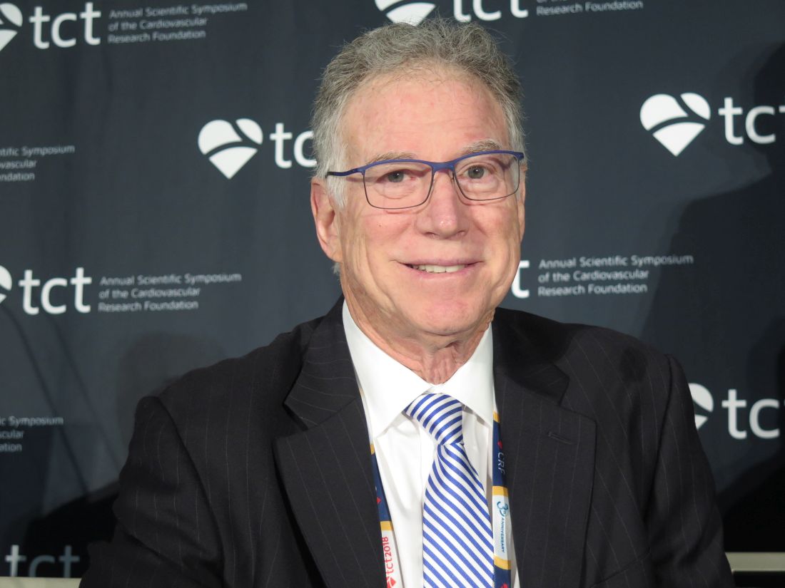
“The fine subgroup analysis of who the best candidates are is still in question,” he elaborated. “The SYNTAXES study told us that surgery for left mains is still pretty good, and even though you can get good results with PCI, the event rates are higher in that three-vessel, high-SYNTAX score group, so we should be careful. Interventionalists need to know their limitations. I think that’s what both studies tell us, actually.”
Study details
The MAIN-COMPARE analyses were based on 2,240 patients with unprotected left main coronary artery disease (stenosis of more than 50% and no coronary artery bypass grafts to the left anterior descending or the left circumflex artery) treated during 2000-2006.
A total of 1,102 patients underwent PCI with stenting: 318 in the era of bare-metal stents and 784 in the era of drug-eluting stents, predominantly sirolimus-eluting stents. A total of 1,138 patients underwent CABG. The minimum follow-up was 10 years in all patients, with a median of 12 years.
In the entire study cohort, PCI and CABG yielded similar rates of death (21.1% vs. 23.2%) and the composite of death, Q-wave myocardial infarction, or stroke (23.8% vs. 26.3%), but the PCI patients had a significantly higher rate of target vessel revascularization (21.1% vs. 5.8%), according to data reported at the meeting, which was sponsored by the New York–based Cardiovascular Research Foundation.
In analyses using a propensity score weighting technique, results for the entire cohort were much the same. But on stratification, PCI with drug-eluting stents versus CABG yielded higher risks of death (hazard ratio, 1.35; P = .05) and the composite adverse outcome (HR, 1.46; P = .009) from 5 years onward, as well as a sharply higher risk of target vessel revascularization for the full duration of follow-up (HR, 5.82; P less than .001).
Dr. Park disclosed that he had no conflicts of interest. The study was supported by the Korean Society of Interventional Cardiology and the CardioVascular Research Foundation of South Korea.
SOURCE: Park SJ et al. J Am Coll Cardiol. 2018 Sep 14. doi: 10.1016/j.jacc.2018.09.012).
SAN DIEGO – In the long run, patients with left main coronary artery disease fare better if they undergo coronary artery bypass grafting (CABG) instead of percutaneous coronary intervention (PCI) with drug-eluting stents, suggest 10-year results of the MAIN-COMPARE trial. Findings were reported at the Transcatheter Cardiovascular Therapeutics annual meeting.
Although CABG is the standard choice for revascularization in this patient population, PCI has been making inroads thanks to advances in stents, antithrombotic drugs, periprocedural management, and operator expertise, noted senior author Seung-Jung Park, MD, PhD, chairman of the Heart Institute at Asan Medical Center in Seoul and professor of medicine at University of Ulsan, South Korea. “Indeed, many studies showed that PCI using drug-eluting stents might be a good alternative for selected patients with left main coronary artery disease.”
Two large, randomized, controlled trials, EXCEL and NOBLE, have compared these treatment strategies and helped clarify outcomes at intermediate follow-up periods of 3-5 years. But long-term data, increasingly important as survival improves, are lacking.
Dr. Park reported the 10-year update of a prospective, observational cohort study that analyzed data from more than 2,000 patients with unprotected left main coronary artery disease in the MAIN-COMPARE registry, which captures revascularization procedures performed at 12 Korean cardiac centers.
In the entire cohort, about a fifth of the patients died, and roughly a fourth experienced a composite adverse outcome of death and cardiovascular events regardless of whether they received PCI or CABG, but the former yielded a rate of target vessel revascularization that was more than three times higher, according to results reported at the meeting and simultaneously published (J Am Coll Cardiol. 2018 Sep 14. doi: 10.1016/j.jacc.2018.09.012). Among the subset of patients treated in the more recent drug-eluting stent era, those who underwent PCI were more likely to die and to experience the composite outcome starting at the 5-year mark.
“Drug-eluting stents were associated with higher risks of death and serious composite outcomes compared to CABG after 5 years. The treatment benefit of CABG has diverged over time during continued follow-up,” Dr. Park noted. “The rate of target-vessel failure was consistently higher in the PCI group.”
“We used mainly first-generation drug-eluting stents,” he acknowledged. “However, many studies have demonstrated there is not too much difference between the first- and second-generation stents.”
Data worth the wait
In the same session, investigators reported the 10-year update of the European and U.S. randomized SYNTAX Extended Survival trial, called SYNTAXES. SYNTAX enrolled patients with three-vessel or left main coronary disease. That trial found no significant difference in survival between PCI with drug-eluting stents and CABG overall. In stratified analysis, mortality was higher with PCI among patients with three-vessel disease, but not among patients with left main disease.
Taken together, these trials help clarify the long-term comparative efficacy of PCI and begin to inform patient selection, according to press conference panelist Morton J. Kern, MD, a professor at the University of California, Irvine Medical Center.
“The fine subgroup analysis of who the best candidates are is still in question,” he elaborated. “The SYNTAXES study told us that surgery for left mains is still pretty good, and even though you can get good results with PCI, the event rates are higher in that three-vessel, high-SYNTAX score group, so we should be careful. Interventionalists need to know their limitations. I think that’s what both studies tell us, actually.”
Study details
The MAIN-COMPARE analyses were based on 2,240 patients with unprotected left main coronary artery disease (stenosis of more than 50% and no coronary artery bypass grafts to the left anterior descending or the left circumflex artery) treated during 2000-2006.
A total of 1,102 patients underwent PCI with stenting: 318 in the era of bare-metal stents and 784 in the era of drug-eluting stents, predominantly sirolimus-eluting stents. A total of 1,138 patients underwent CABG. The minimum follow-up was 10 years in all patients, with a median of 12 years.
In the entire study cohort, PCI and CABG yielded similar rates of death (21.1% vs. 23.2%) and the composite of death, Q-wave myocardial infarction, or stroke (23.8% vs. 26.3%), but the PCI patients had a significantly higher rate of target vessel revascularization (21.1% vs. 5.8%), according to data reported at the meeting, which was sponsored by the New York–based Cardiovascular Research Foundation.
In analyses using a propensity score weighting technique, results for the entire cohort were much the same. But on stratification, PCI with drug-eluting stents versus CABG yielded higher risks of death (hazard ratio, 1.35; P = .05) and the composite adverse outcome (HR, 1.46; P = .009) from 5 years onward, as well as a sharply higher risk of target vessel revascularization for the full duration of follow-up (HR, 5.82; P less than .001).
Dr. Park disclosed that he had no conflicts of interest. The study was supported by the Korean Society of Interventional Cardiology and the CardioVascular Research Foundation of South Korea.
SOURCE: Park SJ et al. J Am Coll Cardiol. 2018 Sep 14. doi: 10.1016/j.jacc.2018.09.012).
REPORTING FROM TCT 2018
Key clinical point: CABG had an edge over PCI with drug-eluting stents in patients with left main disease that became evident with longer follow-up.
Major finding: Compared with CABG, PCI with drug-eluting stents carried higher risks of death (hazard ratio, 1.35; P = .05) and a composite adverse outcome (HR, 1.46; P = .009) from 5 years onward.
Study details: Ten-year follow-up of a multicenter prospective cohort study of 2,240 patients with unprotected left main coronary artery disease who underwent either PCI with stenting or CABG (MAIN-COMPARE study).
Disclosures: Dr. Park disclosed that he had no conflicts of interest. The study was supported by the Korean Society of Interventional Cardiology and the CardioVascular Research Foundation of South Korea.
Source: Park S-J et al. J Am Coll Cardiol. 2018 Sep 14. doi: 10.1016/j.jacc.2018.09.012.
Simplified SYNTAX holds promise, but still has doubters
SAN DIEGO – A simplified version of the SYNTAX score for coronary artery disease complexity strongly correlated with the unmodified SYNTAX score and could make it easier for cardiologists to employ it in everyday practice. The modified version can be easily memorized and has simplified values that a clinician can calculate and use to determine an appropriate treatment without breaking their work flow to consult a computerized system.
“It’s primarily simplifying the values for the location of the lesions that has allowed it to be more memorizable. That does make it a slightly blunter tool, but only slightly,” said Sonya Burgess, MBCHB, during a presentation at the Transcatheter Cardiovascular Therapeutics annual meeting. Dr. Burgess is an interventional cardiologist at the University of New South Wales, Sydney.
In common practice, SYNTAX scores often go uncalculated because of their complexity, despite guidelines that recommend it. “My guess might be that 1 in every 10 [cardiologists] do it, and that’s not right for our patients,” said Dr. Burgess.
Others at the session agreed that SYNTAX is underutilized, but not all agreed with Dr. Burgess’ solution. Notable among the attendees was Patrick Serruys, MD, of Erasmus University, Rotterdam, the Netherlands, who originally published the SYNTAX system. He doesn’t like the idea of simplifying SYNTAX, based in part on previous experience. “About 10 years ago, Boston Scientific found the score too difficult and they wanted to simplify it, but the score completely lost its prognostic value. I was very emotional about that and I said, ‘no, we have to keep all of the components until we understand [the risk factors] much better,’ ” said Dr. Serruys.
But Dr. Burgess argued that the simplified system, which she and her coinvestigators created, makes concessions to the realities of an interventional cardiology suite. To obtain a standard SYNTAX score, “they have to stop doing something or rebook something, or there’s a lesion there that they just want to treat. [With the simplified score], at least before they go on to treat that lesion, they don’t have to take their gowns and gloves off. You can even have a card hanging on the bar [to refer to].” Dr. Burgess did not provide details of the system.
Dr. Serruys agreed with the source of the problem. “Dr. Burgess is right, it is somewhat demanding,” he allowed, but he also called on clinicians to consider the needs of the patient, even going so far as picturing the patient as a family member. “People complain that doing the SYNTAX is on average 7 minutes, with a standard deviation of 7, so you could work 15 minutes just looking at film. But when you think that the future of your father or brother is depending on the surgeon and the cardiologist looking carefully, I find the argument absolutely obnoxious,” said Dr. Serruys at the meeting sponsored by the Cardiovascular Research Foundation.
Rather than simplification, he stresses teamwork. With at least three people looking at the angiogram, the results are consistent and useful. “You look at the film with the trainees, and that’s how they learn. Never do a SYNTAX score alone,” said Dr. Serruys.
To assess the simplified SYNTAX score, the researchers conducted a retrospective assessment of both the SYNTAX score and the simplified SYNTAX score in 617 patients who had multivessel disease. They performed both assessments in subgroups of patients with 169 patients with ST-segment elevation MI, 78 patients with chronic total occlusion, and 113 patients with left main coronary artery (LMCA) stenosis. They used a 100-patient derivation cohort to determine cutoffs for patients who would not be suitable for percutaneous coronary intervention. They also looked at the accuracy of the simplified version compared with standard SYNTAX in 517 patients from five tertiary centers.
The Spearman’s rho value was 0.93 overall and at least 0.91 for all subgroups (P less than .001 for all). In patients with LMCA stenosis, the simplified version had a sensitivity of 100%, specificity of 85%, negative predictive of 100%, and an area under the curve of 1.0 (P less than .001). For patients with multivessel disease and no LMCA stenosis, the values were 98%, 82%, 99%, and 0.971, respectively (P less than .001).
Dr. Burgess and Dr. Serruys reported no financial conflicts of interest.
REPORTING FROM TCT 2018
Key clinical point: A simplified SYNTAX score could lead to broader implementation of evidence-based decision making for complex coronary artery disease.
Major finding: The simplified score had a Spearman’s rho value of 0.93 overall.
Study details: A retrospective analysis of 617 patients with multivessel disease.
Disclosures: Dr. Burgess and Dr. Serruys reported no financial conflicts of interest.
ULTIMATE: IVUS-guided stent placement bests angiography
SAN DIEGO – Clinical outcomes were better when stent placement was guided by intravascular ultrasound rather than angiography, based on the results of a trial conducted in China. Unlike previous comparative studies, which focused on patients with more complex lesions, this trial included all patients undergoing drug-eluting stent (DES) placement.
In the ULTIMATE trial, 1,448 all-comer patients receiving DES were randomized to either intravascular ultrasound (IVUS)–guided or angiography-guided implantation, reported Junjie Zhang, PhD, of Nanjing Medical University in China, and colleagues. The study excluded patients who had a life expectancy shorter than 12 months, who were intolerant of dual antiplatelet therapy, and who had severe calcification needing rotational atherectomy.
At 30 days after stent placement, the incidence of target vessel failure was 0.8% in the IVUS group and 1.9% in the angiography group, a nonsignificant trend. However, outcomes were significantly different at 1 year, with target vessel failure occurring in 2.9% of IVUS patients and 5.4% of angiography patients (hazard ratio, 0.530; 95% confidence interval, 0.312-0.901; P = .019).
The work was simultaneously published in the Journal of the American College of Cardiology (2018 Sept. doi: 10.1016/j.jacc.2018.09.013) .
Despite good results in this and prior studies, uptake of the IVUS procedure is not high in the United States or Europe, according to members of a panel that reviewed the results at the Transcatheter Cardiovascular Therapeutics annual meeting.
“How can people continue to ignore the importance of imaging-guided stent optimization? Even with second-generation DES, the results are consistent across the studies. This is just another piece of irrefutable evidence,” said Gary S. Mintz, MD, chief medical officer at the Cardiovascular Research Foundation, and a discussant at the meeting, sponsored by the Cardiovascular Research Foundation.
That sentiment was generally echoed by the rest of the panel. John M. Hodson, MD, a professor of medicine at MetroHealth Medical Center in Cleveland, pointed out that the study included a variety of cases, and angiography was performed to a high standard in that arm of the study. “It shows that, even with a good angiographic approach, IVUS still wins. I’m amazed that there’s still some resistance to [IVUS] image guidance,” said Dr. Hodgson.
The ULTIMATE study also found that the procedural time was longer (60.88 minutes vs. 45.49 minutes; P less than .001), and the contrast volume was higher (178.29 mL vs. 161.96 mL; P less than .001) in the IVUS than in the angiography group.
A postprocedure IVUS assessment was performed to determine whether the stent was optimally deployed. The criteria for optimal deployment included minimal lumen area in the stented segment of at least 5 mm2, or 90% of the minimal lumen area at distal reference segment meeting that criteria; a less than 50% plaque burden at the 5 mm of vessel proximal or distal to the stent edge; and no edge dissection involving media greater than 3 mm in length.
In the IVUS group, 53% of patients had optimal placement. The rate of target vessel failure was 1.6% of patients with optimal placement and 4.4% of patients who failed to achieve all optimal criteria (HR, 0.349; 95% CI, 0.135-0.898; P = 0.029). Compared with angiography guidance, IVUS guidance was of similar benefit for patients with either American College of Cardiology/American Heart Association–defined B2/C lesions or A/B1 lesions in terms of the composite endpoint. The significant reduction of clinically driven target lesion revascularization or definite stent thrombosis (HR, 0.407; 95% CI: 0.188-0.880; P = 0.018) based on lesion-level analysis by IVUS guidance was not achieved when patient-level analysis was performed.
“I’m particularly impressed by the analysis of the optimal versus nonoptimal group. If you don’t use IVUS correctly, you don’t get a benefit. The ones [in the IVUS group] who did not get optimal stenting were very similar to the angiographic group,” said Dr. Mintz.
The study was funded by the National Science Foundation of China, Six Talent Peaks Project, Nanjing Health and Family Planning Commission, Nanjing Health Youth Talent Training Project, and the Nanjing Municipal Commission of Science & Technology. None of the study authors had relevant financial disclosures. Dr. Mintz reported received research support from Abbott Vascular and Boston Scientific. He has been a consultant for Boston Scientific, Volcano, and Infraredx. Dr. Hodgson reported received research support and consulted for Volcano.
REPORTING FROM TCT 2018
Key clinical point: Intravascular ultrasound–guided placement of drug-eluting stents resulted in a lower target vessel failure rate than did angiography guidance.
Major finding: The 1-year target vessel failure rate was 2.9% in the intravascular ultrasound–guided group and 5.4% in the angiography group.
Study details: A randomized, controlled trial of 1,448 all-comer patients.
Disclosures: The study was funded by the National Science Foundation of China, Six Talent Peaks Project, Nanjing Health and Family Planning Commission, Nanjing Health Youth Talent Training Project, and the Nanjing Municipal Commission of Science & Technology. None of the study authors had relevant financial disclosures. Dr. Mintz reported receiving research support from Abbott Vascular and Boston Scientific. He has been a consultant for Boston Scientific, Volcano, and Infraredx. Dr. Hodgson reported receiving research support and consulted for Volcano.