User login
CorMicA: Nonobstructive angina should trigger functional testing
SAN DIEGO – Don’t be satisfied with a diagnosis of angina with no obstructive coronary artery disease; push for acetylcholine testing, the findings from a trial in Scotland suggest.
Patients will have less angina and a better quality of life at 6 months, according to investigators from the University of Glasgow (Scotland).
Invasive angiography usually ends on both sides of the Atlantic when no occlusions are found. There are concerns about the safety of going further with acetylcholine challenges, and until now, there had been no grade A evidence from a randomized trial that it improves outcomes. The Glasgow team filled the evidence gap with their presentation of the Coronary Microvascular Angina (CorMicA) trial at the Transcatheter Cardiovascular Therapeutics (TCT) annual meeting, and there wasn’t a single serious adverse event (J Am Coll Cardiol. 2018 Sep 25. doi: 10.1016/j.jacc.2018.09.006).
“This was a proof-of-concept study, which we believe [should] substantiate a large, multicenter trial,” said senior investigator Colin Berry, PhD, a professor of cardiology and imaging at the university.
Acetylcholine was infused down the pressure wire in 151 subjects diagnosed with angina with no obstructive coronary artery diseases before they left the catheter lab. Of these patients, 76 were randomized to have their results shared with their treating cardiologist, and 75 were randomized to not have their results shared. Coronary functional testing is hardly ever done, so the no-share group was considered the standard-of-care control arm. The idea was to see whether it made a difference when treating physicians knew what was causing chest pain when their patients didn’t have occlusive disease.
It turned out to make a huge difference. The diagnosis of “chest pain of noncardiac origin” almost fell off the map. Once cardiologists knew what was going on, they switched up treatment according to European Society of Cardiology guidelines for functional heart pain. Patients with microvascular angina were given beta-blockers and switched off nitrates because these drugs make angina worse in microvascular disease. Subjects with vasospasms were shifted to calcium channel blockers and long-acting nitrates and away from beta-blockers because beta-blockers make vasospasms worse.
Cardiologists who didn’t know the results kept muddling along with what patients came in on at baseline – beta-blockers in two-thirds, long-acting nitrates in half, and calcium channel blockers in a third.
Subjects who got the right treatment because of acetylcholine testing outpaced the standard care group by almost 12 points on the Seattle Angina Questionnaire at 6 months; they could walk farther and didn’t have crushing angina almost every day (P = .001). They reported a statistically significant improvement in quality of life, and they were much happier with their doctors.
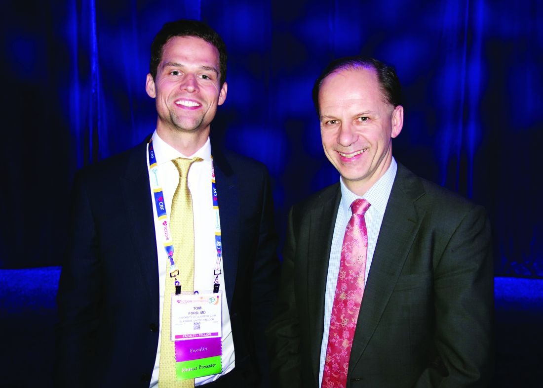
“This is the first randomized, sham-controlled trial in this space”; functional testing “was routinely safe and feasible. Therapy guided by the results of physiologic testing improved outcomes” and “treatment satisfaction,” said University of Glasgow interventional cardiologist Tom Ford, MD.
Acetylcholine was infused down the pressure wire into the radial artery, with the left anterior descending coronary artery as the target vessel. A final bolus of less than 100 mcg checked for coronary artery spasms; a symptomatic constriction of greater than 90% was considered positive. Glyceryl trinitrate was used to reverse the effects.
Three-quarters of the subjects were women, which Dr. Ford noted is unusual in an angina study. The mean age was 61 years, and subjects had about a 20% chance of a heart attack within 10 years. The whole procedure, including the angiogram, randomization, and functional testing, took a median of about 60 minutes.
There were no differences in major adverse cardiac events at 6 months, at 2.6% in both groups.
One patient developed persistent atrial fibrillation with acetylcholine testing that was converted to sinus rhythm with intravenous amiodarone, without a night in the hospital.
The work was funded by the British Heart Foundation. No companies were involved. The investigators didn’t have any relevant disclosures. The TCT meeting is sponsored by the Cardiovascular Research Foundation.
aotto@mdedge.com
SOURCE: Ford TG et al. TCT 2018, Late-Breaking Trial.
SAN DIEGO – Don’t be satisfied with a diagnosis of angina with no obstructive coronary artery disease; push for acetylcholine testing, the findings from a trial in Scotland suggest.
Patients will have less angina and a better quality of life at 6 months, according to investigators from the University of Glasgow (Scotland).
Invasive angiography usually ends on both sides of the Atlantic when no occlusions are found. There are concerns about the safety of going further with acetylcholine challenges, and until now, there had been no grade A evidence from a randomized trial that it improves outcomes. The Glasgow team filled the evidence gap with their presentation of the Coronary Microvascular Angina (CorMicA) trial at the Transcatheter Cardiovascular Therapeutics (TCT) annual meeting, and there wasn’t a single serious adverse event (J Am Coll Cardiol. 2018 Sep 25. doi: 10.1016/j.jacc.2018.09.006).
“This was a proof-of-concept study, which we believe [should] substantiate a large, multicenter trial,” said senior investigator Colin Berry, PhD, a professor of cardiology and imaging at the university.
Acetylcholine was infused down the pressure wire in 151 subjects diagnosed with angina with no obstructive coronary artery diseases before they left the catheter lab. Of these patients, 76 were randomized to have their results shared with their treating cardiologist, and 75 were randomized to not have their results shared. Coronary functional testing is hardly ever done, so the no-share group was considered the standard-of-care control arm. The idea was to see whether it made a difference when treating physicians knew what was causing chest pain when their patients didn’t have occlusive disease.
It turned out to make a huge difference. The diagnosis of “chest pain of noncardiac origin” almost fell off the map. Once cardiologists knew what was going on, they switched up treatment according to European Society of Cardiology guidelines for functional heart pain. Patients with microvascular angina were given beta-blockers and switched off nitrates because these drugs make angina worse in microvascular disease. Subjects with vasospasms were shifted to calcium channel blockers and long-acting nitrates and away from beta-blockers because beta-blockers make vasospasms worse.
Cardiologists who didn’t know the results kept muddling along with what patients came in on at baseline – beta-blockers in two-thirds, long-acting nitrates in half, and calcium channel blockers in a third.
Subjects who got the right treatment because of acetylcholine testing outpaced the standard care group by almost 12 points on the Seattle Angina Questionnaire at 6 months; they could walk farther and didn’t have crushing angina almost every day (P = .001). They reported a statistically significant improvement in quality of life, and they were much happier with their doctors.
“This is the first randomized, sham-controlled trial in this space”; functional testing “was routinely safe and feasible. Therapy guided by the results of physiologic testing improved outcomes” and “treatment satisfaction,” said University of Glasgow interventional cardiologist Tom Ford, MD.
Acetylcholine was infused down the pressure wire into the radial artery, with the left anterior descending coronary artery as the target vessel. A final bolus of less than 100 mcg checked for coronary artery spasms; a symptomatic constriction of greater than 90% was considered positive. Glyceryl trinitrate was used to reverse the effects.
Three-quarters of the subjects were women, which Dr. Ford noted is unusual in an angina study. The mean age was 61 years, and subjects had about a 20% chance of a heart attack within 10 years. The whole procedure, including the angiogram, randomization, and functional testing, took a median of about 60 minutes.
There were no differences in major adverse cardiac events at 6 months, at 2.6% in both groups.
One patient developed persistent atrial fibrillation with acetylcholine testing that was converted to sinus rhythm with intravenous amiodarone, without a night in the hospital.
The work was funded by the British Heart Foundation. No companies were involved. The investigators didn’t have any relevant disclosures. The TCT meeting is sponsored by the Cardiovascular Research Foundation.
aotto@mdedge.com
SOURCE: Ford TG et al. TCT 2018, Late-Breaking Trial.
REPORTING FROM TCT 2018
Key clinical point: A diagnosis of angina with no obstructive coronary artery disease is insufficient; acetylcholine testing should be considered.
Major finding: The diagnosis of “chest pain of noncardiac origin” almost fell off the map. At 6 months, patients could walk farther and didn’t have crushing angina almost every day (P = .001). They reported a statistically significant improvement in quality of life, and they were much happier with their doctors.
Study details: Randomized trial with 151 people who had chest pain but no coronary occlusions on angiography.
Disclosures: There was no industry funding, and the investigators had no relevant industry disclosures.
Source: Ford TG et al. TCT 2018, Late-Breaking Trial.
Pulmonary artery denervation boosts walking capacity in left heart failure
SAN DIEGO – Pulmonary artery denervation is efficacious for treating combined pre- and postcapillary pulmonary hypertension attributable to left heart failure, based on results of the Chinese PADN-5 trial reported at the Transcatheter Cardiovascular Therapeutics annual meeting.

This ablative treatment has been studied among patients with pulmonary hypertension attributable to other etiologies, but not in randomized fashion among this population, noted lead investigator Shao-Liang Chen, MD, of Nanjing (China) First Hospital, Nanjing Medical University. The treatment is an attractive one, as medications recommended for pulmonary arterial hypertension are not recommended for joint pre- and postcapillary pulmonary hypertension (group II pulmonary hypertension).
In PADN-5, 98 patients were randomized to pulmonary artery denervation or to sham denervation plus open-label sildenafil (Viagra), which at the time of trial initiation was thought to be safe and potentially beneficial.
The trial’s main outcome, 6-minute walk distance at 6 months, improved in both groups, according to data reported at the meeting and simultaneously published in JACC Cardiovascular Interventions. But the improvement was about four times greater in the pulmonary artery denervation group. Secondary efficacy outcomes also favored that group, and the rate of fatal pulmonary embolism did not differ for the two groups.
“The PADN-5 trial demonstrates the benefits of pulmonary artery denervation for patients with combined pre- and postcapillary pulmonary hypertension. Patients with preserved and with reduced ejection fraction equally benefited,” summarized Dr. Chen, who pioneered this procedure about 7 years ago. “There was no sign of any harm of sildenafil in patients with combined pre- and postcapillary pulmonary hypertension.”
Trial critique
“This is a very difficult study to conduct, being able to recruit patients and actually have these procedures done,” commented press conference moderator Ori Ben-Yehuda, MD, professor of clinical medicine and director, coronary care unit, UC San Diego Medical Center.
At the same time, he expressed some reservations about the trial. “Sildenafil in the control group might actually be expected to ... decrease your effect size. Also, particularly in men, perhaps even in women, it might unblind them to which group they are in and undermine your sham design,” he noted. In addition, some hemodynamic changes after pulmonary artery denervation – a decrease in wedge pressure and an increase in ejection fraction – were puzzling.
“We need a lot more data here. There are some issues with this trial in terms of design, and we haven’t even gotten into the issue of whether there were core labs, whether the echoes, the hemodynamics, were read blindly,” Dr. Ben-Yehuda maintained. “This issue of secondary or group II pulmonary hypertension due to left heart failure is one that has been very frustrating in terms of actual PA-specific therapies. So this is an important step further, but it needs confirmation in truly sham-controlled trials that have no potential for unblinding.”
The catheter used in PADN-5 is available in China but has not received clearance in the United States, he pointed out. “There are alternative or competing technologies, one using ultrasound, for example, that has a very similar approach. … We’ll have to see how it ends up [performing].”
Trial details
Patients in the PADN-5 pulmonary artery denervation group underwent ablation only in the periconjunctional area between the distal main trunk and the left ostial branch with a multifunction catheter having premounted electrodes. Those in the control group underwent a sham procedure, with catheter positioning at the target sites and connection to a generator but no ablation, and were given open-label sildenafil. All additionally received standard heart failure medical therapy. (No sildenafil placebo was used in the denervation group.)
Trial results reported at the meeting, which is sponsored by the Cardiovascular Research Foundation, showed that most echocardiographic and hemodynamic measures improved more in the pulmonary artery denervation group.
The greater improvement in 6-minute walk test with denervation versus sham sildenafil at 6 months was evident in a variety of measures: absolute median distance walked (432.5 m vs. 358 m) and mean distance walked (434.6 m vs. 359.4 m), and absolute increase (80 m vs. 17.5 m) and relative increase (21.4% vs. 4.9%) The difference was significant for all measures at P less than .001.
The denervation group had a comparatively greater reduction of pulmonary vascular resistance (29.8% vs. 3.4%; P less than .001) and were less likely to experience clinical worsening (16.7% vs. 40.0%; P = .014).
There was a single fatal pulmonary embolism in each treatment group. Of the seven total deaths, two occurred in the denervation group (one attributable to pump failure, one a sudden death) and five occurred in the sham sildenafil group (all but one attributable to pump failure).
Dr. Chen disclosed that he had no relevant conflicts of interest. The trial was sponsored by Nanjing First Hospital, Nanjing Medical University.
SOURCE: Chen S-J et al. TCT 2018. JACC Cardiovasc Interv. 2018 Sep 23.
SAN DIEGO – Pulmonary artery denervation is efficacious for treating combined pre- and postcapillary pulmonary hypertension attributable to left heart failure, based on results of the Chinese PADN-5 trial reported at the Transcatheter Cardiovascular Therapeutics annual meeting.
This ablative treatment has been studied among patients with pulmonary hypertension attributable to other etiologies, but not in randomized fashion among this population, noted lead investigator Shao-Liang Chen, MD, of Nanjing (China) First Hospital, Nanjing Medical University. The treatment is an attractive one, as medications recommended for pulmonary arterial hypertension are not recommended for joint pre- and postcapillary pulmonary hypertension (group II pulmonary hypertension).
In PADN-5, 98 patients were randomized to pulmonary artery denervation or to sham denervation plus open-label sildenafil (Viagra), which at the time of trial initiation was thought to be safe and potentially beneficial.
The trial’s main outcome, 6-minute walk distance at 6 months, improved in both groups, according to data reported at the meeting and simultaneously published in JACC Cardiovascular Interventions. But the improvement was about four times greater in the pulmonary artery denervation group. Secondary efficacy outcomes also favored that group, and the rate of fatal pulmonary embolism did not differ for the two groups.
“The PADN-5 trial demonstrates the benefits of pulmonary artery denervation for patients with combined pre- and postcapillary pulmonary hypertension. Patients with preserved and with reduced ejection fraction equally benefited,” summarized Dr. Chen, who pioneered this procedure about 7 years ago. “There was no sign of any harm of sildenafil in patients with combined pre- and postcapillary pulmonary hypertension.”
Trial critique
“This is a very difficult study to conduct, being able to recruit patients and actually have these procedures done,” commented press conference moderator Ori Ben-Yehuda, MD, professor of clinical medicine and director, coronary care unit, UC San Diego Medical Center.
At the same time, he expressed some reservations about the trial. “Sildenafil in the control group might actually be expected to ... decrease your effect size. Also, particularly in men, perhaps even in women, it might unblind them to which group they are in and undermine your sham design,” he noted. In addition, some hemodynamic changes after pulmonary artery denervation – a decrease in wedge pressure and an increase in ejection fraction – were puzzling.
“We need a lot more data here. There are some issues with this trial in terms of design, and we haven’t even gotten into the issue of whether there were core labs, whether the echoes, the hemodynamics, were read blindly,” Dr. Ben-Yehuda maintained. “This issue of secondary or group II pulmonary hypertension due to left heart failure is one that has been very frustrating in terms of actual PA-specific therapies. So this is an important step further, but it needs confirmation in truly sham-controlled trials that have no potential for unblinding.”
The catheter used in PADN-5 is available in China but has not received clearance in the United States, he pointed out. “There are alternative or competing technologies, one using ultrasound, for example, that has a very similar approach. … We’ll have to see how it ends up [performing].”
Trial details
Patients in the PADN-5 pulmonary artery denervation group underwent ablation only in the periconjunctional area between the distal main trunk and the left ostial branch with a multifunction catheter having premounted electrodes. Those in the control group underwent a sham procedure, with catheter positioning at the target sites and connection to a generator but no ablation, and were given open-label sildenafil. All additionally received standard heart failure medical therapy. (No sildenafil placebo was used in the denervation group.)
Trial results reported at the meeting, which is sponsored by the Cardiovascular Research Foundation, showed that most echocardiographic and hemodynamic measures improved more in the pulmonary artery denervation group.
The greater improvement in 6-minute walk test with denervation versus sham sildenafil at 6 months was evident in a variety of measures: absolute median distance walked (432.5 m vs. 358 m) and mean distance walked (434.6 m vs. 359.4 m), and absolute increase (80 m vs. 17.5 m) and relative increase (21.4% vs. 4.9%) The difference was significant for all measures at P less than .001.
The denervation group had a comparatively greater reduction of pulmonary vascular resistance (29.8% vs. 3.4%; P less than .001) and were less likely to experience clinical worsening (16.7% vs. 40.0%; P = .014).
There was a single fatal pulmonary embolism in each treatment group. Of the seven total deaths, two occurred in the denervation group (one attributable to pump failure, one a sudden death) and five occurred in the sham sildenafil group (all but one attributable to pump failure).
Dr. Chen disclosed that he had no relevant conflicts of interest. The trial was sponsored by Nanjing First Hospital, Nanjing Medical University.
SOURCE: Chen S-J et al. TCT 2018. JACC Cardiovasc Interv. 2018 Sep 23.
REPORTING FROM TCT 2018
Key clinical point: Pulmonary artery denervation is efficacious for treating pulmonary hypertension related to heart failure.
Major finding: Improvement in 6-minute walk distance was greater with pulmonary artery denervation than with sham denervation plus sildenafil (21.4% vs. 4.9%; P less than .001).
Study details: PADN-5 is a randomized controlled trial among 98 patients with combined pre- and postcapillary pulmonary hypertension secondary to left heart failure (group II pulmonary hypertension).
Disclosures: Dr. Chen disclosed that he had no relevant conflicts of interest. The trial was sponsored by Nanjing First Hospital, Nanjing Medical University.
Source: Chen S-L et al. TCT 2018. JACC Cardiovasc Interv. 2018 Sep 23.
Antithrombotic strategy 1 year after stenting in AF patients leans toward oral anticoagulant alone
SAN DIEGO – In patients with atrial fibrillation and stable coronary artery disease, a randomized trial of oral anticoagulation alone versus an anticoagulant plus a single antiplatelet agent failed to establish noninferiority of the single-agent approach. The trial could not demonstrate its primary endpoint of all-cause death, myocardial infarction, stroke, or systemic embolism.
But a secondary endpoint that included major bleeding did demonstrate equivalence, leading the researchers to suggest that oral anticoagulation (OAC) alone may be sufficient in most patients.
“Combined OAC and single antiplatelet therapy is unlikely to provide net clinical benefit over OAC alone. Thus, OAC alone might be reasonable for AF [atrial fibrillation] patients beyond 1 year after coronary stenting,” Yukiko Nakano, MD, of Kyoto (Japan) University Graduate School of Medicine, said during a press conference at the Transcatheter Cardiovascular Therapeutics annual meeting, sponsored by the Cardiovascular Research Foundation. The report was simultaneously published Sept. 24 in Circulation (doi: 10.1161/CIRCULATIONAHA.118.036768).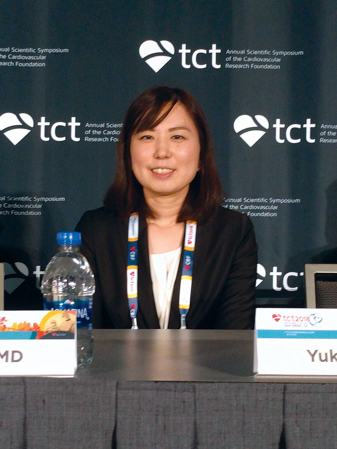
The results support the European Society of Cardiology practice guidelines, which recommend lifelong OAC without antiplatelet therapy. But physicians often continue to prescribe antiplatelet agents out of concern that stent thrombosis could occur if the therapy is stopped.
The study was stopped prematurely because of insufficient recruitment, which may have contributed to the failed primary endpoint. It’s a shortcoming that befalls many such studies, perhaps because cardiologists tend to be set in their ways when it comes to treatment of patients after a stent implant. “Cardiologists just think they know the answer, and they don’t want to expose their patients (to a clinical trial). They say, ‘I have my patients on whatever regimen. It seems to be working, and they’re not bleeding, so I don’t want to change it.’ This study suggests that we probably can stop one of the two (antiplatelet drugs) and get by with a single agent, and in this case they got by with no agent (in the monotherapy arm),” said C. Michael Gibson, MD, chief of clinical research in the division of cardiology at Beth Israel Deaconess Medical Center, Boston, who was a discussant at the press conference.
The study recruited 696 patients who were receiving OAC plus single antiplatelet therapy (SAPT) 1 year after receiving a stent. They were randomized 1:1 to continue combined therapy or to stop SAPT and then followed for a median of 2.5 years. A total of 74% of patients who received OAC alone were taking warfarin, while 26% were taking a direct oral anticoagulant. The SAPT group took aspirin or clopidogrel.
Overall, 15.7% of OAC patients experienced the primary endpoint, compared with 13.6% of the combined group (noninferiority P = .20). None of the individual components of the primary endpoint were statistically significantly different between the groups. International Society on Thrombosis and Haemostasis major bleeding and Thrombolysis in Myocardial Infarction major bleeding trended in favor of OAC alone. The secondary endpoint (primary endpoint plus major bleeding) achieved noninferiority, occurring in 19.5% of the OAC group and 19.4% of the combined therapy group (noninferiority P = .016; superiority P = .96).
Daiichi-Sankyo funded the trial. Dr. Nakano had no conflicts of interest. Dr. Gibson reported numerous financial ties to pharmaceutical companies, including Daiichi-Sankyo.
REPORTING FROM TCT 2018
Key clinical point:
Major finding: A measure that included cardiac events plus major bleeding showed an oral anticoagulant alone was noninferior to an OAC plus antiplatelet therapy.
Study details: Randomized, controlled trial of 696 patients.
Disclosures: Daiichi-Sankyo funded the trial. Dr. Nakano had no conflicts of interest. Dr. Gibson reported numerous financial ties to pharmaceutical companies, including Daiichi-Sankyo.
FAST-FFR: Noninvasive FFR nearly as good as wire
SAN DIEGO – A less-invasive way to measure fractional flow reserve using angiography had a sensitivity of 94% and specificity of 91%, compared with standard wire-based techniques, according to a report at the Transcatheter Cardiovascular Therapeutics annual meeting.
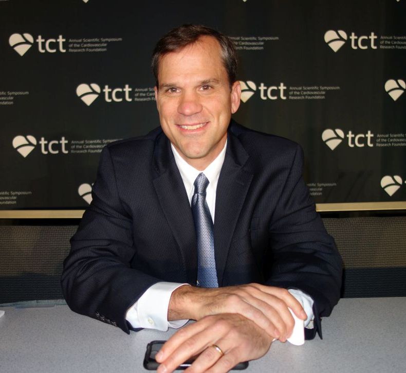
The method may provide an easier and potentially faster method for performing physiology-guided assessment of the overall coronary angiogram than with coronary pressure wire–based FFR, William Fearon, MD, said in presenting results of the FAST-FFR trial.
The added bonus of the FFRangio system, from the Israeli company CathWorks, is that it automatically produces a 3-D reconstruction of the entire coronary tree and can calculate FFR values for all occlusions. It requires high-quality angiograms, which are transferred to a proprietary counsel; the system estimates resistance and flow across stenoses using an algorithm. After a few cases, the process takes less than 5 minutes. CathWorks is working on Food and Drug Administration clearance, Dr. Fearon reported.
Several other companies are also developing noninvasive ways to measure FFR, the pressure gradient across lesions. It helps clinicians make the call on revascularization, since angiograms can be deceiving; occlusions that look bad might not really be causing a problem, and vice-versa.
FFR is recommended in assessment guidelines, but it’s not used much. The problem is that traditional measurement requires threading wires down coronary arteries; the technique is a bit risky and takes time and training. It also has to be repeated for each lesion.
The new system “has the potential to eventually replace wire-based FFR measurement and substantially increase physiologic coronary lesion assessment in the catheterization laboratory, thereby leading to improved patient outcomes,” said Dr. Fearon, professor of cardiology at Stanford (Calif.) University.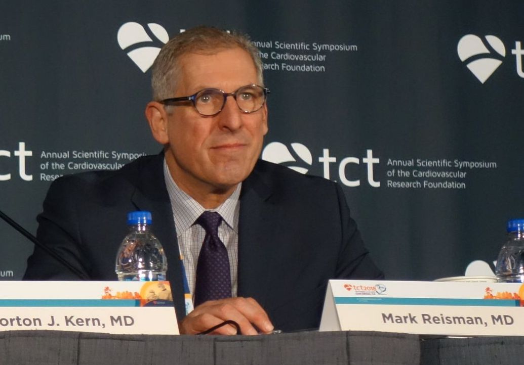
He said that he thought FFRangio could change practice, and several panelists agreed. “This is a real advance in the field. The use of FFR is not as great as it should be. I hope this will improve the ability of the assessor to identify ischemic lesions. I think that’s what’s going to happen. This is going to drive us forward,” said Mark Reisman, MD, head of cardiology and director of the Center for Emerging Cardiovascular Therapies at the University of Washington, Seattle.
The FAST-FFR (FFRangio Accuracy versus Standard FFR) study was conducted at 10 centers in the United States, Europe, and Israel; FFRangio was used to obtain FFRs in 319 vessels among 301 patients; the results were checked against FFR measured by wire. FFRangio operators were blinded to wire results.
The mean FFR was 0.81, and 43% of vessels had an FFR of 0.80 or less, signaling a possible abnormality. The diagnostic accuracy of FFRangio against wire measurement was 92%, and 87% when only gray-zone values between 0.75-0.85 were considered. Correlation with wire measurements was good (r = 0.80; P less than 0.001). Mismatches with the wire were more likely in the right coronary artery. The results were published online at the time of the presentation at TCT, sponsored by the Cardiovascular Research Foundation (Circulation. 2018 Sept 24. doi: 10.1161/circulationaha.118.037350).
“The main issue with this is that we need to do optimal angiographies. We should be doing them on a routine basis, but oftentimes, cardiologists want to be quick; they get lazy. But you do need to fill the entire vessel with contrast,” Dr. Fearon said.
“One of the nice things is you can rotate” the 3-D coronary artery tree reconstruction. “You can get a better idea of the relationship between the lesion and side branches, the length of the lesion, and a lot of additional information you don’t have on the angiogram [alone]. Then, you can pull back the cursor and measure FFR all along the vessel and different branches, all based on one angiography,” he said.
The majority of patients in the study were overweight or obese with complex coronary anatomy, as in daily practice. The investigation was limited to lesions amenable to wire measurement, so it didn’t include left main disease, low ejection fraction, and in-stent restenosis, although assessment may be possible.
Dr. Fearon didn’t know how much FFRangio will cost if it’s cleared or how CathWorks will be made available.
The work was funded by CathWorks. Dr. Fearon disclosed institutional research support from the company. One of the investigators was a cofounder of the company, with shares and intellectual property rights. The TCT meeting is sponsored by the Cardiovascular Research Foundation.
SAN DIEGO – A less-invasive way to measure fractional flow reserve using angiography had a sensitivity of 94% and specificity of 91%, compared with standard wire-based techniques, according to a report at the Transcatheter Cardiovascular Therapeutics annual meeting.
The method may provide an easier and potentially faster method for performing physiology-guided assessment of the overall coronary angiogram than with coronary pressure wire–based FFR, William Fearon, MD, said in presenting results of the FAST-FFR trial.
The added bonus of the FFRangio system, from the Israeli company CathWorks, is that it automatically produces a 3-D reconstruction of the entire coronary tree and can calculate FFR values for all occlusions. It requires high-quality angiograms, which are transferred to a proprietary counsel; the system estimates resistance and flow across stenoses using an algorithm. After a few cases, the process takes less than 5 minutes. CathWorks is working on Food and Drug Administration clearance, Dr. Fearon reported.
Several other companies are also developing noninvasive ways to measure FFR, the pressure gradient across lesions. It helps clinicians make the call on revascularization, since angiograms can be deceiving; occlusions that look bad might not really be causing a problem, and vice-versa.
FFR is recommended in assessment guidelines, but it’s not used much. The problem is that traditional measurement requires threading wires down coronary arteries; the technique is a bit risky and takes time and training. It also has to be repeated for each lesion.
The new system “has the potential to eventually replace wire-based FFR measurement and substantially increase physiologic coronary lesion assessment in the catheterization laboratory, thereby leading to improved patient outcomes,” said Dr. Fearon, professor of cardiology at Stanford (Calif.) University.
He said that he thought FFRangio could change practice, and several panelists agreed. “This is a real advance in the field. The use of FFR is not as great as it should be. I hope this will improve the ability of the assessor to identify ischemic lesions. I think that’s what’s going to happen. This is going to drive us forward,” said Mark Reisman, MD, head of cardiology and director of the Center for Emerging Cardiovascular Therapies at the University of Washington, Seattle.
The FAST-FFR (FFRangio Accuracy versus Standard FFR) study was conducted at 10 centers in the United States, Europe, and Israel; FFRangio was used to obtain FFRs in 319 vessels among 301 patients; the results were checked against FFR measured by wire. FFRangio operators were blinded to wire results.
The mean FFR was 0.81, and 43% of vessels had an FFR of 0.80 or less, signaling a possible abnormality. The diagnostic accuracy of FFRangio against wire measurement was 92%, and 87% when only gray-zone values between 0.75-0.85 were considered. Correlation with wire measurements was good (r = 0.80; P less than 0.001). Mismatches with the wire were more likely in the right coronary artery. The results were published online at the time of the presentation at TCT, sponsored by the Cardiovascular Research Foundation (Circulation. 2018 Sept 24. doi: 10.1161/circulationaha.118.037350).
“The main issue with this is that we need to do optimal angiographies. We should be doing them on a routine basis, but oftentimes, cardiologists want to be quick; they get lazy. But you do need to fill the entire vessel with contrast,” Dr. Fearon said.
“One of the nice things is you can rotate” the 3-D coronary artery tree reconstruction. “You can get a better idea of the relationship between the lesion and side branches, the length of the lesion, and a lot of additional information you don’t have on the angiogram [alone]. Then, you can pull back the cursor and measure FFR all along the vessel and different branches, all based on one angiography,” he said.
The majority of patients in the study were overweight or obese with complex coronary anatomy, as in daily practice. The investigation was limited to lesions amenable to wire measurement, so it didn’t include left main disease, low ejection fraction, and in-stent restenosis, although assessment may be possible.
Dr. Fearon didn’t know how much FFRangio will cost if it’s cleared or how CathWorks will be made available.
The work was funded by CathWorks. Dr. Fearon disclosed institutional research support from the company. One of the investigators was a cofounder of the company, with shares and intellectual property rights. The TCT meeting is sponsored by the Cardiovascular Research Foundation.
REPORTING FROM TCT 2018
Key clinical point:
Major finding: FFRangio, a less-invasive way to measure fractional flow reserve using angiograms, had a sensitivity of 94% and specificity of 91%, compared with standard, wire-based techniques.
Study details: The prospective, multicenter FAST-FFR compared FFRangio with pressure wire–derived fractional flow reserve in 301 subjects.
Disclosures: The work was funded by FFRangio maker, CathWorks. Dr. Fearon disclosed institutional research support from the company. One of the authors was a cofounder of the company, with shares and intellectual property rights.
IVUS guidance cuts target-vessel failure risk by 47%
SAN DIEGO – An all-comers trial conducted in China confirms that intravascular ultrasound (IVUS)–guided implantation of drug-eluting stents (DES) resulted in better clinical outcomes than did angiography-guided procedures, Jun-Jie Zhang, MD, said in reporting results of the ULTIMATE trial at the Transcatheter Cardiovascular Therapeutics annual meeting.
The ULTIMATE trial is the latest in a line of evidence showing the utility of IVUS guidance, though uptake of the procedure is not high in the United States or Europe. Its broader focus also rounds out the findings of earlier studies, which focused on patients with more complex lesions.
“How can people continue to ignore the importance of imaging-guided stent optimization? Even with second-generation DES, the results are consistent across the studies. This is just another piece of irrefutable evidence,” said discussant Gary Mintz, MD.
That sentiment was generally echoed by the rest of the panel. John McB Hodgson, MD, a professor of medicine at MetroHealth Medical Center in Cleveland, also pointed out the consistency of the body of evidence supporting the use of imaging. The study also represented a variety of cases, and the angiography arm of the study showed that the procedure was performed to a high standard. “It shows that even with a good angiographic approach, IVUS still wins. I’m amazed that there’s still some resistance to image guidance,” said Dr. Hodgson.
In ULTIMATE, 1,448 all-comer patients were randomized to either IVUS-guided or angiography-guided DES implantation. Patients with a life-expectancy shorter than 12 months, who were intolerant of dual-antiplatelet therapy, or who had severe calcification needing rotational atherectomy were excluded.
The procedural time was longer in the IVUS group, at 61 min, compared with 45 min in the angiography group, and the contrast volume was higher, at 178 mL and 162 mL, respectively (P less than .001 for both). At 30 days, the incidence of target vessel failure (TVF) was 0.8% in the IVUS group and 1.9% in the angiographic group, though this difference did not reach statistical significance (P = .08). The trend did reach significance at 1 year, with failure occurring in 2.9% of IVUS patients and 5.4% of angiography patients (P = .019). The hazard ratio for TVF in the IVUS group was 0.53 (95% confidence interval, 0.312-0.901), for a relative risk reduction of 47%, reported Dr. Zhang of Nanjing (China) Medical University.
Patients also underwent a postprocedure IVUS assessment to determine whether the stent was deployed optimally or suboptimally. The criteria for optimal deployment included minimal lumen area in the stented segment at least 5.0 mm2, or 90% of the minimal lumen area at distal reference segment meeting that criteria; a less than 50% plaque burden at the 5 mm of vessel proximal or distal to the stent edge; and no edge dissection involving media greater than 3 mm in length.
More than half (53%) of patients in the IVUS group had optimal placement, and 47% did not. Significantly fewer (1.6%) of patients with optimal IVUS experienced TVF at 12 months, compared with 4.4% of the suboptimal group (HR, 0.35; 95% CI, 0.135-0.898). The results were published online simultaneously with the presentation in the Journal of the American College of Cardiology.
“I’m particularly impressed by the analysis of the optimal versus nonoptimal group. If you don’t use IVUS correctly, you don’t get a benefit. The ones who did not get optimal stenting were very similar to the angiographic group,” said Dr. Mintz, chief medical officer at the Cardiovascular Research Foundation, which sponsored the meeting.
ULTIMATE was funded by the National Science Foundation of China and several other research organizations. Dr. Zhang had no relevant disclosures. Dr. Mintz has received research support and/or consulting fees from Abbott Vascular, Boston Scientific, Volcano, and Infraredx. Dr. Hodgson has received research support and consulted for Volcano.
REPORTING FROM TCT 2018
Key clinical point:
Major finding: IVUS guidance reduced target vessel failure by 47%.
Study details: ULTIMATE, a randomized, controlled trial of 1,448 patients.
Disclosures: ULTIMATE was funded by the National Science Foundation of China and several other research organizations. Dr. Zhang had no relevant disclosures. Dr. Mintz has received research support from Abbott Vascular and Boston Scientific. He has been a consultant for Boston Scientific, Volcano, and Infraredx. Dr. Hodgson has received research support and consulted for Volcano.
Rotablation aids in prepping highly calcified lesions: PREPARE-CALC
SAN DIEGO – When it comes to preparing highly calcified coronary lesions for drug-eluting stents, rotational atherectomy has an early procedural advantage over modified balloons that cut or score the lesion, but yields similar clinical outcomes over time, according to results of the randomized PREPARE-CALC trial.
“Because severe calcification is mostly excluded from randomized PCI [percutaneous coronary intervention] trials, there is poor evidence for best PCI practice,” commented presenter Gert Richardt, MD, PhD, chief of cardiology and angiology at the Heart Center Segeberger Kliniken, Bad Segeberg, Germany. “Most interventional cardiologists will agree it is essential to facilitate stent delivery and adequate stent expansion in severely calcified lesions.”
Compared with standard balloon preparation, rotational atherectomy (rotablation) achieves a higher stenting success rate and better acute luminal gain, but its stimulatory effect leads to neointima formation that translates to late lumen loss, he noted. New balloons and drug-eluting stents may alter that calculus, however.
The 200 patients in PREPARE-CALC were randomized evenly to undergo preparation of their severely calcified coronary lesions with rotational atherectomy or a modified cutting/scoring balloon, each followed by placement of drug-eluting stents.
Compared with modified balloons, rotational atherectomy yielded a higher rate of successful stent delivery and expansion, according to data reported at the Transcatheter Cardiovascular Therapeutics annual meeting and simultaneously published online (Circ Cardiovasc Interv. 2018;11:e007415). The two methods did not differ significantly with respect to the 9-month magnitude of restenosis or rate of target vessel failure, at 6% with rotational atherectomy and 8% with modified balloons.
Importantly, about one in six patients in the modified balloon group had to cross over to rotational atherectomy because the balloon could not pass or sufficiently dilate the lesion.
“In patients with severely calcified coronary lesions, elective rotablation is feasible in nearly all patients, and the acute success rate is superior to modified balloons. Both approaches, elective rotational atherectomy and balloon plus bailout rotational atherectomy, are equally safe and effective,” Dr. Richardt summarized at the meeting sponsored by the Cardiovascular Research Foundation. “Use of rotablation is no longer associated with excessive late lumen loss in the era of modern drug-eluting stents.”
An underused tool
“This is a little bit ‘back to the future’ for me,” commented press conference panelist Mark Reisman, MD, director of the Center for Emerging Cardiovascular Therapies and head of UW Cardiology, Seattle. The PREPARE-CALC results are not surprising, but low uptake of rotablation in Europe, at just 1%-2%, is surprising and unexplained.
Importantly, the trial allows a sound comparison of rotational atherectomy with the best available alternatives, he said. “This may significantly impact the behavior and maybe some of the reimbursement in Europe. ... I’m sure that drives a lot of the decisions: cost as well as technique.”
Many of the original rotational atherectomy studies used larger burrs and multiple burrs, whereas the trial investigators applied less aggressive, more refined parameters. It will be interesting to look at their technique as it may have contributed to the favorable findings, according to Dr. Reisman.
“Based on what we just heard, there are very complex lesions that were maybe not being approached historically, which now people will feel a lot more confident in approaching, looking at the durable outcome with the Rotablator [Boston Scientific],” he summarized.
“From the practice of a clinician, it’s very frustrating to hit a calcified lesion and attempt to work on it up front and find you can’t cross it,” commented press conference panelist Morton J. Kern, MD, professor of medicine at the school of medicine and chief of medicine, VA Long Beach Health Care System, University of California, Irvine Medical Center. The high rate of crossover to rotablation in PREPARE-CALC speaks to this problem. Having to resort to this tool after failure of other lesional interventions often translates to a rockier overall procedure and clinical course.
“Lesion preparation is underestimated. I know from my own experience that [in] those calcified long lesions where I didn’t use the Rotablator, I regretted it in a fair number of cases,” Dr. Kern said. “So my view is that we probably don’t use it enough. This trial suggests that you can get away [without it], but I think ultimately, we are going to need this tool, even though it’s not used in a huge number of patients.”
Trial details
Patients enrolled in PREPARE-CALC had severely calcified coronary lesions according to angiographic criteria. For lesion preparation, half underwent high-speed rotational atherectomy (Rotablator), and half underwent treatment with either a cutting balloon (Flextome, Boston Scientific) or a scoring balloon (AngioSculpt, AngioScore, or Scoreflex, OrbusNeich Medical). Thereafter, all received sirolimus-eluting stents (Orsiro, Biotronik).
Results reported at the meeting, which is sponsored by the Cardiovascular Research Foundation, showed that, compared with the modified balloons, rotational atherectomy yielded a higher rate of strategy success, defined as successful stent delivery and expansion with less than 20% in-stent residual stenosis and TIMI 3 flow, without crossover to the other arm or stent failure. The rotational atherectomy patients had a success rate of 98%, compared with 81% for the balloon patients (P = .0001).
“This difference was mainly driven by a high rate of crossovers from balloon to rotablation,” Dr. Richardt, noted, with 16% of patients assigned to the former modality ultimately receiving the latter. “These crossovers were due to noncrossable or nondilated lesions.”
Analyses suggested three subgroups did not benefit from rotational atherectomy when it came to strategy success: women, patients in whom the left anterior descending artery was the target, and patients not having type C lesions.
Quantitative coronary analysis showed that the acute lumen gain, whether in-stent or in-segment, did not differ significantly between the two preparation methods. “This is different from what we know from historical studies where the Rotablator was always achieving more acute gain than standard balloons,” Dr. Richardt commented.
Rotational atherectomy and modified balloons were similar with respect to the magnitude of in-stent late lumen loss (0.22 vs. 0.16 mm, P = .021 for superiority; P = .01 for noninferiority) and the rate of target-vessel failure (6% vs. 8%) at 9 months. Those failure rates were lower than the 10% expected from previous trials, he pointed out. “This might increase over time, but it’s not so far away from what we see in contemporary drug-eluting stent trials.”
Fluoroscopy time was about 4 minutes longer with rotational atherectomy than with modified balloons. “You may say that’s a lot. I would say in these 75-year-old patients, it is acceptable,” Dr. Richardt commented. “But to be honest, we had some cases where we had to place a pacemaker, and this increases the duration of the fluoroscopy time.”
The PREPARE-CALC findings should improve the rate of rotational atherectomy use in Europe, he said. “If you believe my data, then the rate should go up to 10% or 12%.”
Dr. Richardt disclosed that he receives speaker’s honoraria from Boston Scientific and Biotronik. The trial was sponsored by the Herz-Kreislauf-Zentrum Segeberger Kliniken GmbH.
REPORTING FROM TCT 2018
Key clinical point:
Major finding: Rotational atherectomy preparation and cutting/scoring balloon preparation did not differ significantly with respect to 9-month in-stent late lumen loss (0.22 vs. 0.16 mm) and target vessel failure (6% vs. 8%).
Study details: PREPARE-CALC, an open-label, randomized, controlled trial in 200 patients with severely calcified coronary lesions undergoing implantation of drug-eluting stents.
Disclosures: Dr. Richardt disclosed that he receives speaker’s honoraria from Boston Scientific and Biotronik. The trial was sponsored by the Herz-Kreislauf-Zentrum Segeberger Kliniken GmbH.
Prosthesis-patient mismatch post TAVR ups death risk 19%
SAN DIEGO – Severe prosthesis-patient mismatch (PPM) after transcatheter aortic valve replacement (TAVR) increases risk of adverse outcomes and may be preventable in some cases with careful preprocedural planning, suggests a registry-based retrospective cohort study of 62,125 patients treated in the contemporary era.
The study – the largest to date of this patient population – determined that about one in every eight patients undergoing TAVR ultimately had a severe mismatch between the hemodynamics of the valve prosthesis and the requirements for cardiac output. Compared with counterparts that have moderate or no PPM, these patients with severe PPM had a 12% higher adjusted risk of heart failure rehospitalization and a 19% higher adjusted risk of death, according to results reported at the Transcatheter Cardiovascular Therapeutics annual meeting and simultaneously published online (J Am Coll Cardiol. 2018 Sep 23. doi: 10.1016/j.jacc.2018.09.001).
Notably, some of the predictors of severe PPM, such as use of smaller-diameter valves and performance of a valve-in-valve procedure, were potentially modifiable.
“Our findings suggest that efforts should be made to identify this problem and limit the risk for PPM after TAVR,” concluded lead investigator Howard C. Herrmann, MD, a professor at the University of Pennsylvania and director of the cardiac catheterization laboratories, Hospital of the University of Pennsylvania, both in Philadelphia. “Awareness is really the first step in trying to fix it.”
“We spend a lot of time in the heart-team meetings looking at the CT scans for annular dimensions and the vascular access, but we don’t really talk too much about severe PPM or the risk of that,” he elaborated. “This [study] allows us to start to predict it, based on patient factors and what prosthesis we might be choosing for a patient, and it allows us to have that conversation and think about alternatives.
“There are alternatives to try to avoid PPM, everything from which prosthesis we choose to the size of the prosthesis, to whether we fracture a patient’s valve if we are doing a valve-in-valve procedure. In the future, in some situations, we might even choose a low-risk or low-intermediate-risk patient for surgery with an enlargement operation in order to get a larger effective orifice area. So there are choices that we can make, and we should start thinking about that in the heart-team approach.”
Findings in context
The new study reinforces the message “that hemodynamics matter,” he said. “To the extent that we can get larger valves in and get better results from those valves, it will reduce the frequency of PPM. That’s something as operators we don’t spend as much time focusing on, and this will refocus our attention in trying to prevent PPM by being more diligent in terms of prosthesis choice and some operator characteristics, to try to reduce the gradients and improve the effective orifice areas as much as we can.”
Panelist Jeffrey J. Popma, MD, an interventional cardiologist at Beth Israel Deaconess Medical Center, Boston, noted that he and his colleagues have observed similar trends in their smaller studies but had difficulty teasing out contributors. “It really goes back to the preprocedural planning about what valve you can get in, and larger orifice area is certainly better,” he concurred. “So I do think that this is a phenomenal addition.”
Study details
For the study, Dr. Herrmann and his colleagues analyzed 2014-2107 data from the STS/ACC Transcatheter Valve Therapy Registry, a national surveillance and quality improvement system. They identified enrollees aged 65 years or older at the time of their TAVR procedure who had fee-for-service Medicare and linked them to Centers for Medicare & Medicaid Services claims data to assess outcomes.
Overall, 12.1% of patients had severe PPM, defined as an effective valve orifice area indexed to body surface area of less than 0.65 cm2/m2 on discharge echocardiography, and another 24.6% had moderate PPM, Dr. Herrmann reported at the meeting, sponsored by the Cardiovascular Research Foundation.
The strongest multivariate predictors of severe PPM were small prosthetic valve size (up to 23 mm in diameter) (odds ratio, 2.77), a valve-in-valve procedure (OR, 2.78), larger body surface area (OR, 1.71 per 0.2-U increase), and female sex (OR, 1.46). Odds also increased with decreasing age and were elevated for patients of nonwhite/Hispanic race and those having a lower ejection fraction, atrial fibrillation or flutter, or severe mitral or tricuspid regurgitation.
It was not possible to assess specific valves as predictors of mismatch because the registry prohibits comparisons across manufacturers, according to Dr. Herrmann.
One-year mortality, the study’s primary endpoint, was 17.2% in patients with severe PPM, compared with 15.8% in patients with moderate or no PPM (adjusted hazard ratio, 1.19; P less than .001). Findings were similar across subgroups.
The 1-year rate of heart failure rehospitalization was 14.7% in patients with severe PPM, compared with 12.2% in patients with moderate or no PPM (AHR, 1.12; P = .017).
“I would point out that these [outcome] curves are divergent at 1 year,” Dr. Herrmann noted. “So if we look at low-intermediate-risk and low-risk patients and younger patients, who may be more active and who see the effects of PPM more commonly and who are going to be living more than 1 year, we are going to have to consider this going forward in a more important way.”
Severe PPM did not significantly influence the rate of stroke (which stood at about 4% in each group) or worsen quality of life score at 1 year.
Dr. Herrmann disclosed receiving institutional grant/research support from Abbott Vascular, Bayer, Boston Scientific, Corvia Medical, Edwards Lifesciences, Medtronic, and St. Jude Medical, as well as consulting fees/honoraria from Edwards, Medtronic, and Siemens Healthineers.
REPORTING FROM TCT 2018
Key clinical point:
Major finding: Patients with severe PPM after TAVR had elevated risks of heart failure rehospitalization (adjusted hazard ratio, 1.12) and death (AHR, 1.19).
Study details: A retrospective cohort study of 62,125 patients aged 65 years or older who underwent TAVR and were captured in the national STS/ACC Transcatheter Valve Therapy Registry.
Disclosures: Dr. Herrmann disclosed receiving institutional grant/research support from Abbott Vascular, Bayer, Boston Scientific, Corvia Medical, Edwards Lifesciences, Medtronic, and St. Jude Medical, as well as consulting fees/honoraria from Edwards, Medtronic, and Siemens Healthineers.
Novel device improves mitral regurgitation 30% in REDUCE-FMR
SAN DIEGO – In patients with heart failure and functional mitral regurgitation, implantation of an investigational device led to reduced MR and improved left ventricular remodeling at 1 year, compared with patients who received sham treatment in the REDUCE-FMR trial.
The device showed promise in this trial, despite a small sample size, and its nature makes it possible to follow up with other procedures if the disease progresses. “The advantage of this technique is that all other options are still open,” Horst Sievert, MD, director of the CardioVascular Center in Frankfurt, said during a press conference at the Transcatheter Cardiovascular Therapeutics annual meeting.
The Carillon Mitral Counter System includes two anchors, one in the great cardiac vein and one in the coronary sinus, connected by a shaping ribbon. The tension of the ribbon bolsters the mitral annulus, which in turn reduces mitral regurgitation.
REDUCE-FMR recruited 120 patients from centers in eight countries and randomized 87 to the Carillon device (73 implanted) and 33 to a sham procedure. Sham patients were sedated and received a coronary sinus angiogram. Patients were included if they had dilated ischemic or nonischemic cardiomyopathy and moderate to severe functional MR, among other requirements. Exclusion criteria included existing coronary artery stents in the implant target zone, severe mitral annular calcification, and significant organic mitral valve pathology.
The primary endpoint was the mean reduction of regurgitant volume at 1 year. The treated patients had a 22% reduction of 7.1 mL, while the sham group on average had an 8% increase of 3.3 mL (P = .03). In the as-treated subpopulation, which comprised 45 patients in the treatment group and 13 controls, the values were –7.5 mL and +3.3 mL (P = .02). A per-protocol analysis, which excluded patients who did not meet protocol criteria, led to an amplification of the effect when the study design was adhered to (–12.5 mL vs. +1.3 mL), though this result did not achieve statistical significance owing to the small sample size.
For the safety endpoints, the researchers examined the frequency of major adverse events (MAE), including death, myocardial infarction, cardiac perforation, device embolism, and surgery or percutaneous coronary intervention related to the device at 1 year. In the treatment group, 16.1% experienced a MAE, compared with 18.2% of control patients, a statistically nonsignificant difference.
A secondary efficacy endpoint of change in left ventricular end-diastolic volume showed improvements in the treatment group at 6 months (–12.4 mL) and 12 months (–8.6 mL), compared with increases in the sham group at 6 months (+5.4 mL) and 12 months (+6.5 mL). A similar trend occurred in left ventricular end-systolic volume (–7.8 mL and –4.8 mL; +3.4 mL and +6.1 mL, respectively).
The study was conducted in a patient population similar to that of the COAPT trial, which examined implantation of Abbott’s MitraClip. That study, presented here at TCT 2018 and simultaneously published in the New England Journal of Medicine, also examined patients with heart failure and secondary MR.
However, in REDUCE-FMR, many of the patients had milder heart failure than the researchers had expected: 44.8% in the treatment group had NYHA class II, as did 48.5% in the sham group. That surprise may help identify an appropriate patient population. “I think this device may have a nice spot in between medical therapy and MitraClip implantation, because we have, by chance, a patient population with mild heart insufficiency and mild MR,” said Dr. Sievert.
The two devices also showed different physiologic effects, Michael Mack, MD, said at a press conference. “One subtle difference is that, in this trial, the difference is due to both positive left ventricular remodeling in the treatment arm and continued progression in the sham control. In COAPT, the difference in improvement that we saw was totally due to prevention of progression of disease. We just stabilized the disease to where it was at. So that’s an intriguing difference here, that you actually were able to demonstrate positive left ventricular remodeling,” noted Dr. Mack, medical director for cardiovascular surgery at Baylor Scott & White Medical Center, Plano, Tex. He was a coinvestigator in the COAPT trial.
REDUCE-FMR was funded by Cardiac Dimensions. Dr. Sievert has received consulting fees, travel expenses, and study honoraria from Cardiac Dimensions, and 35 other companies. Dr. Mack has received grant support or had a research contract with Abbott Vascular, Medtronic, and Edwards Lifesciences.
REPORTING FROM TCT 2018
Key clinical point: The device led to improvement in mitral regurgitation as well as ventricular remodeling.
Major finding:
Study details: REDUCE-FMR, a randomized, sham controlled trial of 120 patients from 8 countries.
Disclosures: REDUCE-FMR was funded by Cardiac Dimensions. Dr. Sievert has received consulting fees, travel expenses, and study honoraria from Cardiac Dimensions, and 35 other companies. Dr. Mack has received grant support or had a research contract with Abbott Vascular, Medtronic, and Edwards Lifesciences.
COAPT: MitraClip prolongs life in selected HF patients
SAN DIEGO – Among a carefully selected subset of heart failure patients and severe secondary mitral regurgitation, transcatheter mitral valve repair with the MitraClip reduced hospitalizations for heart failure by 47%, and death from any cause by 38% over 24 months, compared with maximal medical therapy alone.
That’s according to a randomized, open-label trial presented at the Transcatheter Cardiovascular Therapeutics annual meeting.
The number needed to treat to prevent one heart failure (HF) hospitalization within 2 years was three; the number needed to treat to save one life was six. Only about 3% of patients had a device complication within 12 months of placement in the study, dubbed COAPT (the Heart Failure Patients with Functional Mitral Regurgitation Trial).
COAPT patients had grade 3+ or 4+ secondary mitral regurgitation, with a mean effective regurgitant orifice area (EROA) of 41 mm2. Their left ventricles were dilated, but not huge, with a mean left ventricular end-diastolic volume of 101 mL/m2. “We estimate that’s about 10% of heart failure patients,” said lead investigator and interventional cardiologist Gregg W. Stone, MD, a professor of medicine at Columbia University, New York.
MitraClip placement was performed in high-volume centers by experienced operators, and patients were on maximally tolerated doses of guideline-directed medical therapy, as per the 2013 American College of Cardiology/American Heart Association heart failure management guidelines. There was very little variation in treatment regimens during the 2-year trial (J Am Coll Cardiol. 2017;70:776-803).
Those parameters matter. Among HF patients who did not fit them in the recent Mitra-FR trial in France, MitraClip did not reduce rates of death or unplanned hospitalization (N Engl J Med. 2018 Aug 27. doi: 10.1056/NEJMoa1805374).
COAPT and Mitra-FR investigators said at the meeting that the studies are complimentary, not conflicting, because together, they define secondary mitral regurgitation (MR) patients who will and will not benefit from the device.
MR was less severe in Mitra-FR, with a mean EROA of 31 mm2, but left ventricles were more dilated, with a mean left ventricular end-diastolic volume of 135 mL/m2. Patients were on more real-world drug regimens that varied over the course of the trial. Also, the lower implantation rates and higher complication rates in Mitra-FR “suggests perhaps greater experience of the COAPT operators,” said Dr. Stone, who also is the director of cardiovascular research and education at the Center for Interventional Vascular Therapy at New York-Presbyterian Hospital/Columbia University Medical Center.
In short, “they were a different patient population than were enrolled in COAPT,” he said at the meeting, sponsored by the Cardiovascular Research Foundation, which Dr. Stone also codirects.
There was great excitement at TCT about COAPT because there was a startling benefit for patients who previously had few options. But many speakers worried that the hype surrounding the trial will drown out the critically important message about patient selection and that the clip will be used in HF patients who don’t fit the COAPT profile.
They also said that the emerging picture of benefit in patients with less ventricular dilation but more mitral regurgitation needs to be fleshed out and better quantified.
COAPT randomized 302 patients to MitraClip on a background of guideline-directed therapy and 312 to guideline-directed therapy alone. Participants who had mitral regurgitation caused by left ventricular dysfunction, were not surgical candidates, and remained symptomatic despite optimal treatment.
The annualized rate of all hospitalizations for HF within 24 months was 35.8% per patient-year in the device group, as compared with 67.9% per patient-year in the control group, for a relative reduction of 47% (P less than .001).
Death from any cause within 24 months occurred in 29.1% of the patients in the device group and 46.1% in the control group, yielding a reduction of 38% (P less than .001).
“We didn’t cure patients by fixing their MR. They still had 29% 2-year mortality, but we did markedly improve their quality of life. The only subgroup that didn’t benefit were patients that had an EORA of less than 30 mm2 and end diastolic volume greater than the median” of 96 mL/m2, which was “fascinating,” Dr. Stone said, and fit the emerging picture.
Mitral regurgitation grade fell to 1+ or lower in 82% of patients after clip placement and remained there in the majority of survivors at 2 years.
For a long time, “HF experts thought MR was just a marker of severe left ventricular dysfunction. What I think we see here is that secondary MR is not just a bystander. It contributes to the abnormal pathophysiology of these patients,” he said.
The trial was sponsored by MitraClip’s maker, Abbott. The company participated in site selection, management, and data analysis. Dr. Stone disclosed that his employer, Columbia University, receives royalties from Abbott for sale of the clip. Several fellow investigators disclosed grants, fees, and other financial ties to the company.
Simultaneously with the COAPT presentation, the results were published online (N Engl J Med. 2018 Sep 23. doi: 10.1056/NEJMoa1806640).
Mitra-FR was funded by the French Ministry of Health and Research and Abbott.
This is really a blockbuster trial, because you see a statistically significant reduction in cardiovascular endpoints, which is something we almost never see in device-based trials. I think this is going to change clinical practice, but the question of generalizability is tricky. This was such a well-conducted trial; it may be difficult to generalize this to the practicing public. I was impressed by the MitraClip performance: the reduction in MR [mitral regurgitation], the lack of recurrence, and the small number of complications. Perhaps more than anything else, the difference between Mitra-FR and COAPT was the quality of the operators.
Martin B. Leon, MD , is the director of the Center for Interventional Vascular Therapy at Columbia University, N.Y., and the Cardiovascular Research Foundation’s founder and codirector of medical research and education. He was not involved in COAPT, and made his comments after the study presentation.
SAN DIEGO – Among a carefully selected subset of heart failure patients and severe secondary mitral regurgitation, transcatheter mitral valve repair with the MitraClip reduced hospitalizations for heart failure by 47%, and death from any cause by 38% over 24 months, compared with maximal medical therapy alone.
That’s according to a randomized, open-label trial presented at the Transcatheter Cardiovascular Therapeutics annual meeting.
The number needed to treat to prevent one heart failure (HF) hospitalization within 2 years was three; the number needed to treat to save one life was six. Only about 3% of patients had a device complication within 12 months of placement in the study, dubbed COAPT (the Heart Failure Patients with Functional Mitral Regurgitation Trial).
COAPT patients had grade 3+ or 4+ secondary mitral regurgitation, with a mean effective regurgitant orifice area (EROA) of 41 mm2. Their left ventricles were dilated, but not huge, with a mean left ventricular end-diastolic volume of 101 mL/m2. “We estimate that’s about 10% of heart failure patients,” said lead investigator and interventional cardiologist Gregg W. Stone, MD, a professor of medicine at Columbia University, New York.
MitraClip placement was performed in high-volume centers by experienced operators, and patients were on maximally tolerated doses of guideline-directed medical therapy, as per the 2013 American College of Cardiology/American Heart Association heart failure management guidelines. There was very little variation in treatment regimens during the 2-year trial (J Am Coll Cardiol. 2017;70:776-803).
Those parameters matter. Among HF patients who did not fit them in the recent Mitra-FR trial in France, MitraClip did not reduce rates of death or unplanned hospitalization (N Engl J Med. 2018 Aug 27. doi: 10.1056/NEJMoa1805374).
COAPT and Mitra-FR investigators said at the meeting that the studies are complimentary, not conflicting, because together, they define secondary mitral regurgitation (MR) patients who will and will not benefit from the device.
MR was less severe in Mitra-FR, with a mean EROA of 31 mm2, but left ventricles were more dilated, with a mean left ventricular end-diastolic volume of 135 mL/m2. Patients were on more real-world drug regimens that varied over the course of the trial. Also, the lower implantation rates and higher complication rates in Mitra-FR “suggests perhaps greater experience of the COAPT operators,” said Dr. Stone, who also is the director of cardiovascular research and education at the Center for Interventional Vascular Therapy at New York-Presbyterian Hospital/Columbia University Medical Center.
In short, “they were a different patient population than were enrolled in COAPT,” he said at the meeting, sponsored by the Cardiovascular Research Foundation, which Dr. Stone also codirects.
There was great excitement at TCT about COAPT because there was a startling benefit for patients who previously had few options. But many speakers worried that the hype surrounding the trial will drown out the critically important message about patient selection and that the clip will be used in HF patients who don’t fit the COAPT profile.
They also said that the emerging picture of benefit in patients with less ventricular dilation but more mitral regurgitation needs to be fleshed out and better quantified.
COAPT randomized 302 patients to MitraClip on a background of guideline-directed therapy and 312 to guideline-directed therapy alone. Participants who had mitral regurgitation caused by left ventricular dysfunction, were not surgical candidates, and remained symptomatic despite optimal treatment.
The annualized rate of all hospitalizations for HF within 24 months was 35.8% per patient-year in the device group, as compared with 67.9% per patient-year in the control group, for a relative reduction of 47% (P less than .001).
Death from any cause within 24 months occurred in 29.1% of the patients in the device group and 46.1% in the control group, yielding a reduction of 38% (P less than .001).
“We didn’t cure patients by fixing their MR. They still had 29% 2-year mortality, but we did markedly improve their quality of life. The only subgroup that didn’t benefit were patients that had an EORA of less than 30 mm2 and end diastolic volume greater than the median” of 96 mL/m2, which was “fascinating,” Dr. Stone said, and fit the emerging picture.
Mitral regurgitation grade fell to 1+ or lower in 82% of patients after clip placement and remained there in the majority of survivors at 2 years.
For a long time, “HF experts thought MR was just a marker of severe left ventricular dysfunction. What I think we see here is that secondary MR is not just a bystander. It contributes to the abnormal pathophysiology of these patients,” he said.
The trial was sponsored by MitraClip’s maker, Abbott. The company participated in site selection, management, and data analysis. Dr. Stone disclosed that his employer, Columbia University, receives royalties from Abbott for sale of the clip. Several fellow investigators disclosed grants, fees, and other financial ties to the company.
Simultaneously with the COAPT presentation, the results were published online (N Engl J Med. 2018 Sep 23. doi: 10.1056/NEJMoa1806640).
Mitra-FR was funded by the French Ministry of Health and Research and Abbott.
REPORTING FROM TCT 2018
Key clinical point: MitraClip reduced death and heart failure hospitalizations in certain patients, but
Major finding: Among a subset of heart failure patients with moderately dilated left ventricles and severe secondary mitral regurgitation, transcatheter mitral valve repair with the MitraClip reduced hospitalizations for heart failure within 24 months by 47%, and death from any cause within 24 months by 38%, compared with maximal medical therapy alone.
Study details: COAPT, a randomized, open-label trial with 614 subjects
Disclosures: The work was funded by MitraClip maker, Abbott. The company participated in site selection, management, and data analysis. The lead and several other investigators disclosed financial ties to the company.
Cheaper drug-eluting stent fares as well as Xience
SAN DIEGO – The Supraflex drug-eluting stent is noninferior to Abbott’s Xience stent, according to results of the TALENT trial. Supfraflex elutes sirolimus from a bioabsorbable polymer, while Xience elutes everolimus from a durable polymer coating.

Stent technology has reached a “safety plateau,” which is leading to decisions based on economic factors, said Patrick Serruys, MD, during a presentation of the results at Transcatheter Cardiovascular Therapeutics annual meeting. Dr. Serruys is a professor of cardiology at the National Heart and Lung Institute of Imperial College in London.
The Indian government’s policy also requires companies to prove that their product is noninferior to proven stents. To satisfy that requirement, researchers conducted the Thin Strut Sirolimus-Eluting Stent in All Comers Population vs Everolimus-eluting Stent (TALENT) study. In this trial (NCT02870140), 1,435 patients from 23 sites were randomized to receive either the Supraflex stent, manufactured by India-based SMT, or the Xience stent.
The population was “all comers” who had symptomatic coronary artery disease that qualified for percutaneous coronary interventions, including chronic stable angina, silent ischemia, and acute coronary syndromes. The researchers followed patients out to 3 years.
The two groups were similar in their baseline characteristics, with the exception of previous coronary artery bypass graft, which was more frequent in the Xience group (7.7% versus 4.6%).
The primary endpoint of the study was a composite of cardiac death, target vessel myocardial infarction, and clinically indicated target lesion revascularization (CI-TLR). At 3 years, 5.3% of the Xience group experienced the composite outcome versus 4.9% of the Supraflex group, a nonsignificant difference. The result confirmed the noninferiority of Supraflex at a margin of 4% (P less than .001). When the adverse events were broken down individually in a per-protocol analysis, the only significant difference between the two groups was with respect to CI-TLR, which favored Supraflex (1.2% versus 3.1%; P = .021). There were seven deaths in the Supraflex arm versus two in the Xience group. However, there was no apparent explanation for the difference, and it may have been because of chance, according to Dr. Serruys.
“The performance of Xience was excellent in this trial, at 5.3% it had one of the lowest (adverse outcome rates) I have seen so far, but the Supraflex also did very well,” said Dr. Serruys at a press conference.
The results of the study have implications in countries with capped stent prices or where there are models for competitive pricing. “Market competitiveness may influence future decisions on which stent you use,” said Dr. Serruys.
Because the trial was designed with India’s regulatory environment in mind, it is unlikely to be applicable to marketing approval in the United States, but such inexpensive stents could be coming. They may drive down prices, but they could also have a dark side, according to Dr. Cohen. “The companies that make these kinds of stents don’t do the kind of innovation we’ve come to expect in the field. I don’t know if they can support the R&D programs that the companies that we currently use [can support],” he said.
TALENT was sponsored by SMT, which manufactures Supraflex, and by the European Clinical Research Institute. Dr. Serruys has consulted for, received honoraria from, or served on the speaker’s bureau for Abbott. Dr. Cohen has received grant support from Abbott Vascular, Boston Scientific, Edwards Lifesciences, Medtronic, Corvia Medical, and Svelte. He has consulted for, received honoraria, or served on the speaker’s bureau for Edwards Lifesciences and Medtronic.
SAN DIEGO – The Supraflex drug-eluting stent is noninferior to Abbott’s Xience stent, according to results of the TALENT trial. Supfraflex elutes sirolimus from a bioabsorbable polymer, while Xience elutes everolimus from a durable polymer coating.
Stent technology has reached a “safety plateau,” which is leading to decisions based on economic factors, said Patrick Serruys, MD, during a presentation of the results at Transcatheter Cardiovascular Therapeutics annual meeting. Dr. Serruys is a professor of cardiology at the National Heart and Lung Institute of Imperial College in London.
The Indian government’s policy also requires companies to prove that their product is noninferior to proven stents. To satisfy that requirement, researchers conducted the Thin Strut Sirolimus-Eluting Stent in All Comers Population vs Everolimus-eluting Stent (TALENT) study. In this trial (NCT02870140), 1,435 patients from 23 sites were randomized to receive either the Supraflex stent, manufactured by India-based SMT, or the Xience stent.
The population was “all comers” who had symptomatic coronary artery disease that qualified for percutaneous coronary interventions, including chronic stable angina, silent ischemia, and acute coronary syndromes. The researchers followed patients out to 3 years.
The two groups were similar in their baseline characteristics, with the exception of previous coronary artery bypass graft, which was more frequent in the Xience group (7.7% versus 4.6%).
The primary endpoint of the study was a composite of cardiac death, target vessel myocardial infarction, and clinically indicated target lesion revascularization (CI-TLR). At 3 years, 5.3% of the Xience group experienced the composite outcome versus 4.9% of the Supraflex group, a nonsignificant difference. The result confirmed the noninferiority of Supraflex at a margin of 4% (P less than .001). When the adverse events were broken down individually in a per-protocol analysis, the only significant difference between the two groups was with respect to CI-TLR, which favored Supraflex (1.2% versus 3.1%; P = .021). There were seven deaths in the Supraflex arm versus two in the Xience group. However, there was no apparent explanation for the difference, and it may have been because of chance, according to Dr. Serruys.
“The performance of Xience was excellent in this trial, at 5.3% it had one of the lowest (adverse outcome rates) I have seen so far, but the Supraflex also did very well,” said Dr. Serruys at a press conference.
The results of the study have implications in countries with capped stent prices or where there are models for competitive pricing. “Market competitiveness may influence future decisions on which stent you use,” said Dr. Serruys.
Because the trial was designed with India’s regulatory environment in mind, it is unlikely to be applicable to marketing approval in the United States, but such inexpensive stents could be coming. They may drive down prices, but they could also have a dark side, according to Dr. Cohen. “The companies that make these kinds of stents don’t do the kind of innovation we’ve come to expect in the field. I don’t know if they can support the R&D programs that the companies that we currently use [can support],” he said.
TALENT was sponsored by SMT, which manufactures Supraflex, and by the European Clinical Research Institute. Dr. Serruys has consulted for, received honoraria from, or served on the speaker’s bureau for Abbott. Dr. Cohen has received grant support from Abbott Vascular, Boston Scientific, Edwards Lifesciences, Medtronic, Corvia Medical, and Svelte. He has consulted for, received honoraria, or served on the speaker’s bureau for Edwards Lifesciences and Medtronic.
REPORTING FROM TCT 2018
Key clinical point: Equivalent, cheaper stents could put economic pressure on choice of stent.
Major finding: At 1 year, there was no significant difference in a composite of death and cardiac events.
Study details: TALENT, a multicenter, randomized, controlled trial of 1,435 patients.
Disclosures: TALENT was sponsored by SMT, which manufactures Supraflex, and by the European Clinical Research Institute. Dr. Serruys has consulted for, received honoraria from, or served on the speaker’s bureau for Abbott. Dr. Cohen has received grant support from Abbott Vascular, Boston Scientific, Edwards Lifesciences, Medtronic, Corvia Medical, and Svelte. He has consulted for, received honoraria from, or served on the speaker’s bureau for Edwards Lifesciences and Medtronic.