User login
ASCT or novel therapies in early relapsing follicular lymphoma?
NEW YORK – For patients with newly diagnosed follicular lymphoma and other indolent non-Hodgkin lymphoma, the combination of bendamustine (Treanda) and rituximab is associated with significantly better progression-free survival (PFS) and longer time-to-next treatment than is rituximab plus CHOP chemotherapy, results of the BRIGHT study indicate.
But That was the question taken on in a debate at an international congress on hematologic malignancies by Carla Casulo, MD of the James P. Wilmot Cancer Institute at the University of Rochester (N.Y.), and Brian K. Link, MD, of the University of Iowa Hospitals & Clinics in Iowa City.
Dr. Casulo: ASCT
“Follicular lymphoma with a short remission duration has been established as a poor prognostic marker for survival, and the optimal therapy for these patients is really not known,” Dr. Casulo said.
“Of course, [novel] therapies can be considered, I think, for the appropriate patient, and hopefully in the context of a clinical study,” she added.
To lay out her argument for ASCT, Dr. Casulo pointed to four studies suggesting that about 20% of patients with follicular lymphoma will experience disease progression within 24 months of chemoimmunotherapy. Similar patterns of progression at 24 months were seen with R-CHOP in the SWOG S0016 trial, with both bendamustine-rituximab and R-CHOP in the StiL Study, with lenalidomide (Revlimid) and rituximab in a phase 3 clinical trial, and with one of three rituximab-based immunochemotherapy regimens in the PRIMA trial.
The results from these trials suggest that “there is an inherent biology to this population that relapses early, regardless of what induction strategy is used. However, what’s not known, until now, is whether early relapse implies poor survival in this disease,” she said.
To examine this question Dr. Casulo and her colleagues performed an analysis of time to progression among patients with newly-diagnosed follicular lymphoma treated with R-CHOP who were enrolled in the National LymphoCare Study (NLCS). “What we found was that there were very poor outcomes associated with early-relapsing follicular lymphoma,” she said.
Overall survival (OS) at 8 years was 50% for patients with disease progression within 24 months of therapy, compared with 90% for patients who did not have early progression, a finding that was validated in a cohort of patients from the University of Iowa and the Mayo Clinic in which 8-year OS for early progressors was 34%, compared with 90% for other patients. The results held up when the researchers controlled for Follicular Lymphoma International Prognostic Index scores and in patients treated with rituximab and the cyclophosphamide, vincristine, and prednisone regimen rather than CHOP, Dr. Casulo noted.
“So, given these findings, how does one navigate the treatment landscape for patients with early relapsing follicular lymphoma? The reality is that there is really no standard of care or best approach,” she said.
“Ultimately, the goal of therapy, at least in my opinion, should be overcoming the chemoresistance that’s inherent to this biology, and establishing durable disease control, and there are a couple of strategies that might be able to achieve that,” she added.
There have been only two clinical trials of ASCT in patients with relapsed follicular lymphoma.
In the CUP trial, initiated prior to the introduction of rituximab, 89 patients with relapsed follicular lymphoma were treated with three cycles of CHOP, and those with a response were then randomized to either purged or unpurged ASCT, or to three additional cycles of CHOP. Four-year OS in this study was 70% for patients who underwent ASCT vs. 50% for those who received six cycles of CHOP.
In the EBMT LYM1 trial, 280 rituximab-naive patients with relapsed follicular lymphoma after a partial or complete remission were randomized to rituximab-purged or unpurged ASCT, followed by randomization to observation or rituximab maintenance. In this trial, the 10-year OS with ASCT ranged from 68% to 73%.
A Spanish registry study presented in a poster session at the 14th International Conference on Malignant Lymphoma in Lugano, Italy, showed long-term efficacy of ASCT in relapsed follicular lymphoma, with plateaus in both PFS and OS about 9 years after transplant for both rituximab-exposed and rituximab-naive patients, “suggesting that perhaps a subset of patients with relapsed follicular lymphoma can be cured with this approach,” Dr. Casulo said.
Similarly, a trial from the German Low Grade Lymphoma Studies group, presented at the 2016 American Society of Hematology annual meeting, showed 5-year OS of 77% with ASCT vs. 46% for patients who did not receive a transplant.
Dr. Casulo and her colleagues collaborated with investigators at the Center for International Blood and Marrow Transplant Research and the NLCS to see whether ASCT can improve OS compared with no transplant in patients with early-relapsed follicular lymphoma. They found that patients who received ASCT within 1 year of therapy failure had a 5-year OS of 73%, compared with 60% for those who did not receive ASCT (P = .02).
She acknowledged that toxicities associated with ASCT are a concern, pointing to a 2007 study looking at long-term follow-up of myeloablative ASCT for follicular lymphoma at the time of second or subsequent remission. The investigators found that rates of myelodysplasia were as high as 20% at 10 years, especially among patients who had undergone total body irradiation, a practice that has since fallen out of favor.
A separate study led by Matt Kalaycio, MD, of the Cleveland Clinic, showed that more lines of prior therapy (4-6 vs. 1-3) and radiation were both risk factors for subsequent myelodysplastic syndrome and acute myeloid leukemia.
“I hope we have demonstrated that autologous transplant can have durable response in these patients, with possibly a cure in a subset; but, ultimately, I think strategies that combine novel agents and autologous transplant in a clinical trial are the way to go to improve outcomes,” she said.
Dr. Link: Novel agents
“I actually happen to agree with very much of what Carla had to share, but I do have a couple of caveats,” Dr. Link said.
“The focus of this discussion is on patients with high-risk follicular lymphoma, as Carla very carefully defined in her analysis of the NLCS. These are the early progressors,” he said.
He cited data from the University of Iowa/Mayo Clinic series, validated in a cohort of patients from Lyon, France, showing that high-risk patients with early progression after immunochemotherapy had “especially poor outcomes.” In contrast, patients who were not early progressors fared quite well.
“It suggested that with agents that were available as of 2015, if you’re not an early progressor, your survival at least matches, or essentially matches with statistical power, that of the expected age- and gender-matched populations. So, novel agents are not required necessarily nor are clinical trials necessarily required for people who have good early outcomes,” Dr. Link said.
The best snapshot of current practice for high-risk patients comes from unpublished data from the NLCS showing that after a median follow-up of 8 years, 889 of 2,652 patients had received a second line of therapy, with the choice of agents or approaches generally similar between early progressors and others.
Early progressors were slightly less likely to receive rituximab monotherapy (30% vs. 36%) or an investigational agent (4.4% vs. 5.5%), whereas they were slightly more likely to receive an anthracycline (18% vs. 13%) or to undergo ASCT (3.5% vs. 1.1%).
In the treatment of patients with high-risk follicular lymphoma, a novel agent can be considered as one that was either not available or had not been used in follicular lymphoma when the previously mentioned survival data were generated, including immunomodulators such as thalidomide analogues, targeted kinase inhibitors, new anti-CD20 antibodies such as obinutuzumab (Gazyva), and immune checkpoint inhibitors.
For example, in Alliance 50803, a phase 2 trial in patients with previously untreated stage II-IV follicular lymphoma, the combination of lenalidomide (Revlimid) and rituximab was associated with a 95% overall response rate (ORR), including 72% complete response, and 5-year PFS rate of 70%, comparable to trials with rituximab plus bendamustine, CHOP, or cyclophosphamide-vincristine-prednisone, Dr. Link said.
In the phase 2 GALEN study, the combination of lenalidomide and obinutuzumab was associated with an ORR of 74% among 86 patients with relapsed/refractory follicular lymphoma, with a 1-year PFS rate of 76%.
An analysis of responses by time to relapse in GALEN showed that the ORR among 24 patients with disease progression within 24 months was 70.8%, including 33.3% complete or unconfirmed complete responses by the 1999 International Working Group criteria, and 66.7% with 54.2% complete or unconfirmed complete responses by the 2007 criteria.
Idelalisib, an inhibitor of the delta isoform of phosphatidylinositol 3-kinase (PI3K), was granted accelerated approval in 2014 for treatment of patients with follicular lymphoma after two or more prior lines of therapy, but toxicities associated with this agent caused the drug maker Gilead to dial back its development of this agent.
“But idelalisib is not the only PI3 kinase inhibitor on the block,” Dr. Link said, noting that more than a dozen similar agents are currently in development.
In clinical trials, PI3 kinase inhibitors have been associated with ORRs of about 60% in patients who experience early disease progression on other therapies, “suggesting an uncoupling between the paradigm that says that early progressors are going to have a less effective outcome than late progressors, perhaps, with targeted therapies.
The best evidence for the Bruton’s tyrosine kinase inhibitor ibrutinib (Imbruvica) comes from the DAWN study, a phase 2 trial in patients with follicular lymphoma refractory to immunochemotherapy. The drug showed some biologic activity, but only a 21% ORR.
Dr. Link noted that the S1608 trial, currently recruiting patients, may give clinicians a better idea of which novel agent is most effective. The phase 2 trial is enrolling patients with early-progressing or refractory follicular lymphoma who will be randomized to receive obinutuzumab with either the investigational PI3 kinase inhibitor TGR-1202, lenalidomide, or CHOP chemotherapy.
“High-risk follicular lymphoma is a bad hombre,” Dr. Link said. “If we want to be any smarter as a society 10 years from now, we should incorporate clinical trials with novel therapies as standard operating practice into this setting of high-risk follicular lymphoma.”
Dr. Casulo reported serving on the speakers bureau for Gilead. Dr. Link reported serving as a consultant to AbbVie, Celgene, Genentech, and Gilead.
EXPERT ANALYSIS AT LYMPHOMA & MYELOMA
Studies need to address best follow-on therapy to ibrutinib in CLL
Reporting AT LYMPHOMA & MYELOMA 2017
NEW YORK – Clinical trials are needed to determine the best follow-on therapies when patients discontinue the ibrutinib due to adverse events or disease progression, according to a leading expert on chronic lymphocytic leukemia (CLL).
Anthony Mato, MD, MSCE, from the Abramson Cancer Center at the University of Pennsylvania in Philadelphia, discussed how real-world experience with the use of ibrutinib (Imbruvica) can fill the gaps in knowledge left by clinical trials and point to the need for further study.
“Regulatory bodies around the world are more and more interested in what’s going on in the clinic, and there is a question about whether or not the experiences for patients that we take care of might actually answer some important questions that aren’t easily answered in the context of clinical research,” he said at the annual Lymphoma & Myeloma International Congress on Hematologic Malignancies here.
“Are the experiences in practice with novel agents similar to experiences from clinical trials? I think that’s very important,” he added.
Other important questions that real-world experience may help to answer include whether it’s possible to refine adverse event profiles and reasons for ibrutinib discontinuation, what therapies should be prescribed after ibrutinib, and what is the optimal sequencing of therapies for CLL.
For example, in the RESONATE-2 trial, an open-label, international phase 3 study comparing ibrutinib with chlorambucil in previously untreated patients 65 and older, ibrutinib was found to be superior to chlorambucil in terms of progression-free survival (PFS), overall survival (OS), response rate, and improvements in hematologic variables.
However, this trial excluded patients with the deleterious chromosome 17p deletion (del17p) and included only patients 65 and older, a population that does not necessarily reflect clinical experience.
To get a better sense of how ibrutinib is used to treat CLL in the front-line setting Dr. Mato and colleagues conducted a retrospective cohort study of 391 patients treated in 19 US and international academic and community centers.
The median age of the sample was 68 years, but 41% of the patients were younger than 65. In all, 62% were male, and 80% had Rai stage 2 or greater disease. Genetic analyses showed that 30% of the patients were positive for del17p, and 17% had both del17p and the 11q deletion (del11q). Mutations in TP53 were seen in 20% of patients, 23% had a complex karyotype, and 67% had an unmutated immuglobulin heavy chain variable region (IGHV). Only 57 patients (14.5%) were classified as genetically low risk.
Additionally, only 79 of the 391 patients had complete data for CLL International Prognostic Index (CLL-IPI) scoring, “which goes, I think, to show how often this is actually being tested and utilized in clinical practice,” Dr. Mato said.
Off-label use of ibrutinib in combination therapy was given to 16% of patients, most commonly with an anti-CD20 inhibitor such as rituximab.
In all, 17% of patients required permanent dose reductions; and 42% had a dose interruption, with a median hiatus of 12 days.
Grade 3 or 4 adverse events were uncommon, but more than 20% of patients experienced arthralgias or myalgias of any kind, about 19% reported fatigue, 18% had dermatologic toxicities, 18% reported bruising, 17% had diarrhea or colitis and 15% had infections.
The toxicities seen in RESONATE-2 were somewhat similar, but generally occurred in higher frequencies in the trial than in real-world practice.
Dr. Mato and colleagues found that at a median of 12 months of follow-up, 24% of patients had discontinued ibrutinib. In contrast, in RESONATE-2, after 18 months of follow-up, 13% of patients had discontinued the drug.
The most common reasons for discontinuation in clinical practice were for toxicities (59.5% of 94 discontinuations) including atrial fibrillation in 20% of the patients who discontinued, arthralgias/myalgias and skin toxicities in 14.5% each, and bleeding in 9.1%.
Other reasons for discontinuation included Richter’s transformation in 9.6%, doctor or patient preference in 7.4%, and deaths that were not secondary to CLL progression in 3.2%.
“We also tried to get a sense of whether or not cost was a factor for patients, and in this series and the relapsed refractory setting, 1% or less of patients discontinued due to financial issues,” Dr. Mato said.
Outcomes in the real word were quite good, he noted, with an overall response rate (ORR) of 81.7%, which included 17.4% complete responses (CR), Neither median PFS nor OS have been reached and the respective PFS and OS at 12 months were 92% and 95%. The respective PFS and OS rates for patients with del17p were 87% and 89%. An analysis of predictors of survival showed that only the presence of del17p was associated with inferior PFS (odds ratio 1.91, P = .035)
Dr. Mato noted that there was no clear standard treatment approach for patients who discontinued ibrutinib or for whom ibrutinib did not work. The top three second-line approaches used included an anti-CD20 agent combined with chlorambucil, venetoclax (Venclexta), or a different kinase inhibitor. Chemoimmunotherapy with either fludarabine, cyclophosphamide, and rituximab or bendamustine and rituximab was given to only 5 patients as a second line therapy.
Dr. Mato disclosed serving as a consultant for AbbVie, AstraZeneca, Janssen/Pharmacyclics, and TG Therapeutics.
Debate: Is MRD ready for prime time in multiple myeloma?
NEW YORK – Evidence of minimal residual disease (MRD) has been shown to be an important prognostic factor in several different hematologic malignancies, including acute and chronic myeloid leukemias, but its clinical utility in day-to-day practice in multiple myeloma is still uncertain.
At the annual Lymphoma & Myeloma International Congress on Hematologic Malignancies, C. Ola Landgren, MD, PhD, chief of the myeloma service at Memorial Sloan Kettering Cancer Center in New York, and Paul G. Richardson, MD, clinical program leader and director of clinical research at the Jerome Lipper Myeloma Center at the Dana-Farber Cancer Institute in Boston, debated the question: “Is MRD ready for prime time?”
Yes: Dr. Landgren
“As we all know, with older drugs for myeloma, only a small proportion of patients reached a complete response, so there was really no reason to talk about MRD. But this belongs to the past: using the modern combination therapies, about 100% of our patients have a response, an overall response, and up to 80% of patients are reaching a complete response. So it’s really necessary, a logical step to go forward, to look at MRD,” Dr. Landgren said.
He cited evidence from two meta-analyses showing that MRD negativity is a strong predictor of clinical outcomes, including long-term survival.
“Our results show that MRD negativity is a strong predictor of clinical outcomes, supportive of MRD becoming a regulatory end point for drug approval in newly diagnosed multiple myeloma,” they wrote.
In a second meta-analysis, Nikhil Munshi, MD, from the Dana-Farber Cancer Institute, and his colleagues, reviewed PFS data from 14 studies with a total of 1,273 patients, and OS data from 12 studies with a total of 1,100 patients.
This second meta-analysis found that MRD negativity was associated with significantly better PFS (HR, 0.41; P less than .001), including among patients in studies looking specifically at complete response (CR) (HR, 0.44; P less than .001).
Munshi et al. also saw a significant benefit for MRD negativity among all patients in trials with OS as the endpoint (HR, 0.57; P less than .001) and in those focusing on patients with a CR (HR, 0.47; P less than .001).
They concluded that MRD-negative status after treatment for new newly diagnosed multiple myeloma is associated with long-term survival, and like Landgren et al. contended that their findings “provide quantitative evidence to support the integration of MRD assessment as an end point in clinical trials of multiple myeloma.”
Dr. Landgren noted that 2016 International Myeloma Working Group consensus criteria for response and minimal residual disease assessment in multiple myeloma, coauthored by both Dr. Landgren and Dr. Richardson, now incorporate MRD.
In addition, in the IFM/DFCI 2009 trial comparing induction therapy for patients with newly diagnosed multiple myeloma with or without autologous stem cell transplant after three cycles of lenalidomide, bortezomib, and dexamethasone, patients in each trial arm who were MRD negative had significantly better PFS than patients who were MRD positive after consolidation, regardless of assigned treatment, Dr. Landgren noted.
In the relapsed/refractory multiple myeloma setting, MRD negativity was associated with better PFS for patients in the POLLUX trial, whether subjects were assigned to receive daratumumab plus lenalidomide and dexamethasone, or len-dex alone.
“This raises an important question: Is MRD more important than treatment modality?” Dr. Landgren said.
“The debate is: Is MRD ready for prime time? And as I have shown you with all the data, the answer is ‘Yes’,” he concluded.
No: Dr. Richardson
“My position on this is that MRD testing is absolutely ready for prime time in the research and regulatory arena. The question for me as a clinician in my clinic is: ‘Do I apply it to everyday practice?’ And I would simply suggest to you at this point we’re not ready for that, and we’re not ready for that for a variety of complex reasons,” Dr. Richardson said in his rebuttal.
He cited a definition of MRD offered by Simone Ferrero, MD, and his colleagues from the University of Turin (Italy) in 2011 in Hematological Oncology: “Any approach aimed at detecting and possibly quantifying residual tumor cells beyond the sensitivity level of routine imaging and laboratory techniques.”
“We recognize in hematologic malignancies in particular, and increasingly in myeloma, that achievement of complete clinical remission and assessing this needs a variety of different scenarios,” he said.
These scenarios may include establishing the full eradication of the neoplastic clone, determining the long-term persistence of quiescent or nonclonogenic or immunologically regulated tumor cells, or persistence of clonogenic cells capable of giving rise to a full clinical relapse within a number of years.
Myeloma specialists recognize that MRD is a real phenomenon, made more challenging by the “extraordinary” heterogeneity of myeloma, he said.
Determination of MRD using sensitive molecular techniques may allow analysis of treatments that induce a greater depth of response than others, and may also identify patients who are experiencing early relapse and will need further treatment, Dr. Richardson acknowledged.
“The question is, should it dictate what you and I do every afternoon in the clinic with a particular patient, for example, outside of a clinical trial?”
He noted that MRD is still a secondary endpoint in trials for acute lymphoblastic leukemia, acute myeloid leukemia, acute promyelocytic leukemia, and chronic lymphocytic leukemia, although it has been accepted by the FDA as a primary endpoint to assess molecular responses to second-generation tyrosine kinase inhibitors.
MRD is also still a secondary endpoint in trials for therapies against follicular and mantle cell lymphomas as well.
“So my hypothesis, or suggestion to you this morning, is that whilst MRD clearly is a vital area of research – and I especially applaud Ola for being on the forefront of this, and I fully support all the points he made – I would just simply suggest to you that it’s less advanced than in leukemia and lymphoma, and we are currently at the point where MRD assessments are clearly secondary endpoints and an important research tool,” he said,
MRD remains a research tool in multiple myeloma because, despite the wealth of new therapies and combinations approved in just the past few years, “we’re not able to eradicate it completely, and cure remains in myeloma, frankly, evasive,” he added.
Immunotherapy, for example, is not the “mutationally agnostic” approach that clinicians had hoped for, with recent evidence suggesting that it cannot overcome every genetic variation that may give rise to multiple myeloma.
For MRD to become a useful clinical tool rather than a research/regulatory tool, standardization of testing methods will be needed. Flow cytometry until recently has been the mainstay for detecting MRD, but molecular strategies are currently being investigated, and rapid next-generation sequencing has the potential to become a gold standard, with its ability to identify and quantify all clonotypes in a sample.
“What’s critical is, therapeutic adjustment for what? What is the difference? For example, [if] one arm of a trial has 20% MRD positivity vs. 40% in another, what does that really mean for overall survival? These are enormous challenges that we still face,” Dr. Richardson said.
“I do think the lack of standardization broadly across the country is a challenge, and so with that in mind, I would simply suggest that it is not yet a standard of care in clinical practice, but may be,” he concluded.
Dr. Landgren disclosed serving as a consultant to AbbVie, Amgen, Bristol-Myers Squibb, Celgene and Janssen. Dr. Richardson disclosed consulting for Celgene and Takeda.
AT LYMPHOMA & MYELOMA 2017
Is MRD ready for prime time in multiple myeloma?

NEW YORK, NY—Speakers faced off over the issue of minimal residual disease (MRD) testing in multiple myeloma (MM) at Lymphoma & Myeloma 2017.
Ola Landgren, MD, PhD, of Weill Cornell Medicine in New York, New York, said, “it’s really a necessary and logical step forward to look at MRD.”
On the other hand, Paul Richardson, MD, of Dana-Farber Cancer Institute in Boston, Massachusetts, took the clinicians’ perspective and suggested that, at this point, “we’re not yet ready to apply it to everyday practice.”
“[P]atients who have a complete response (CR) and are MRD negative have longer progression-free survival (PFS),” Dr Landgren pointed out, “and there are indications that their overall survival (OS) is better than in those patients who are just CR and MRD positive.”
“My position on this is that MRD testing is absolutely ready for prime time in the research and regulatory arena,” Dr Richardson contended. “The question for me, as a clinician, in my clinic, is ‘Do I apply it to everyday practice?’ And I would simply suggest to you, at this point, we’re not ready for that.”
Yes—MRD is ready for prime time
Dr Landgren based his argument on 2 meta-analyses published in 2016 and 2017 that outline the importance of MRD status in newly diagnosed MM patients.
The first analysis (Landgren et al 2016) showed that MRD negativity was associated with better PFS (hazard ratio [HR]=0.35] and OS (HR=0.48) than MRD positivity.
“So using more simple language,” Dr Landgren said, “this means that MRD negativity reduces the risk of progression by 65%, and it also reduces the risk of dying by 52%.”
The second analysis (Munshi et al 2017) also associated MRD-negative status with superior survival outcomes for both PFS (HR=0.41) and OS (HR=0.57).
As further confirmation of the importance of MRD status, the International Myeloma Working Group last year published response definitions that include MRD negativity at a sensitivity of 1 in 105 cells or higher as the deepest level of treatment response in MM.
Dr Landgren drew on additional studies to support routine MRD testing in patient care.
The IFM Study Group found that, in newly diagnosed patients treated with lenalidomide, bortezomib, and dexamethasone followed by 1 year of lenalidomide maintenance, patients who received a subsequent transplant achieved superior outcomes compared to non-transplanted patients, in terms of CR (58% vs 46%) and 3-year PFS (61% vs 48%).
However, in patients who were MRD negative in both arms, the PFS rates were very similar, Dr Landgren said. And in terms of 3-year OS, there was no difference, at 88% in both arms.
The experience with daratumumab in relapsed/refractory patients exhibited a similar pattern.
The phase 3 POLLUX trial first showed that adding daratumumab to lenalidomide and dexamethasone was superior to lenalidomide and dexamethasone only, with a PFS at 18 months of 78% and 52%, respectively. This amounted to a 63% reduction in the risk of disease progression.
Investigators then took one more step forward, Dr Landgren said, and looked at MRD.
At a sensitivity of 10-5, almost 25% of patients on the 3-drug regimen were MRD negative, “which is kind of amazing,” Dr Landgren said. “This is a very big step forward.”
“If you break down the results by MRD status, which is not the primary endpoint of the study, you see very similar patterns for PFS for MRD negative patients in each of the 2 arms,” he continued.
This raises the question of whether attaining MRD negativity is more important than the treatment modality.
MRD negativity has implications for speeding drug approvals, developing more sensitive assays, and future treatment management, Dr Landgren said.
No—MRD is not ready for prime time
Dr Richardson acknowledged that MRD assessment is important. However, he pointed out a number of caveats regarding how MRD assessment would be applied in clinical practice to support his position.
“I’d simply suggest to you that, in day-to-day practice, the definition [of MRD] is somewhat fluid,” he said. “And it varies, obviously, between diseases and technology used.”
For most malignancies, Dr Richardson said, 109 to 1010 malignant cells are undetectable with conventional methods. These may or may not lead to a full clinical relapse within months or even years.
Using a sensitive technique to determine the presence of MRD could permit analysis of treatments that induce a greater depth of response or identify patients at risk of early relapse who need further treatment.
Dr Richardson enumerated hematologic malignancies that utilize MRD as secondary endpoints—acute lymphoblastic leukemia, acute myeloid leukemia, acute promyelocytic leukemia, chronic lymphocytic leukemia, follicular lymphoma, and mantle cell lymphoma.
In chronic myeloid leukemia, MRD is used as a primary endpoint that dictates practice.
“And I would applaud the field in that area because, obviously, molecular response accepted as an endpoint by FDA for second-generation TKIs has been a bedrock of that approval process, and it now applies in clinical practice,” Dr Richardson said.
“Obviously, that’s where we’d like to be, but I’d suggest to you, just again, with a certain amount of moderation and a certain amount of caution, that we may not be quite there yet.”
Dr Richardson suggested that MRD assessment in MM is less advanced than in leukemia and lymphoma.
“[W]e are currently at the point where MRD assessments are clearly secondary endpoints, an important research tool,” he said.
Some “remarkable combination therapies,” he added, have abrogated some of the “extraordinary genetic complexity” in MM.
“The critical point here, though, is that, while we’re more successful in terms of these triplets and quadruplets and now with the introduction of monoclonal antibodies and similar approaches, we’re able to throw a bigger net around the disease,” Dr Richardson said.
“We’re not able to eradicate it completely, and cure remains, in myeloma, frankly, evasive. And I think that’s a critical point.”
Dr Richardson reviewed various strategies for molecular response monitoring, from flow cytometry to polymerase chain reaction and next-generation sequencing, noting that there is variance in applicability and sensitivity.
For example, the limits of detection among 91 labs ranged from 0.10% to 0.001%.
Dr Richardson returned to the “very robust” meta-analysis by Munshi and colleagues discussed by Dr Landgren.
While the authors’ analysis demonstrated that MRD is predictive of both longer PFS and OS, they concluded that the evidence supported MRD as an endpoint and research tool in clinical trials.
“So I would humbly suggest perhaps it’s not ready for clinical prime time yet,” Dr Richardson said.
He also referred to the IFM Study Group trial described by Dr Landgren, calling it a “critical forward effort.”
“[W]hat’s so interesting is that there was no difference in overall survival,” Dr Richardson said. “Now, that’s a very important point as we soberly look at these data and judge what they mean for each patient.”
And so Dr Richardson stood by his assessment that MRD is not yet a standard of care but may be one day. ![]()
Ibrutinib sustains efficacy in CLL at 4-year follow-up

NEW YORK, NY—The 4-year follow-up of the RESONATE trial suggests ibrutinib may provide long-term efficacy in previously treated patients with chronic lymphocytic leukemia (CLL).
The median progression-free survival (PFS) has not yet been reached in this trial, regardless of high-risk cytogenetics, according to Jennifer Brown, MD, PhD, of the Dana-Farber Cancer Institute in Boston, Massachusetts.
She presented the update at Lymphoma & Myeloma 2017. The follow-up study was awarded the best clinical CLL abstract of the meeting.
In the phase 3 RESONATE study, investigators compared ibrutinib—the first-in-class, once-daily, oral inhibitor of Bruton tyrosine kinase—to ofatumumab in previously treated CLL/small lymphocytic lymphoma (SLL).
The primary analysis showed ibrutinib significantly improved survival, with a 78% reduction in the risk of progression and a 57% reduction in the risk of death.
The phase 3 trial randomized 195 CLL/SLL patients to oral ibrutinib at 420 mg once daily and 196 patients to intravenous ofatumumab at an initial dose of 300 mg followed by 2000 mg for 11 doses over 24 weeks.
One hundred thirty-three patients progressed on ofatumumab and crossed over to receive once-daily ibrutinib.
Patient characteristics
In each arm, the median patient age was 67, more than half of patients had an ECOG status of 1, and more than half had advanced-stage disease.
High-risk genetic abnormalities were common, Dr Brown said, with deletion 11q in a third of patients in the ibrutinib arm and 31% in the ofatumumab arm. Another third in each arm had deletion 17p, while 51% in the ibrutinib arm and 46% in the ofatumumab arm had TP53 mutation.
About a quarter of the patients in each arm had complex karyotype, and 73% and 63% in the ibrutinib and ofatumumab arms, respectively, were IGHV-unmutated.
Survival
Ibrutinib significantly extended PFS compared with ofatumumab. At a median follow-up for ibrutinib of 44 months (range, 0.33 – 53), ibrutinib led to an 87% reduction in the risk of progression or death. The 3-year PFS rate was 59% with ibrutinib and 3% with ofatumumab.
Ibrutinib conferred a benefit in PFS across all baseline patient characteristics.
Among ibrutinib-treated patients, the 3-year PFS was 53% for patients with deletion 17p, 66% for those with deletion 11q but not deletion 17p, and 58% for those with neither abnormality.
Dr Brown noted how closely complex karyotype associates with high-risk cytogenetics. Forty-two percent of patients with 17p deletion had a complex karyotype, as did 23% of patients with 11q deletion and 15% of patients with neither 17p nor 11q deletion.
For IGHV-mutation status, Dr Brown said there is no difference in PFS with this degree of follow-up.
In terms of TP53 mutation status, Dr Brown pointed out a trend toward a worse PFS in those patients with the mutation.
“We actually looked by individual p53 mutation versus 17p deletion, versus both, versus neither, in the 2-year follow-up paper and found that p53 with 17p, both abnormalities, did have worse PFS than neither,” she said.
“This may require further follow-up because we do know that most 17p patients also have a p53 mutation, particularly in the relapsed setting.”
As expected, Dr Brown said, those patients with more than 2 prior therapies had a worse PFS compared to patients with 2 or fewer prior therapies.
Multivariate analysis demonstrated that more than 2 prior lines of therapy or an elevated ß2 microglobulin were associated with decreased PFS with ibrutinib.
When the investigators adjusted the overall survival data for cross-over, ibrutinib was projected to continue the overall survival benefit compared with ofatumumab, with a hazard ratio of 0.37.
Response rates
Dr Brown noted that, early on, there’s quite a significant rate of partial response with lymphocytosis observed in patients on ibrutinib.
This “diminishes dramatically,” she said, but about 5% of patients at 3 and 4 years still have ongoing lymphocytosis.
“Similarly, initially, there’s a very low rate of complete remission, which has risen steadily to 9% at this follow-up,” she said.
And the overall response rate is 91%.
Treatment exposure and toxicity
The median duration of ibrutinib treatment is 41 months, and 46% of patients continue on treatment. Twenty-seven percent of patients discontinued due to progression, and 12% because of adverse events (AEs).
Of the 53 patients who discontinued therapy, 14 had transformation as their primary reason, 9 with diffuse large B-cell lymphoma, 3 with Hodgkin disease, and 2 with prolymphocytic lymphoma.
The most frequent AEs leading to discontinuation included pneumonia (n=3), anemia (n=2), thrombocytopenia (n=2), diarrhea (n=2), and anal incontinence (n=2).
AEs leading to discontinuation decreased over time—6% in year 0 to 1 and 4% in years 2 to 3.
“The most frequent cumulative AEs are similar to what we’ve seen in most prior studies,” Dr Brown said, including diarrhea, fatigue, and cough.
In terms of grade 3 or higher AEs, about a quarter of patients had neutropenia, 17% had pneumonia, and 8% had hypertension.
Six percent of patients had major hemorrhage, and all-grade atrial fibrillation occurred in 11% of patients.
“Now, many of the grade 3 and higher AEs did decline over time during the study,” Dr Brown noted. “You can see this is quite evident for neutropenia as well as pneumonia, and all infections declined from year 1 to subsequent years.”
Hypertension, in contrast, has been fairly steady over the later years, she said, and atrial fibrillation is highest in the first 6 months but then continues at a low rate thereafter.
The investigators believe these long-term results demonstrate that ibrutinib is tolerable and continues to show sustained efficacy in previously treated and high-genomic-risk patients with CLL. In addition, no long-term safety signals have emerged.
This study was sponsored by Pharmacyclics, LLC, an AbbVie company. ![]()
BCMA emerging as a promising target in MM
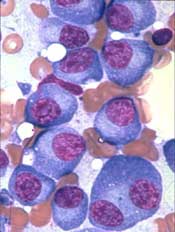
NEW YORK, NY—The B-cell maturation antigen (BCMA) is emerging as a promising target in multiple myeloma (MM), according to Adam D. Cohen, MD, of the University of Pennsylvania in Philadelphia.
BCMA is highly expressed on MM cells and, with its 2 ligands, is responsible for maintaining normal plasma cell homeostasis.
“And it’s really not expressed on any other normal tissues of the body,” Dr Cohen said.
“Importantly, BCMA is not just sitting on the cell surface as a target but actually promotes myeloma pathogenesis.”
Dr Cohen reviewed the progress being made using BCMA as a target in MM, paying particular attention to chimeric antigen receptor (CAR) T cells. He presented the update at Lymphoma & Myeloma 2017.
NCI BCMA-specific CARs
The first CAR to specifically target BCMA in MM was developed at the National Cancer Institute (NCI). It consisted of a murine single-chain variable fragment (scFv), CD3/CD28 signaling domains, and a gamma-retroviral vector.
Investigators conducted the first-in-human trial of this CAR T-cell therapy in 12 relapsed/refractory MM patients.
All patients received a lymphodepleting conditioning regimen of cyclophosphamide and fludarabine and a single infusion of 1 of 4 doses of the CAR T-cell therapy.
At higher dose levels, the BCMA-CAR produced objective responses “even in these highly refractory patients,” Dr Cohen said. “Some responses lasted 4 to 6 months.”
Patients who had the greatest degree of expansion of CAR T cells were the ones who had the best responses.
The BCMA-CAR is associated with the same toxicities as the CD19-directed CAR T-cell therapies now approved in acute lymphoblastic leukemia and non-Hodgkin lymphoma—cytokine release syndrome (CRS) and neurotoxicity.
The NCI study (NCT02215967) is ongoing.
Penn BCMA-specific CAR
A different BCMA CAR is being investigated at the University of Pennsylvania. It is a fully human CAR that consists of a human scFv, CD3/4-1BB costimulatory domains, and a lentiviral vector.
Investigators designed the first-in-human trial* (NCT02546167) with 3 different cohorts.
Patients in cohort 1 received 5 x 10⁸ CAR T cells without any lymphodepleting chemotherapy.
The remaining patients received cyclophosphamide, followed by 5 x 10⁷ CAR T cells in cohort 2 and 5 x 10⁸ CAR T cells in cohort 3.
Dr Cohen reviewed current data from cohort 1, which included 9 patients. They were a median age of 57 (range, 44-70), and 67% were male. They were heavily pretreated with a median of 9 prior lines of therapy (range, 4-11).
All had high-risk cytogenetics, 67% had deletion 17p or TP53 mutation, and they had a median of 80% bone marrow plasma cells (range, 15%-95%).
“Despite this,” Dr Cohen said, “we were able to generate, successfully, CAR T cells from all patients, although 1 patient did require a second apheresis and manufacturing attempt.”
Four of the 9 patients achieved very good partial responses, and an additional 2 patients had minimal responses.
One patient had a stringent CR (sCR) for close to 2 years without having any intervening therapy.
“[The sCR] shows the potential for this [therapy] to create a durable remission in a patient without any other therapy,” Dr Cohen said. “And this patient still has circulating CAR cells detectable.”
Most of the other patients did not have as durable a response. Responses lasted a median of 3 to 5 months before the patients relapsed.
Dr Cohen noted that the Penn data confirm the NCI experience showing proof of principle.
“You can target BCMA with these cells and get objective responses that can lead to a durable one in a subset of patients,” he added.
Dr Cohen is scheduled to report the preliminary data on the cyclophosphamide cohorts (cohorts 2 and 3) at ASH 2017. Those cohorts, he said, are showing a bit more persistence and increased frequency of responses.
Other BCMA-specific CAR trials
Another BCMA CAR, bb2121, consists of a human anti-BCMA scFv, a lentiviral vector, and a CD3/4-1BB costimulatory domain.
In the first-in-human trial of bb2121* (NCT02658929), all 21 patients received cyclophosphamide and fludarabine conditioning first.
There were “very impressive response rates in this study,” Dr Cohen said.
Once patients were dosed at the higher levels of 150 million cells or greater, every patient responded.
“Many responses are still durable,” he pointed out, “some approaching a year.”
LCAR-B38M is structurally different from the other BCMA-specific CARs. It has 2 binding sites for BCMA.
Thirty-five patients enrolled on the trial of LCAR-B38M (NCT03090659), and 19 were evaluable for response.
Patients had a median of 3 or 4 lines of prior therapy. All received cyclophosphamide alone as conditioning.
All 19 evaluable patients responded, and 14 (74%) achieved an sCR.
Dr Cohen noted the differences among the 4 trials: costimulatory domains varied, the Penn study did not require a pre-existing level of BCMA on the MM cells, other studies excluded patients who didn’t meet a certain threshold level of BCMA expression, median lines of prior therapy differed, and conditioning regimens differed as well.
All trials, however, presented data on fewer than 20 patients.
“And so I think it’s a little too early to compare head-to-head between all of these, but they all are really demonstrating promising results so far,” he observed.
Toxicities
“Unfortunately, there’s no free lunch with CAR T cells,” Dr Cohen stated, “and these extraordinary responses do come at the cost of several somewhat unique toxicities that can be serious.”
Toxicities have included:
- Tumor lysis syndrome, which is expected and manageable
- B-cell aplasia, which is not as much of an issue with BCMA as with CD19-directed CAR Ts, since most B cells don’t express BCMA
- Hypogammaglobulinemia, which can be mitigated with IVIG
- CRS, which can be alleviated with tocilizumab
- Neurotoxicity and encephalopathy, which do not occur as frequently as CRS.
“These, I think, are things we still obviously need to learn more about and try to mitigate before this [BCMA-directed CAR therapy] is expanded and brought earlier to patients,” Dr Cohen said.
“The other issues with CAR T cells are logistical. Limited access (the procedure is available at only a few centers); manufacturing can take 2 to 4 weeks, during which time it is difficult to maintain disease control; and the cost is significant.”
“We are really in the early days of CAR cells for myeloma, but, certainly, this appears very bright in terms of its future.” ![]()
* Data from the presentation differ from the abstract.
Speaker advises caution in adding mAbs upfront in MM

NEW YORK, NY—Despite the attraction of incorporating monoclonal antibodies (mAbs) into upfront therapy for multiple myeloma (MM), a speaker at Lymphoma & Myeloma 2017 suggested mAbs are “not quite ready” for this use.
MAbs, particularly daratumumab, have shown single-agent activity in refractory MM and have been feasibly added to proteasome inhibitors and immunomodulatory drugs.
MAbs may even have the potential to enhance induction and shorten the time to minimal residual disease negativity.
“So the tendency is to simply add them to frontline therapy,” said the speaker, Joseph Mikhael, MD, from Mayo Clinic Arizona in Scottsdale.
However, he noted that there is little long-term experience with these agents.
“I’m going to suggest to you that they’re not quite ready [for upfront use] but will likely be ready in the future,” Dr Mikhael said. “We’ve had such a revolution in myeloma the last decade that it’s just easy for us to say, ‘Oh, throw it in there, just like we did, frankly, with rituximab in the lymphoma days. We added it to CVP, we added it to CHOP, we added it to bendamustine. It didn’t matter what we added it to, it just upgraded the response.”
“And, sometimes, I think we have the same approach with daratumumab or elotuzumab or some of the other mAbs that we have. I think we just have to do so cautiously.”
At present, the combination of a proteasome inhibitor and an immunomodulatory drug are the standard of care upfront in transplant-eligible and -ineligible MM patients.
Daratumumab plus KRd
Dr Mikhael described the experience of daratumumab added to carfilzomib, lenalidomide, and dexamethasone (KRd) in patients with newly diagnosed MM in the MMY1001 study.
Twenty-two patients were enrolled on the study, 91% achieved a very good partial response (VGPR) or better, and 43% achieved a complete response (CR). The depth of response improved with the duration of treatment.
Eight patients (36%) discontinued treatment.
Dr Mikhael emphasized that the preliminary data included very small numbers.
“There is a little bit of a yellow flag that pops up here,” he added, “when I see that 36% discontinued treatment, even in small numbers.”
The safety profile was consistent with previous reports for daratumumab or KRd.
The most common hematologic grade 3-4 treatment-emergent adverse events (AEs) occurring in 30% or more of patients were lymphopenia (64%), thrombocytopenia (9%), anemia (9%), leukopenia (9%), and neutropenia (14%).
Diarrhea (14%), cough (5%), fatigue (5%), insomnia (5%), and increased ALT (9%) were the most common grade 3-4 nonhematologic treatment-emergent AEs occurring in 30% or more of patients.
The treatment had no adverse impact on stem cell collection.
Elotuzumab plus VRd
Turning to elotuzumab in combination with bortezomib, lenalidomide, and dexamethasone (VRd), Dr Mikhael reviewed the phase 2a study (NCT02375555) presented at ASCO 2017 (abstract 8002).
Forty-one patients were enrolled on the study.
The overall response rate after 4 cycles was 100%, with 24% achieving a CR, 47% achieving a VGPR, and 29% a partial response.
Fatigue (60%), neuropathy (55%), musculoskeletal/joint pain (55%), infection (50%), back/neck pain (48%), diarrhea (45%), edema (38%), constipation (38%), cough (35%), mood alteration (35%), rash (35%), and insomnia (30%) occurred in 30% or more of patients.
“So again, not shocking,” Dr Mikhael said, “there was fatigue, there was neuropathy, and there were infections in 50% of patients.”
Grade 4 or greater AEs included thrombocytopenia, hyperglycemia, sepsis, cardiac arrest, and respiratory failure.
“However, here, [we have] maybe not even a yellow flag but a red flag of caution that there were 2 patients who died,” Dr Mikhael noted.
One patient died on study due to respiratory failure and sepsis that arose during cycle 2.
The other patient died more than 30 days after discontinuing study therapy due to febrile neutropenia and hypotension related to sepsis, followed by renal failure.
“Again, in a study that has such small numbers, I don’t want to overstate the case . . ., we don’t want to overreact, but whenever there is death involved, obviously, we have to be particularly cautious,” Dr Mikhael said.
Put into the context of 3 other VRd studies, he noted, the response rate with elotuzumab and VRd is relatively similar but not as good as the phase 3 study of VRd, which was a much larger study of 350 patients.
The situation with daratumumab and KRd is similar to elotuzumab, Dr Mikhael pointed out.
The initial response rates are impressive, but, when compared to other studies, “71% VGPR is good, only after 4 cycles, but we know that, in other studies, after a few more cycles, it was significantly higher.”
Cost
Dr Mikhael also considered cost in his assessment of daratumumab and elotuzumab integrated into frontline regimens.
Adding elotuzumab to VRd would almost double the cost of 12 weeks of therapy. And adding daratumumab to KRd would increase the cost even more.
“These costs are real,” Dr Mikhael said, “and, ultimately, if it’s the best thing for our patients, that’s what we are going to do. But until we have that convincing evidence, I think it’s critical to keep that in perspective. I would suggest that VRd, in many respects, is the standard of care for most patients.”
In terms of adding a mAb upfront, he said, “I don’t think we’re there yet. Do I think, in time, we will be? Quite likely, but I don’t think we are there yet.” ![]()
X-GEM finds drug to have economic value in MM
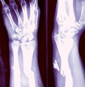
NEW YORK, NY—Investigators have developed a model that suggests the clinical benefits of denosumab translate into economic value.
The investigators designed their model, X-GEM (Exgeva-Global Economic Model), using results from a large multiple myeloma (MM) trial that showed denosumab to be non-inferior to zoledronic acid (ZA) for skeletal-related events (SRE).
Noopur S. Raje, MD, of Massachusetts General Hospital Cancer Center in Boston, discussed this work at Lymphoma & Myeloma 2017.
The abstract was selected as the best clinical myeloma abstract of the meeting.
“I do think this is a very timely topic because people are not just concerned about the clinical outcomes in patients,” Dr Raje said. “[T]here’s a lot of buzz around the economic evaluation of what we do.”
Trial results
The phase 3 study (NCT01345019) on which X-GEM is based enrolled 1718 MM patients and randomized them to either denosumab or ZA.
The results, reported this year at ASCO, showed denosumab to be non-inferior to ZA in time to first on-study SRE, the primary endpoint.
Denosumab was also found to be non-inferior to ZA for the secondary endpoint of overall survival.
For the exploratory endpoint of progression-free survival (PFS), denosumab-treated patients experienced a significant benefit in terms of PFS. The median PFS was 46.09 months with denosumab and 35.38 months with ZA (difference, 10.71 months).
“Now, we’ve never really seen a survival difference or progression-free survival difference in patients, even in treatment studies, amounting to about 10.7 months,” Dr Raje said. “So this, we found, was quite remarkable in the study.”
Dr Raje also highlighted some safety features from the study. There was significantly less renal toxicity with denosumab than with ZA.
“And in patients who had a creatinine clearance of less than 60 [mL/min], there was almost a doubling of renal toxicity in patients getting zoledronic acid,” she said.
“[D]enosumab may, in fact, be the safer alternative, specifically, in our patients with multiple myeloma who we all know have this problem of renal toxicity throughout the course of their lifetime with myeloma.”
X-GEM
Investigators based X-GEM on the original model published by Stopeck et al. in 2012, which evaluated the cost-effectiveness of denosumab to prevent SREs compared with ZA in patients with solid tumors—prostate, breast, and lung cancer.
“[Stopeck’s] data did show that denosumab was, in fact, cost-effective when compared to zoledronic acid in respect to SREs,” Dr Raje noted.
The timeline for the economic analysis in MM patients spanned the time from diagnosis to death, and patients were evaluated for SREs every 4 weeks on study.
Investigators used 2 cost scenarios. The first was based on an average sales price for 28 days, which was $1928 for denosumab and $45 for ZA. The second was based on the wholesale acquisition cost, which was $2155 for denosumab and $922 for ZA.
“No surprise to anybody,” Dr Raje noted, “denosumab is a lot more expensive. But obviously, this does not tell the whole story. Built into the X-GEM model are a whole host of other factors.”
These include the costs of administration, adverse events, the number of SREs, treatment of an MM patient, and the quality-adjusted life year (QALY) gain with either denosumab or zoledronic acid.
“When you count up all these costs and calculate them based on the data set from the 1800 patients, we found that there was really a difference of zoledronic acid costing a little bit more than denosumab,” Dr Raje said.
From the payer perspective, when clinical outcomes were monetized, the net monetary benefit of denosumab compared with ZA was $5959.
And from a societal perspective, the model calculated the net monetary benefit for denosumab to be $10,259.
The societal perspective included SRE direct costs (hospital, outpatient, and emergency department visits, long-term care, hospice, physical therapy and devices, skilled nursing facility, and strong opioids), direct non-medical costs (driving for treatment, parking, and caregiver costs), and indirect costs (short-term disability and productivity loss).
The investigators concluded that denosumab is cost-effective below a willingness-to-pay threshold of $150,000/QALY regardless of ZA price, whether wholesale or average sales price.
“The bone-specific benefits and observed prolongation of progression-free survival in combination with the economic analysis provides denosumab as a valuable option for patients with multiple myeloma,” Dr Raje said. “In total, we still think it’s more cost-effective to use denosumab when compared to zoledronic acid in this patient population.”
At present, treatment options to prevent bone complications in MM patients are limited to bisphosphonates, including ZA. Denosumab is under review by the US Food and Drug Administration for an expanded indication to include MM.
Both the phase 3 and cost-effectiveness studies were funded by Amgen, Inc. Dr Raje and co-investigators of the study have either consulted for or are employed by Amgen, Inc. ![]()
Idelalisib efficacy against CLL tarnished by toxicity
NEW YORK – PI3K inhibitors are highly active against B-cell malignancies, but this class of drugs, led by
Idelalisib is a potent inhibitor of the delta isoform of phosphatidylinositol 3-kinase (PI3K) that in a phase 1 trial was associated at higher dose levels with a median progression-free survival (PFS) of 32 months in patients with CLL who had received a median of five prior lines of therapy, noted Jennifer R. Brown, MD, PhD, director of the CLL center at the Dana-Farber Cancer Institute in Boston.
“This is really a very effective drug. So what’s happened? Why aren’t we using it more?” she asked rhetorically at an international congress on hematologic malignancies.
“This relates to a pattern of toxicities that has becoming increasingly familiar to us,” she added.
There is increasing evidence to suggest that the toxicities associated with idelalisib are immune mediated, indicating both the need for caution among clinicians who think about prescribing the drug, and a potential future use for this and other PI3K inhibitors as immunomodulatory agents, Dr. Brown said.
Registration trial toxicities
Among 760 patients enrolled in trials for the idelalisib registration programs, grade 3 or greater diarrhea and/or colitis and transaminitis each occurred in 14% of patients, rash occurred in 6%, and pneumonitis of any grade was seen in 3%.
Among patients with relapsed disease, transaminitis was often self-limiting and usually resolved when the drug was withheld, and about 75% of patients were successfully restarted on idelalisib at the same or lower dose, Dr. Brown noted.
Rashes, which can occur any time with therapy, were also successfully managed by withholding drug and then rechallenging, with the addition of corticosteroids as necessary.
Patients who developed drug-related pneumonitis were less likely than those with other toxicities to be rechallenged, and most required steroids until the infections resolved.
“The steroid responsiveness of many of these side effects suggested that they were autoimmune,” Dr. Brown said.
Drugs only work when you take them
The toxicities seen with idelalisib have had a marked effect on the use of the drug. In registration trials for idelalisib in combination with rituximab or ofatumumab (Arzerra), each of which had at least 2 years of follow-up, only 22.5% of 369 patients remained on idelalisib, primarily because of toxicities rather than disease progression. The combined 2-year progression in these trials was 13.3% In contrast, 40.7% of patients discontinued idelalisib because of adverse events.
Out to about 7 months, survival rates for patients who discontinued idelalisib because of disease progression or adverse events were roughly similar, but survival for the patients who stopped because of side effects began to plateau out to 2 years, Dr. Brown noted.
As of March 2016, 23.2% of patients who received idelalisib in clinical trials in combination with other agents as second- or third-line therapy had died, compared with 31% of controls, indicating a clear survival benefit with the drug.
“This is probably because the benefit of disease control in that setting overwhelmed the adverse event or infections problem,” she said.
Many of the deaths in registration trials were related to opportunistic infections, including Pneumocystis jiroveci pneumonia, fungal infection, and cytomegalovirus.
“Idelalisib, I think, is a prototypical delta inhibitor with a pattern of immune-mediated toxicity that remains unpredictable and can be severe. We now have pretty good data, based on the Gilead [sponsor] trials, that younger age and less prior therapy predispose to this toxicity,” Dr. Brown said.
Evidence is less robust, but growing, that mutated IGHV and a decrease in regulatory T cells may be also be risk factors for immune-mediated toxicities with idelalisib. Immune modulation with the drug may also account for associated neutropenia, sepsis, and opportunistic infections seen with idelalisib therapy, she added.
So how to use it?
Currently, the best uses for idelalisib and other PI3K inhibitors in CLL appear to be in single-agent therapy in patients with relapsed disease who cannot tolerate a Bruton’s tyrosine kinase (BTK) inhibitor such as ibrutinib (Imbruvica) or in patients whose disease has progressed on a BTK inhibitor.
“Where I think about this drug is in older, more heavily pretreated patients, who are generally at less risk for toxicities, and if they have significant comorbidities that may impact BTK-inhibitor tolerability, usually cardiac,” Dr. Brown said.
Future expansion of PI3K inhibitors in B-cell malignancies may require identifying a biomarker for tolerance, alternative dosing schedules, or identification of an idelalisib/drug X combination that might mitigate the toxicity, she said.
The immune-activation properties of PI3K-delta inhibitors suggests that they might also play a role as antitumor immunomodulatory agents in treatment of both hematologic malignancies and solid tumors, Dr. Brown concluded.
Idelalisib trials were sponsored by Gilead Sciences. Dr. Brown disclosed serving as a consultant for Gilead and other companies.
EXPERT ANALYSIS FROM LYMPHOMA & MYELOMA