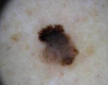
While dermoscopy can enhance the diagnosis of a number of skin conditions, it is especially useful in determining whether neoplasms should undergo biopsy.
"Patients are becoming aware of the technique and more and more are expecting their dermatologists to be skilled in its application," Dr. David L. Swanson said in an interview.
However, fewer than half of academic dermatologists and less than a quarter of practicing dermatologists use dermoscopy in the United States, according to Dr. Swanson, chief of medical dermatology at the Mayo Clinic in Scottsdale, Ariz. In comparison, dermoscopy is taught to primary care physicians in Europe and Australia.
He discussed methods for assessing whether a lesion should be biopsied.
With the two-step pattern recognition method, the dermoscopist must first determine whether a neoplasm is a melanocytic proliferative one, such as a nevus or melanoma. "The reason that the first step is to consider nevi or melanomas is the importance of identifying the latter – especially melanomas that are not obvious with the naked eye," said Dr. Swanson at a seminar on women's and pediatric dermatology sponsored by Skin Disease Education Foundation.
If the lesion is melanocytic proliferative, the dermoscopist then applies the methods of interpretation for nevi or melanoma – such as pattern recognition or an algorithm. "If it isn't, then there are other diagnostic features that are used to lead to a clinical impression," he said.
The dermoscopy three-point rule has a high sensitivity for melanoma, he said. If a pigmented lesion has any two of the three criteria – atypical asymmetry, atypical pigment network, or blue-white structures – there is a likelihood of melanoma, and the lesion should be biopsied.
"The three-point rule is easier to learn and apply but as with all algorithms, there are a lot of exceptions to the rules. So one has to be willing to learn those exceptions and step out of the algorithm if they aren't certain," said Dr. Swanson. "One specific problem with the three-point method is that it doesn't take into account an atypical vasculature. Any dermoscopist using the three-point rule has to keep that in mind as an additional feature."
The seven-point algorithm – developed by Dr. Giuseppe Argenziano and others – is aimed at helping the novice dermoscopist. "It is a fairly reproducible method with very good specificity and sensitivity for making a decision to biopsy a melanocytic lesion. The three-point algorithm actually was derived from it as a method to simplify," said Dr. Swanson. "Most experienced dermoscopists only use algorithms occasionally, because with experience they become comfortable with pattern recognition."
The seven-point algorithm includes the criteria of the three-point rule, plus four minor criteria: streaks, regression pattern, irregular diffuse pigmentation, and irregular dot and globules.
Disclosures: Dr. Swanson reported having none. SDEF and this news organization are owned by Elsevier.


