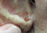
The lesion on the face of this 72-year-old woman has slowly grown over a period of years but has never caused pain. On several occasions, she has consulted primary care providers (PCPs), most of whom prescribed oral antibiotics for possible infection. However, these medications never helped.
She recently developed oral health problems and established care with a new PCP, who insisted she see a dermatologist about the facial lesion. The patient, who is “terrified” of needles, resisted this advice at first but finally agreed once her family got involved.
The patient reports a history of extensive sun exposure in childhood, when she worked in the fields, that continued into adulthood; she maintained a garden until recently. Her response to the sun is invariably a deep tan, which lasts all summer.
The lesion, almost 6 cm wide and more than 4 cm long, is located in the right infraorbital facial cheek area, well below the eyelid margin. It is focally eroded, peripherally erythematous, and quite firm to the touch. Fortunately, it is still mobile and not fixed to the underlying tissue. There are no palpable lymph nodes in the area and no change in sensation from one side of the face to the other.
A punch biopsy is performed under local infiltrate with 1% lidocaine with epinephrine. The 4-mm defect is closed with 2 x 5-0 nylon sutures and the specimen submitted to pathology. The report confirms the clinical suspicion of basal cell carcinoma (BCC).
Continue for Joe Monroe's discussion >>
