User login
Infectious Mononucleosis
CE/CME No: CR-1306
PROGRAM OVERVIEW
Earn credit by reading this article and successfully completing the posttest. Successful completion is defined as a cumulative score of at least 70% correct.
EDUCATIONAL OBJECTIVES
• Discuss the pathophysiology of the Epstein-Barr virus (EBV) and its manifestation in infectious mononucleosis (IM).
• Name specific factors that distinguish IM from each of several conditions to be considered in the differential diagnosis.
• Describe the use of heterophile antibody testing and EBV serology testing to confirm a diagnosis of IM.
• Explain supportive management, activity restrictions, potential complications, and essentials of patient education for IM.
FACULTY
Scott J. Saccomano is an Assistant Professor in the Family Nurse Practitioner Program, Department of Nursing, at Herbert H. Lehman College in Bronx, New York. Lucille R. Ferrara is an Assistant Professor and Director of the Family Nurse Practitioner Program in the Department of Graduate Studies at Pace University, College of Health Professions, Lienhard School of Nursing, in Pleasantville, New York.
The authors have no significant financial relationships to disclose.
ACCREDITATION STATEMENT
![]()
Article begins on next page >>
Although 15- to 24-year-olds account for the greatest incidence of infectious mononucleosis (IM), antibodies to the Epstein-Barr virus, the causative organism, may be present in 95% of the population. Learn to distinguish between IM and other illnesses with similar presentations—and to watch for the potentially severe complications of IM.
Infectious mononucleosis (IM), commonly known as kissing disease, is a viral syndrome resulting from an acute infection with Epstein-Barr virus (EBV). Mononucleosis typically occurs between early childhood and early adulthood and is ordinarily self-limiting as the patient develops EBV-specific immunity; in some cases, however, IM can lead to severe complications.1-4
Worldwide, the EBV may be present in 95% of adults between 35 and 40.2,5,6 In patients previously infected during early adulthood, the EBV remains dormant in the B-lymphocytes, and those affected may continue to carry asymptomatic infection lifelong.1,7
EPIDEMIOLOGY
IM antibodies may be present in 90% to 95% of the population; however, epidemiologic data regarding infection vary among age-groups and by geographic location.2,4 IM occurring in patients between ages 1 and 5 years is very limited in industrialized countries and within higher socioeconomic groups; in these settings, infection onset occurs primarily in the second decade of life. EBV is rarely found in patients younger than 1 year, possibly because of serologic protection from maternal antibodies.6
Early childhood infection with EBV is predominantly found in the developing countries and in lower socioeconomic groups. EBV infection in young children usually presents with nonspecific symptoms. Wherever improvements in hygiene have been made during recent years, EBV infection in early childhood has become increasingly rare.5,8
Susceptibility to EBV becomes more pronounced during adolescence and early adulthood; in the United States, the incidence of IM is 500 cases per 100,000 persons per year, with persons between ages 15 and 24 accounting for the greatest incidence.5 Among college freshmen who are initially seronegative for EBV (estimates range from 30% to 70%), between 10% and 20% will become infected with the virus; of these, 30% to 50% will develop IM. IM is not seasonal or cyclical and has no sexual predisposition.5,8,9
PATHOPHYSIOLOGY
EBV, a gamma herpes virus, is spread via intimate contact from persons recently infected with EBV infection to other susceptible humans.5 EBV is typically transmitted through oropharyngeal secretions, mainly saliva. After its transmission, the EBV enters epithelial B-lymphocytes, which contain receptors for EBV. Infected B-lymphocytes target the salivary glands, the lymphoid cells, and the oropharynx, causing pharyngitis and other early manifestations of IM. After infection, the host immune response is an activation of T cells against infected B cells, producing larger, atypical lymphocytes called Downey cells.10
The infected B cells then enter the blood stream, carrying the virus throughout the body to the spleen, liver, and peripheral lymph nodes. The spread of these infected lymphocytes elicits a significant cellular immunologic response to the viral infection. This immunologic response is largely responsible for the clinical presentation of IM in the lymph nodes, spleen, and possibly the liver.10
Once acute infection occurs, antibodies are produced against both EBV and unrelated viral species. Recent-onset infection produces heterophile antibodies, the presence of which is helpful in the diagnosis of IM. Specific antibodies to the EBV also develop, including immunoglobulin G (IgG) and IgM; these and others can be useful for serologic identification of EBV. Once an individual is acutely infected with EBV, he or she becomes a lifelong carrier of the virus.10
The incubation period of the EBV is between 30 and 50 days,5,10 making it difficult to pinpoint an exact date of exposure. Transmission of the virus to others may be possible for three months or longer; even after symptoms are resolved, the virus is shed in the saliva for months. Additionally, the EBV has been found in semen and cervical secretions, indicating the possibility of sexual transmission.5,9,10,11
CLINICAL MANIFESTATIONS
Patients with IM usually seek medical treatment for worsening sore throat and increasing fever. The clinical presentation of IM can vary, depending on time between symptom onset and that of presentation.4 However, the triad of most common clinical symptoms, which ordinarily resolve in one to three weeks, are sore throat, fever, and lymphadenopathy.5,10 During the one- or two-week-long prodromal period, patients may also have nonspecific symptoms of malaise, fatigue, and myalgia. About half of patients report headache.10,12
Triad of Common Symptoms
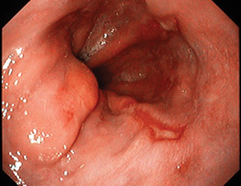
Pharyngitis, the most prominent physical finding, can be severe. It presents in the initial weeks of illness, with pharyngeal exudates present in about half of cases.10,13 The pharyngeal exudates in IM may be difficult to distinguish from those associated with streptococcal pharyngitis. With inflammation of the lymphoid tissue, tonsillitis develops; tonsillar ulceration has been reported in 20% of patients with IM.13 “Kissing tonsils,” with enlargement of the tonsillar pillars causing the tonsils to touch, can lead to airway compromise.14 In addition to pharyngitis and tonsillitis, oral palatal petechiae may develop.5,8,10
Low-grade fever, rarely exceeding 102°F, usually lasts for one to two weeks but may persist for as long as five weeks.11
Cervical lymphadenopathy usually presents with symmetrically enlarged, firm, mobile and tender anterior and posterior lymph nodes, as well as the submandibular lymph nodes; however, clinicians should be aware that more generalized adenopathy may be present, affecting the axillary and inguinal nodes.2,10,11 Lymphadenopathy usually resolves within one to two weeks.
Additional Manifestations of IM
Skin rashes have been reported in 3% to 15% of patients with IM,5 although results from a recent retrospective study suggest that one-third of patients with IM who are initially treated with amoxicillin experience rash.15 This maculopapular, pruritic, copper-colored or tan and brown rash usually begins about 5 to 10 days after antibiotic therapy is initiated. The rash, which is not considered an allergic reaction, usually resolves once the antibiotic is discontinued.10,12,16
Hepatosplenomegaly, particularly splenomegaly, is common—although this is usually a late finding in patients with IM, developing during the second to third week of illness.11,12,16 Abdominal pain is typically absent. However, when patients complain of severe left upper quadrant abdominal pain, splenic ultrasound should be considered to assess for splenic enlargement or possible rupture.8
On the next page: Differential diagnosis >>
DIFFERENTIAL DIAGNOSIS
Not all patients with symptoms suggestive of IM are found to be infected with EBV. Other pathogens may be responsible for primary infection in these patients10 (see Table 15,10,12,17-20).
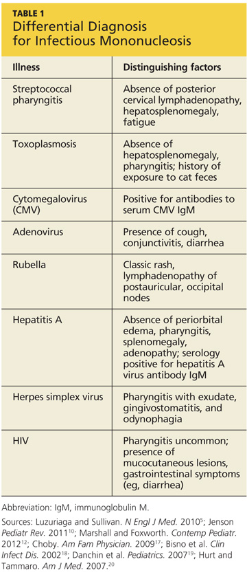
Streptococcal pharyngitis is the illness most likely to be confused with IM because of the similarity of symptoms; one in four children with acute sore throat (as well as 5% to 20% of affected adults) may be infected with group A beta-hemolytic streptococcus (GABHS).17-19 Although, in addition to sore throat, patients with GABHS may experience localized swelling of the anterior cervical lymph nodes, GABHS is not usually characterized by posterior cervical lymphadenopathy. Additionally, the hepatosplenomegaly and fatigue common to IM would not be present in patients with streptococcal pharyngitis.5
GABHS is confirmed by a positive throat culture for group A streptococcus17; in patients with negative results on a rapid antigen test, IM should be considered in the differential diagnosis.
Toxoplasmosis can manifest with symptoms similar to those of EBV-associated IM. The classic IM symptoms of hepatosplenomegaly and pharyngitis are not often seen in cases of toxoplasmosis, however. The history should reveal contact with cat droppings or cat litter.12,20
Patients infected with cytomegalovirus (CMV) can present asymptomatically or with the common triad of symptoms of IM. Splenomegaly, malaise, and lymphadenopathy may be present. A diagnosis of CMV is confirmed by the presence of antibodies to serum CMV IgM.12,20
Adenovirus typically manifests with cold-like symptoms, cough, conjunctivitis, and diarrhea. The triad of symptoms typically seen with IM may be present with decreased severity in patients with adenovirus. Culture of nasopharyngeal swabbings can confirm a diagnosis of adenovirus.12,20
Symptoms of rubella can mimic those of IM, but the classic rubella rash should be a distinguishing factor. Lymphadenopathy most commonly affects the postauricular and occipital nodes. Parents may acknowledge that the patient has not been immunized against rubella.12
Manifestations of hepatitis A include hepatomegaly, fever, jaundice, abdominal pain, and fatigue. Symptoms associated with IM, including periorbital edema, pharyngitis, splenomegaly, and adenopathy, are not typically seen in patients with hepatitis A. Laboratory findings in the patient with hepatitis would include elevations in alanine aminotransferase and aspartate aminotransferase. Serology would be positive for hepatitis A virus antibody IgM and negative for EBV heterophile antibodies.12,16
Herpes simplex virus, commonly known as a cold sore, manifests with pharyngitis with exudate, gingivostomatitis, and odynophagia (painful swallowing). The appropriate polymerase chain reaction (PCR) test can be used to confirm the presence of herpes virus.12,20
HIV screening should be conducted in at-risk patients, as IM and HIV can present with similar symptoms of fever, malaise, lymphadenopathy, and rashes, especially in the early phase of primary HIV infection. Pharyngitis is less common in patients with HIV infection than in those affected by IM; rather, mucocutaneous lesions and gastrointestinal symptoms, such as diarrhea, are likely to be present.5 Diagnostic findings include a positive ELISA HIV antibody test and positive plasma viral load.12,20
On the next page: Diagnosis >>
DIAGNOSIS
Pertinent information elicited in the patient history should include signs and symptoms, particularly the common triad of pharyngitis, fever, and lymphadenopathy, and possible exposure or sick contacts. Certainly, patients with symptoms of pharyngitis and continued fatigue that are unresolved within 48 to 72 hours should be screened for IM.12,13
In addition to the in-depth history, routine hematologic testing is conducted, as well as serologic testing to confirm the diagnosis. A complete blood count will reveal hyperlymphocytosis (≥ 50%), an elevated white blood cell count (usually ranging from 10,000 to 20,000/mL in most patients), and at least 10% atypical lymphocytes.2,6,8,11 Elevations in liver enzymes are found in 80% to 90% of cases.2,11
Heterophile Antibody Testing
In the symptomatic patient, a positive result on the monospot, a rapid test for heterophile antibodies, is 85% sensitive and 94% specific for IM.5,6,8 Heterophile serology is elevated for the first four weeks of infection, then declines rapidly; thus, this test should not be used beyond one month of symptom onset. Additionally, false-negative monospot test results have been reported in about one-fourth of patients during the first week of symptoms.5,6 For that reason, when suspicion for IM is high but the monospot yields negative results, additional serology with greater sensitivity and specificity is warranted.
Of note, false-negative results on heterophile serology are common in children younger than 12.11,21
EBV Serology
Because IM represents infection with EBV, serologic testing for EBV is accurate, highly specific, and recommended for use when IM is strongly suspected, particularly in infants and younger children. Serologic tests are more reliable, though more expensive and time-consuming, than heterophile antibody testing.11,22
Various EBV serologic tests are used for diagnosis, and it is recommended by the CDC6 that these tests be performed concurrently because the antibody response in EBV is rapid. A blood sample obtained during the acute phase of illness will reveal antibodies representing acute, recent, past, or reactive disease.6
Testing for EBV-specific IgM and IgG antibodies against viral capsid antigen (VCA), early antigen (EA), and the Epstein-Barr virus nuclear antigen proteins (EBNA) is 97% sensitive and 94% specific for a diagnosis of IM.5,11,23 EBV antigen testing can be performed using the following laboratory tests: IgM VCA, IgG VCA, EA IgM, and EBNA IgG. The decision to perform antigen testing is based on timing and onset of symptoms (see Table 25,11 for possible findings from certain tests). Primary EBV infection is indicated by the presence of IgG antibodies against VCA or of IgM antibodies against VCA, in addition to the absence of EBNA antibodies.6,23
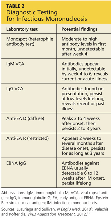
As with any laboratory testing, the EBV serology should be correlated with patient presentation and the overall clinical picture. Early antigen testing, it should be noted, is often recommended for patients who have significant and persistent symptoms of IM but who are past the four-week time frame in which heterophile antibody testing is effective.11
A quantitative real-time PCR assay, which detects EBV viral load, can also be used to test blood or plasma for acute infection.11 PCR testing is usually reserved for young children with significant symptoms but negative results on the monospot test; or for patients presenting with IM-like symptoms but having more complex conditions, such as HIV, in which further differentiation of EBV DNA subtypes is needed.24,25
On the next page: Considerations in specific patient populations and complications >>
CONSIDERATIONS IN SPECIFIC PATIENT POPULATIONS
Adolescents and young adults. When caring for adolescents and young adults, clinicians are advised to obtain an in-depth sexual history. In patients who are considered at high risk for sexually transmitted diseases (STDs), testing for HIV and other STDs is also recommended.26
Adults and elderly adults. Epidemiology does not support the likelihood of IM in these age-groups; this can lead to a missed diagnosis or a misdiagnosis with one of the more common adult infectious diseases, including those of a hepatic or hematologic nature.27 Adults older than 40 account for 7.5% of cases of IM.28
In these patients, manifestation of the clinical signs of IM may be altered. Rather than displaying the classic triad of symptoms, older patients may present with nonspecific complaints of fever, myalgias, malaise, and fatigue.16,28 Serologic testing should be considered to avoid misdiagnosis in this age-group.
COMPLICATIONS
Most patients with IM recover and are able to return to normal activity within two to three months.5 Several complications can develop, however (see Table 33,5,10,12,14,29,30).
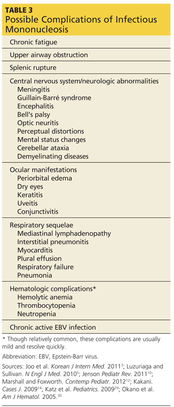
Chronic fatigue is the most commonly reported sequela of IM. In one study of 12- to 18-year-olds who had had IM, chronic fatigue was reported in 13%, 7%, and 4% of patients at six, 12, and 24 months, respectively.29 The study authors concluded that IM during adolescence may be a risk factor for chronic fatigue syndrome.
A more serious but less common complication of IM is upper airway obstruction, occurring in perhaps 1% to 5% of patients.5,10 The result of IM-associated tonsillar enlargement and edema in the pharyngeal tissue, upper airway obstruction can require hospitalization. Appropriate interventions include IV corticosteroids, tonsilloadenoidectomy, and endotracheal intubation.10,14
Also less common but extremely serious is splenic rupture, which develops in 0.5% to 1% of patients with IM.5 Affected patients usually require emergency surgery.8,12
Unusual complications include central nervous system effects, such as meningitis, Guillain-Barré syndrome, encephalitis, Bell’s palsy, optic neuritis, perceptual distortions, and mental status changes. Other neurologic abnormalities, such as cerebellar ataxia and demyelinating diseases, have been reported.10,14 Ocular manifestations sometimes associated with IM are periorbital edema, dry eyes, keratitis, uveitis, and conjunctivitis.5,16
On occasion, clinicians may see patients with IM complicated by respiratory involvement, such as mediastinal lymphadenopathy, interstitial pneumonitis, myocarditis, and plural effusion. Incidences of respiratory failure and pneumonia have been documented, primarily in immunocompromised patients.3,10
Potential hematologic complications of IM include hemolytic anemia, thrombocytopenia, and mild neutropenia.10,11 Although these conditions have been reported in 25% to 50% of patients with IM, they typically present in a mild form and resolve within a few weeks.5,10
Another serious complication of IM, though rare, is chronic active EBV infection, a persistent syndrome with manifestations that may include fever, significant lymphadenopathy, persistent hepatitis, and a high viral load in the peripheral blood.3,30
If symptoms of IM persist for longer than 6 months, further evaluation should take place, including investigation for other chronic disease states, such as HIV, lupus erythematosus, or chronic fatigue syndrome.
The most common complication of IM misdiagnosed as GABHS is a rash resulting from treatment with penicillin (which is an appropriate treatment for GABHS but not IM).15 Although penicillin use is the most common cause of drug-induced rash in patients with IM, extremely rare cases have been reported in which a rash developed after treatment with a macrolide, specifically azithromycin.31
On the next page: Management of IM >>
MANAGEMENT OF IM
The goals of therapy are to minimize complications and restore the patient to full capacity. Supportive care is the mainstay, including bed rest, fluids, and administration of NSAIDs.
Although the incidence of splenic rupture is 0.5% to 1%,5 activity should be restricted to minimize the risk. Patients are usually advised to restrict activity (especially contact sports) for at least three to four weeks. Strenuous activity should be avoided for three weeks to two months.8,32
A scaffold plan to return to full activity after week 4 should be provided to asymptomatic patients who receive adequate hydration and are unaffected by splenomegaly.32,33 If patients experience respiratory involvement related to tonsillar enlargement and tissue hypertrophy causing increased respiratory obstruction, oral corticosteroids can be used.
Corticosteroids are also appropriate for hematologic complications such as hemolytic anemia and thrombocytopenia. Other than these or respiratory complications, there is insufficient evidence for corticosteroid use as supportive treatment for IM.34 Similarly, data to support the use of antivirals, such as acyclovir, are inconclusive.35
PATIENT EDUCATION
Patients should be educated about progression of the disease as well as the timing and length of expected clinical symptoms during the disease course. Patients infected with IM do not need to be isolated but should avoid exposing others to infected oropharyngeal secretions by kissing or sexual contact.5,11 Patients should avoid sharing items that may harbor the EBV, such as drinking containers or eating utensils, particularly during the febrile period.
Individuals must also be advised to curtail active physical activities, specifically strenuous or contact sports, until given clearance by their provider.8
CONCLUSION
Once a patient is infected with EBV, the virus remains present for life. EBV infection is dormant in the B-lymphocytes and can occasionally become reactivated. In these cases, the patient becomes infectious, though rarely displaying symptoms of latent B-lymphocyte infection. In certain circumstances, given the right environment in susceptible contacts, the reactivated virus may produce subclinical symptoms, and the virus can be spread to others.
1. Jain N, Bhatia V, Lattoo S. Epstein-Barr virus and associated head and neck manifestations. Ann Nigerian Med. 2011;5:38-41.
2. World Health Organization, Initiative for Vaccine Research. Viral cancers. www.who.int/vaccine_research/diseases/viral_cancers/en/index1.html. Accessed May 8, 2013.
3. Joo EJ, Ha YE, Jung DS, et al. An adult case of chronic active Epstein-Barr virus infection with interstitial pneumonitis. Korean J Intern Med. 2011;26:466-469.
4. Çeltik C, Küçükugurluoglu Y, Balci DB, et al. Evaluation of clinical and laboratory features of Epstein-Barr virus–associated acute infectious mononucleosis in children. Trakya Universitesi Tip Fakultesi Dergisi. 2008;25:221-227.
5. Luzuriaga K, Sullivan JL. Infectious mononucleosis. N Engl J Med. 2010;362:1993-2000.
6. CDC. Epstein-Barr virus and mononucleosis (2006). www.CDC.gov/ncidod/diseases/ebv.htm. Accessed May 8, 2013.
7. Macsween KF, Higgins CD, McAulay KA, et al. Infectious mononucleosis in university students in the United Kingdom: evaluation of the clinical features and consequences of the disease. Clin infect Dis. 2010;50:699-706.
8. Bennett NJ. Pediatric mononucleosis and Epstein-Barr virus infection (2012). http://emedicine.medscape.com/article/963894. Accessed May 8, 2013.
9. Crawford DH, Macsween KF, Higgins CD, et al. A cohort study among university students: identification of risk factors for Epstein-Barr virus seroconversion and infectious mononucleosis. Clin Infect Dis. 2006;43:276-82.
10. Jenson HB. Epstein-Barr virus. Pediatr Rev. 2011;32:375-384.
11. Valachis A, Kofteridis DP. Mononucleosis and Epstein-Barr virus infection: treatment and medication. Virus Adaptation Treatment. 2012; 4:23-28. www.dovepress.com/getfile.php?fileID=12299. Accessed May 8, 2013.
12. Marshall BC, Foxworth MK II. Epstein-Barr virus–associated infectious mononucleosis (2012). Contemp Pediatr. http://digital.health caregroup.advanstar.com/nxtbooks/advanstar/cntped_201210/index.php?start id=52. Accessed May 8, 2013.
13. Kutuya N, Kurosaki Y, Suzuki K, et al. Pharynigitis of infectious mononucleosis: computed tomography findings. Radiat Med. 2008;26:248-251.
14. Kakani S. Airway compromise in infectious mononucleosis: a case report. Cases J. 2009;2;6736.
15. Chovel-Sella A, Ben Tov A, Lahav E, et al. Incidence of rash after amoxicillin treatment in children with infectious mononucleosis. Pediatrics. 2013 Apr 15. [Epub ahead of print]
16. Cunha BA. Infectious mononucleosis (2013). http://emedicine.medscape.com/article/222040-overview. Accessed May 8, 2013.
17. Choby BA. Diagnosis and treatment of streptococcal pharyngitis. Am Fam Physician. 2009;79:383-390.
18. Bisno AL, Gerber MA, Gwaltney JM Jr, et al; Infectious Diseases Society of America. Practice guidelines for the diagnosis and management of group A streptococcal pharyngitis. Clin Infect Dis. 2002;35:113-125.
19. Danchin MH, Rogers S, Kelpie L, et al. Burden of acute sore throat and group A streptococcal pharyngitis in school-aged children and their families in Australia. Pediatrics. 2007;120:950-957.
20. Hurt C, Tammaro D. Diagnostic evaluation of mononucleosis-like illnesses. Am J Med. 2007;120;911.e1-e8.
21. Dohno S, Maeda A, Ishiura Y, et al. Diagnosis of infectious mononucleosis caused by Epstein-Barr virus in infants. Pediatr Int. 2010;52:536-540.
22. Tamaro G, Donato M, Princi T, Parco S. Correlation between the immunological condition and the results of immunoenzymatic tests in diagnosing infectious mononucleosis. Acta Biomed. 2009;80:47-50.
23. de Ory F, Guisasola ME, Sanz JC, García-Bermejo I. Evaluation of four commercial systems for the diagnosis of Epstein-Barr virus primary infections. Clin Vaccine Immunol. 2011;18:444-448.
24. Kimura H, Ito Y, Suzuki R, Nishiyama Y. Measuring Epstein-Barr virus (EBV) load: the significance and application for each EBV-associated disease. Rev Med Virol. 2008;18:305-319.
25. Gatto F, Cassina G, Broccolo F, et al. A multiplex calibrated real-time PCR assay for quantitation of DNA of EBV-1 and 2. J Virol Methods. 2011;178:98-105.
26. Simpson T, Ivey J. Fever and sore throat in a 16-year-old female. Pediatr Nurs. 2007;33:245-246.
27. Malfuson JV, Dutasta F, Konopacki J, et al. Infectious mononucleosis and monoclonal B lymphocytosis in an elderly man. J Am Geriatr Soc. 2011;59:2156-2157.
28. Dourakis SP, Alexopoulou A, Stamoulis N, et al. Acute Epstein-Barr virus infection in two elderly individuals. Age Ageing. 2006;35:196-198.
29. Katz BZ, Shiraishi Y, Mears CJ, et al. Chronic fatigue syndrome after infectious mononucleosis in adolescents. Pediatrics. 2009;124:189-193.
30. Okano M, Kawa K, Kimura H, et al. Proposed guidelines for diagnosing chronic active Epstein-Barr virus infection. Am J Hematol. 2005;80:64-69.
31. Dakdouki GK, Obeid KH, Kanj SS. Azithromycin-induced rash in infectious mononucleosis. Scand J Infect Dis. 2002;34:939-941.
32. Waninger KN, Harcke HT. Determination of safe return to play for athletes recovering from infectious mononucleosis: a review of the literature. Clin J Sport Med. 2005;15:410-416.
33. Hosey RG, Rodenberg RE. Infectious disease and the collegiate athlete. Clin Sports Med. 2007;26:449-471.
34. Candy B, Hotopf M. Steroids for symptom control in infectious mononucleosis. Cochrane Database Syst Rev. 2006;(3):CD004402.
35. Torre D, Tambini R. Acyclovir for treatment of infectious mononucleosis: a meta-analysis. Scand J Infect Dis. 1999;31:543-547.
CE/CME No: CR-1306
PROGRAM OVERVIEW
Earn credit by reading this article and successfully completing the posttest. Successful completion is defined as a cumulative score of at least 70% correct.
EDUCATIONAL OBJECTIVES
• Discuss the pathophysiology of the Epstein-Barr virus (EBV) and its manifestation in infectious mononucleosis (IM).
• Name specific factors that distinguish IM from each of several conditions to be considered in the differential diagnosis.
• Describe the use of heterophile antibody testing and EBV serology testing to confirm a diagnosis of IM.
• Explain supportive management, activity restrictions, potential complications, and essentials of patient education for IM.
FACULTY
Scott J. Saccomano is an Assistant Professor in the Family Nurse Practitioner Program, Department of Nursing, at Herbert H. Lehman College in Bronx, New York. Lucille R. Ferrara is an Assistant Professor and Director of the Family Nurse Practitioner Program in the Department of Graduate Studies at Pace University, College of Health Professions, Lienhard School of Nursing, in Pleasantville, New York.
The authors have no significant financial relationships to disclose.
ACCREDITATION STATEMENT
![]()
Article begins on next page >>
Although 15- to 24-year-olds account for the greatest incidence of infectious mononucleosis (IM), antibodies to the Epstein-Barr virus, the causative organism, may be present in 95% of the population. Learn to distinguish between IM and other illnesses with similar presentations—and to watch for the potentially severe complications of IM.
Infectious mononucleosis (IM), commonly known as kissing disease, is a viral syndrome resulting from an acute infection with Epstein-Barr virus (EBV). Mononucleosis typically occurs between early childhood and early adulthood and is ordinarily self-limiting as the patient develops EBV-specific immunity; in some cases, however, IM can lead to severe complications.1-4
Worldwide, the EBV may be present in 95% of adults between 35 and 40.2,5,6 In patients previously infected during early adulthood, the EBV remains dormant in the B-lymphocytes, and those affected may continue to carry asymptomatic infection lifelong.1,7
EPIDEMIOLOGY
IM antibodies may be present in 90% to 95% of the population; however, epidemiologic data regarding infection vary among age-groups and by geographic location.2,4 IM occurring in patients between ages 1 and 5 years is very limited in industrialized countries and within higher socioeconomic groups; in these settings, infection onset occurs primarily in the second decade of life. EBV is rarely found in patients younger than 1 year, possibly because of serologic protection from maternal antibodies.6
Early childhood infection with EBV is predominantly found in the developing countries and in lower socioeconomic groups. EBV infection in young children usually presents with nonspecific symptoms. Wherever improvements in hygiene have been made during recent years, EBV infection in early childhood has become increasingly rare.5,8
Susceptibility to EBV becomes more pronounced during adolescence and early adulthood; in the United States, the incidence of IM is 500 cases per 100,000 persons per year, with persons between ages 15 and 24 accounting for the greatest incidence.5 Among college freshmen who are initially seronegative for EBV (estimates range from 30% to 70%), between 10% and 20% will become infected with the virus; of these, 30% to 50% will develop IM. IM is not seasonal or cyclical and has no sexual predisposition.5,8,9
PATHOPHYSIOLOGY
EBV, a gamma herpes virus, is spread via intimate contact from persons recently infected with EBV infection to other susceptible humans.5 EBV is typically transmitted through oropharyngeal secretions, mainly saliva. After its transmission, the EBV enters epithelial B-lymphocytes, which contain receptors for EBV. Infected B-lymphocytes target the salivary glands, the lymphoid cells, and the oropharynx, causing pharyngitis and other early manifestations of IM. After infection, the host immune response is an activation of T cells against infected B cells, producing larger, atypical lymphocytes called Downey cells.10
The infected B cells then enter the blood stream, carrying the virus throughout the body to the spleen, liver, and peripheral lymph nodes. The spread of these infected lymphocytes elicits a significant cellular immunologic response to the viral infection. This immunologic response is largely responsible for the clinical presentation of IM in the lymph nodes, spleen, and possibly the liver.10
Once acute infection occurs, antibodies are produced against both EBV and unrelated viral species. Recent-onset infection produces heterophile antibodies, the presence of which is helpful in the diagnosis of IM. Specific antibodies to the EBV also develop, including immunoglobulin G (IgG) and IgM; these and others can be useful for serologic identification of EBV. Once an individual is acutely infected with EBV, he or she becomes a lifelong carrier of the virus.10
The incubation period of the EBV is between 30 and 50 days,5,10 making it difficult to pinpoint an exact date of exposure. Transmission of the virus to others may be possible for three months or longer; even after symptoms are resolved, the virus is shed in the saliva for months. Additionally, the EBV has been found in semen and cervical secretions, indicating the possibility of sexual transmission.5,9,10,11
CLINICAL MANIFESTATIONS
Patients with IM usually seek medical treatment for worsening sore throat and increasing fever. The clinical presentation of IM can vary, depending on time between symptom onset and that of presentation.4 However, the triad of most common clinical symptoms, which ordinarily resolve in one to three weeks, are sore throat, fever, and lymphadenopathy.5,10 During the one- or two-week-long prodromal period, patients may also have nonspecific symptoms of malaise, fatigue, and myalgia. About half of patients report headache.10,12
Triad of Common Symptoms
Pharyngitis, the most prominent physical finding, can be severe. It presents in the initial weeks of illness, with pharyngeal exudates present in about half of cases.10,13 The pharyngeal exudates in IM may be difficult to distinguish from those associated with streptococcal pharyngitis. With inflammation of the lymphoid tissue, tonsillitis develops; tonsillar ulceration has been reported in 20% of patients with IM.13 “Kissing tonsils,” with enlargement of the tonsillar pillars causing the tonsils to touch, can lead to airway compromise.14 In addition to pharyngitis and tonsillitis, oral palatal petechiae may develop.5,8,10
Low-grade fever, rarely exceeding 102°F, usually lasts for one to two weeks but may persist for as long as five weeks.11
Cervical lymphadenopathy usually presents with symmetrically enlarged, firm, mobile and tender anterior and posterior lymph nodes, as well as the submandibular lymph nodes; however, clinicians should be aware that more generalized adenopathy may be present, affecting the axillary and inguinal nodes.2,10,11 Lymphadenopathy usually resolves within one to two weeks.
Additional Manifestations of IM
Skin rashes have been reported in 3% to 15% of patients with IM,5 although results from a recent retrospective study suggest that one-third of patients with IM who are initially treated with amoxicillin experience rash.15 This maculopapular, pruritic, copper-colored or tan and brown rash usually begins about 5 to 10 days after antibiotic therapy is initiated. The rash, which is not considered an allergic reaction, usually resolves once the antibiotic is discontinued.10,12,16
Hepatosplenomegaly, particularly splenomegaly, is common—although this is usually a late finding in patients with IM, developing during the second to third week of illness.11,12,16 Abdominal pain is typically absent. However, when patients complain of severe left upper quadrant abdominal pain, splenic ultrasound should be considered to assess for splenic enlargement or possible rupture.8
On the next page: Differential diagnosis >>
DIFFERENTIAL DIAGNOSIS
Not all patients with symptoms suggestive of IM are found to be infected with EBV. Other pathogens may be responsible for primary infection in these patients10 (see Table 15,10,12,17-20).
Streptococcal pharyngitis is the illness most likely to be confused with IM because of the similarity of symptoms; one in four children with acute sore throat (as well as 5% to 20% of affected adults) may be infected with group A beta-hemolytic streptococcus (GABHS).17-19 Although, in addition to sore throat, patients with GABHS may experience localized swelling of the anterior cervical lymph nodes, GABHS is not usually characterized by posterior cervical lymphadenopathy. Additionally, the hepatosplenomegaly and fatigue common to IM would not be present in patients with streptococcal pharyngitis.5
GABHS is confirmed by a positive throat culture for group A streptococcus17; in patients with negative results on a rapid antigen test, IM should be considered in the differential diagnosis.
Toxoplasmosis can manifest with symptoms similar to those of EBV-associated IM. The classic IM symptoms of hepatosplenomegaly and pharyngitis are not often seen in cases of toxoplasmosis, however. The history should reveal contact with cat droppings or cat litter.12,20
Patients infected with cytomegalovirus (CMV) can present asymptomatically or with the common triad of symptoms of IM. Splenomegaly, malaise, and lymphadenopathy may be present. A diagnosis of CMV is confirmed by the presence of antibodies to serum CMV IgM.12,20
Adenovirus typically manifests with cold-like symptoms, cough, conjunctivitis, and diarrhea. The triad of symptoms typically seen with IM may be present with decreased severity in patients with adenovirus. Culture of nasopharyngeal swabbings can confirm a diagnosis of adenovirus.12,20
Symptoms of rubella can mimic those of IM, but the classic rubella rash should be a distinguishing factor. Lymphadenopathy most commonly affects the postauricular and occipital nodes. Parents may acknowledge that the patient has not been immunized against rubella.12
Manifestations of hepatitis A include hepatomegaly, fever, jaundice, abdominal pain, and fatigue. Symptoms associated with IM, including periorbital edema, pharyngitis, splenomegaly, and adenopathy, are not typically seen in patients with hepatitis A. Laboratory findings in the patient with hepatitis would include elevations in alanine aminotransferase and aspartate aminotransferase. Serology would be positive for hepatitis A virus antibody IgM and negative for EBV heterophile antibodies.12,16
Herpes simplex virus, commonly known as a cold sore, manifests with pharyngitis with exudate, gingivostomatitis, and odynophagia (painful swallowing). The appropriate polymerase chain reaction (PCR) test can be used to confirm the presence of herpes virus.12,20
HIV screening should be conducted in at-risk patients, as IM and HIV can present with similar symptoms of fever, malaise, lymphadenopathy, and rashes, especially in the early phase of primary HIV infection. Pharyngitis is less common in patients with HIV infection than in those affected by IM; rather, mucocutaneous lesions and gastrointestinal symptoms, such as diarrhea, are likely to be present.5 Diagnostic findings include a positive ELISA HIV antibody test and positive plasma viral load.12,20
On the next page: Diagnosis >>
DIAGNOSIS
Pertinent information elicited in the patient history should include signs and symptoms, particularly the common triad of pharyngitis, fever, and lymphadenopathy, and possible exposure or sick contacts. Certainly, patients with symptoms of pharyngitis and continued fatigue that are unresolved within 48 to 72 hours should be screened for IM.12,13
In addition to the in-depth history, routine hematologic testing is conducted, as well as serologic testing to confirm the diagnosis. A complete blood count will reveal hyperlymphocytosis (≥ 50%), an elevated white blood cell count (usually ranging from 10,000 to 20,000/mL in most patients), and at least 10% atypical lymphocytes.2,6,8,11 Elevations in liver enzymes are found in 80% to 90% of cases.2,11
Heterophile Antibody Testing
In the symptomatic patient, a positive result on the monospot, a rapid test for heterophile antibodies, is 85% sensitive and 94% specific for IM.5,6,8 Heterophile serology is elevated for the first four weeks of infection, then declines rapidly; thus, this test should not be used beyond one month of symptom onset. Additionally, false-negative monospot test results have been reported in about one-fourth of patients during the first week of symptoms.5,6 For that reason, when suspicion for IM is high but the monospot yields negative results, additional serology with greater sensitivity and specificity is warranted.
Of note, false-negative results on heterophile serology are common in children younger than 12.11,21
EBV Serology
Because IM represents infection with EBV, serologic testing for EBV is accurate, highly specific, and recommended for use when IM is strongly suspected, particularly in infants and younger children. Serologic tests are more reliable, though more expensive and time-consuming, than heterophile antibody testing.11,22
Various EBV serologic tests are used for diagnosis, and it is recommended by the CDC6 that these tests be performed concurrently because the antibody response in EBV is rapid. A blood sample obtained during the acute phase of illness will reveal antibodies representing acute, recent, past, or reactive disease.6
Testing for EBV-specific IgM and IgG antibodies against viral capsid antigen (VCA), early antigen (EA), and the Epstein-Barr virus nuclear antigen proteins (EBNA) is 97% sensitive and 94% specific for a diagnosis of IM.5,11,23 EBV antigen testing can be performed using the following laboratory tests: IgM VCA, IgG VCA, EA IgM, and EBNA IgG. The decision to perform antigen testing is based on timing and onset of symptoms (see Table 25,11 for possible findings from certain tests). Primary EBV infection is indicated by the presence of IgG antibodies against VCA or of IgM antibodies against VCA, in addition to the absence of EBNA antibodies.6,23
As with any laboratory testing, the EBV serology should be correlated with patient presentation and the overall clinical picture. Early antigen testing, it should be noted, is often recommended for patients who have significant and persistent symptoms of IM but who are past the four-week time frame in which heterophile antibody testing is effective.11
A quantitative real-time PCR assay, which detects EBV viral load, can also be used to test blood or plasma for acute infection.11 PCR testing is usually reserved for young children with significant symptoms but negative results on the monospot test; or for patients presenting with IM-like symptoms but having more complex conditions, such as HIV, in which further differentiation of EBV DNA subtypes is needed.24,25
On the next page: Considerations in specific patient populations and complications >>
CONSIDERATIONS IN SPECIFIC PATIENT POPULATIONS
Adolescents and young adults. When caring for adolescents and young adults, clinicians are advised to obtain an in-depth sexual history. In patients who are considered at high risk for sexually transmitted diseases (STDs), testing for HIV and other STDs is also recommended.26
Adults and elderly adults. Epidemiology does not support the likelihood of IM in these age-groups; this can lead to a missed diagnosis or a misdiagnosis with one of the more common adult infectious diseases, including those of a hepatic or hematologic nature.27 Adults older than 40 account for 7.5% of cases of IM.28
In these patients, manifestation of the clinical signs of IM may be altered. Rather than displaying the classic triad of symptoms, older patients may present with nonspecific complaints of fever, myalgias, malaise, and fatigue.16,28 Serologic testing should be considered to avoid misdiagnosis in this age-group.
COMPLICATIONS
Most patients with IM recover and are able to return to normal activity within two to three months.5 Several complications can develop, however (see Table 33,5,10,12,14,29,30).
Chronic fatigue is the most commonly reported sequela of IM. In one study of 12- to 18-year-olds who had had IM, chronic fatigue was reported in 13%, 7%, and 4% of patients at six, 12, and 24 months, respectively.29 The study authors concluded that IM during adolescence may be a risk factor for chronic fatigue syndrome.
A more serious but less common complication of IM is upper airway obstruction, occurring in perhaps 1% to 5% of patients.5,10 The result of IM-associated tonsillar enlargement and edema in the pharyngeal tissue, upper airway obstruction can require hospitalization. Appropriate interventions include IV corticosteroids, tonsilloadenoidectomy, and endotracheal intubation.10,14
Also less common but extremely serious is splenic rupture, which develops in 0.5% to 1% of patients with IM.5 Affected patients usually require emergency surgery.8,12
Unusual complications include central nervous system effects, such as meningitis, Guillain-Barré syndrome, encephalitis, Bell’s palsy, optic neuritis, perceptual distortions, and mental status changes. Other neurologic abnormalities, such as cerebellar ataxia and demyelinating diseases, have been reported.10,14 Ocular manifestations sometimes associated with IM are periorbital edema, dry eyes, keratitis, uveitis, and conjunctivitis.5,16
On occasion, clinicians may see patients with IM complicated by respiratory involvement, such as mediastinal lymphadenopathy, interstitial pneumonitis, myocarditis, and plural effusion. Incidences of respiratory failure and pneumonia have been documented, primarily in immunocompromised patients.3,10
Potential hematologic complications of IM include hemolytic anemia, thrombocytopenia, and mild neutropenia.10,11 Although these conditions have been reported in 25% to 50% of patients with IM, they typically present in a mild form and resolve within a few weeks.5,10
Another serious complication of IM, though rare, is chronic active EBV infection, a persistent syndrome with manifestations that may include fever, significant lymphadenopathy, persistent hepatitis, and a high viral load in the peripheral blood.3,30
If symptoms of IM persist for longer than 6 months, further evaluation should take place, including investigation for other chronic disease states, such as HIV, lupus erythematosus, or chronic fatigue syndrome.
The most common complication of IM misdiagnosed as GABHS is a rash resulting from treatment with penicillin (which is an appropriate treatment for GABHS but not IM).15 Although penicillin use is the most common cause of drug-induced rash in patients with IM, extremely rare cases have been reported in which a rash developed after treatment with a macrolide, specifically azithromycin.31
On the next page: Management of IM >>
MANAGEMENT OF IM
The goals of therapy are to minimize complications and restore the patient to full capacity. Supportive care is the mainstay, including bed rest, fluids, and administration of NSAIDs.
Although the incidence of splenic rupture is 0.5% to 1%,5 activity should be restricted to minimize the risk. Patients are usually advised to restrict activity (especially contact sports) for at least three to four weeks. Strenuous activity should be avoided for three weeks to two months.8,32
A scaffold plan to return to full activity after week 4 should be provided to asymptomatic patients who receive adequate hydration and are unaffected by splenomegaly.32,33 If patients experience respiratory involvement related to tonsillar enlargement and tissue hypertrophy causing increased respiratory obstruction, oral corticosteroids can be used.
Corticosteroids are also appropriate for hematologic complications such as hemolytic anemia and thrombocytopenia. Other than these or respiratory complications, there is insufficient evidence for corticosteroid use as supportive treatment for IM.34 Similarly, data to support the use of antivirals, such as acyclovir, are inconclusive.35
PATIENT EDUCATION
Patients should be educated about progression of the disease as well as the timing and length of expected clinical symptoms during the disease course. Patients infected with IM do not need to be isolated but should avoid exposing others to infected oropharyngeal secretions by kissing or sexual contact.5,11 Patients should avoid sharing items that may harbor the EBV, such as drinking containers or eating utensils, particularly during the febrile period.
Individuals must also be advised to curtail active physical activities, specifically strenuous or contact sports, until given clearance by their provider.8
CONCLUSION
Once a patient is infected with EBV, the virus remains present for life. EBV infection is dormant in the B-lymphocytes and can occasionally become reactivated. In these cases, the patient becomes infectious, though rarely displaying symptoms of latent B-lymphocyte infection. In certain circumstances, given the right environment in susceptible contacts, the reactivated virus may produce subclinical symptoms, and the virus can be spread to others.
1. Jain N, Bhatia V, Lattoo S. Epstein-Barr virus and associated head and neck manifestations. Ann Nigerian Med. 2011;5:38-41.
2. World Health Organization, Initiative for Vaccine Research. Viral cancers. www.who.int/vaccine_research/diseases/viral_cancers/en/index1.html. Accessed May 8, 2013.
3. Joo EJ, Ha YE, Jung DS, et al. An adult case of chronic active Epstein-Barr virus infection with interstitial pneumonitis. Korean J Intern Med. 2011;26:466-469.
4. Çeltik C, Küçükugurluoglu Y, Balci DB, et al. Evaluation of clinical and laboratory features of Epstein-Barr virus–associated acute infectious mononucleosis in children. Trakya Universitesi Tip Fakultesi Dergisi. 2008;25:221-227.
5. Luzuriaga K, Sullivan JL. Infectious mononucleosis. N Engl J Med. 2010;362:1993-2000.
6. CDC. Epstein-Barr virus and mononucleosis (2006). www.CDC.gov/ncidod/diseases/ebv.htm. Accessed May 8, 2013.
7. Macsween KF, Higgins CD, McAulay KA, et al. Infectious mononucleosis in university students in the United Kingdom: evaluation of the clinical features and consequences of the disease. Clin infect Dis. 2010;50:699-706.
8. Bennett NJ. Pediatric mononucleosis and Epstein-Barr virus infection (2012). http://emedicine.medscape.com/article/963894. Accessed May 8, 2013.
9. Crawford DH, Macsween KF, Higgins CD, et al. A cohort study among university students: identification of risk factors for Epstein-Barr virus seroconversion and infectious mononucleosis. Clin Infect Dis. 2006;43:276-82.
10. Jenson HB. Epstein-Barr virus. Pediatr Rev. 2011;32:375-384.
11. Valachis A, Kofteridis DP. Mononucleosis and Epstein-Barr virus infection: treatment and medication. Virus Adaptation Treatment. 2012; 4:23-28. www.dovepress.com/getfile.php?fileID=12299. Accessed May 8, 2013.
12. Marshall BC, Foxworth MK II. Epstein-Barr virus–associated infectious mononucleosis (2012). Contemp Pediatr. http://digital.health caregroup.advanstar.com/nxtbooks/advanstar/cntped_201210/index.php?start id=52. Accessed May 8, 2013.
13. Kutuya N, Kurosaki Y, Suzuki K, et al. Pharynigitis of infectious mononucleosis: computed tomography findings. Radiat Med. 2008;26:248-251.
14. Kakani S. Airway compromise in infectious mononucleosis: a case report. Cases J. 2009;2;6736.
15. Chovel-Sella A, Ben Tov A, Lahav E, et al. Incidence of rash after amoxicillin treatment in children with infectious mononucleosis. Pediatrics. 2013 Apr 15. [Epub ahead of print]
16. Cunha BA. Infectious mononucleosis (2013). http://emedicine.medscape.com/article/222040-overview. Accessed May 8, 2013.
17. Choby BA. Diagnosis and treatment of streptococcal pharyngitis. Am Fam Physician. 2009;79:383-390.
18. Bisno AL, Gerber MA, Gwaltney JM Jr, et al; Infectious Diseases Society of America. Practice guidelines for the diagnosis and management of group A streptococcal pharyngitis. Clin Infect Dis. 2002;35:113-125.
19. Danchin MH, Rogers S, Kelpie L, et al. Burden of acute sore throat and group A streptococcal pharyngitis in school-aged children and their families in Australia. Pediatrics. 2007;120:950-957.
20. Hurt C, Tammaro D. Diagnostic evaluation of mononucleosis-like illnesses. Am J Med. 2007;120;911.e1-e8.
21. Dohno S, Maeda A, Ishiura Y, et al. Diagnosis of infectious mononucleosis caused by Epstein-Barr virus in infants. Pediatr Int. 2010;52:536-540.
22. Tamaro G, Donato M, Princi T, Parco S. Correlation between the immunological condition and the results of immunoenzymatic tests in diagnosing infectious mononucleosis. Acta Biomed. 2009;80:47-50.
23. de Ory F, Guisasola ME, Sanz JC, García-Bermejo I. Evaluation of four commercial systems for the diagnosis of Epstein-Barr virus primary infections. Clin Vaccine Immunol. 2011;18:444-448.
24. Kimura H, Ito Y, Suzuki R, Nishiyama Y. Measuring Epstein-Barr virus (EBV) load: the significance and application for each EBV-associated disease. Rev Med Virol. 2008;18:305-319.
25. Gatto F, Cassina G, Broccolo F, et al. A multiplex calibrated real-time PCR assay for quantitation of DNA of EBV-1 and 2. J Virol Methods. 2011;178:98-105.
26. Simpson T, Ivey J. Fever and sore throat in a 16-year-old female. Pediatr Nurs. 2007;33:245-246.
27. Malfuson JV, Dutasta F, Konopacki J, et al. Infectious mononucleosis and monoclonal B lymphocytosis in an elderly man. J Am Geriatr Soc. 2011;59:2156-2157.
28. Dourakis SP, Alexopoulou A, Stamoulis N, et al. Acute Epstein-Barr virus infection in two elderly individuals. Age Ageing. 2006;35:196-198.
29. Katz BZ, Shiraishi Y, Mears CJ, et al. Chronic fatigue syndrome after infectious mononucleosis in adolescents. Pediatrics. 2009;124:189-193.
30. Okano M, Kawa K, Kimura H, et al. Proposed guidelines for diagnosing chronic active Epstein-Barr virus infection. Am J Hematol. 2005;80:64-69.
31. Dakdouki GK, Obeid KH, Kanj SS. Azithromycin-induced rash in infectious mononucleosis. Scand J Infect Dis. 2002;34:939-941.
32. Waninger KN, Harcke HT. Determination of safe return to play for athletes recovering from infectious mononucleosis: a review of the literature. Clin J Sport Med. 2005;15:410-416.
33. Hosey RG, Rodenberg RE. Infectious disease and the collegiate athlete. Clin Sports Med. 2007;26:449-471.
34. Candy B, Hotopf M. Steroids for symptom control in infectious mononucleosis. Cochrane Database Syst Rev. 2006;(3):CD004402.
35. Torre D, Tambini R. Acyclovir for treatment of infectious mononucleosis: a meta-analysis. Scand J Infect Dis. 1999;31:543-547.

Pediatric GERD
CE/CME No: CR-1305
PROGRAM OVERVIEW
Earn credit by reading this article and successfully completing the posttest. Successful completion is defined as a cumulative score of at least 70% correct.
EDUCATIONAL OBJECTIVES
• Differentiate between gastroesophageal reflux and gastroesophageal reflux disease (GER and GERD, respectively) in the pediatric patient, including symptomatology and risk factors.
• Explain typical and atypical presentations of GERD as factors in the differential diagnosis.
• Describe diagnostic testing options for GERD and their appropriate use in infants and children with suspected GERD.
• Discuss age-appropriate strategies to reduce the symptoms of GERD in children, including lifestyle changes and pharmacologic and surgical options.
FACULTY
Ellen D. Mandel is Clinical Associate Professor in the Pace University Physician Assistant Program in New York City, and Associate Professor in the Physician Assistant Program at Seton Hall University in South Orange, New Jersey. Claudia Ashforth and Kristine Daugherty are students in the Pace University Lenox Hill Hospital Physician Assistant Program.
The authors have no significant financial relationships to disclose.
ACCREDITATION STATEMENT
![]()
Article begins on next page >>
As with US adults, infants and children appear to be at increased risk for gastroesophageal reflux disease (GERD). Lacking a cardinal symptom in children and often linked with confounding extra-esophageal symptoms, pediatric GERD challenges the primary care clinician to make an early diagnosis, preventing progressive damage and possible complications. Management begins with conservative lifestyle changes; pharmacologic and surgical options are reserved for specific pediatric patients.
Traditionally, gastroesophageal reflux disease (GERD) has been viewed as an adult disease, but it is now recognized as a disorder that also occurs in children. A teenager with heartburn, a child with complaints of chest pain, and a coughing infant refusing to feed may all be experiencing it. Review of the literature reveals an increased incidence of GERD in both adults and children, making it one of the five most common gastrointestinal (GI) conditions in the United States.1
US pediatric hospitalization rates associated with GERD significantly increased from 1995 to 2000, accounting for 4% of these admissions.1 In a 2009 review of ICD-9 codes in a large claims database, GERD was diagnosed in 12.3% of North American infants and in 1% of other pediatric age-groups.2,3 In another recent study in which pediatric endoscopy data from 1999 to 2002 were analyzed, 9.5% of children age 1 year and 7.6% of children age 2 had erosive esophagitis.4
It is unclear whether the increased frequency in diagnosis of GERD should be attributed to improved diagnostic strategies or to an actual increase in disease prevalence.1 In any event, a timely diagnosis of GERD is essential to allow for appropriate intervention and early symptom management to reduce the risk for complications.
Overall, GERD is primarily a Western disease, affecting an estimated 10% to 15% of this geographic population. It is associated with obesity, recent dietary trends, and other causes.5 Prevalence of GERD in the general population has been shown to vary across ethnic groups. In the US, persons of Hispanic descent are more likely to be affected than whites, with symptoms of GERD least common among Asian-Americans.5
Since pediatric GERD is seen in primary care settings in the same rising numbers as associated hospital admissions, clinicians who provide primary care must be aware of its contributing factors, treatment modalities that are most effective in reducing symptoms, and strategies to prevent this disease. Treatment choices vary according to patient age-groups: infants (younger than 1 year), children ages 1 to 11 years, and adolescents, 12 to 18.
DISTINGUISHING GERD FROM GER
Gastroesophageal reflux (GER) is the term used to describe the passage of gastric contents, including stomach acid, fluids, and food, into the esophagus, with or without regurgitation or vomiting.6 GER is a normal, common physiologic process among infants, in whom frequent feedings, small stomach size, and predominance of the recumbent position allow reflux to occur during transient relaxation of the lower esophageal sphincter.6-9
Healthy infants and children may have asymptomatic episodes of GER; or episodes may be short, lasting less than three minutes, and occurring postprandially.6 Only when GER involves blood loss, esophagitis, strictures, nutritional deficits, and/or apnea should GERD be considered.9
The pathologic process known as GERD involves persistent, troublesome symptoms resulting from continued mucosal exposure to stomach acid, damaging the lining of the esophagus and possibly leading to erosive esophagitis.6,10,11 Endoscopic findings indicating GERD-associated esophageal damage include visible tears in the esophageal mucosa near the gastroesophageal junction6 (see Figure 1). In one single-center US study, almost 30% of patients with pediatric GERD who underwent endoscopy had erosive esophagitis.4
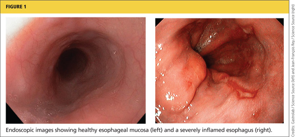
Continued on next page >>
SIGNS AND SYMPTOMS
Symptoms of GERD vary among adults and children in different age-groups. According to the Montreal definition, which was developed and modified by an international panel of pediatric gastroenterologists,10,12 GERD should be suspected in infants and toddlers who fail to thrive and exhibit the symptoms listed in Table 1.10,12 Clinicians should also consider GERD in older children and adolescents who present with heartburn, since it is the most common initial presenting symptom.10,13 Of note, a 2010 database study of UK children with GERD revealed a high incidence before age 1 year and the greatest incidence among 16- to 17-year-olds.5
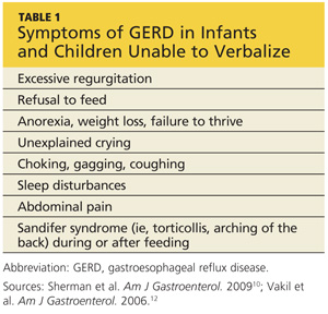
GERD should also be considered in pediatric patients who complain of vague symptoms of “stomachache” or nausea, abdominal pain or chest pain, since children may have trouble describing the sensation of heartburn.6,10 Children may also present with extra-esophageal complaints, such as dry cough, asthma-like symptoms, sore throat, hoarseness, sleep apnea, or dysphagia, all of which can be complications of GERD.10,11 Researchers have suggested that GERD contributes to and/or exacerbates pulmonary fibrosis, asthma, and chronic cough.6,10 Therefore, clinicians should consider GERD in children with these seemingly unrelated illnesses.
Continued on next page >>
RISK FACTORS
Certain pediatric groups are at increased risk for severe GERD, with or without complications. Neurologic impairment can cause dysphagia, and anatomic abnormalities such as hiatal hernia can impair lower esophageal sphincter function, allowing acid to rise into the esophagus.6,11 Table 26,10,13 lists illnesses and congenital conditions that are considered predisposing factors for severe, chronic GERD.
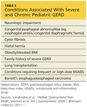
When seeing patients in these populations, clinicians should specifically focus on relevant GI symptoms during the history and physical exam. Clinicians should also consider long-term monitoring for complications or changes that might indicate new-onset GERD.
Overweight and Obesity
Although data are limited on the relationship between pediatric GERD and obesity,14 associated research findings seem to conflict with the established relationship in adult patients. While certain study groups found an association between obesity, elevated BMI, and increased waist circumference with an increase in symptoms of GERD (eg, regurgitation, heartburn),15,16 others found no significant correlation between overweight and reflux esophagitis.14,17 Notably, one analysis found a significant correlation between male gender and incidence of GERD.17
Although the evidence is not conclusive, clinicians are encouraged to counsel the older children among their patients on the benefits of weight and BMI reduction—encouraging them to achieve an overall healthier lifestyle and avoid diseases associated with excess weight.
DIFFERENTIAL DIAGNOSIS
In infants with unusual symptoms, certain GI conditions such as obstructive disorders (eg, pyloric stenosis), motility disorders, and peptic ulcer disease must be ruled out with further diagnostic testing.6,7 Red-flag symptoms that warrant further investigation include hematemesis, hematochezia, diarrhea, abdominal tenderness or distention, constipation, bilious vomiting, onset of vomiting after age 6 months, failure to thrive, macrocephaly or microcephaly, fever, lethargy, and hepatosplenomegaly.6,7 More common differentials are described below.
Eosinophilic Esophagitis
Eosinophilic esophagitis is an inflammatory condition of the esophagus, an apparent manifestation of food allergy (eg, milk protein), characterized by infiltrating mucosal eosinophils.6,18 First identified in children (though also occurring in adults), eosinophilic esophagitis is recognized as a more common cause of dysphagia than GERD.6
This recently discovered disease is often mistaken for GERD. While symptoms including heartburn and dysphagia occur in both conditions, eosinophilic esophagitis can only be diagnosed via multiple endoscopic mucosal biopsies. Corticosteroid therapy has been found to be a more effective treatment for this condition than acid suppression therapy.19
Current research is focused on a possible link between eosinophilic esophagitis and autoimmune disease, as many affected patients also have asthma, allergic rhinitis, and/or eczema.19 The increasing prevalence of childhood allergies should prompt clinicians to place eosinophilic esophagitis on the short list of differentials when evaluating a child for GERD-type symptoms. Referral for evaluation by an allergist may also be beneficial.
Asthma
GER may actually trigger asthma in some patients, even without symptoms of GERD.6 While there is support for a possible link between GERD and asthma in infants and children, a definitive relationship cannot be confirmed without more reliable studies.20 However, clinicians must not overlook the possibility of GERD in a child who presents with symptoms indicative of asthma, such as wheezing, shortness of breath, coughing, and chest tightness.
Other Extra-Esophageal Diseases
Concern exists over apparent associations between GERD and other extra-esophageal illnesses. In addition to asthma, the Montreal consensus group10,12 found a connection between GERD and other respiratory conditions, including chronic cough and chronic laryngitis. These conditions usually represent multifactorial disease processes, researchers state, and GER can be an exacerbating factor rather than an actual cause of these conditions.5,10
Further manifestations of extra-esophageal GERD include pneumonia, bronchiectasis, any apparent life-threatening event, laryngotracheitis, sinusitis, and dental erosion. Again, causality has not been clearly established, and the shortage of high-quality studies with adequate sample sizes makes it impossible to confirm clear relationships.21 It is therefore prudent for clinicians to evaluate any child with these non-GI conditions for reflux disease.
Continued on next page >>
EVALUATION AND DIAGNOSIS
In 2009, the North American Society for Pediatric Gastroenterology, Hepatology, and Nutrition and the European Society for Pediatric Gastroenterology, Hepatology, and Nutrition (NASPGHAN and ESPGHAN)6 released international guidelines for the management of pediatric GERD, including evaluation, diagnosis, and treatment. Making a diagnosis of GERD in an infant or a toddler can be challenging, since no reliable pathognomonic symptoms are known. Older children and adolescents are better able to articulate their presenting symptoms (which resemble those of adults); thus, a detailed history and physical exam are ordinarily adequate to diagnose GERD and introduce treatment in these patients.6
The diagnostic tool that is considered the gold standard for pediatric patients, including infants, is 24-hour esophageal pH monitoring. This test directly measures the quantity of acid present in the esophagus by way of an internal probe that is passed through the mouth or nose and worn for 24 hours. Esophageal pH monitoring quantifies the amount of acid to which the esophagus is exposed over this time period, compared with standard pediatric values.6,13
However, a newer combined multichannel intraluminal impedance and pH monitoring (MII/pH) tool offers advantages over esophageal pH monitoring, as it detects nonacidic or weakly acidic reflux events, in addition to more obvious episodes of acidic reflux.6,13,22
Clinical benefits of the MII/pH are:
• Better efficiency than pH monitoring alone in the evaluation of respiratory symptoms and GERD
• Ability to correlate timing of reflux episodes with symptoms, including chronic cough, apnea, and other respiratory symptoms
• Improved accuracy in monitoring postprandial reflux episodes, which are typically less acidic.13,23
Additionally, MII/pH provides a graphic readout from which the duration, height, and frequency of the reflux episodes can be analyzed. Currently, the chief disadvantage to its use is the absence of standardized pediatric values.13
Endoscopic biopsy should not be used to establish whether esophagitis is due to reflux. Endoscopically visible breaks in the distal esophageal mucosa are the most reliable indicators of reflux esophagitis. However, because other signs, such as mucosal erythema, pallor, and vascular patterns have normal variations, they cannot be considered evidence of reflux esophagitis.6 Endoscopic biopsy is mainly recommended for confirmation of suspected Barrett’s esophagus (which is rare in children) or for eosinophilic esophagitis, as it can confirm infiltrating mucosal eosinophils.6,19
Nuclear scintigraphy uses imaging to time the passage of a radioisotope-labeled meal through the upper GI tract. It can provide information about gastric emptying, which may be delayed in children with GERD. It may also be useful in diagnosing aspiration in patients with chronic intractable respiratory symptoms.6 Because standardized techniques and age-specific norms are lacking, however, nuclear scintigraphy is not recommended for patients with other potentially reflux-related symptoms. The sensitivity of this test is low, and negative results may not exclude the possibility of reflux and aspiration.6
Barium contrast radiography (upper GI series) is not recommended due to its poor sensitivity and specificity. This test is useful for confirming anatomic anomalies, however.6
For infants and toddlers with symptoms suggestive of GERD, the NASPGHAN/ESPGHAN authors6 find no evidence to support an empiric trial of pharmacologic treatment to confirm the diagnosis. For older, verbal children and adolescents who present with heartburn and/or chest pain, a short-term trial (as long as 4 weeks) of acid suppressants may be used to identify acid reflux as the cause of these symptoms.
Based on the guidelines by Vandenplas et al,6 a clinician’s initial approach to evaluation for GERD and its diagnosis should begin with a thorough history and physical exam and the least invasive diagnostic process possible. The child’s specific symptoms and suspected involvement of other organ systems will dictate the use of progressively invasive diagnostic strategies.
MANAGEMENT OF GERD
Conservative Treatment
Nonpharmacologic, age-appropriate approaches focus on diet and lifestyle changes. An effective treatment option for infants uses dry rice cereal to thicken the formula, resulting in decreased visible reflux and regurgitation.6,8 Recommended amounts of thickened formula are 4 oz/kg/d, divided into four to eight daily feedings, depending on the age of the patient; infants close to 1 year require only four feedings.8 For breastfed infants, expressed breast milk can be thickened with rice cereal and given at comparable volumes.8
Recommendations from the NASPGHAN/ESPGHAN clinical practice guidelines6 include a two- to four-week trial of an extensively hydrolyzed protein formula for formula-fed infants who vomit frequently. These formulas are considered hypoallergenic and contain shorter protein particles, allowing for easier digestion.8
Lifestyle changes recommended for adults with GERD can also be tailored for use in pediatric patients (see Table 38,11). They include avoidance of overfeeding by giving smaller portions at greater frequency (as described above), avoidance of foods known to cause GERD symptoms, avoidance of cigarette smoke, restriction of eating and drinking close to bedtime, elevation of the head of the bed or crib (use of a pillow is not recommended in children younger than 1 year), and a left-sided sleeping position for adolescents.6,11
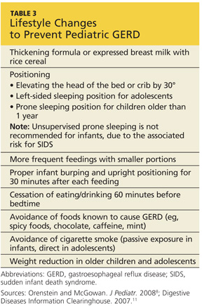
Holding infants upright for 30 minutes after feeding with ample burping may reduce reflux.11 In infants, the prone position provides the greatest benefit for reducing acid reflux, according to findings from one study based on pH monitoring.13 Nonetheless, the association between prone positioning and sudden infant death syndrome (SIDS) has led to recommendations of supine position when infants sleep unobserved.6 Perhaps supervised “tummy time” several times per day (eg, after feeding) can help reduce GERD symptoms in infants.
Finally, breastfeeding mothers are advised to avoid consuming cow’s milk, eggs, and soy products, as their presence in breast milk may promote reflux in an infant with unrecognized food allergy.6,8
Pharmacologic Treatment
Since esophagitis develops as a result of continuous acid exposure from the refluxate, the primary pharmacologic therapy for the treatment of GERD is aimed at acid reduction in the upper GI tract. Current pharmacologic options include histamine2-receptor antagonists (H2RAs) and proton pump inhibitors (PPIs).6 H2RAs have been shown to alleviate symptoms and promote mucosal healing. To their disadvantage, long-term use of H2RAs may lead to drug tolerance.
PPIs are superior to H2RAs in symptom relief and esophageal healing without causing tolerance; however, although certain agents are FDA approved for treatment of children age 1 year or older with GERD (including esomeprazole, lansoprazole, and omeprazole), use of PPIs in infants younger than 1 year is controversial.6,13 (See “New Solution for Pediatric GERD?”24,25)
According to results from existing studies of PPI use in infants with GERD ages 34 weeks to 1 year, PPIs are no more effective at symptom reduction than placebo.2,26 Further, data to demonstrate efficacy of long-term PPI use in infants and toddlers are scant.6,27 Safety results from various trials are inconsistent, ranging from no reported adverse effects to rare but severe adverse events, including necrotizing enterocolitis in infants, and lower respiratory tract infections, community-acquired pneumonia, gastric polyps, and acute gastroenteritis in children.2,26
The most common adverse effects of esomeprazole use in infants and children are nausea, vomiting, diarrhea, pyrexia, and headache.4,6 Because of these potential risks, none of the PPIs is approved for use in infants younger than 1 year. However, a short-term PPI trial for as long as 4 weeks (along with lifestyle changes) for symptom reduction in older children is recommended by the NASPGHAN/ESPGHAN clinical practice guidelines.2,6,13
Also based on the NASPGHAN/ESPGHAN guidelines,6 empiric PPI therapy is not recommended in pediatric patients presenting with wheezing or asthma. One research team has reported that asthma symptoms may be validly treated with a PPI in patients who do not respond to standard asthma treatment and who have a high reflux index.28 However, an absence of studies to support this finding makes a firm recommendation impossible.
According to the NASPGHAN/ESPGHAN guidelines,6 there is insufficient evidence to justify the use of prokinetic agents (eg, metoclopramide, erythromycin, bethanechol, domperidone) for pediatric GERD, as their potential risks outweigh their potential benefits. Neither are alginates nor sucralfate recommended for long-term therapy, because PPIs and H2RAs are considered more effective.6
Surgical Treatment
As with adults, surgery should be a last resort for treatment. Antireflux surgery is deemed appropriate only in children who cannot tolerate long-term medical therapy due to life-threatening complications, who cannot comply with the treatment schedule, or in whom medications have been found ineffective.6
The gold standard for the surgical treatment of GERD is laparoscopic Nissen fundoplication, a procedure in which the shape of the stomach fundus is modified to provide strength and functional support to the lower esophageal sphincter.29 Although children with heartburn, asthma, nocturnal asthma symptoms, or steroid-dependent asthma have been found to benefit clinically from long-term medical therapy or antireflux surgery, the comparable benefits of surgical versus medical treatment in these children is unknown.6
Continued on next page >>
PROGNOSIS AND FOLLOW-UP
The large majority of infants respond well to conservative nonpharmacologic treatment and outgrow their reflux symptoms by age 1 year, with maturation of muscle control and lower esophageal sphincter function.7 Further testing and intervention is generally not required in healthy infants, and parents should be reassured by this information.6,7 If regurgitation does not resolve by age 12 to 18 months, or if red-flag symptoms develop, referral to a pediatric gastroenterologist is recommended.6
For older children and adolescents with GERD, lifestyle changes should be implemented first, followed by short-term pharmacologic intervention, as recommended by appropriate guidelines.
Children with other illnesses or complications, such as neurologic impairment, premature birth, or a strong family history of severe GERD, have a poorer prognosis and may require more aggressive diagnostic evaluation and management.7 Complications such as esophageal stricture and Barrett’s esophagus, which poses an increased risk for adenocarcinoma, require referral to a specialist for further evaluation.10
CONCLUSION
GERD is no longer a condition found only in adults. Since primary care practitioners are increasingly likely to see GERD in their pediatric patients, it is important for these clinicians to become familiar with the contributing factors, definitive signs and symptoms, diagnosis, and treatment of GERD. Children at high risk for GERD should be followed closely and introduced to appropriate lifestyle modifications to avoid the troublesome symptoms of GERD and its complications, whenever possible. In the differential diagnosis, respiratory illnesses and other extra-esophageal diseases, as well as eosinophilic esophagitis in allergic patients, should be considered.
Practitioners should consult the 2009 NASPGHAN/ESPGHAN clinical practice guidelines for further details regarding the evaluation, diagnosis, preferred treatment, management of pediatric GERD, and indications for specialist referral. Research is ongoing in many areas, including the suitability of PPI use in pediatrics. More research is needed to clarify the theorized link between GERD and asthma.
1. Diaz DM, Winter HS, Colletti RB, et al. Knowledge, attitudes and practice styles of North American pediatricians regarding gastroesophageal reflux disease. J Pediatr Gastroenterol Nutr. 2007;45:56-64.
2. van der Pol RJ, Smits MJ, van Wijk MP, et al. Efficacy of proton-pump inhibitors in children with gastroesophageal reflux disease: a systematic review. Pediatrics. 2011;127:925-935.
3. Nelson SP, Kothari S, Wu EQ, et al. Pediatric gastroesophageal reflux disease and acid-related conditions: trends in incidence of diagnosis and acid suppression therapy. J Med Econ. 2009;12:348-355.
4. Tolia V, Gilger MA, Barker PN, Illueca M. Healing of erosive esophagitis and improvement of symptoms of gastroesophageal reflux disease after esomeprazole treatment in children 12 to 36 months old. J Pediatr Gastroenterol Nutr. 2010;51:593-598.
5. Vakil N. Disease definition, clinical manifestations, epidemiology and natural history of GERD. Best Pract Res Clin Gastroenterol. 2010;24:759-764.
6. Vandenplas Y, Rudolph CD, Di Lorenzo C, et al. Pediatric gastroesophageal reflux clinical practice guidelines: joint recommendations of the North American Society for Pediatric Gastroenterology, Hepatology, and Nutrition (NASPGHAN) and the European Society for Pediatric Gastroenterology, Hepatology, and Nutrition (ESPGHAN). J Pediatr Gastroenterol Nutr. 2009;49:498-547.
7. Michail S. Gastroesophageal reflux. Pediatr Rev. 2007;28:101-111.
8. Orenstein SR, McGowan JD. Efficacy of conservative therapy as taught in the primary care setting for symptoms suggesting infant gastroesophageal reflux. J Pediatr. 2008;152:310-314.
9. Vandenplas Y, Lifshitz JZ, Orenstein S, et al. Nutritional management of regurgitation in infants. J Am Coll Nutr. 1998;17:308-316.
10. Sherman PM, Hassall E, Fagundes-Neto U, et al. A global, evidence-based consensus on the definition of gastroesophageal reflux disease in the pediatric population. Am J Gastroenterol. 2009;104:1278-1295.
11. Digestive Diseases Information Clearinghouse. National Institute of Diabetes and Digestive and Kidney Diseases, NIH. Heartburn, gastroesophageal reflux (GER), and gastroesophageal reflux disease (GERD)(2007). http://digestive.niddk.nih.gov/ddiseases/pubs/gerd. Accessed April 4, 2013.
12. Vakil N, van Zanten SV, Kahrilas P, et al; Global Consensus Group. The Montreal definition and classification of gastroesophageal reflux disease: a global evidence-based consensus. Am J Gastroenterol. 2006;101:1900-1920.
13. Bharwani S. Gastroesophageal reflux disease in children: from infancy to adolescence. J Med Sci. 2011;4:25-39.
14. Patel NR, Ward MJ, Beneck D, et al. The association between childhood overweight and reflux esophagitis. J Obes. 2010;2010.
15. Quitadamo P, Buonavolontà R, Miele E, et al. Total and abdominal obesity are risk factors for gastroesophageal reflux symptoms in children. J Pediatr Gastroenterol Nutr. 2012;55:72-75.
16. Pashankar DS, Corbin Z, Shah SK, Caprio S. Increased prevalence of gastroesophageal reflux symptoms in obese children evaluated in an academic medical center. J Clin Gastroenterol. 2009;43:410-413.
17. Elitsur Y, Dementieva Y, Elitsur R, Rewalt M. Obesity is not a risk factor in children with reflux esophagitis: a retrospective analysis of 738 children. Metab Syndr Relat Disord. 2009;7:211-214.
18. Hill DJ, Heine RG, Cameron DJ, et al. Role of food protein intolerance in infants with persistent distress attributed to reflux esophagitis. J Pediatr. 2000;136:641-647.
19. Franciosi JP, Liacouras CA. Eosinophilic esophagitis. Immunol Allergy Clin N America. 2009;29:19-27.
20. Thakkar K, Boatright RO, Gilger MA, El-Serag HB. Gastroesophageal reflux and asthma in children: a systematic review. Pediatrics. 2010; 125:e925-e930.
21. Tolia V, Vandenplas Y. Systematic review: the extra-oesophageal symptoms of gastro-oesophageal reflux disease in children. Aliment Pharmacol Ther. 2009;29:258-272.
22. Cresi F, Locatelli E, Marinaccio C, et al. Prognostic values of multichannel intraluminal impedance and pH monitoring in newborns with symptoms of gastroesophageal reflux disease. J Pediatr. 2012 Nov 10. [Epub ahead of print]
23. Dalby K, Nielsen RG, Markoew S, et al. Reproducibility of 24-hour combined multiple intraluminal impedance (MII) and pH measurements in infants and children: evaluation of a diagnostic procedure for gastroesophageal reflux disease. Dig Dis Sci. 2007;52:2159-2165.
24. Tucker ME. FDA approves pediatric formulation of proton-pump inhibitor. www.medscape.com/viewarticle/781615. Accessed April 4, 2013.
25. Zannikos PN, Doose DR, Leitz GJ, et al. Pharmacokinetics and tolerability of rabeprazole in children 1 to 11 years old with gastroesophageal reflux disease. J Pediatr Gastroenterol Nutr. 2011;52:691-701.
26. Orenstein SR, Hassall E. Infants and proton pump inhibitors: tribulations, no trials. J Pediatr Gastroenterol Nutr. 2007;45:395-398.
27. Barron JJ, Tan H, Spalding J, et al. Proton pump inhibitor utilization patterns in infants. J Pediatr Gastroenterol Nutr. 2007;45:421-427.
28. Sopo SM, Radzik D, Calvani M. Does treatment with proton pump inhibitors for gastroesophageal reflux disease (GERD) improve asthma symptoms in children with asthma and GERD? A systematic review. J Investig Allergol Clin Immunol. 2009;19:1-5.
29. Anvari M, Allen C, Marshall J, et al. A randomized controlled trial of laparoscopic Nissen fundoplication versus proton pump inhibitors for the treatment of patients with chronic gastroesophageal reflux disease (GERD): 3-year outcomes. Surg Endosc. 2011;25:2547-2554
CE/CME No: CR-1305
PROGRAM OVERVIEW
Earn credit by reading this article and successfully completing the posttest. Successful completion is defined as a cumulative score of at least 70% correct.
EDUCATIONAL OBJECTIVES
• Differentiate between gastroesophageal reflux and gastroesophageal reflux disease (GER and GERD, respectively) in the pediatric patient, including symptomatology and risk factors.
• Explain typical and atypical presentations of GERD as factors in the differential diagnosis.
• Describe diagnostic testing options for GERD and their appropriate use in infants and children with suspected GERD.
• Discuss age-appropriate strategies to reduce the symptoms of GERD in children, including lifestyle changes and pharmacologic and surgical options.
FACULTY
Ellen D. Mandel is Clinical Associate Professor in the Pace University Physician Assistant Program in New York City, and Associate Professor in the Physician Assistant Program at Seton Hall University in South Orange, New Jersey. Claudia Ashforth and Kristine Daugherty are students in the Pace University Lenox Hill Hospital Physician Assistant Program.
The authors have no significant financial relationships to disclose.
ACCREDITATION STATEMENT
![]()
Article begins on next page >>
As with US adults, infants and children appear to be at increased risk for gastroesophageal reflux disease (GERD). Lacking a cardinal symptom in children and often linked with confounding extra-esophageal symptoms, pediatric GERD challenges the primary care clinician to make an early diagnosis, preventing progressive damage and possible complications. Management begins with conservative lifestyle changes; pharmacologic and surgical options are reserved for specific pediatric patients.
Traditionally, gastroesophageal reflux disease (GERD) has been viewed as an adult disease, but it is now recognized as a disorder that also occurs in children. A teenager with heartburn, a child with complaints of chest pain, and a coughing infant refusing to feed may all be experiencing it. Review of the literature reveals an increased incidence of GERD in both adults and children, making it one of the five most common gastrointestinal (GI) conditions in the United States.1
US pediatric hospitalization rates associated with GERD significantly increased from 1995 to 2000, accounting for 4% of these admissions.1 In a 2009 review of ICD-9 codes in a large claims database, GERD was diagnosed in 12.3% of North American infants and in 1% of other pediatric age-groups.2,3 In another recent study in which pediatric endoscopy data from 1999 to 2002 were analyzed, 9.5% of children age 1 year and 7.6% of children age 2 had erosive esophagitis.4
It is unclear whether the increased frequency in diagnosis of GERD should be attributed to improved diagnostic strategies or to an actual increase in disease prevalence.1 In any event, a timely diagnosis of GERD is essential to allow for appropriate intervention and early symptom management to reduce the risk for complications.
Overall, GERD is primarily a Western disease, affecting an estimated 10% to 15% of this geographic population. It is associated with obesity, recent dietary trends, and other causes.5 Prevalence of GERD in the general population has been shown to vary across ethnic groups. In the US, persons of Hispanic descent are more likely to be affected than whites, with symptoms of GERD least common among Asian-Americans.5
Since pediatric GERD is seen in primary care settings in the same rising numbers as associated hospital admissions, clinicians who provide primary care must be aware of its contributing factors, treatment modalities that are most effective in reducing symptoms, and strategies to prevent this disease. Treatment choices vary according to patient age-groups: infants (younger than 1 year), children ages 1 to 11 years, and adolescents, 12 to 18.
DISTINGUISHING GERD FROM GER
Gastroesophageal reflux (GER) is the term used to describe the passage of gastric contents, including stomach acid, fluids, and food, into the esophagus, with or without regurgitation or vomiting.6 GER is a normal, common physiologic process among infants, in whom frequent feedings, small stomach size, and predominance of the recumbent position allow reflux to occur during transient relaxation of the lower esophageal sphincter.6-9
Healthy infants and children may have asymptomatic episodes of GER; or episodes may be short, lasting less than three minutes, and occurring postprandially.6 Only when GER involves blood loss, esophagitis, strictures, nutritional deficits, and/or apnea should GERD be considered.9
The pathologic process known as GERD involves persistent, troublesome symptoms resulting from continued mucosal exposure to stomach acid, damaging the lining of the esophagus and possibly leading to erosive esophagitis.6,10,11 Endoscopic findings indicating GERD-associated esophageal damage include visible tears in the esophageal mucosa near the gastroesophageal junction6 (see Figure 1). In one single-center US study, almost 30% of patients with pediatric GERD who underwent endoscopy had erosive esophagitis.4
Continued on next page >>
SIGNS AND SYMPTOMS
Symptoms of GERD vary among adults and children in different age-groups. According to the Montreal definition, which was developed and modified by an international panel of pediatric gastroenterologists,10,12 GERD should be suspected in infants and toddlers who fail to thrive and exhibit the symptoms listed in Table 1.10,12 Clinicians should also consider GERD in older children and adolescents who present with heartburn, since it is the most common initial presenting symptom.10,13 Of note, a 2010 database study of UK children with GERD revealed a high incidence before age 1 year and the greatest incidence among 16- to 17-year-olds.5
GERD should also be considered in pediatric patients who complain of vague symptoms of “stomachache” or nausea, abdominal pain or chest pain, since children may have trouble describing the sensation of heartburn.6,10 Children may also present with extra-esophageal complaints, such as dry cough, asthma-like symptoms, sore throat, hoarseness, sleep apnea, or dysphagia, all of which can be complications of GERD.10,11 Researchers have suggested that GERD contributes to and/or exacerbates pulmonary fibrosis, asthma, and chronic cough.6,10 Therefore, clinicians should consider GERD in children with these seemingly unrelated illnesses.
Continued on next page >>
RISK FACTORS
Certain pediatric groups are at increased risk for severe GERD, with or without complications. Neurologic impairment can cause dysphagia, and anatomic abnormalities such as hiatal hernia can impair lower esophageal sphincter function, allowing acid to rise into the esophagus.6,11 Table 26,10,13 lists illnesses and congenital conditions that are considered predisposing factors for severe, chronic GERD.
When seeing patients in these populations, clinicians should specifically focus on relevant GI symptoms during the history and physical exam. Clinicians should also consider long-term monitoring for complications or changes that might indicate new-onset GERD.
Overweight and Obesity
Although data are limited on the relationship between pediatric GERD and obesity,14 associated research findings seem to conflict with the established relationship in adult patients. While certain study groups found an association between obesity, elevated BMI, and increased waist circumference with an increase in symptoms of GERD (eg, regurgitation, heartburn),15,16 others found no significant correlation between overweight and reflux esophagitis.14,17 Notably, one analysis found a significant correlation between male gender and incidence of GERD.17
Although the evidence is not conclusive, clinicians are encouraged to counsel the older children among their patients on the benefits of weight and BMI reduction—encouraging them to achieve an overall healthier lifestyle and avoid diseases associated with excess weight.
DIFFERENTIAL DIAGNOSIS
In infants with unusual symptoms, certain GI conditions such as obstructive disorders (eg, pyloric stenosis), motility disorders, and peptic ulcer disease must be ruled out with further diagnostic testing.6,7 Red-flag symptoms that warrant further investigation include hematemesis, hematochezia, diarrhea, abdominal tenderness or distention, constipation, bilious vomiting, onset of vomiting after age 6 months, failure to thrive, macrocephaly or microcephaly, fever, lethargy, and hepatosplenomegaly.6,7 More common differentials are described below.
Eosinophilic Esophagitis
Eosinophilic esophagitis is an inflammatory condition of the esophagus, an apparent manifestation of food allergy (eg, milk protein), characterized by infiltrating mucosal eosinophils.6,18 First identified in children (though also occurring in adults), eosinophilic esophagitis is recognized as a more common cause of dysphagia than GERD.6
This recently discovered disease is often mistaken for GERD. While symptoms including heartburn and dysphagia occur in both conditions, eosinophilic esophagitis can only be diagnosed via multiple endoscopic mucosal biopsies. Corticosteroid therapy has been found to be a more effective treatment for this condition than acid suppression therapy.19
Current research is focused on a possible link between eosinophilic esophagitis and autoimmune disease, as many affected patients also have asthma, allergic rhinitis, and/or eczema.19 The increasing prevalence of childhood allergies should prompt clinicians to place eosinophilic esophagitis on the short list of differentials when evaluating a child for GERD-type symptoms. Referral for evaluation by an allergist may also be beneficial.
Asthma
GER may actually trigger asthma in some patients, even without symptoms of GERD.6 While there is support for a possible link between GERD and asthma in infants and children, a definitive relationship cannot be confirmed without more reliable studies.20 However, clinicians must not overlook the possibility of GERD in a child who presents with symptoms indicative of asthma, such as wheezing, shortness of breath, coughing, and chest tightness.
Other Extra-Esophageal Diseases
Concern exists over apparent associations between GERD and other extra-esophageal illnesses. In addition to asthma, the Montreal consensus group10,12 found a connection between GERD and other respiratory conditions, including chronic cough and chronic laryngitis. These conditions usually represent multifactorial disease processes, researchers state, and GER can be an exacerbating factor rather than an actual cause of these conditions.5,10
Further manifestations of extra-esophageal GERD include pneumonia, bronchiectasis, any apparent life-threatening event, laryngotracheitis, sinusitis, and dental erosion. Again, causality has not been clearly established, and the shortage of high-quality studies with adequate sample sizes makes it impossible to confirm clear relationships.21 It is therefore prudent for clinicians to evaluate any child with these non-GI conditions for reflux disease.
Continued on next page >>
EVALUATION AND DIAGNOSIS
In 2009, the North American Society for Pediatric Gastroenterology, Hepatology, and Nutrition and the European Society for Pediatric Gastroenterology, Hepatology, and Nutrition (NASPGHAN and ESPGHAN)6 released international guidelines for the management of pediatric GERD, including evaluation, diagnosis, and treatment. Making a diagnosis of GERD in an infant or a toddler can be challenging, since no reliable pathognomonic symptoms are known. Older children and adolescents are better able to articulate their presenting symptoms (which resemble those of adults); thus, a detailed history and physical exam are ordinarily adequate to diagnose GERD and introduce treatment in these patients.6
The diagnostic tool that is considered the gold standard for pediatric patients, including infants, is 24-hour esophageal pH monitoring. This test directly measures the quantity of acid present in the esophagus by way of an internal probe that is passed through the mouth or nose and worn for 24 hours. Esophageal pH monitoring quantifies the amount of acid to which the esophagus is exposed over this time period, compared with standard pediatric values.6,13
However, a newer combined multichannel intraluminal impedance and pH monitoring (MII/pH) tool offers advantages over esophageal pH monitoring, as it detects nonacidic or weakly acidic reflux events, in addition to more obvious episodes of acidic reflux.6,13,22
Clinical benefits of the MII/pH are:
• Better efficiency than pH monitoring alone in the evaluation of respiratory symptoms and GERD
• Ability to correlate timing of reflux episodes with symptoms, including chronic cough, apnea, and other respiratory symptoms
• Improved accuracy in monitoring postprandial reflux episodes, which are typically less acidic.13,23
Additionally, MII/pH provides a graphic readout from which the duration, height, and frequency of the reflux episodes can be analyzed. Currently, the chief disadvantage to its use is the absence of standardized pediatric values.13
Endoscopic biopsy should not be used to establish whether esophagitis is due to reflux. Endoscopically visible breaks in the distal esophageal mucosa are the most reliable indicators of reflux esophagitis. However, because other signs, such as mucosal erythema, pallor, and vascular patterns have normal variations, they cannot be considered evidence of reflux esophagitis.6 Endoscopic biopsy is mainly recommended for confirmation of suspected Barrett’s esophagus (which is rare in children) or for eosinophilic esophagitis, as it can confirm infiltrating mucosal eosinophils.6,19
Nuclear scintigraphy uses imaging to time the passage of a radioisotope-labeled meal through the upper GI tract. It can provide information about gastric emptying, which may be delayed in children with GERD. It may also be useful in diagnosing aspiration in patients with chronic intractable respiratory symptoms.6 Because standardized techniques and age-specific norms are lacking, however, nuclear scintigraphy is not recommended for patients with other potentially reflux-related symptoms. The sensitivity of this test is low, and negative results may not exclude the possibility of reflux and aspiration.6
Barium contrast radiography (upper GI series) is not recommended due to its poor sensitivity and specificity. This test is useful for confirming anatomic anomalies, however.6
For infants and toddlers with symptoms suggestive of GERD, the NASPGHAN/ESPGHAN authors6 find no evidence to support an empiric trial of pharmacologic treatment to confirm the diagnosis. For older, verbal children and adolescents who present with heartburn and/or chest pain, a short-term trial (as long as 4 weeks) of acid suppressants may be used to identify acid reflux as the cause of these symptoms.
Based on the guidelines by Vandenplas et al,6 a clinician’s initial approach to evaluation for GERD and its diagnosis should begin with a thorough history and physical exam and the least invasive diagnostic process possible. The child’s specific symptoms and suspected involvement of other organ systems will dictate the use of progressively invasive diagnostic strategies.
MANAGEMENT OF GERD
Conservative Treatment
Nonpharmacologic, age-appropriate approaches focus on diet and lifestyle changes. An effective treatment option for infants uses dry rice cereal to thicken the formula, resulting in decreased visible reflux and regurgitation.6,8 Recommended amounts of thickened formula are 4 oz/kg/d, divided into four to eight daily feedings, depending on the age of the patient; infants close to 1 year require only four feedings.8 For breastfed infants, expressed breast milk can be thickened with rice cereal and given at comparable volumes.8
Recommendations from the NASPGHAN/ESPGHAN clinical practice guidelines6 include a two- to four-week trial of an extensively hydrolyzed protein formula for formula-fed infants who vomit frequently. These formulas are considered hypoallergenic and contain shorter protein particles, allowing for easier digestion.8
Lifestyle changes recommended for adults with GERD can also be tailored for use in pediatric patients (see Table 38,11). They include avoidance of overfeeding by giving smaller portions at greater frequency (as described above), avoidance of foods known to cause GERD symptoms, avoidance of cigarette smoke, restriction of eating and drinking close to bedtime, elevation of the head of the bed or crib (use of a pillow is not recommended in children younger than 1 year), and a left-sided sleeping position for adolescents.6,11
Holding infants upright for 30 minutes after feeding with ample burping may reduce reflux.11 In infants, the prone position provides the greatest benefit for reducing acid reflux, according to findings from one study based on pH monitoring.13 Nonetheless, the association between prone positioning and sudden infant death syndrome (SIDS) has led to recommendations of supine position when infants sleep unobserved.6 Perhaps supervised “tummy time” several times per day (eg, after feeding) can help reduce GERD symptoms in infants.
Finally, breastfeeding mothers are advised to avoid consuming cow’s milk, eggs, and soy products, as their presence in breast milk may promote reflux in an infant with unrecognized food allergy.6,8
Pharmacologic Treatment
Since esophagitis develops as a result of continuous acid exposure from the refluxate, the primary pharmacologic therapy for the treatment of GERD is aimed at acid reduction in the upper GI tract. Current pharmacologic options include histamine2-receptor antagonists (H2RAs) and proton pump inhibitors (PPIs).6 H2RAs have been shown to alleviate symptoms and promote mucosal healing. To their disadvantage, long-term use of H2RAs may lead to drug tolerance.
PPIs are superior to H2RAs in symptom relief and esophageal healing without causing tolerance; however, although certain agents are FDA approved for treatment of children age 1 year or older with GERD (including esomeprazole, lansoprazole, and omeprazole), use of PPIs in infants younger than 1 year is controversial.6,13 (See “New Solution for Pediatric GERD?”24,25)
According to results from existing studies of PPI use in infants with GERD ages 34 weeks to 1 year, PPIs are no more effective at symptom reduction than placebo.2,26 Further, data to demonstrate efficacy of long-term PPI use in infants and toddlers are scant.6,27 Safety results from various trials are inconsistent, ranging from no reported adverse effects to rare but severe adverse events, including necrotizing enterocolitis in infants, and lower respiratory tract infections, community-acquired pneumonia, gastric polyps, and acute gastroenteritis in children.2,26
The most common adverse effects of esomeprazole use in infants and children are nausea, vomiting, diarrhea, pyrexia, and headache.4,6 Because of these potential risks, none of the PPIs is approved for use in infants younger than 1 year. However, a short-term PPI trial for as long as 4 weeks (along with lifestyle changes) for symptom reduction in older children is recommended by the NASPGHAN/ESPGHAN clinical practice guidelines.2,6,13
Also based on the NASPGHAN/ESPGHAN guidelines,6 empiric PPI therapy is not recommended in pediatric patients presenting with wheezing or asthma. One research team has reported that asthma symptoms may be validly treated with a PPI in patients who do not respond to standard asthma treatment and who have a high reflux index.28 However, an absence of studies to support this finding makes a firm recommendation impossible.
According to the NASPGHAN/ESPGHAN guidelines,6 there is insufficient evidence to justify the use of prokinetic agents (eg, metoclopramide, erythromycin, bethanechol, domperidone) for pediatric GERD, as their potential risks outweigh their potential benefits. Neither are alginates nor sucralfate recommended for long-term therapy, because PPIs and H2RAs are considered more effective.6
Surgical Treatment
As with adults, surgery should be a last resort for treatment. Antireflux surgery is deemed appropriate only in children who cannot tolerate long-term medical therapy due to life-threatening complications, who cannot comply with the treatment schedule, or in whom medications have been found ineffective.6
The gold standard for the surgical treatment of GERD is laparoscopic Nissen fundoplication, a procedure in which the shape of the stomach fundus is modified to provide strength and functional support to the lower esophageal sphincter.29 Although children with heartburn, asthma, nocturnal asthma symptoms, or steroid-dependent asthma have been found to benefit clinically from long-term medical therapy or antireflux surgery, the comparable benefits of surgical versus medical treatment in these children is unknown.6
Continued on next page >>
PROGNOSIS AND FOLLOW-UP
The large majority of infants respond well to conservative nonpharmacologic treatment and outgrow their reflux symptoms by age 1 year, with maturation of muscle control and lower esophageal sphincter function.7 Further testing and intervention is generally not required in healthy infants, and parents should be reassured by this information.6,7 If regurgitation does not resolve by age 12 to 18 months, or if red-flag symptoms develop, referral to a pediatric gastroenterologist is recommended.6
For older children and adolescents with GERD, lifestyle changes should be implemented first, followed by short-term pharmacologic intervention, as recommended by appropriate guidelines.
Children with other illnesses or complications, such as neurologic impairment, premature birth, or a strong family history of severe GERD, have a poorer prognosis and may require more aggressive diagnostic evaluation and management.7 Complications such as esophageal stricture and Barrett’s esophagus, which poses an increased risk for adenocarcinoma, require referral to a specialist for further evaluation.10
CONCLUSION
GERD is no longer a condition found only in adults. Since primary care practitioners are increasingly likely to see GERD in their pediatric patients, it is important for these clinicians to become familiar with the contributing factors, definitive signs and symptoms, diagnosis, and treatment of GERD. Children at high risk for GERD should be followed closely and introduced to appropriate lifestyle modifications to avoid the troublesome symptoms of GERD and its complications, whenever possible. In the differential diagnosis, respiratory illnesses and other extra-esophageal diseases, as well as eosinophilic esophagitis in allergic patients, should be considered.
Practitioners should consult the 2009 NASPGHAN/ESPGHAN clinical practice guidelines for further details regarding the evaluation, diagnosis, preferred treatment, management of pediatric GERD, and indications for specialist referral. Research is ongoing in many areas, including the suitability of PPI use in pediatrics. More research is needed to clarify the theorized link between GERD and asthma.
1. Diaz DM, Winter HS, Colletti RB, et al. Knowledge, attitudes and practice styles of North American pediatricians regarding gastroesophageal reflux disease. J Pediatr Gastroenterol Nutr. 2007;45:56-64.
2. van der Pol RJ, Smits MJ, van Wijk MP, et al. Efficacy of proton-pump inhibitors in children with gastroesophageal reflux disease: a systematic review. Pediatrics. 2011;127:925-935.
3. Nelson SP, Kothari S, Wu EQ, et al. Pediatric gastroesophageal reflux disease and acid-related conditions: trends in incidence of diagnosis and acid suppression therapy. J Med Econ. 2009;12:348-355.
4. Tolia V, Gilger MA, Barker PN, Illueca M. Healing of erosive esophagitis and improvement of symptoms of gastroesophageal reflux disease after esomeprazole treatment in children 12 to 36 months old. J Pediatr Gastroenterol Nutr. 2010;51:593-598.
5. Vakil N. Disease definition, clinical manifestations, epidemiology and natural history of GERD. Best Pract Res Clin Gastroenterol. 2010;24:759-764.
6. Vandenplas Y, Rudolph CD, Di Lorenzo C, et al. Pediatric gastroesophageal reflux clinical practice guidelines: joint recommendations of the North American Society for Pediatric Gastroenterology, Hepatology, and Nutrition (NASPGHAN) and the European Society for Pediatric Gastroenterology, Hepatology, and Nutrition (ESPGHAN). J Pediatr Gastroenterol Nutr. 2009;49:498-547.
7. Michail S. Gastroesophageal reflux. Pediatr Rev. 2007;28:101-111.
8. Orenstein SR, McGowan JD. Efficacy of conservative therapy as taught in the primary care setting for symptoms suggesting infant gastroesophageal reflux. J Pediatr. 2008;152:310-314.
9. Vandenplas Y, Lifshitz JZ, Orenstein S, et al. Nutritional management of regurgitation in infants. J Am Coll Nutr. 1998;17:308-316.
10. Sherman PM, Hassall E, Fagundes-Neto U, et al. A global, evidence-based consensus on the definition of gastroesophageal reflux disease in the pediatric population. Am J Gastroenterol. 2009;104:1278-1295.
11. Digestive Diseases Information Clearinghouse. National Institute of Diabetes and Digestive and Kidney Diseases, NIH. Heartburn, gastroesophageal reflux (GER), and gastroesophageal reflux disease (GERD)(2007). http://digestive.niddk.nih.gov/ddiseases/pubs/gerd. Accessed April 4, 2013.
12. Vakil N, van Zanten SV, Kahrilas P, et al; Global Consensus Group. The Montreal definition and classification of gastroesophageal reflux disease: a global evidence-based consensus. Am J Gastroenterol. 2006;101:1900-1920.
13. Bharwani S. Gastroesophageal reflux disease in children: from infancy to adolescence. J Med Sci. 2011;4:25-39.
14. Patel NR, Ward MJ, Beneck D, et al. The association between childhood overweight and reflux esophagitis. J Obes. 2010;2010.
15. Quitadamo P, Buonavolontà R, Miele E, et al. Total and abdominal obesity are risk factors for gastroesophageal reflux symptoms in children. J Pediatr Gastroenterol Nutr. 2012;55:72-75.
16. Pashankar DS, Corbin Z, Shah SK, Caprio S. Increased prevalence of gastroesophageal reflux symptoms in obese children evaluated in an academic medical center. J Clin Gastroenterol. 2009;43:410-413.
17. Elitsur Y, Dementieva Y, Elitsur R, Rewalt M. Obesity is not a risk factor in children with reflux esophagitis: a retrospective analysis of 738 children. Metab Syndr Relat Disord. 2009;7:211-214.
18. Hill DJ, Heine RG, Cameron DJ, et al. Role of food protein intolerance in infants with persistent distress attributed to reflux esophagitis. J Pediatr. 2000;136:641-647.
19. Franciosi JP, Liacouras CA. Eosinophilic esophagitis. Immunol Allergy Clin N America. 2009;29:19-27.
20. Thakkar K, Boatright RO, Gilger MA, El-Serag HB. Gastroesophageal reflux and asthma in children: a systematic review. Pediatrics. 2010; 125:e925-e930.
21. Tolia V, Vandenplas Y. Systematic review: the extra-oesophageal symptoms of gastro-oesophageal reflux disease in children. Aliment Pharmacol Ther. 2009;29:258-272.
22. Cresi F, Locatelli E, Marinaccio C, et al. Prognostic values of multichannel intraluminal impedance and pH monitoring in newborns with symptoms of gastroesophageal reflux disease. J Pediatr. 2012 Nov 10. [Epub ahead of print]
23. Dalby K, Nielsen RG, Markoew S, et al. Reproducibility of 24-hour combined multiple intraluminal impedance (MII) and pH measurements in infants and children: evaluation of a diagnostic procedure for gastroesophageal reflux disease. Dig Dis Sci. 2007;52:2159-2165.
24. Tucker ME. FDA approves pediatric formulation of proton-pump inhibitor. www.medscape.com/viewarticle/781615. Accessed April 4, 2013.
25. Zannikos PN, Doose DR, Leitz GJ, et al. Pharmacokinetics and tolerability of rabeprazole in children 1 to 11 years old with gastroesophageal reflux disease. J Pediatr Gastroenterol Nutr. 2011;52:691-701.
26. Orenstein SR, Hassall E. Infants and proton pump inhibitors: tribulations, no trials. J Pediatr Gastroenterol Nutr. 2007;45:395-398.
27. Barron JJ, Tan H, Spalding J, et al. Proton pump inhibitor utilization patterns in infants. J Pediatr Gastroenterol Nutr. 2007;45:421-427.
28. Sopo SM, Radzik D, Calvani M. Does treatment with proton pump inhibitors for gastroesophageal reflux disease (GERD) improve asthma symptoms in children with asthma and GERD? A systematic review. J Investig Allergol Clin Immunol. 2009;19:1-5.
29. Anvari M, Allen C, Marshall J, et al. A randomized controlled trial of laparoscopic Nissen fundoplication versus proton pump inhibitors for the treatment of patients with chronic gastroesophageal reflux disease (GERD): 3-year outcomes. Surg Endosc. 2011;25:2547-2554