Take-Home Points
- Technique provides optimal fixation while simultaneously protecting open growth plates.
- Self tensioning feature insures both optimal ACL tension and fracture reduction.
- No need for future hardware removal.
- 10Cross suture configuration optimizes strength of fixation for highly consistent results.
- Use fluoroscopy to avoid violation of tibial physis.
Generally occurring in the 8- to 14-year-old population, tibial eminence avulsion (TEA) fractures are a common variant of anterior cruciate ligament (ACL) ruptures and represent 2% to 5% of all knee injuries in skeletally immature individuals.1,2 Compared with adults, children likely experience this anomaly more often because of the weakness of their incompletely ossified tibial plateau relative to the strength of their native ACL.3
The open repair techniques that have been described have multiple disadvantages, including open incisions, difficult visualization of the fracture owing to the location of the fat pad, and increased risk for arthrofibrosis. Arthroscopic fixation is considered the treatment of choice for TEA fractures because it allows for direct visualization of injury, accurate reduction of fracture fragments, removal of loose fragments, and easy treatment of associated soft-tissue injuries.4-6Several fixation techniques for ACL-TEA fractures were recently described: arthroscopic reduction and internal fixation (ARIF) with Kirschner wires,7 cannulated screws,4 the Meniscus Arrow device (Bionx Implants),8 pull-out sutures,9,10 bioabsorbable nails,11 Herbert screws,12 TightRope fixation (Arthrex),13 and various other rotator cuff and meniscal repair systems.14,15 These approaches tend to have good outcomes for TEA fractures, but there are risks associated with ACL tensioning and potential tibial growth plate violation or hardware problems. Likewise, there are no studies with large numbers of patients treated with these new techniques, so the optimal method of reduction and fixation is still unknown.
In this article, we describe a new ARIF technique that involves 2 absorbable anchors with adjustable suture-tensioning technology. This technique optimizes reduction and helps surgeons avoid proximal tibial physeal damage, procedure-related morbidity, and additional surgery.
Case Report
History
The patient, an 8-year-old boy, sustained a noncontact twisting injury of the left knee during a cutting maneuver in a flag football game. He experienced immediate pain and subsequent swelling. Clinical examination revealed a moderate effusion with motion limitations secondary to swelling and irritability. The patient’s Lachman test result was 2+. Pivot shift testing was not possible because of guarding. The knee was stable to varus and valgus stress at 0° and 30° of flexion. Limited knee flexion prohibited placement of the patient in the position needed for anterior and posterior drawer testing. His patella was stable on lateral stress testing at 20° of flexion with no apprehension. Neurovascular status was intact throughout the lower extremity.
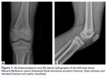
Anteroposterior and lateral radiographs showed a minimally displaced Meyers-McKeever type II TEA fracture (Figures 1A, 1B).
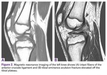
Distal femoral and proximal tibial growth plates were wide open. Magnetic resonance imaging confirmed the displaced type II TEA fracture and showed good signal quality in the attached ACL (Figures 2A, 2B). The remaining ligamentous structures appeared without injury or signal change. No tear signal was seen in the imaging sequences of the medial and lateral meniscus.After discussing potential treatment options with the parents, Dr. Smith proceeded with arthroscopic surgery for definitive reduction and internal fixation of the patient’s left knee displaced ACL-TEA fracture. The new adjustable suture-tensioning fixation technique was used. The patient’s guardian provided written informed consent for print and electronic publication of this case report.
Examination Under Anesthesia
Examination with the patient under general anesthesia revealed 3+ Lachman, 2+ pivot shift with foot in internal and external rotation, and 1+ anterior drawer with foot in neutral and internal rotation. The knee was stable to varus and valgus stress testing.
Surgical Technique
Proper patient positioning and padding of bony prominences were ensured, and the limb was sterilely prepared and draped.
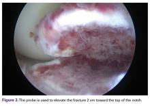
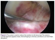
A standard lateral parapatellar portal was established for arthroscope placement; a medial parapatellar working portal was established as well. Thorough joint inspection revealed normal articular surfaces of patella, femur, and tibial plateau. Similarly, both menisci were intact without evidence of injury. With use of the probe, the ACL-TEA fracture could be elevated up to 2 cm toward the top of the notch (Figure 3). Further inspection of the ACL fibers revealed minimal hemorrhaging and no frank tearing (Figure 4).Given the young age of the patient, it was imperative to avoid the open proximal tibial growth plate. The surgical plan for stabilization involved use of two 3.0-mm BioComposite Knotless SutureTak anchors (Arthrex). This anchor configuration is based on a No. 2 FiberWire suture shuttled through itself to create a locking splice mechanism that allows for adjustable tensioning. The anchors were placed on each side of the tibial bony avulsion site with two No. 2 FiberWire sutures and were then crossed about the avulsion fracture fragment in an “x-type” configuration to secure the ACL back down to the bony bed.
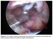
First, a curette was used to débride fibrous tissue on the underside of the fracture fragment and on the fracture bed. Minimal amounts of cancellous bone were débrided from the tibial fracture bed to optimize fracture reduction by slightly recessing the fracture fragment to ensure optimal ACL tensioning (Figure 5).
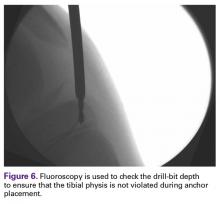
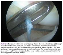
Next, an 18-gauge needle was used to establish an accessory superior medial percutaneous portal to ensure a satisfactory drilling trajectory just medial to the fracture site. Under fluoroscopic guidance, a drill guide was placed, and a 2.4-mm bit was used to drill to a depth of 16 mm to accommodate the 12.7-mm anchor. Avoidance of the proximal tibial physis was confirmed with fluoroscopy (Figure 6). One of the SutureTak anchors was secured in this drill hole along the anteromedial avulsion fracture site. From the anteromedial portal, a curved needle tip suture passer was placed medially through the ACL fibers and bone, with the wire retrieved out of the superior medial accessory portal. Then, the drill guide was introduced through the lateral portal and positioned just lateral to the tibial avulsion site, a hole was drilled 16 mm deep, and fluoroscopy was used to confirm the physis was not violated. The second SutureTak anchor was placed in this anterolateral location. From the anterolateral portal, the curved needle tip suture passer was placed laterally through the ACL fibers and avulsion fragment, and the wire was passed and retrieved out the anteromedial portal and shuttled back to the anterolateral portal.Next, from the accessory superior medial portal, the end of the wire that had been passed through the medial aspect of the bony avulsion was retrieved through the lateral portal. This wire was used to shuttle the repair suture from the laterally positioned SutureTak anchor over and through the medial aspect of the bony fragment out of the accessory superior medial (Figure 7).
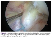
This suture was passed through the shuttling loop of the medially positioned SutureTak anchor to create the splice in the anchor for the adjustable fixation. This process was repeated through the lateral aspect of the bony fragment—the medial SutureTak repair suture was passed over the bone here. Thus, the lateral suture was over and through the bony fragment secured to the medial SutureTak anchor, and the medial suture was crossed over and through bone to the lateral SutureTak anchor. With the knee held in full extension, the bony avulsion fracture was easily reduced by alternating tension on the SutureTak limbs, which enabled controlled reduction of the TEA fracture (Figures 8A, 8B). An arthroscopic knot pusher was used for final tightening of the SutureTak fixation. An arthroscopic probe was used to confirm anatomical reduction of the fracture and restoration of ACL fiber tension (Figure 9). The knee was ranged from 0° to 120° of flexion with visual affirmation of the construct and maintenance of the reduction. Fluoroscopy confirmed anatomical reduction of the TEA fracture. The patient was immobilized in a long leg brace locked in 30° of flexion.
