User login
‘Sobering’ high 10-year mortality post-MI after age 65
WASHINGTON – Patients who experience an acute MI at age 65 or older have unsettlingly high 5- and 10-year mortality in community practice settings despite excellent rates of evidence-based medications being prescribed at discharge, Ajar Kochar, MD, reported at the annual meeting of the American College of Cardiology.
This observation is based upon more than 22,000 patients aged 65 years or older treated for an acute MI during 2004-2006 at 344 U.S. hospitals participating in the CRUSADE registry. Their median age at the time of MI was 77 years. But 10-year all-cause mortality remained high even among relatively younger patients aged 65-74 whom one would expect to have a favorable long-term prognosis because they had additional survival-enhancing factors working in their favor, including having undergone coronary revascularization during their index hospitalization and surviving their first year post-MI, observed Dr. Kochar of the Duke Clinical Research Institute in Durham, N.C.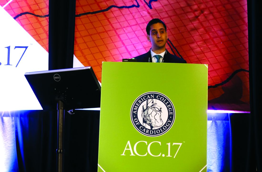
This unmet need will increasingly clamor for attention as the aging of the American population accelerates like a runaway freight train. By 2030, an estimated 20% of Americans will be aged 65 or older. That’s more than 71 million people. And more than half of all MIs occur in individuals above age 65, he noted.
Dr. Kochar presented a CRUSADE analysis which included 19,755 older Americans with a non–ST elevation MI (NSTEMI) and 2,540 with a STEMI. The overall group’s 1-year mortality was 24%, with a 5-year cumulative mortality of 51% and a whopping 10-year mortality of 72%.
According to the Centers for Disease Control and Prevention, the expected additional lifespan of someone who was 65 years old in 2015 is 19 years. In contrast, the median survival of patients in the CRUSADE registry who were 65-69 at the time of their MI was less than half of that, at 8.3 years.
Among the key findings from the CRUSADE analysis:
• Unadjusted 10-year all-cause mortality was significantly greater in patients with NSTEMI than STEMI, by a margin of 73% versus 60%. Notably, however, NSTEMI patients were far less likely to undergo coronary revascularization: 32% of them had percutaneous coronary intervention during their index hospitalization, and 8.7% underwent coronary artery bypass grafting, in contrast to rates of 65.5% for PCI and 8.0% for CABG in the STEMI patients. After adjustment for these and other differences in care, NSTEMI patients actually had a 7% lower risk of long-term mortality than the STEMI group.
• Even after limiting the analysis to the youngest elderly – patients aged 65-74 when their MI occurred – 10-year mortality remained high, at 53%.
• After excluding the 24% of patients who died within 1 year after MI, 10-year mortality was still quite high, at 63%. Dr. Kochar and his coinvestigators chose to reanalyze the data in this way because the 1-year mark is an important time point clinically, since it’s when decisions regarding extended dual-antiplatelet therapy are made.
Patients who underwent coronary revascularization during their index hospitalization had a much-improved long-term prognosis, compared with those with medical management only. The 10-year cumulative mortality rate was 57% in patients who had PCI, identical at 57% in those who received CABG, and 84% in medically managed patients.
Ninety-five percent of patients were discharged on aspirin, 94% on a beta blocker, 81% on a statin, and 73% on clopidogrel. Discharge prescriptions for statins and clopidogrel were more common for the STEMI group. Unfortunately, the CRUSADE registry doesn’t include data on long-term medication adherence or prescription refill rates.
Dr. Kochar named several potential strategies aimed at reducing the high long-term mortality rates in older patients with MI as documented in this study. These include structured efforts to improve adherence to evidence-based medications for secondary prevention, as well as making percutaneous revascularization more widely available for older patients with NSTEMI. He noted that while in 2004-2006, 32% of CRUSADE participants with NSTEMI underwent PCI during their index hospitalization, by 2011-2012 that rate had inched upward only to 36%.
Several physicians commented that the high long-term all-cause mortality rates in older CRUSADE participants may paint a grim picture, in part because the aged face growing risks of cancer and other noncardiovascular competing causes of death. But Dr. Kochar replied that while the lack of information on specific causes of death is a study limitation, he and his coinvestigators are convinced based upon data from other studies that most of the deaths in CRUSADE were cardiovascular in nature.
He reported having no financial conflicts regarding his study.
AT ACC 17
Key clinical point:
Major finding: The 10-year cumulative mortality rate in patients who had an MI at age 65-74 is 53%.
Data source: This was an analysis of 10-year cumulative mortality in more than 22,000 patients aged 65 or older treated for an acute MI during 2004-2006 at 344 U.S. community hospitals participating in the prospective CRUSADE registry.
Disclosures: The study presenter reported having no financial conflicts.
Intensive ventilation precedes lesser pulmonary complications
Addition of 10 cm H2O to positive end-expiratory volume (PEEP) during mechanical ventilation was followed by significantly lessened pulmonary complications in hospitalized patients who developed hypoxemia after cardiac surgery, participating in a single-center, randomized trial.
This “intensive” alveolar recruitment strategy yielded a median pulmonary complications score of 1.7 (interquartile range, 1.0-2.0), compared with 2.0 (IQR, 1.5-3.0) among patients who underwent ventilation with a PEEP of 20 cm H2O, Alcino Costa Leme, RRT, PhD, said at the International Symposium on Intensive Care and Emergency Medicine. The report was published simultaneously online March 21 in JAMA.
Intensive alveolar recruitment nearly doubled the odds of a lower pulmonary complications score (common odds ratio, 1.9; 95% confidence interval, 1.2-2.8; P = .003), Dr. Leme and his associates reported.
The study comprised 320 adults who developed hypoxemia immediately after undergoing elective cardiac surgery at the Heart Institute (Incor) of the University of São Paulo. The median age of the patients was 62 years, and none had a history of lung disease. Pulmonary complications were scored between 0 (no signs or symptoms) and 5 (death), the investigators noted (JAMA. 2017 Mar 21. doi: 10.1001/jama.2017.2297).
The intensive alveolar recruitment strategy consisted of three 60-second cycles of lung inflation with a positive end-expiratory pressure (PEEP) of 30 cm H2O, pressure-controlled ventilation, driving pressure of 15 cm H2O, respiratory rate of 15/min, inspiratory time of 1.5 seconds, and FIO2 of 0.40. Between and after inflations, patients received assist-controlled or pressure-controlled ventilation, with driving pressures set to achieve a tidal volume of 6 mL/kg of predicted body weight, an inspiratory time of 1 second, PEEP of 13 cm H2O, and minimum respiratory rate to maintain PaCO2 between 35 and 45 mm Hg.
The “moderate strategy” consisted of three 30-second inflations under continuous positive airway pressure mode at 20 cm H2O and FIO2 of 0.60. Between and after inflations, patients received assist or control volume-controlled ventilation (decelerating-flow waveform), tidal volume of 6 mL/kg of predicted body weight, inspiratory time of 1 second, PEEP of 8 cm H2O, and FIO2 of 0.60, at a minimum respiratory rate that maintained PaCO2 at 35-45 mm Hg.
“[The] use of an intensive alveolar recruitment strategy compared with a moderate recruitment strategy resulted in less severe pulmonary complications during the hospital stay,” the investigators wrote. On average, intensively managed patients had shorter stays in the hospital (10.9 vs. 12.4 days; P = .04) and in the intensive care unit (3.8 vs. 4.8 days; P = .01) than did moderately managed patients. Intensive management also was associated with lower rates of hospital mortality and barotrauma, but the differences in these less common outcomes did not reach statistical significance.
“To our knowledge, this is the first study to show a significant effect of lung recruitment maneuvers on clinical outcomes, which objectively resulted in modest reductions in ICU and hospital length of stay,” the researchers wrote. “This is especially noteworthy considering that the control group was also receiving protective lung ventilation with low [tidal volume] and moderate PEEP levels. Thus, the major difference between treatment groups was the intensity of lung recruitment.”
FAPESP (Fundação de Amparo e Pesquisa do Estado de São Paulo) and FINEP (Financiadora de Estudos e Projetos) provided partial funding. Dr. Leme had no disclosures. Senior author Marcelo Britto Passos Amato, MD, PhD, disclosed research funding from Covidien/Medtronics, Dixtal Biomedica Ltd, and Timpel SA.
High PEEP “not only recruits collapsed lung tissue, but can also lead to lung overdistension. If lung collapse is extensive, as in patients with ARDS [acute respiratory distress syndrome], and maybe also in patients with postoperative ARDS, the balance between benefit (i.e., recruitment of lung tissue), and harm (i.e., lung overdistension), tips toward benefit. If there is very little lung collapse, as in critically ill patients without ARDS or patients during surgery, this balance could go in the other direction.”
The clinical trial by Leme and his colleagues “provides another brick in the evidence wall of lung protection. However, it remains unclear which patients benefit most from ventilation with a high [positive end-expiratory pressure] level.”
Ary Serpa Neto, MD, MSc, PhD, and Marcus J. Schultz, MD, PhD, are at the Academic Medical Center, Amsterdam. They reported having no conflicts of interest. These comments are from their editorial (JAMA. 2017 Mar 21. doi: 10.1001/jama.2017.2570).
Addition of 10 cm H2O to positive end-expiratory volume (PEEP) during mechanical ventilation was followed by significantly lessened pulmonary complications in hospitalized patients who developed hypoxemia after cardiac surgery, participating in a single-center, randomized trial.
This “intensive” alveolar recruitment strategy yielded a median pulmonary complications score of 1.7 (interquartile range, 1.0-2.0), compared with 2.0 (IQR, 1.5-3.0) among patients who underwent ventilation with a PEEP of 20 cm H2O, Alcino Costa Leme, RRT, PhD, said at the International Symposium on Intensive Care and Emergency Medicine. The report was published simultaneously online March 21 in JAMA.
Intensive alveolar recruitment nearly doubled the odds of a lower pulmonary complications score (common odds ratio, 1.9; 95% confidence interval, 1.2-2.8; P = .003), Dr. Leme and his associates reported.
The study comprised 320 adults who developed hypoxemia immediately after undergoing elective cardiac surgery at the Heart Institute (Incor) of the University of São Paulo. The median age of the patients was 62 years, and none had a history of lung disease. Pulmonary complications were scored between 0 (no signs or symptoms) and 5 (death), the investigators noted (JAMA. 2017 Mar 21. doi: 10.1001/jama.2017.2297).
The intensive alveolar recruitment strategy consisted of three 60-second cycles of lung inflation with a positive end-expiratory pressure (PEEP) of 30 cm H2O, pressure-controlled ventilation, driving pressure of 15 cm H2O, respiratory rate of 15/min, inspiratory time of 1.5 seconds, and FIO2 of 0.40. Between and after inflations, patients received assist-controlled or pressure-controlled ventilation, with driving pressures set to achieve a tidal volume of 6 mL/kg of predicted body weight, an inspiratory time of 1 second, PEEP of 13 cm H2O, and minimum respiratory rate to maintain PaCO2 between 35 and 45 mm Hg.
The “moderate strategy” consisted of three 30-second inflations under continuous positive airway pressure mode at 20 cm H2O and FIO2 of 0.60. Between and after inflations, patients received assist or control volume-controlled ventilation (decelerating-flow waveform), tidal volume of 6 mL/kg of predicted body weight, inspiratory time of 1 second, PEEP of 8 cm H2O, and FIO2 of 0.60, at a minimum respiratory rate that maintained PaCO2 at 35-45 mm Hg.
“[The] use of an intensive alveolar recruitment strategy compared with a moderate recruitment strategy resulted in less severe pulmonary complications during the hospital stay,” the investigators wrote. On average, intensively managed patients had shorter stays in the hospital (10.9 vs. 12.4 days; P = .04) and in the intensive care unit (3.8 vs. 4.8 days; P = .01) than did moderately managed patients. Intensive management also was associated with lower rates of hospital mortality and barotrauma, but the differences in these less common outcomes did not reach statistical significance.
“To our knowledge, this is the first study to show a significant effect of lung recruitment maneuvers on clinical outcomes, which objectively resulted in modest reductions in ICU and hospital length of stay,” the researchers wrote. “This is especially noteworthy considering that the control group was also receiving protective lung ventilation with low [tidal volume] and moderate PEEP levels. Thus, the major difference between treatment groups was the intensity of lung recruitment.”
FAPESP (Fundação de Amparo e Pesquisa do Estado de São Paulo) and FINEP (Financiadora de Estudos e Projetos) provided partial funding. Dr. Leme had no disclosures. Senior author Marcelo Britto Passos Amato, MD, PhD, disclosed research funding from Covidien/Medtronics, Dixtal Biomedica Ltd, and Timpel SA.
FROM ISICEM
Key clinical point: Intensive lung recruitment during mechanical ventilation of hypoxemic cardiac surgery patients was followed by less severe pulmonary complications, compared with moderate lung recruitment.
Major finding: Compared with moderate alveolar recruitment, intensive recruitment nearly doubled the odds that patients had a lower pulmonary complications score (odds ratio, 1.9; 95% confidence interval, 1.2 to 2.8; P = .003).
Data source: A single-center randomized trial of 320 adults with no history of pulmonary disease who developed hypoxemia after undergoing elective cardiac surgery.
Disclosures: FAPESP (Fundação de Amparo e Pesquisa do Estado de São Paulo) and FINEP (Financiadora de Estudos e Projetos) provided partial funding. Dr. Leme had no disclosures. Senior author Marcelo Britto Passos Amato, MD, PhD, disclosed research funding from Covidien/Medtronics, Dixtal Biomedica Ltd, and Timpel SA.
Levosimendan does not improve 30-day mortality following cardiac surgery
Adding a low dose of levosimendan to the standard care for patients on perioperative hemodynamic support does not improve outcomes to any significant extent, according to the findings of a new study presented at the annual congress of the European Society of Intensive Care Medicine and published simultaneously online in the New England Journal of Medicine.
“Levosimendan (Simdax, Orion) is an inotropic agent that has been shown to be associated with a higher rate of survival than other inotropic agents in meta-analyses, especially those involving patients undergoing cardiac surgery,” wrote the authors of the study, led by Giovanni Landoni, MD, of Vita-Salute San Raffaele University in Milan, adding, “Considering the pharmacologic properties of levosimendan and the results of previous studies, we hypothesized that the administration of levosimendan, in addition to standard treatment, might result in lower mortality in this context.”
Dr. Landoni and his colleagues conducted the Levosimendan to Reduce Mortality in High Risk Cardiac Surgery Patients: A Multicenter Randomized Controlled Trial, known more commonly as CHEETAH. Written consent was given by 4,725 patients from 14 centers located in Italy, Russia, and Brazil between November 2009 and April 2016. The investigators recruited a total of 506 patients. All subjects included in the study underwent cardiac surgery and experienced a perioperative cardiovascular dysfunction (N Engl J Med. 2017 Mar 21. doi: 10.1056/NEJMoa1616325).
Subjects were then randomized into a cohort receiving either a placebo or a low dose of levosimendan, which varied between 0.025 to 0.2 mcg/kg of body weight per minute continuously for up to 48 hours. A total of 248 subjects received levosimendan and 258 received placebo. All patients were administered the treatment while in the ICU; patients were taken off their regimen prior to 48 hours if they were discharged from the ICU. Anyone screened for inclusion who already had experienced a negative reaction to levosimendan was excluded from the study.
“We collected preoperative data on baseline characteristics and coexisting conditions, intraoperative and postoperative treatment data, postoperative laboratory values, duration of mechanical ventilation, durations of ICU and hospital stays, and major outcomes,” the authors explained, adding that “telephone follow-up was performed at 30 days and 180 days after randomization by an investigator who was unaware of the trial group assignments.”
The primary outcome of the study was 30-day mortality. The levosimendan cohort experienced 32 deaths (12.9%), while the placebo cohort saw 33 (12.8%), a nonsignificant difference (P = .97) between the two groups. Similarly, the median time spent on mechanical ventilation was 19 hours for those on levosimendan, versus 21 hours for those on placebo (P = .48), and median length of hospital stay was 14 days in both groups (P = .39).
“Previous meta-analyses of randomized, controlled trials showed a higher rate of survival with levosimendan than with other treatment regimens among patients undergoing cardiac surgery,” Dr. Landoni and his coauthors noted. Previous studies also had a number of key differences, such as the number of subjects who underwent coronary artery bypass grafting, the dosing of levosimendan, and inclusion of patients who had reduced preoperative ejection fraction instead of ones with myocardial dysfunction who needed inotropic support, which the current study used.
The study was funded by the Italian Ministry of Health. Dr. Landoni reported receiving nonfinancial support from the Orion Corporation while the study was ongoing; several other coauthors reported similar disclosures.
Key clinical point:
Major finding: Among other things, 30-day mortality rates between levosimendan (12.9%) and placebo (12.8%) cohorts were not significantly different (P = .97).
Data source: The CHEETAH study is a multicenter, randomized, double-blind, placebo-controlled trial of 506 cardiac surgery patients recommended for perioperative hemodynamic support.
Disclosures: Funded by the Italian Ministry of Health. Several coauthors reported potentially relevant conflicts of interest.
VIDEO: Subclinical leaflet thrombosis frequent after TAVR, SAVR
WASHINGTON – Subclinical leaflet thrombosis, as evidenced by reduced leaflet motion and corresponding hypoattenuating lesions on high-resolution CT, occurs frequently in bioprosthetic aortic valves implanted either by the transcatheter route or surgically, according to a report presented at the annual meeting of the American College of Cardiology and simultaneously published March 19 in the Lancet.
In an observational cohort study involving 890 patients enrolled in two single-center registries, reduced leaflet motion signaling the presence of thrombosis was detected in 106 patients (12%) by four-dimensional, volume-rendered CT. The thrombosis was hemodynamically silent and undetected by transthoracic echocardiograms done in a large subgroup of the study participants. Mean aortic valve gradients were numerically higher in affected patients, but still fell within the normal range in most of them, said Raj R. Makkar, MD, of Cedars-Sinai Heart Institute, Los Angeles.
The video associated with this article is no longer available on this site. Please view all of our videos on the MDedge YouTube channel
Dr. Makkar and his associates studied this issue by analyzing data in the RESOLVE (Assessment of Transcatheter and Surgical Aortic Bioprosthetic Valve Thrombosis and its Treatment with Anticoagulation) registry at Cedars-Sinai and the SAVORY (Subclinical Aortic Valve Biosprosthesis Thrombosis Assessed with Four-Dimensional CT) registry at the Rigshospitalet Heart Center in Copenhagen. Their study focused on enrolled patients who underwent either TAVR (626 patients) or SAVR (264 patients) and then had high-resolution CT scans done at various times after implantation. All the CT scans were analyzed in blinded fashion, and the study participants were followed for a mean of 540 days.
The prevalence of subclinical leaflet thrombosis was significantly higher in TAVR valves (13%) than in SAVR valves (4%). The severity of the thrombosis also was significantly higher in TAVR valves, when measured by the extent of leaflet motion restriction (71.0% vs. 56.9%).
One possible reason for this discrepancy is that traumatic injury to the pericardial leaflets, which theoretically could predispose to thrombus formation, may be more likely to occur during crimping and deployment of balloon-expandable and self-expanding stent valves in TAVR. Another possibility is that resection of the calcified native aortic valves during SAVR might improve flow dynamics after valve replacement, compared with leaving native aortic valve cusps in situ during TAVR. A third possibility is that inadvertent incomplete expansion or overexpansion of transcatheter valves might alter mechanical stress on the leaflets, predisposing them to thrombus formation, compared with the uniform expansion of valves placed surgically, the investigator said.
“Nevertheless, these findings should be interpreted in the context of findings from multiple randomized controlled trials showing similar mortality and stroke rates, better hemodynamics, and equivalent durability of transcatheter aortic valves at 5 years, compared with surgical valves,” he noted.
Patients taking novel oral anticoagulants (NOACs) or warfarin for anticoagulation before valve replacement were less likely to develop subclinical leaflet thrombosis than patients not taking those anticoagulants. And the thrombosis appeared to resolve in all patients treated with NOACs or warfarin for 30 days after it was found on CT, but it persisted or progressed in those not treated with anticoagulation. Thus, “anticoagulation with NOACs or warfarin was effective in prevention and treatment of reduced leaflet motion, but dual-antiplatelet therapy, which is the standard of care, was not,” the investigators said (Lancet 2017 Mar 19. doi: 10.1016/S0140-6736(17)30757-2.
“Our study challenges the American College of Cardiology and American Heart Association and European Society of Cardiology and European Association for Cardio-Thoracic Surgery guidelines, which recommend dual-antiplatelet therapy after TAVR and do not recommend routine anticoagulation after TAVR,” he noted.
However, routine anticoagulation for all TAVR and SAVR patients, or even for all who develop leaflet thrombosis, cannot be recommended on the basis of results from a single observational study. The benefit of anticoagulation may not offset the risk of bleeding in this predominantly elderly population with multiple comorbidities, he said.
There were no significant differences between patients who developed subclinical leaflet thrombosis and those who did not regarding rates of death, MI, or stroke. However, “reduced leaflet wall motion was significantly associated with increased rates of all TIAs [transient ischemic attacks], nonprocedural TIAs, and post-CT TIAs.” Dr. Makkar said.
Based on his new findings, Dr. Makkar suggested that clinicians keep possible leaflet thrombosis on a replaced aortic valve in mind if they see clinical evidence for it, such as an increase in the valve’s pressure gradient or development of a TIA or stroke.* These events should trigger further investigation of the valve with CT angiography, and if thrombosis is then confirmed, the patient should receive treatment with an anticoagulant, although the appropriate duration of anticoagulant treatment remains unclear, he said in a video interview. Dr. Makkar also said that trials are needed to determine whether routine CT angiography assessment or routine prophylactic treatment with an anticoagulant is warranted because the antiplatelet therapy that patients currently receive after aortic valve replacement seems unable to prevent leaflet thrombosis.
Since this was an observational study based on data from nonrandomized registries, the findings cannot prove causality but only an association between leaflet thrombosis and TIA. They must be substantiated in further studies, including “the current Food and Drug Administration–mandated imaging substudies in randomized clinical trials.”
Dr. Makkar has received consultant fees and/or honoraria from Abbott Vascular, Medtronic, Cordis, and Entourage Medical, and research grants from Edwards Lifesciences and St. Jude Medical.
Mitchel L. Zoler contributed to this report.
Correction, 3/20/17: An earlier version of this article misstated the clinical evidence for possible leaflet thrombosis on a replaced aortic valve.
The study by Dr. Makkar and his coinvestigators provides important new information to guide future research, but changing clinical practice or guidelines on the basis of this information would be premature.
In particular, whether to recommend anticoagulation therapy to TAVR and SAVR patients is unknown, given the risks of chronic anticoagulation. We do not yet know whether such a recommendation should be guided by CT or echocardiographic imaging, and, if so, at what intervals? We don’t yet know the optimal duration of anticoagulation treatment, or whether NOAC’s are as good as or better than warfarin in this setting. And we don’t yet know whether the risk of bleeding is offset by a reduction in stroke or any other meaningful clinical sequelae.
Jeroen J. Bax, MD, is in the department of cardiology at Leiden (the Netherlands) University Medical Center. Gregg W. Stone, MD, is at Columbia University Medical Center and the Cardiovascular Research Foundation, both in New York. Dr. Bax’s institution has received unrestricted research grants from Biotronik, Medtronic, Boston Scientific, and Edwards LifeScience; Dr. Stone reported ties to numerous industry sources. Dr. Bax and Dr. Stone made these remarks in an editorial accompanying the Lancet report (Lancet 2017 Mar 19. doi: 10.1016/S0140-6736(17)30757-2).
WASHINGTON – Subclinical leaflet thrombosis, as evidenced by reduced leaflet motion and corresponding hypoattenuating lesions on high-resolution CT, occurs frequently in bioprosthetic aortic valves implanted either by the transcatheter route or surgically, according to a report presented at the annual meeting of the American College of Cardiology and simultaneously published March 19 in the Lancet.
In an observational cohort study involving 890 patients enrolled in two single-center registries, reduced leaflet motion signaling the presence of thrombosis was detected in 106 patients (12%) by four-dimensional, volume-rendered CT. The thrombosis was hemodynamically silent and undetected by transthoracic echocardiograms done in a large subgroup of the study participants. Mean aortic valve gradients were numerically higher in affected patients, but still fell within the normal range in most of them, said Raj R. Makkar, MD, of Cedars-Sinai Heart Institute, Los Angeles.
The video associated with this article is no longer available on this site. Please view all of our videos on the MDedge YouTube channel
Dr. Makkar and his associates studied this issue by analyzing data in the RESOLVE (Assessment of Transcatheter and Surgical Aortic Bioprosthetic Valve Thrombosis and its Treatment with Anticoagulation) registry at Cedars-Sinai and the SAVORY (Subclinical Aortic Valve Biosprosthesis Thrombosis Assessed with Four-Dimensional CT) registry at the Rigshospitalet Heart Center in Copenhagen. Their study focused on enrolled patients who underwent either TAVR (626 patients) or SAVR (264 patients) and then had high-resolution CT scans done at various times after implantation. All the CT scans were analyzed in blinded fashion, and the study participants were followed for a mean of 540 days.
The prevalence of subclinical leaflet thrombosis was significantly higher in TAVR valves (13%) than in SAVR valves (4%). The severity of the thrombosis also was significantly higher in TAVR valves, when measured by the extent of leaflet motion restriction (71.0% vs. 56.9%).
One possible reason for this discrepancy is that traumatic injury to the pericardial leaflets, which theoretically could predispose to thrombus formation, may be more likely to occur during crimping and deployment of balloon-expandable and self-expanding stent valves in TAVR. Another possibility is that resection of the calcified native aortic valves during SAVR might improve flow dynamics after valve replacement, compared with leaving native aortic valve cusps in situ during TAVR. A third possibility is that inadvertent incomplete expansion or overexpansion of transcatheter valves might alter mechanical stress on the leaflets, predisposing them to thrombus formation, compared with the uniform expansion of valves placed surgically, the investigator said.
“Nevertheless, these findings should be interpreted in the context of findings from multiple randomized controlled trials showing similar mortality and stroke rates, better hemodynamics, and equivalent durability of transcatheter aortic valves at 5 years, compared with surgical valves,” he noted.
Patients taking novel oral anticoagulants (NOACs) or warfarin for anticoagulation before valve replacement were less likely to develop subclinical leaflet thrombosis than patients not taking those anticoagulants. And the thrombosis appeared to resolve in all patients treated with NOACs or warfarin for 30 days after it was found on CT, but it persisted or progressed in those not treated with anticoagulation. Thus, “anticoagulation with NOACs or warfarin was effective in prevention and treatment of reduced leaflet motion, but dual-antiplatelet therapy, which is the standard of care, was not,” the investigators said (Lancet 2017 Mar 19. doi: 10.1016/S0140-6736(17)30757-2.
“Our study challenges the American College of Cardiology and American Heart Association and European Society of Cardiology and European Association for Cardio-Thoracic Surgery guidelines, which recommend dual-antiplatelet therapy after TAVR and do not recommend routine anticoagulation after TAVR,” he noted.
However, routine anticoagulation for all TAVR and SAVR patients, or even for all who develop leaflet thrombosis, cannot be recommended on the basis of results from a single observational study. The benefit of anticoagulation may not offset the risk of bleeding in this predominantly elderly population with multiple comorbidities, he said.
There were no significant differences between patients who developed subclinical leaflet thrombosis and those who did not regarding rates of death, MI, or stroke. However, “reduced leaflet wall motion was significantly associated with increased rates of all TIAs [transient ischemic attacks], nonprocedural TIAs, and post-CT TIAs.” Dr. Makkar said.
Based on his new findings, Dr. Makkar suggested that clinicians keep possible leaflet thrombosis on a replaced aortic valve in mind if they see clinical evidence for it, such as an increase in the valve’s pressure gradient or development of a TIA or stroke.* These events should trigger further investigation of the valve with CT angiography, and if thrombosis is then confirmed, the patient should receive treatment with an anticoagulant, although the appropriate duration of anticoagulant treatment remains unclear, he said in a video interview. Dr. Makkar also said that trials are needed to determine whether routine CT angiography assessment or routine prophylactic treatment with an anticoagulant is warranted because the antiplatelet therapy that patients currently receive after aortic valve replacement seems unable to prevent leaflet thrombosis.
Since this was an observational study based on data from nonrandomized registries, the findings cannot prove causality but only an association between leaflet thrombosis and TIA. They must be substantiated in further studies, including “the current Food and Drug Administration–mandated imaging substudies in randomized clinical trials.”
Dr. Makkar has received consultant fees and/or honoraria from Abbott Vascular, Medtronic, Cordis, and Entourage Medical, and research grants from Edwards Lifesciences and St. Jude Medical.
Mitchel L. Zoler contributed to this report.
Correction, 3/20/17: An earlier version of this article misstated the clinical evidence for possible leaflet thrombosis on a replaced aortic valve.
AT ACC 17
Key clinical point: Subclinical leaflet thrombosis occurs frequently in bioprosthetic aortic valves implanted either by the transcatheter route or surgically.
Major finding: Reduced leaflet motion signaling the presence of thrombosis was detected in 106 patients (12%).
Data source: An observational cohort study involving 890 patients enrolled in two registries who underwent high-resolution CT to detect leaflet thrombosis after either TAVR or SAVR.
Disclosures: The RESOLVE study was funded by Cedars-Sinai Heart Institute and the SAVORY study was funded by Rigshospitalet. Dr. Makkar has received consultant fees and/or honoraria from Abbott Vascular, Medtronic, Cordis, and Entourage Medical, and research grants from Edwards Lifesciences and St. Jude Medical.
VIDEO: SURTAVI results ‘tremendously important’ for decision making
Washington – Heart teams will have more options when making treatment decisions for patients with severe aortic stenosis who are at intermediate risk, now that the results of the SURTAVI (Surgical Replacement and Transcatheter Aortic Valve Implantation) trial have been presented.
Those results showed that transcatheter aortic valve replacement (TAVR) in patients with severe aortic stenosis at intermediate risk was not only noninferior to surgical replacement, “but in my mind superior, because you don’t get your chest cracked, you get home earlier, and you have fewer strokes [in this trial], Roxana Mehran, MD, professor and director of interventional cardiovascular research at Mount Sinai Hospital in New York, said in a video interview at the annual meeting of the American College of Cardiology.
In SURTAVI, which used Medtronic’s self-expanding CoreValve and Evolut-R bioprostheses in 863 patients randomized to TAVR, 12.6% met the study’s primary endpoint – death from any cause or disabling stroke at 24 months – versus 14% of the 796 patients randomized to surgery, a statistically nonsignificant difference. Notably, the risk of any type of stroke at 30 days was statistically superior for TAVR, 3.4%, compared with 5.6% for surgical replacement.
“It’s tremendously important to see the change in how we’re going to be evaluating patients with aortic stenosis,” Dr. Mehran said.
The video associated with this article is no longer available on this site. Please view all of our videos on the MDedge YouTube channel
AT ACC 17
Can better operations improve CABG in women?
Worse outcomes after cardiovascular arterial bypass grafting (CABG) in women have been attributed to a number of clinical and nonclinical factors: older age, delayed diagnosis and treatment, more comorbidities, smaller body size, underuse of arterial grafts, and referral bias. However, a team of Cleveland Clinic researchers reported that women were less likely than men to have bilateral–internal thoracic artery (ITA) grafting and complete revascularization, both of which are linked to better long-term survival.
“Women had less favorable preoperative characteristics than men but received fewer bilateral-ITA grafts and less all-arterial grafting as part of their revascularization strategy,” Tamer Attia, MD, MSc, and his coauthors said in the study published in the Journal of Thoracic and Cardiovascular Surgery. They also found that women were more likely to die in the hospital after CABG and had lower risk-adjusted long-term survival than men (2017 March;153:571-9).
Survival was lowest in women who were not completely revascularized and had no ITA grafting, while survival was highest in men who were completely revascularized and had bilateral grafting, Dr. Attia and coauthors said.
The researchers’ goal was to use extensive risk adjustment to evaluate how differences in revascularization strategies influenced survival of men and women after CABG. They analyzed 57,943 primary, isolated CABG procedures performed at Cleveland Clinic from 1972 to 2011, including 11,009 procedures in women.
The researchers identified differences between sexes in three key areas: revascularization strategies, in-hospital outcomes, and time-related survival.
With regard to revascularization strategies, while men were significantly more likely to have incomplete revascularization than were women, women received significantly fewer arterial grafts (8.4% vs. 9.3% for men). This included fewer bilateral-ITA grafts and radial artery grafts, less use of total arterial revascularization, and greater use of saphenous vein grafts (SVGs) to the left anterior descending artery. Women were also significantly more likely to receive only SVGs for revascularization (32% vs. 30% for men; P less than .0001). Most operations for both women and men were done with cardiopulmonary bypass, but significantly more women had off-pump procedures (5.4% vs. 2.9%).
In the hospital after operations, women were significantly more prone to postoperative deep sternal–wound infections and septicemias, as well as strokes and renal failure, including dialysis-dependent renal failure. Women also had higher rates of new-onset atrial fibrillation (15% vs. 13% in men), and higher rates of mechanical ventilation for more than 24 hours (13% vs. 9.2%). Women spent more time in the ICU and hospital overall, and their in-hospital death rate was more than double that of men (2.7% vs. 1.1%).
Women also had shorter long-term survival, “and this has persisted since the beginning of CABG at Cleveland Clinic,” Dr. Attia and coauthors said. What’s more, the survival gap between women in the study and women in the general population post CABG was wider than that in men. “After we adjusted for patient and revascularization strategy differences, female sex remained an independent risk factor for death overall and both early and late after CABG.”
Incomplete revascularization had greater consequences for women than for men. At 10 years, 58% of women with incomplete revascularization survived, vs. 70% of men. At 20 years, the rates were 25% for women and 35% for men. “Use of ITA grafts was associated with better survival than use of SVGs alone and was best when bilateral-ITA grafting was performed,” Dr. Attia and coauthors said. However, the study determined that bilateral grafting seems less effective in women long-term. “Hence, a patient’s sex deserves special consideration in operative planning.”
Many questions about CABG in women remain: Identifying which female patients would benefit from more meticulous conduit harvesting, from better coronary artery selection, and from bilateral-ITA grafting and which are less susceptible to sternal would infections could increase appropriate use of bilateral-ITA grafting, the researchers noted. “The difference in effectiveness of bilateral-ITA grafting needs to be considered in women at elevated risk for bilateral-ITA harvesting complications.”
Coauthor Ellen Mayer Sabik, MD, is a principal investigator for Abbott Laboratories and is on the scientific advisory board of Medtronic. Dr. Attia and all other coauthors reported having no relevant financial relationships to disclose.
As with other long-term analyses, the answer in this study “is in the shadows as opposed to the spotlight,” George L. Hicks Jr., MD, said in his invited commentary (J Thorac Cardiovasc Surg. 2017;153[3]:580-1).
Dr. Hicks of the University of Rochester (N.Y.) noted key limitations of the study: use of all-cause mortality, substandard use of bilateral– or single–internal thoracic artery grafting, little data about postdischarge cholesterol levels, diabetes incidence, or blood pressure, among others. However, “the authors raise the banner for the continued need for increased use of arterial revascularization with the eventual hope that the Arterial Revascularization Trial will reinforce the survival benefits manifested by that strategy,” he said.
Reducing risks and not changing the type of operation will even out the differences in postoperative survival between genders, he indicated. “Furthermore, the extension of similar therapies – for example, [bilateral–internal thoracic artery] or all-arterial grafting and improved long-term risk modification in both men and women – may improve the inequality but not eliminate the differences until we know that both men and women come from the same planet,” he said.
Dr. Hicks reported having no relevant financial relationships to disclose.
Worse outcomes after cardiovascular arterial bypass grafting (CABG) in women have been attributed to a number of clinical and nonclinical factors: older age, delayed diagnosis and treatment, more comorbidities, smaller body size, underuse of arterial grafts, and referral bias. However, a team of Cleveland Clinic researchers reported that women were less likely than men to have bilateral–internal thoracic artery (ITA) grafting and complete revascularization, both of which are linked to better long-term survival.
“Women had less favorable preoperative characteristics than men but received fewer bilateral-ITA grafts and less all-arterial grafting as part of their revascularization strategy,” Tamer Attia, MD, MSc, and his coauthors said in the study published in the Journal of Thoracic and Cardiovascular Surgery. They also found that women were more likely to die in the hospital after CABG and had lower risk-adjusted long-term survival than men (2017 March;153:571-9).
Survival was lowest in women who were not completely revascularized and had no ITA grafting, while survival was highest in men who were completely revascularized and had bilateral grafting, Dr. Attia and coauthors said.
The researchers’ goal was to use extensive risk adjustment to evaluate how differences in revascularization strategies influenced survival of men and women after CABG. They analyzed 57,943 primary, isolated CABG procedures performed at Cleveland Clinic from 1972 to 2011, including 11,009 procedures in women.
The researchers identified differences between sexes in three key areas: revascularization strategies, in-hospital outcomes, and time-related survival.
With regard to revascularization strategies, while men were significantly more likely to have incomplete revascularization than were women, women received significantly fewer arterial grafts (8.4% vs. 9.3% for men). This included fewer bilateral-ITA grafts and radial artery grafts, less use of total arterial revascularization, and greater use of saphenous vein grafts (SVGs) to the left anterior descending artery. Women were also significantly more likely to receive only SVGs for revascularization (32% vs. 30% for men; P less than .0001). Most operations for both women and men were done with cardiopulmonary bypass, but significantly more women had off-pump procedures (5.4% vs. 2.9%).
In the hospital after operations, women were significantly more prone to postoperative deep sternal–wound infections and septicemias, as well as strokes and renal failure, including dialysis-dependent renal failure. Women also had higher rates of new-onset atrial fibrillation (15% vs. 13% in men), and higher rates of mechanical ventilation for more than 24 hours (13% vs. 9.2%). Women spent more time in the ICU and hospital overall, and their in-hospital death rate was more than double that of men (2.7% vs. 1.1%).
Women also had shorter long-term survival, “and this has persisted since the beginning of CABG at Cleveland Clinic,” Dr. Attia and coauthors said. What’s more, the survival gap between women in the study and women in the general population post CABG was wider than that in men. “After we adjusted for patient and revascularization strategy differences, female sex remained an independent risk factor for death overall and both early and late after CABG.”
Incomplete revascularization had greater consequences for women than for men. At 10 years, 58% of women with incomplete revascularization survived, vs. 70% of men. At 20 years, the rates were 25% for women and 35% for men. “Use of ITA grafts was associated with better survival than use of SVGs alone and was best when bilateral-ITA grafting was performed,” Dr. Attia and coauthors said. However, the study determined that bilateral grafting seems less effective in women long-term. “Hence, a patient’s sex deserves special consideration in operative planning.”
Many questions about CABG in women remain: Identifying which female patients would benefit from more meticulous conduit harvesting, from better coronary artery selection, and from bilateral-ITA grafting and which are less susceptible to sternal would infections could increase appropriate use of bilateral-ITA grafting, the researchers noted. “The difference in effectiveness of bilateral-ITA grafting needs to be considered in women at elevated risk for bilateral-ITA harvesting complications.”
Coauthor Ellen Mayer Sabik, MD, is a principal investigator for Abbott Laboratories and is on the scientific advisory board of Medtronic. Dr. Attia and all other coauthors reported having no relevant financial relationships to disclose.
FROM THE JOURNAL OF THORACIC AND CARDIOVASCULAR SURGERY
Key clinical point: Survival rates after CABG are worse for women than for men.
Major finding: In both men and women, complete revascularization and use of bilateral-ITA grafting achieve better long-term survival than incomplete revascularization and single-ITA grafting.
Data source: Analysis of 57,943 adults who had primary isolated CABG from 1972 to 2011 at Cleveland Clinic.
Disclosure: Coauthor Ellen Mayer Sabik, MD, is a principal investigator for Abbott Laboratories and is on the scientific advisory board of Medtronic. Dr. Attia and all other coauthors reported having no relevant financial disclosures.
TAVR can be performed safely within 30 days of PCI
WASHINGTON – The risk of adverse events from a transcatheter aortic valve replacement (TAVR) does not appear to be significantly increased in those who have undergone a recent percutaneous intervention (PCI), according to a matched retrospective analysis.
“PCI prior to TAVR in patients with severe aortic stenosis and significant coronary artery disease appears to be feasible and safe,” reported Ashwat S. Dhillon, MD, a cardiology fellow at the University of Southern California, Los Angeles, at CRT 2017 sponsored by the Cardiovascular Research Institute at Washington Hospital Center.
The conclusion that PCI can be performed safely prior to TAVR was drawn from a series of 286 patients treated with TAVR over a nearly 5-year period. Within this group, 29 patients underwent PCI for CAD within 30 days prior to TAVR. They were matched in a 1:1 fashion based on age, sex, history of prior myocardial infarction, and left ventricular ejection fraction to patients undergoing PCI without subsequent TAVR.
The primary endpoint of the analysis was a composite of major in-hospital adverse cardiovascular events (MACE) that included MI and stroke. In addition, the two groups were compared for mortality and readmission rates 30 days after TAVR.
Most of the patients (69%) were male, and the mean age was 77 years. About 20% had a prior MI, roughly 30% had a prior coronary artery bypass graft procedure, and approximately 30% had a prior PCI. There were numerical differences in the rates of hypertension, chronic kidney disease, and diabetes when the two groups were compared, but none were statistically significant.
The procedural details of the PCI were also similar, according to Dr. Dhillon. Although there was a significantly greater proportion of patients treated for lesions in the left circumflex artery in the group that did not undergo TAVR (31.03% vs. 3.45%; P = .02), there were no significant differences in procedures performed in other arteries. There were also no significant differences in the average number of stents and the average total stent length for those who underwent TAVR relative to those who did not.
The rate of in-hospital MI was 14% in both groups. No patient in either group had a stroke. At 30 days, mortality was 3% in each group. Although 30-day readmissions were higher in the group that underwent both PCI and TAVR than those who underwent PCI alone (10% vs. 0%), the difference did not reach significance.
Data evaluating the safety of performing PCI and TAVR procedures in close proximity is needed because “a significant proportion of patients with severe aortic stenosis have coexisting and significant CAD,” Dr. Dhillon explained. Although he suggested that a larger pool of data is needed to confirm the preliminary findings of this study, he suggested that these data are reassuring.
AT CRT 2017
Key clinical point: Adverse outcomes from transcatheter aortic valve replacement are not increased in patients who had a recent percutaneous intervention.
Major finding: Rates of in-hospital MI (14%), stroke (0%), and 30-day mortality (3%) were exactly the same for those with or without prior PCI.
Data source: A nonrandomized, retrospective, matched analysis.
Disclosures: Dr. Dhillon reported no financial relationships to disclose.
Arteriovenous coupler for structural hypertension poised for sham-controlled study
WASHINGTON – A new device may be the best opportunity yet to achieve sustained blood pressure control in individuals aged 60 years and older, according to Paul Sobotka, MD, Chief Scientific Officer at ROX Medical, the maker of the device.
The experimental device is an arteriovenous coupler that provides an anastomosis between artery and vein to lower arterial volume and reduce blood pressure due to a structural cause. The device has already performed well in initial clinical studies, including a controlled, open-label trial, he reported at CRT 2017 sponsored by the Cardiovascular Research Institute at Washington Hospital Center.
While neurohormonally driven elevations drive increases in diastolic and systolic blood pressures in younger patients, aortic stiffness and loss of vascular elasticity characterize the structurally driven hypertension of older patients, he said. While diuretics can lower arterial volume to achieve reductions in structurally related systolic hypertension, large doses are typically required to have meaningful results and are likely to be accompanied by unacceptable side effects in a large proportion of patients, he said.
“Somewhere between the age of 50 and 60 years, the majority of hypertensive patients will no longer be principally responsive to drugs acting on neurohormonal pathways or to renal denervation strategies.” In the elderly, cardiovascular risk is largely driven by hypertension principally related to the loss of aortic elasticity, which does not respond to most antihypertensive drugs.
The investigational arteriovenous coupler being developed by ROX Medical is intended to lower systolic blood pressure by reducing vascular resistance and therefore arterial volume. The coupler can be placed during cardiac catheterization, does not require sedation, takes about 1 hour to insert, and can be removed if necessary.
In a randomized, open-label study (Lancet. 2015 Apr;385:1634-41), mean systolic blood pressure was reduced by 26.9 mmHg on average in the 44 patients who received the arteriovenous coupler (P less than .001 vs. baseline) and by 3.7 mmHg (P = .31 vs. baseline) in the 39 patients who were maintained on antihypertensive medication, said Dr. Sobotka, who was a coauthor on this and several other clinical studies testing the efficacy and safety of the coupler. Systolic blood pressure falls almost immediately after the device is positioned, and blood pressure control has been sustained for up to 2 years of follow-up so far.
The procedure has been effective in patients resistant to antihypertensive medications and in those who have failed renal denervation, he said.
“The most significant adverse event observed has been venous stenosis related to turbulence, which occurs within in the first 12 months” after device placement, Dr. Sobotka reported. He said that venous stenting has resolved the problem in all affected patients. “Adverse events beyond that have been trivial.”
EXPERT ANALYSIS AT CRT 2017
When STEMI door-to-balloon times deteriorate
WASHINGTON – The interventional cardiology team at Geisinger Medical Center, Danville, Pa., reorganized their care for ST segment elevation MI (STEMI) in 2004 to improve their door-to-balloon angioplasty times. Their highly successful efforts soon streamlined the process, placing Geisinger in the top 10% nationally for low average door-to-balloon (DTB) times.
Then, Geisinger began to see their DTB times creep up.
After witnessing a 5- to 10-minute increase in DTB times for STEMI patients – whether picked up by ambulance, appearing in the emergency department (ED), or referred from neighboring hospitals – James Blankenship, MD, director of the division of cardiology, and fellow Geisinger researchers took on an institutional analysis, which was presented at CRT 2017 sponsored by the Cardiovascular Research Institute at Washington Hospital Center.
Their analysis failed to identify a single explanation, but it did reinforce the importance of constantly reinvigorating processes, Dr. Blankenship said. And their analysis indicated that delays don’t necessarily translate into poorer outcomes.
One proposed explanation was that delays had been caused by an increased reliance on radial rather than femoral catheter access. Dr. Blankenship cited studies suggesting that radial access increases DTB times by 1 to 12 minutes. The proportion of percutaneous coronary interventions performed radially at Geisinger had risen from 2% to 85% over the time period that DTB times had risen.
“There is evidence of benefit from radial access, so even if it slows you down a few minutes, it may be worth doing,” Dr. Blankenship noted. However, when this variable was evaluated, the DTB times were, if anything, slightly faster with radial relative to femoral access.
Another theory was that the decision to provide fellows with a greater role in STEMI management had produced treatment delays. In the cath lab at Geisinger, the increased fellow participation “correlated perfectly” with the decline in DTB times, according to Dr. Blankenship. However, a close look at this variable failed to show any meaningful impact on DTB times.
Changes in process were also examined. For one example, a form must now be completed documenting airway assessment. However, Dr. Blankenship found that filling out this form only takes about a minute and could account for only a small part of the observed loss.
The most significant cause for the increased DTB times among STEMI patients presenting in the ED may well have been a 2012 change in the configuration of the hospital. Prior to 2012, the distance from the ED to the cath lab was less than 100 yards and a 1-minute walk. After the change in the configuration, the cath lab was approximately 7 minutes away, a change that “correlated somewhat” to a prolongation in DTB times.
Similarly, regional hospital STEMI referral patterns changed when a hospital in relatively close proximity opened a cath lab. Up until that time, most referrals had been a 5-minute helicopter transfer, according to Dr. Blankenship. Afterwards, some helicopter transfer times rose to 25 minutes.
Yet, no explanation seemed to be more important than simply ensuring that new staff understand and adhere to the processes. Recounting his experience with a “secret shopper” approach in which he called Geisinger posing as a referring physician to report a potential STEMI, he was disappointed to reach a staff member uncertain of the meaning of the term STEMI.
“STEMI systems need a lot of maintenance,” Dr. Blankenship said. He cautioned that staff changes are common and frequent, making it important to continually assess whether all the participants in delivering STEMI care understand their role.
Some published studies challenge the importance of small changes in DTB times as a predictor of STEMI survival, according to Dr. Blankenship, citing one national survey unable to link a reduction in DTB times with a change in in-hospital mortality (N Engl J Med. 2013;369:901-09). However, he has been more convinced by those studies that do show a relationship. He cited one large study that found an 8% loss in the mortality benefit for each 10-minute delay in DTB (Lancet. 2015;385:1114-22).
Rapid DTB times cannot be divorced from quality of care, Dr. Blankenship said. He cited an experience at one center in which an aggressive program for reducing DTB times led to an increase in mortality among false-positive patients. “It is okay to sacrifice time for quality.”
It was at the 2016 CRT meeting that Dr. Blankenship first provided data showing the decline in DTB times at his institution. At that time and prior to the more comprehensive evaluation of the reasons for the delay presented at this year’s meeting, he speculated that it may be just a question of fatigue from sustaining a rapid response posture over several years.
“My original observation, that after a while you just get tired, is probably still true,” he said.
AT CRT 2017
Key clinical point: With small deviations in process over time, delays build in the emergency treatment of ST segment elevation MI (STEMI).
Major finding: Processes must be regularly reinvigorated, a systematic evaluation of deteriorating door-to-balloon times for STEMI care suggests.
Data source: Nonrandomized retrospective analysis.
Disclosures: Dr. Blankenship reported no financial relationships to disclose.
The percutaneous mitral valve replacement pipe dream
SNOWMASS, COLO. – Percutaneous mitral valve replacement is unlikely to ever catch on in any way remotely approaching that of transcatheter aortic valve replacement for the treatment of aortic stenosis, Blase A. Carabello, MD, predicted at the Annual Cardiovascular Conference at Snowmass.
“We’ve spent $2 billion looking for methods of percutaneous mitral valve replacement, and yet, I have to wonder if that makes any sense,” said Dr. Carabello, professor of medicine and chief of cardiology at East Carolina University in Greenville, N.C.
“If repair is superior to replacement in primary MR [mitral regurgitation], which I think we all agree is true, and you don’t need to get rid of every last molecule of blood going backward across the mitral valve when you’ve got a good left ventricle, then a percutaneous replacement in primary MR would have only the niche of patients who are inoperable and whose leaflets can’t be grabbed by the MitraClip or some new percutaneous device down the road. And, in secondary MR, it doesn’t seem to matter whether you replace or repair the valve, so why not just repair it with a clip?” he argued.
Numerous nonrandomized studies have invariably demonstrated superior survival for surgical repair versus replacement in patients with primary MR.
“There’s never going to be a randomized controlled trial of repair versus replacement; there’s no equipoise there. We all believe that, in primary MR, repair is superior to replacement. There are no data anywhere to suggest the opposite. It’s essentially sacrosanct,” according to the cardiologist.
In contrast, a major randomized trial of surgical repair versus replacement has been conducted in patients with severe secondary MR. This NIH-funded study conducted by the Cardiothoracic Surgical Trials Network found no difference in survival between the two groups (N Engl J Med. 2016 Jan 28; 374[4]:344-53). That’s not a surprising result, Dr. Carabello said, since the underlying cause of this type of valve disease is a sick left ventricle. But, since surgical repair entails less morbidity than replacement – and a percutaneous repair with a leaflet-grasping device such as the MitraClip is simpler and safer than a surgical repair – it seems likely that the future treatment for secondary MR will be a percutaneous device, he said.
That future could depend upon the results of the ongoing COAPT trial (Cardiovascular Outcomes Assessment of the MitraClip Percutaneous Therapy), in which the MitraClip is being studied as an alternative to surgical repair for significant secondary MR. The MitraClip, which doesn’t entail a concomitant annuloplasty, is currently approved by the Food and Drug Administration only for patients with primary, degenerative mitral regurgitation not amenable to surgical repair. But, if COAPT yields positive results, the role of the MitraClip will greatly expand.
An intriguing and poorly understood difference exists in the significance of residual mitral regurgitation following surgical repair as opposed to percutaneous MitraClip repair, Dr. Carabello observed.
“I go to the OR a lot, and I know of no surgeon [who] will leave 2+ MR behind. Most surgeons won’t leave 1+ MR behind. They’ll put the patient back on the pump to repair even mild residual MR, accepting only trace MR or zero before they leave the OR because they know that the best predictor of a failed mitral repair is the presence of residual MR in the OR,” he said.
In contrast, following successful deployment of the MitraClip most patients are left with 1-2+ MR. Yet, as was demonstrated in the 5-year results of the randomized EVEREST II trial (Endovascular Valve Edge-to-Edge Repair Study), this residual MR wasn’t a harbinger of poor outcomes long-term (J Am Coll Cardiol. 2015 Dec 29;66[25]:2844-54).
“You would have expected, with that much residual MR, there would be a perpetually increasing failure rate over time, but that didn’t happen. In Everest II, there was an early failure rate for percutaneous repair, where the MitraClip didn’t work and those patients required surgical mitral valve repair. But, after the first 6 months, the failure rate for the clip was exactly the same as the surgical failure rate, even though, with the clip, you start with more MR to begin with,” the cardiologist noted.
The MitraClip procedure is modeled after the surgical Alfieri double-orifice end-to-end stitch technique, which has been shown to have durable results when performed in conjunction with an annuloplasty ring for primary MR.
“The MitraClip essentially joins the valve in the middle the way the Alfieri stitch does, but it doesn’t appear to behave the same way. Why is that? Maybe the clip does something different than the Alfieri stitch on which it was modeled. Maybe that bar in the middle of the mitral valve does something in terms of scarring or stabilization that we don’t know about yet,” he speculated.
As for the prospects for percutaneous mitral valve replacement, Dr. Carabello said that this type of procedure “is a very difficult thing to do, and so far, has been met with a fair amount of failure. It’ll be very interesting to see what percentage of market share it gets 10 years down the road. My prediction is that, for mitral regurgitation, repair is always going to be it.”
Dr. Carabello reported serving on a data safety monitoring board for Edwards Lifesciences.
The author provides valuable insight into how the definition of “success” of a procedure can change depending on the approach to the problem. While the gold standard of open mitral valve repair is 1+ regurgitation or less, those promoting percutaneous valve replacement are willing to accept long term 1+ to 2+ regurgitation. New technology and innovation is critical in medicine, provided the results are at least equivalent or superior to the standard techniques.
The author provides valuable insight into how the definition of “success” of a procedure can change depending on the approach to the problem. While the gold standard of open mitral valve repair is 1+ regurgitation or less, those promoting percutaneous valve replacement are willing to accept long term 1+ to 2+ regurgitation. New technology and innovation is critical in medicine, provided the results are at least equivalent or superior to the standard techniques.
The author provides valuable insight into how the definition of “success” of a procedure can change depending on the approach to the problem. While the gold standard of open mitral valve repair is 1+ regurgitation or less, those promoting percutaneous valve replacement are willing to accept long term 1+ to 2+ regurgitation. New technology and innovation is critical in medicine, provided the results are at least equivalent or superior to the standard techniques.
SNOWMASS, COLO. – Percutaneous mitral valve replacement is unlikely to ever catch on in any way remotely approaching that of transcatheter aortic valve replacement for the treatment of aortic stenosis, Blase A. Carabello, MD, predicted at the Annual Cardiovascular Conference at Snowmass.
“We’ve spent $2 billion looking for methods of percutaneous mitral valve replacement, and yet, I have to wonder if that makes any sense,” said Dr. Carabello, professor of medicine and chief of cardiology at East Carolina University in Greenville, N.C.
“If repair is superior to replacement in primary MR [mitral regurgitation], which I think we all agree is true, and you don’t need to get rid of every last molecule of blood going backward across the mitral valve when you’ve got a good left ventricle, then a percutaneous replacement in primary MR would have only the niche of patients who are inoperable and whose leaflets can’t be grabbed by the MitraClip or some new percutaneous device down the road. And, in secondary MR, it doesn’t seem to matter whether you replace or repair the valve, so why not just repair it with a clip?” he argued.
Numerous nonrandomized studies have invariably demonstrated superior survival for surgical repair versus replacement in patients with primary MR.
“There’s never going to be a randomized controlled trial of repair versus replacement; there’s no equipoise there. We all believe that, in primary MR, repair is superior to replacement. There are no data anywhere to suggest the opposite. It’s essentially sacrosanct,” according to the cardiologist.
In contrast, a major randomized trial of surgical repair versus replacement has been conducted in patients with severe secondary MR. This NIH-funded study conducted by the Cardiothoracic Surgical Trials Network found no difference in survival between the two groups (N Engl J Med. 2016 Jan 28; 374[4]:344-53). That’s not a surprising result, Dr. Carabello said, since the underlying cause of this type of valve disease is a sick left ventricle. But, since surgical repair entails less morbidity than replacement – and a percutaneous repair with a leaflet-grasping device such as the MitraClip is simpler and safer than a surgical repair – it seems likely that the future treatment for secondary MR will be a percutaneous device, he said.
That future could depend upon the results of the ongoing COAPT trial (Cardiovascular Outcomes Assessment of the MitraClip Percutaneous Therapy), in which the MitraClip is being studied as an alternative to surgical repair for significant secondary MR. The MitraClip, which doesn’t entail a concomitant annuloplasty, is currently approved by the Food and Drug Administration only for patients with primary, degenerative mitral regurgitation not amenable to surgical repair. But, if COAPT yields positive results, the role of the MitraClip will greatly expand.
An intriguing and poorly understood difference exists in the significance of residual mitral regurgitation following surgical repair as opposed to percutaneous MitraClip repair, Dr. Carabello observed.
“I go to the OR a lot, and I know of no surgeon [who] will leave 2+ MR behind. Most surgeons won’t leave 1+ MR behind. They’ll put the patient back on the pump to repair even mild residual MR, accepting only trace MR or zero before they leave the OR because they know that the best predictor of a failed mitral repair is the presence of residual MR in the OR,” he said.
In contrast, following successful deployment of the MitraClip most patients are left with 1-2+ MR. Yet, as was demonstrated in the 5-year results of the randomized EVEREST II trial (Endovascular Valve Edge-to-Edge Repair Study), this residual MR wasn’t a harbinger of poor outcomes long-term (J Am Coll Cardiol. 2015 Dec 29;66[25]:2844-54).
“You would have expected, with that much residual MR, there would be a perpetually increasing failure rate over time, but that didn’t happen. In Everest II, there was an early failure rate for percutaneous repair, where the MitraClip didn’t work and those patients required surgical mitral valve repair. But, after the first 6 months, the failure rate for the clip was exactly the same as the surgical failure rate, even though, with the clip, you start with more MR to begin with,” the cardiologist noted.
The MitraClip procedure is modeled after the surgical Alfieri double-orifice end-to-end stitch technique, which has been shown to have durable results when performed in conjunction with an annuloplasty ring for primary MR.
“The MitraClip essentially joins the valve in the middle the way the Alfieri stitch does, but it doesn’t appear to behave the same way. Why is that? Maybe the clip does something different than the Alfieri stitch on which it was modeled. Maybe that bar in the middle of the mitral valve does something in terms of scarring or stabilization that we don’t know about yet,” he speculated.
As for the prospects for percutaneous mitral valve replacement, Dr. Carabello said that this type of procedure “is a very difficult thing to do, and so far, has been met with a fair amount of failure. It’ll be very interesting to see what percentage of market share it gets 10 years down the road. My prediction is that, for mitral regurgitation, repair is always going to be it.”
Dr. Carabello reported serving on a data safety monitoring board for Edwards Lifesciences.