User login
Gluconolactone
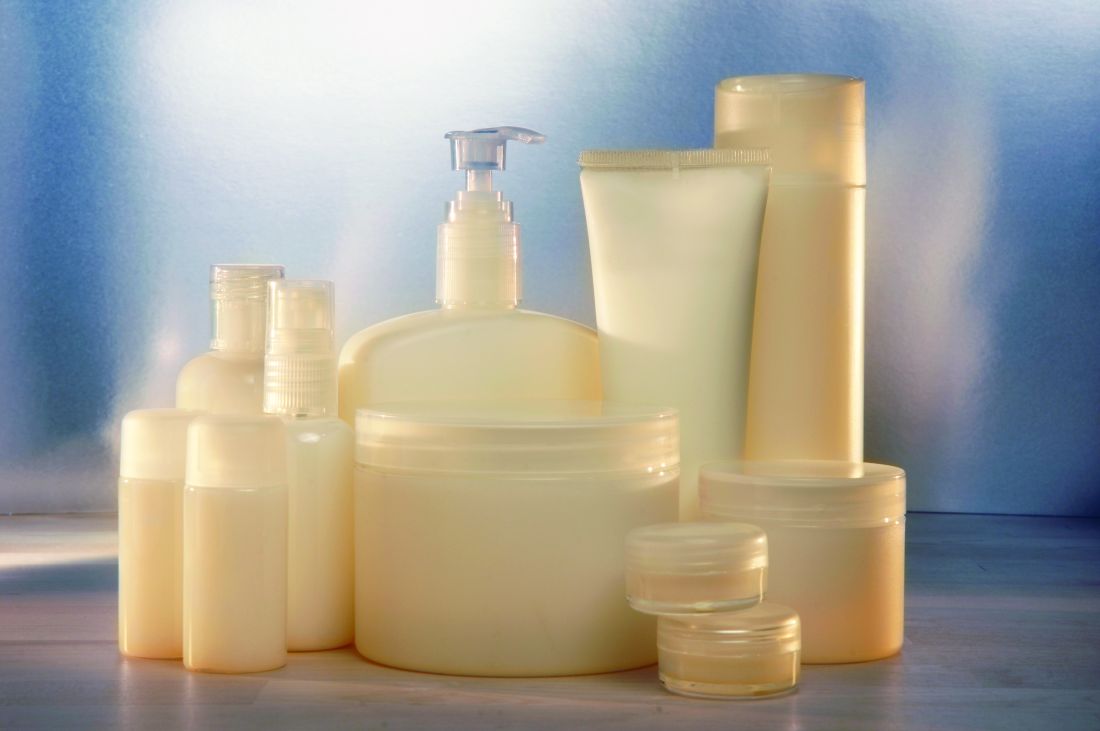
This derivative of oxidized glucose lactone is present naturally in bread, cheese, fruit juices, honey, tofu, and wine, and is used as a food additive in Europe.1,2 In dermatology, it is most often used in chemical peels.
Polyhydroxy acids (PHAs) were discovered about 3 decades ago to exert similar functions as alpha hydroxy acids without provoking sensory irritation reactions. Gluconolactone along with lactobionic acid were the identified PHAs and further characterized as delivering more humectant and moisturizing activity than alpha hydroxy acids and effective in combination with retinoic acid to treat adult acne and with retinyl acetate to confer antiaging benefits.3 It is typically added to products for its skin-conditioning qualities, resulting in smoother, brighter, more toned skin.4 This column focuses on recent studies using this bioactive agent for dermatologic purposes.
Split-Face Studies Show Various Benefits
In 2023, Jarząbek-Perz and colleagues conducted a split-face evaluation to assess the effects on various skin parameters (ie, hydration, pH, sebum, and transepidermal water loss [TEWL]) of gluconolactone and oxybrasion, compared with gluconolactone and microneedling. Twenty-one White women underwent a series of three split-face treatments at 1-week intervals. Chemical peels with 10% gluconolactone were performed on the whole face. The right side of the face was also treated with oxybrasion and the left with microneedle mesotherapy. Skin parameters were measured before the first and third treatments and 2 weeks following the final treatment. Photos were taken before and after the study. Both treatments resulted in improved hydration and reductions in sebum, pH, and TEWL. No statistically significant differences were noted between the treatment protocols. The researchers concluded that gluconolactone peels can be effectively combined with oxybrasion or microneedle mesotherapy to enhance skin hydration and to secure the hydrolipid barrier.5
Later that year, the same team evaluated pH, sebum levels, and TEWL before, during, and after several applications of 10% and 30% gluconolactone chemical peels in a split-face model in 16 female participants. The investigators conducted three procedures on both sides of the face, taking measurements on the forehead, periorbital area, on the cheek, and on the nose wing before, during, and 7 days after the final treatment. They found statistically significant improvements in sebum levels in the cheeks after the treatment series. Also, pH values were lower at each measurement site after each procedure. TEWL levels were significantly diminished around the eyes, as well as the left forehead and right cheek, with no significant discrepancy between gluconolactone concentrations. The researchers concluded that gluconolactone plays a major role in reducing cutaneous pH and TEWL and imparts a regulatory effect on sebum.1
Two years earlier, Jarząbek-Perz and colleagues assessed skin moisture in a split-face model in 16 healthy women after the application of 10% and 30% gluconolactone. Investigators measured skin moisture before and after each of three treatments and a week after the final treatment from the forehead, periorbital area, and on the cheek. They observed no significant discrepancies between the 10% and 30% formulations, but a significant elevation in facial skin hydration was found to be promoted by gluconolactone. The investigators concluded that gluconolactone is an effective moisturizer for care of dry skin.6
Topical Formulation
In 2023, Zerbinati and colleagues determined that a gluconolactone-based lotion that they had begun testing 2 years earlier was safe and effective for dermatologic applications, with the noncomedogenic formulation found suitable as an antiaging agent, particularly as it treats aging-related pore dilatation.7,8
Acne Treatment
In 2019, Kantikosum and colleagues conducted a double-blind, within-person comparative study to assess the efficacy of various cosmeceutical ingredients, including gluconolactone, glycolic acid, licochalcone A, and salicylic acid, combined with the acne treatment adapalene vs adapalene monotherapy for mild to moderate acne. Each of 25 subjects over 28 days applied a product mixed with 0.1% adapalene on one side of the face, and 0.1% adapalene alone on the other side of the face once nightly. The VISIA camera system spot score pointed to a statistically significant improvement on the combination sides. Differences in lesion reduction and severity were within acceptable margins, the authors reported. They concluded that the cosmeceutical combinations yielded similar benefits as adapalene alone, with the combination formulations decreasing acne complications.9
Potential Use as an Antifibrotic Agent

In 2018, Jayamani and colleagues investigated the antifibrotic characteristics of glucono-delta-lactone, a known acidifier, to ascertain if it could directly suppress collagen fibrils or even cause them to disintegrate. The researchers noted that collagen fibrillation is pH dependent, and that glucono-delta-lactone was found to exert a concentration-dependent suppression of fibrils and disintegration of preformed collagen fibrils with the antifibrotic function of the compound ascribed to its capacity to decrease pH. Further, glucono-delta-lactone appeared to emerge as an ideal antifibrotic agent as it left intact the triple helical structure of collagen after treatment. The investigators concluded that glucono-delta-lactone provides the foundation for developing antifibrotic agents intended to treat disorders characterized by collagen deposition.10
Conclusion
Gluconolactone emerged in the 1990s as a PHA useful in skin peels as an alternative to alpha hydroxy acids because of its nonirritating qualities. Since then, its soothing, hydrating, and, in particular, antiacne and antiaging qualities have become established. Wider applications of this versatile agent for dermatologic purposes are likely to be further investigated.
Dr. Baumann is a private practice dermatologist, researcher, author, and entrepreneur in Miami. She founded the division of cosmetic dermatology at the University of Miami in 1997. The third edition of her bestselling textbook, “Cosmetic Dermatology,” was published in 2022. Dr. Baumann has received funding for advisory boards and/or clinical research trials from Allergan, Galderma, Johnson & Johnson, and Burt’s Bees. She is the CEO of Skin Type Solutions, a SaaS company used to generate skin care routines in office and as a ecommerce solution. Write to her at dermnews@mdedge.com.
References
1. Jarząbek-Perz S et al. J Cosmet Dermatol. 2023 Dec;22(12):3305-3312..
2. Qin X et al. Front Physiol. 2022 Mar 14;13:856699.
3. Grimes PE et al. Cutis. 2004 Feb;73(2 Suppl):3-13.
4. Glaser DA. Facial Plast Surg Clin North Am. 2003 May;11(2):219-227.
5. Jarząbek-Perz S et al. Skin Res Technol. 2023 Jun;29(6):e13353.
6. Jarząbek-Perz S et al. Skin Res Technol. 2021 Sep;27(5):925-930.
7. Zerbinati N et al. Molecules. 2021 Dec 15;26(24):7592.
8. Zerbinati Net al. Pharmaceuticals (Basel). 2023 Apr 27;16(5):655.
9. Kantikosum K et al. Clin Cosmet Investig Dermatol. 2019 Feb 19;12:151-161.
10. Jayamani J et al. Int J Biol Macromol. 2018 Feb;107(Pt A):175-185.
This derivative of oxidized glucose lactone is present naturally in bread, cheese, fruit juices, honey, tofu, and wine, and is used as a food additive in Europe.1,2 In dermatology, it is most often used in chemical peels.
Polyhydroxy acids (PHAs) were discovered about 3 decades ago to exert similar functions as alpha hydroxy acids without provoking sensory irritation reactions. Gluconolactone along with lactobionic acid were the identified PHAs and further characterized as delivering more humectant and moisturizing activity than alpha hydroxy acids and effective in combination with retinoic acid to treat adult acne and with retinyl acetate to confer antiaging benefits.3 It is typically added to products for its skin-conditioning qualities, resulting in smoother, brighter, more toned skin.4 This column focuses on recent studies using this bioactive agent for dermatologic purposes.
Split-Face Studies Show Various Benefits
In 2023, Jarząbek-Perz and colleagues conducted a split-face evaluation to assess the effects on various skin parameters (ie, hydration, pH, sebum, and transepidermal water loss [TEWL]) of gluconolactone and oxybrasion, compared with gluconolactone and microneedling. Twenty-one White women underwent a series of three split-face treatments at 1-week intervals. Chemical peels with 10% gluconolactone were performed on the whole face. The right side of the face was also treated with oxybrasion and the left with microneedle mesotherapy. Skin parameters were measured before the first and third treatments and 2 weeks following the final treatment. Photos were taken before and after the study. Both treatments resulted in improved hydration and reductions in sebum, pH, and TEWL. No statistically significant differences were noted between the treatment protocols. The researchers concluded that gluconolactone peels can be effectively combined with oxybrasion or microneedle mesotherapy to enhance skin hydration and to secure the hydrolipid barrier.5
Later that year, the same team evaluated pH, sebum levels, and TEWL before, during, and after several applications of 10% and 30% gluconolactone chemical peels in a split-face model in 16 female participants. The investigators conducted three procedures on both sides of the face, taking measurements on the forehead, periorbital area, on the cheek, and on the nose wing before, during, and 7 days after the final treatment. They found statistically significant improvements in sebum levels in the cheeks after the treatment series. Also, pH values were lower at each measurement site after each procedure. TEWL levels were significantly diminished around the eyes, as well as the left forehead and right cheek, with no significant discrepancy between gluconolactone concentrations. The researchers concluded that gluconolactone plays a major role in reducing cutaneous pH and TEWL and imparts a regulatory effect on sebum.1
Two years earlier, Jarząbek-Perz and colleagues assessed skin moisture in a split-face model in 16 healthy women after the application of 10% and 30% gluconolactone. Investigators measured skin moisture before and after each of three treatments and a week after the final treatment from the forehead, periorbital area, and on the cheek. They observed no significant discrepancies between the 10% and 30% formulations, but a significant elevation in facial skin hydration was found to be promoted by gluconolactone. The investigators concluded that gluconolactone is an effective moisturizer for care of dry skin.6
Topical Formulation
In 2023, Zerbinati and colleagues determined that a gluconolactone-based lotion that they had begun testing 2 years earlier was safe and effective for dermatologic applications, with the noncomedogenic formulation found suitable as an antiaging agent, particularly as it treats aging-related pore dilatation.7,8
Acne Treatment
In 2019, Kantikosum and colleagues conducted a double-blind, within-person comparative study to assess the efficacy of various cosmeceutical ingredients, including gluconolactone, glycolic acid, licochalcone A, and salicylic acid, combined with the acne treatment adapalene vs adapalene monotherapy for mild to moderate acne. Each of 25 subjects over 28 days applied a product mixed with 0.1% adapalene on one side of the face, and 0.1% adapalene alone on the other side of the face once nightly. The VISIA camera system spot score pointed to a statistically significant improvement on the combination sides. Differences in lesion reduction and severity were within acceptable margins, the authors reported. They concluded that the cosmeceutical combinations yielded similar benefits as adapalene alone, with the combination formulations decreasing acne complications.9
Potential Use as an Antifibrotic Agent
In 2018, Jayamani and colleagues investigated the antifibrotic characteristics of glucono-delta-lactone, a known acidifier, to ascertain if it could directly suppress collagen fibrils or even cause them to disintegrate. The researchers noted that collagen fibrillation is pH dependent, and that glucono-delta-lactone was found to exert a concentration-dependent suppression of fibrils and disintegration of preformed collagen fibrils with the antifibrotic function of the compound ascribed to its capacity to decrease pH. Further, glucono-delta-lactone appeared to emerge as an ideal antifibrotic agent as it left intact the triple helical structure of collagen after treatment. The investigators concluded that glucono-delta-lactone provides the foundation for developing antifibrotic agents intended to treat disorders characterized by collagen deposition.10
Conclusion
Gluconolactone emerged in the 1990s as a PHA useful in skin peels as an alternative to alpha hydroxy acids because of its nonirritating qualities. Since then, its soothing, hydrating, and, in particular, antiacne and antiaging qualities have become established. Wider applications of this versatile agent for dermatologic purposes are likely to be further investigated.
Dr. Baumann is a private practice dermatologist, researcher, author, and entrepreneur in Miami. She founded the division of cosmetic dermatology at the University of Miami in 1997. The third edition of her bestselling textbook, “Cosmetic Dermatology,” was published in 2022. Dr. Baumann has received funding for advisory boards and/or clinical research trials from Allergan, Galderma, Johnson & Johnson, and Burt’s Bees. She is the CEO of Skin Type Solutions, a SaaS company used to generate skin care routines in office and as a ecommerce solution. Write to her at dermnews@mdedge.com.
References
1. Jarząbek-Perz S et al. J Cosmet Dermatol. 2023 Dec;22(12):3305-3312..
2. Qin X et al. Front Physiol. 2022 Mar 14;13:856699.
3. Grimes PE et al. Cutis. 2004 Feb;73(2 Suppl):3-13.
4. Glaser DA. Facial Plast Surg Clin North Am. 2003 May;11(2):219-227.
5. Jarząbek-Perz S et al. Skin Res Technol. 2023 Jun;29(6):e13353.
6. Jarząbek-Perz S et al. Skin Res Technol. 2021 Sep;27(5):925-930.
7. Zerbinati N et al. Molecules. 2021 Dec 15;26(24):7592.
8. Zerbinati Net al. Pharmaceuticals (Basel). 2023 Apr 27;16(5):655.
9. Kantikosum K et al. Clin Cosmet Investig Dermatol. 2019 Feb 19;12:151-161.
10. Jayamani J et al. Int J Biol Macromol. 2018 Feb;107(Pt A):175-185.
Dermatologists and the history of skin care and beauty devices: Part 4
In this series on the role dermatologists have played in the history of skin care, I have covered dermatologists who developed cosmeceutical ingredients, dermatologists who consulted for the skin care industry, and those who developed a novel and successful skin care line. In this column, part 4 of the series, I will continue to discuss .
Dermatologists and Stiefel Laboratories
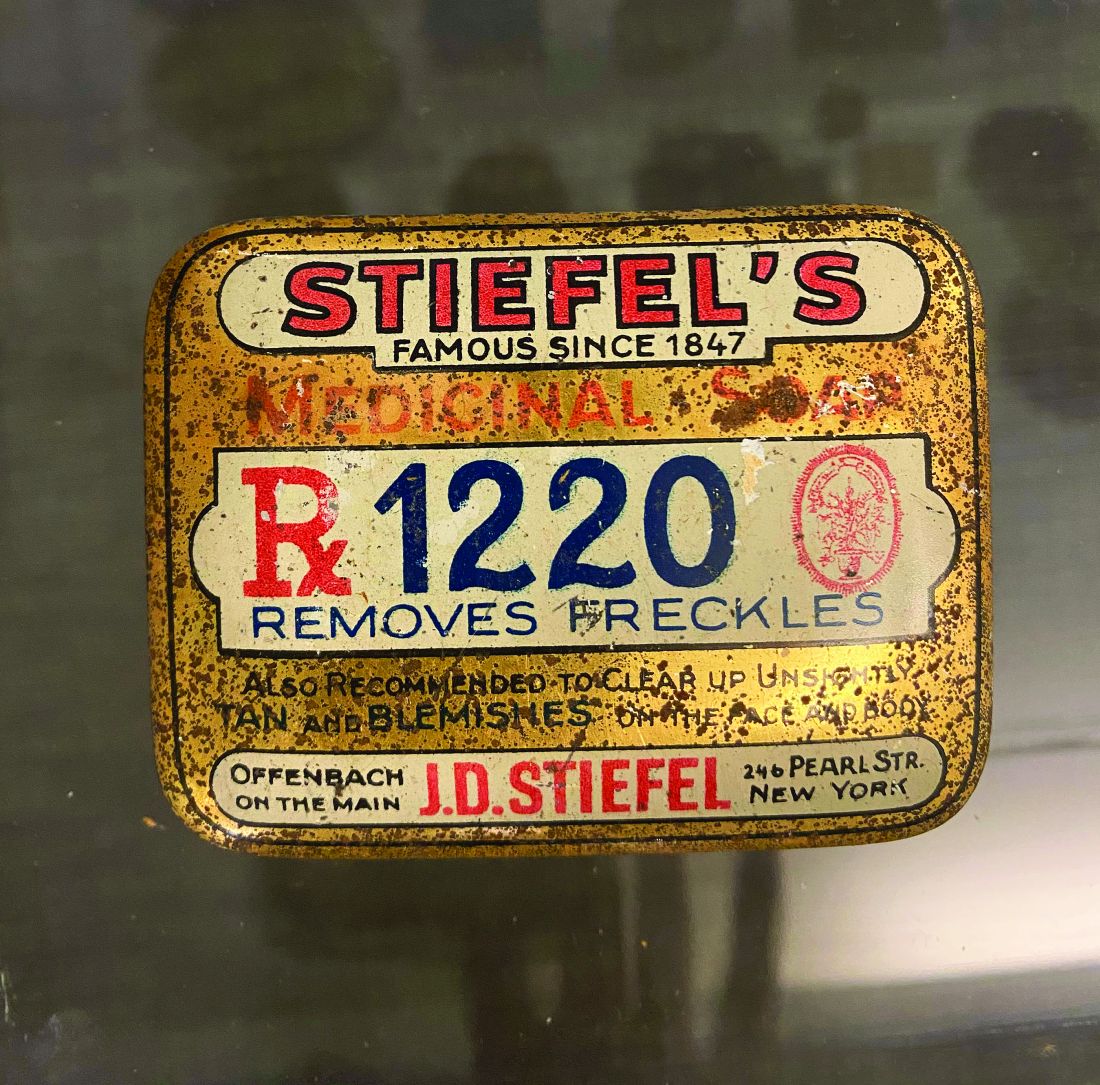
The Stiefel Medicinal Soap Company, founded in 1847, later became Stiefel Laboratories and was sold to GlaxoSmithKline in 2009. Stiefel Laboratories made many contributions over the years to the field of dermatology as chronicled in the excellent book, “Skin Saga” written by Charles Stiefel and published in 2018. The company was first known for soaps and groundbreaking products, such as “Freckle Soap” that sped epidermal turnover, resulting in a more even toned complexion.
Many dermatologists were involved in developing products and providing advice to the company. Herman Sharlit, MD, in New York, had the idea for a moisturizing soap (Oilatum), a detergent soap (Acne Aid detergent soap), and a coal tar soap (Polytar). Eugene Farber, MD, who was professor and chairman of the department of dermatology at Stanford (Calif.) University, consulted for Stiefel Laboratories and helped them identify and develop many products over the years.1 Stiefel Labs came out with the first facial scrub called Brasivol, an abrasive cream with aluminum oxide particles – the predecessor to modern day microdermabrasion. This facial scrub was conceived by dermatologist Rose Saperstein, MD, Los Angeles, who published a report2 on this in 1960 and also received a patent for it in 1963.3 Brasivol became the company’s first million dollar product.1
Stiefel Laboratories worked with many dermatologists to help them develop their ideas. They included Cleveland White, MD, who patented a highly absorbent foot and body powder known as Zeasorb powder. William Pace, MD, was a Canadian dermatologist who patented an acne treatment containing benzoyl peroxide and sulfur that Stiefel Labs marketed as Sulfoxyl Lotion. Dr. Pace is lovingly referred to as “the father of benzoyl peroxide” because his idea led Stiefel Labs to develop more benzoyl peroxide products. Benzoyl peroxide remains the most popular OTC ingredient to treat acne.
Comedone extractors
Many dermatologists have developed ways to extract comedones. There are publications on using paper clips,4,5safety pins,6 and medicine droppers,7 but some dermatologists have developed special comedone extractors, which include the following: Jay Schamberg, MD, developed a comedone extractor with a loop at each end. He disapproved of cutting a comedone, so did not include a needle or scalpel in his extractor.8
- Leonard Savitt, MD,9 attached a scalpel to one end of the Schamberg extractor.
- Alan Shalita, MD, developed a comedone extractor with a large, keyhole-shaped extracting orifice that made the tool easier to clean.10
The Saalfield comedone extractor combines a fixed pointed blade at one end and a small spoon-shaped expressor foot at the other end. (However, I have not been able to determine if Saalfield was a dermatologist.)
Dermatologist who developed methods for lesion excisions
Robert Segal, MD, a dermatologist at the University of Arizona, Tucson, invented the Dermablade. Although this is technically not a beauty device, I am including it because it has made the removal of unsightly moles and lesions much easier. He holds six patents on this device.
Dermatologists developed dermabrasion and microneedling
Ernst Kromayer, MD,11 a dermatologist in Germany, first described microneedling in 1905 when he mounted dental burrs on motor-driven flexible cord equipment to treat scars. Abner Kurtin, MD, a New York dermatologist, learned about Dr. Kromayer’s technique and modified it using stainless wireless brushes. Dr. Kurtin is known as the “father of dermabrasion.” His work was noted by Nobel Laureate Alexis Carrel, MD, who moved to New York City and began using the technique. Dr. Carrel’s protege, New York dermatologist, Norman Orentreich, MD, began using hypodermic needles instead of wire brushes. Microneedling has gained much popularity over the last decade and has been combined with platelet rich plasma injections.
Dermatologist-developed injection to shrink fat
Adam Rotunda, MD, was a dermatology resident at the University of California, Los Angeles, when he and his professor Michael Kolodney, MD, PhD, had the idea to develop deoxycholate as an injectable to reduce fat deposits. They filed a patent in 2004, conducted clinical trials, and it worked! In 2009, the patent for deoxycholic acid (ATX-10), marketed as Kybella, was granted. The rights to the drug were purchased by Aestherx, which later became Kythera Biopharmaceuticals. Kybella received Food and Drug Administration approval in 2015, and 6 months later, Kythera was acquired by Allergan.
Development of FDA-approved drugs to improve skin appearance
In 2004, dermatologists Stuart Shanler, MD, and Andrew Ondo, MD, filed a patent for the use of topical oxymetazoline for the treatment of the erythema of rosacea. They published their observations in 2007, noting that oxymetazoline improved facial flushing and erythema.11 Dr. Shanler then teamed up with dermatologist Neal Walker, MD, to form a start-up pharmaceutical company, Vicept Therapeutics, and took this compound through phase 2 clinical trials, while Dr. Shanler filed additional patents on oxymetazoline compositions and their uses. Once they successfully demonstrated the efficacy of topical oxymetazoline for rosacea, Allergan acquired the rights of the drug, successfully completed the phase 3 clinical trials, and Rhofade was approved by the FDA in 2017. It is the only topical drug invented and developed by a dermatologist to receive FDA approval since tretinoin (Renova) was developed by Albert Kligman, MD, and approved by the FDA for the improvement in appearance of fine wrinkling, mottled hyperpigmentation and roughness associated with photodamage in 1992.
The development of lasers
The last dermatologist I will discuss in this history series is R. Rox Anderson, MD, professor of dermatology at Harvard University, and director of the Wellman Center for Photomedicine at Massachusetts General Hospital, Boston. It is impossible to list all his contributions in such a limited space. It would take a book. Building on efforts pioneered by Leon Goldman, MD, Dr. Anderson and his associates pioneered the use of lasers in dermatology and invented the idea of photothermolysis when they filed a patent on using light to remove hair in 1995.Dieter Manstein, MD, PhD,Dr. Anderson and others filed many patents that led to devices such as hair removal lasers, resurfacing lasers, and Fraxel lasers. They also made discoveries related to using cold to shrink fat. One of their inventions is known as CoolSculpting. They were so influential in the development of cosmetic dermatology that it is hard to imagine the field without their contributions.
This concludes my four-part series on the history of dermatologists’ role in the development of the skin care industry. I hope I have not forgotten anyone; if I did, I apologize. I have asked for ideas on Dermchat, Facebook and LinkedIn. Feel free to reach out if I missed one of your contributions. I will be giving lectures on this topic in the future and would be happy to include anyone I missed.
As the year 2020 ends, I want to say, Happy 50th Anniversary Dermatology News! I hope you enjoyed this historical series in honor of this anniversary.
Dr. Baumann is a private practice dermatologist, researcher, author, and entrepreneur who practices in Miami. She founded the Cosmetic Dermatology Center at the University of Miami in 1997. Dr. Baumann has written two textbooks and a New York Times Best Sellers book for consumers. Dr. Baumann has received funding for advisory boards and/or clinical research trials from Allergan, Galderma, Revance, Evolus, and Burt’s Bees. She is the CEO of Skin Type Solutions Inc., a company that independently tests skin care products and makes recommendations to physicians on which skin care technologies are best. Write to her at dermnews@mdedge.com.
References
1. Stiefel, CW. (n.d.). Skin Saga: How a Tiny Family Soap Business Evolved Over Six Generations Into the #1 Dermatology Company in the World. United States: Smart Business Network.
2. Saperstein, RB. Arch Dermatol. 1960 Apr;81:601.
3. Saperstein, RB, and Stiefel, WK (1963). U.S. Patent No. 3,092,111. Washington, DC: U.S. Patent and Trademark Office.
4. George DE et al. J Am Acad Dermatol. 2006 Feb;54(2):326.
5. Cvancara JL, Meffert JJ. J Am Acad Dermatol. 1999 Mar;40(3):477-8.
6. Mukhtar M., Sharma R. Int J Dermatol. 2004 Dec;43(12):967-8.
7. Shellow, H. JAMA. 1951;147(18):1777.
8. Wright CS. Arch Dermatol. 1961;84(3):515.
9. Savitt LE. Arch Dermatol. 1961 Apr;83:660-1.
10. Shalita AR, Harris H. Arch Dermatol. 1972 May;105(5):759-60.
11. Shanler SD, Ondo AL. Arch Dermatol. 2007 Nov;143(11):1369-71.
In this series on the role dermatologists have played in the history of skin care, I have covered dermatologists who developed cosmeceutical ingredients, dermatologists who consulted for the skin care industry, and those who developed a novel and successful skin care line. In this column, part 4 of the series, I will continue to discuss .
Dermatologists and Stiefel Laboratories
The Stiefel Medicinal Soap Company, founded in 1847, later became Stiefel Laboratories and was sold to GlaxoSmithKline in 2009. Stiefel Laboratories made many contributions over the years to the field of dermatology as chronicled in the excellent book, “Skin Saga” written by Charles Stiefel and published in 2018. The company was first known for soaps and groundbreaking products, such as “Freckle Soap” that sped epidermal turnover, resulting in a more even toned complexion.
Many dermatologists were involved in developing products and providing advice to the company. Herman Sharlit, MD, in New York, had the idea for a moisturizing soap (Oilatum), a detergent soap (Acne Aid detergent soap), and a coal tar soap (Polytar). Eugene Farber, MD, who was professor and chairman of the department of dermatology at Stanford (Calif.) University, consulted for Stiefel Laboratories and helped them identify and develop many products over the years.1 Stiefel Labs came out with the first facial scrub called Brasivol, an abrasive cream with aluminum oxide particles – the predecessor to modern day microdermabrasion. This facial scrub was conceived by dermatologist Rose Saperstein, MD, Los Angeles, who published a report2 on this in 1960 and also received a patent for it in 1963.3 Brasivol became the company’s first million dollar product.1
Stiefel Laboratories worked with many dermatologists to help them develop their ideas. They included Cleveland White, MD, who patented a highly absorbent foot and body powder known as Zeasorb powder. William Pace, MD, was a Canadian dermatologist who patented an acne treatment containing benzoyl peroxide and sulfur that Stiefel Labs marketed as Sulfoxyl Lotion. Dr. Pace is lovingly referred to as “the father of benzoyl peroxide” because his idea led Stiefel Labs to develop more benzoyl peroxide products. Benzoyl peroxide remains the most popular OTC ingredient to treat acne.
Comedone extractors
Many dermatologists have developed ways to extract comedones. There are publications on using paper clips,4,5safety pins,6 and medicine droppers,7 but some dermatologists have developed special comedone extractors, which include the following: Jay Schamberg, MD, developed a comedone extractor with a loop at each end. He disapproved of cutting a comedone, so did not include a needle or scalpel in his extractor.8
- Leonard Savitt, MD,9 attached a scalpel to one end of the Schamberg extractor.
- Alan Shalita, MD, developed a comedone extractor with a large, keyhole-shaped extracting orifice that made the tool easier to clean.10
The Saalfield comedone extractor combines a fixed pointed blade at one end and a small spoon-shaped expressor foot at the other end. (However, I have not been able to determine if Saalfield was a dermatologist.)
Dermatologist who developed methods for lesion excisions
Robert Segal, MD, a dermatologist at the University of Arizona, Tucson, invented the Dermablade. Although this is technically not a beauty device, I am including it because it has made the removal of unsightly moles and lesions much easier. He holds six patents on this device.
Dermatologists developed dermabrasion and microneedling
Ernst Kromayer, MD,11 a dermatologist in Germany, first described microneedling in 1905 when he mounted dental burrs on motor-driven flexible cord equipment to treat scars. Abner Kurtin, MD, a New York dermatologist, learned about Dr. Kromayer’s technique and modified it using stainless wireless brushes. Dr. Kurtin is known as the “father of dermabrasion.” His work was noted by Nobel Laureate Alexis Carrel, MD, who moved to New York City and began using the technique. Dr. Carrel’s protege, New York dermatologist, Norman Orentreich, MD, began using hypodermic needles instead of wire brushes. Microneedling has gained much popularity over the last decade and has been combined with platelet rich plasma injections.
Dermatologist-developed injection to shrink fat
Adam Rotunda, MD, was a dermatology resident at the University of California, Los Angeles, when he and his professor Michael Kolodney, MD, PhD, had the idea to develop deoxycholate as an injectable to reduce fat deposits. They filed a patent in 2004, conducted clinical trials, and it worked! In 2009, the patent for deoxycholic acid (ATX-10), marketed as Kybella, was granted. The rights to the drug were purchased by Aestherx, which later became Kythera Biopharmaceuticals. Kybella received Food and Drug Administration approval in 2015, and 6 months later, Kythera was acquired by Allergan.
Development of FDA-approved drugs to improve skin appearance
In 2004, dermatologists Stuart Shanler, MD, and Andrew Ondo, MD, filed a patent for the use of topical oxymetazoline for the treatment of the erythema of rosacea. They published their observations in 2007, noting that oxymetazoline improved facial flushing and erythema.11 Dr. Shanler then teamed up with dermatologist Neal Walker, MD, to form a start-up pharmaceutical company, Vicept Therapeutics, and took this compound through phase 2 clinical trials, while Dr. Shanler filed additional patents on oxymetazoline compositions and their uses. Once they successfully demonstrated the efficacy of topical oxymetazoline for rosacea, Allergan acquired the rights of the drug, successfully completed the phase 3 clinical trials, and Rhofade was approved by the FDA in 2017. It is the only topical drug invented and developed by a dermatologist to receive FDA approval since tretinoin (Renova) was developed by Albert Kligman, MD, and approved by the FDA for the improvement in appearance of fine wrinkling, mottled hyperpigmentation and roughness associated with photodamage in 1992.
The development of lasers
The last dermatologist I will discuss in this history series is R. Rox Anderson, MD, professor of dermatology at Harvard University, and director of the Wellman Center for Photomedicine at Massachusetts General Hospital, Boston. It is impossible to list all his contributions in such a limited space. It would take a book. Building on efforts pioneered by Leon Goldman, MD, Dr. Anderson and his associates pioneered the use of lasers in dermatology and invented the idea of photothermolysis when they filed a patent on using light to remove hair in 1995.Dieter Manstein, MD, PhD,Dr. Anderson and others filed many patents that led to devices such as hair removal lasers, resurfacing lasers, and Fraxel lasers. They also made discoveries related to using cold to shrink fat. One of their inventions is known as CoolSculpting. They were so influential in the development of cosmetic dermatology that it is hard to imagine the field without their contributions.
This concludes my four-part series on the history of dermatologists’ role in the development of the skin care industry. I hope I have not forgotten anyone; if I did, I apologize. I have asked for ideas on Dermchat, Facebook and LinkedIn. Feel free to reach out if I missed one of your contributions. I will be giving lectures on this topic in the future and would be happy to include anyone I missed.
As the year 2020 ends, I want to say, Happy 50th Anniversary Dermatology News! I hope you enjoyed this historical series in honor of this anniversary.
Dr. Baumann is a private practice dermatologist, researcher, author, and entrepreneur who practices in Miami. She founded the Cosmetic Dermatology Center at the University of Miami in 1997. Dr. Baumann has written two textbooks and a New York Times Best Sellers book for consumers. Dr. Baumann has received funding for advisory boards and/or clinical research trials from Allergan, Galderma, Revance, Evolus, and Burt’s Bees. She is the CEO of Skin Type Solutions Inc., a company that independently tests skin care products and makes recommendations to physicians on which skin care technologies are best. Write to her at dermnews@mdedge.com.
References
1. Stiefel, CW. (n.d.). Skin Saga: How a Tiny Family Soap Business Evolved Over Six Generations Into the #1 Dermatology Company in the World. United States: Smart Business Network.
2. Saperstein, RB. Arch Dermatol. 1960 Apr;81:601.
3. Saperstein, RB, and Stiefel, WK (1963). U.S. Patent No. 3,092,111. Washington, DC: U.S. Patent and Trademark Office.
4. George DE et al. J Am Acad Dermatol. 2006 Feb;54(2):326.
5. Cvancara JL, Meffert JJ. J Am Acad Dermatol. 1999 Mar;40(3):477-8.
6. Mukhtar M., Sharma R. Int J Dermatol. 2004 Dec;43(12):967-8.
7. Shellow, H. JAMA. 1951;147(18):1777.
8. Wright CS. Arch Dermatol. 1961;84(3):515.
9. Savitt LE. Arch Dermatol. 1961 Apr;83:660-1.
10. Shalita AR, Harris H. Arch Dermatol. 1972 May;105(5):759-60.
11. Shanler SD, Ondo AL. Arch Dermatol. 2007 Nov;143(11):1369-71.

The role of oleuropein, the primary phenol in olives, in skin health
Olives and olive oil have long been known to confer salutary effects to the skin.1 Leaves and fruits of the olive plant (Olea europaea) have been used as external emollients to treat skin ulcers and inflammatory wounds.2 The phenolic compound oleuropein, the most abundant phenolic found in olive leaves and oil, has been shown to exhibit antioxidant and free radical–scavenging activities.3,4 Also present in the stems and flowers of the plant, oleuropein, an ester of elenolic acid and 3,4-dihydroxyphenyl ethanol and the primary glycoside in olives,5 is thought to be the major contributor to its antioxidant and antimelanogenesis activities.6 Notably, olive leaves, which contain a copious supply of oleuropein, are thought to exert significantly more antioxidant activity than olive fruit.7
Hydroxytyrosol is an ortho-diphenolic substance and essential constituent of oleuropein that has been shown in vitro to prevent apoptotic cell death caused by UVB in HaCaT cells.8,9 Both oleuropein and hydroxytyrosol impart various anticancer properties at the initiation, promotion, and metastasis stages and yield protection against multiple cancers, including skin tumors.10 The antioxidant activity of both compounds, which has been found to be more potent than that of vitamin E, is attributed to their phenolic content.11,12 In addition, oleuropein and lipophilic olive mill wastewater derivatives have been useful as active ingredients for stabilizing cosmetic formulations.13 This column revisits oleuropein after 10 years to focus on its dermatologic potential.
Protection against UV damage
A hairless mouse study by Kimura and Sumiyoshi in 2009 revealed that olive leaf extract and its primary constituent oleuropein exert a skin-protective effect against chronic UVB-induced skin damage and carcinogenesis, as well as tumor growth. This is likely caused by reducing cutaneous cyclooxygenase (COX)-2 levels, thus suppressing the expression of vascular endothelial growth factor (VEGF) and various matrix metalloproteinases, specifically MMP-2, MMP-9, and MMP-13.14
A year later, the same researchers examined the potential protective effects of olive leaf extract and oleuropein on acute damage induced by UVB exposure in C57BL/6J mice. Both oral extract (300 mg/kg or 1,000 mg/kg) and oral oleuropein (25mg/kg or 85 mg/kg) hindered skin thickness increases engendered by daily doses of UVB (120 mJ/cm2 for 5 days, then every other day for 9 days). Olive leaf extract and oleuropein also suppressed increases in Ki-67- and 8-hydroxy-2’-deoxyguanosine–positive cell numbers, melanin granule area, and MMP-13 expression, the investigators noted.15 Preinitiation with oleuropein also appears to have prevented skin tumor formation in a two-stage carcinogenesis model in mice, which the investigators ascribed to the antioxidant and antiapoptotic properties of the olive protein.16
The cosmetic characteristics of oleuropein against UVB-induced erythema in healthy volunteers were assessed by Perugini et al. in 2008. Using an emulsion and emulgel containing oleuropein and vitamin E as a reference compound, the investigators found that the botanical ingredient was responsible for decreases in erythema (22%), transepidermal water loss (35%), and blood flow (30%). They suggested that the use of oleuropein in cosmetic formulations warrants further investigation for its potential to help mitigate UV damage.3
Wound healing
Koca et al. assessed the wound healing activity of O. europaea leaf extracts using in vivo wound models and the reference ointment Madecassol (Bayer; Istanbul) for comparison, in 2011. The results showed that the aqueous extract exhibited wound healing properties, with secoiridoid oleuropein (4.6059%) found to be the primary active constituent.2
In a 2014 skin wound–healing investigation in aged male Balb/c mice, Mehraein et al. divided 24 mice, 16 months of age, into control and experimental groups. On days 3 and 7 after incision, collagen fiber deposition was significantly increased and reepithelialization more advanced in the oleuropein group (administered via an intradermal injection once a day), which also experienced decreased cell infiltration. The investigators concluded that oleuropein speeds cutaneous wound healing in mice and may have potential for clinical applications in human would healing from surgery.17
Later that year, the same team investigated the therapeutic effects of oleuropein on the wounded skin of young male Balb/c mice, finding similar results, with the phenolic compound again accelerating reepithelialization, improving collagen fiber synthesis, and augmenting blood flow to wound areas via up-regulating VEGF protein expression.4
Hair growth
In 2015, Tong et al. reported that topically applied oleuropein spurred the anagen hair growth phase in telogenic C57BL/6N mouse skin.18 An O. europaea subcutaneous immunotherapy has also demonstrated reductions in cutaneous reactivity, safety, and tolerability in patients with rhinoconjunctivitis.19
Conclusion
The benefits of consuming olives and olive oil are well established and continue to be studied. backed by many years of anecdotal reporting and use in traditional medicine. While the emerging data on the dermatologic uses of the olive phenolic constituent oleuropein are encouraging, much more information, particularly derived from randomized, controlled trials in humans, is necessary to establish the full potential of oleuropein for indications such as wound healing and protection against UV damage.
Dr. Baumann is a private practice dermatologist, researcher, author, and entrepreneur who practices in Miami. She founded the Cosmetic Dermatology Center at the University of Miami in 1997. Dr. Baumann wrote two textbooks: “Cosmetic Dermatology: Principles and Practice” (New York: McGraw-Hill, 2002), and “Cosmeceuticals and Cosmetic Ingredients” (New York: McGraw-Hill, 2014), and a New York Times Best Sellers book for consumers, “The Skin Type Solution” (New York: Bantam Dell, 2006). Dr. Baumann has received funding for advisory boards and/or clinical research trials from Allergan, Evolus, Galderma, and Revance. She is the founder and CEO of Skin Type Solutions Franchise Systems. Write to her at dermnews@mdedge.com.
References
1. Baumann LS, Weisberg EM. “Olive oil in botanical cosmeceuticals.” Olives and Olive Oil in Health and Disease Prevention. New York: Academic Press, 2010.
2. Koca U et al. J Med Food. 2011 Jan-Feb;14(1-2):140-6.
3. Perugini P et al. Int J Cosmet Sci. 2008 Apr;30(2):113-20.
4. Mehraein F et al. Wounds. 2014 Mar;26(3):83-8.
5. Imran M et al. J Food Sci. 2018 Jul;83(7):1781-91.
6. Kishikawa A et al. Phytother Res. 2015 Jun;29(6):877-86.
7. Zheng J et al. Zhongguo Zhong Yao Za Zhi. 2016 Feb;41(4):613-8.
8. Salucci S et al. J Dermatol Sci. 2015 Oct;80(1):61-8.
9. Jeon S, Choi M. Biomed Dermatol. 2018;2:21.
10. Imran M et al. J Food Sci. 2018 Jul;83(7):1781-91.
11. Visioli F et al. Biochem Biophys Res Commun. 1998 Jun 9;247(1):60-4.
12. Polišak N et al. Phytother Res. 2019 Oct 27. doi: 10.1002/ptr.6524.
13. Aissa I et al. Biotechnol Appl Biochem. 2017 Jul;64(4):579-89.
14. Kimura Y, Sumiyoshi M. J Nutr. 2009 Nov;139(11):2079-86.
15. Sumiyoshi M, Kimura Y. Phytother Res. 2010 Jul;24(7):995-1003.
16. John DNS et al. JKIMSU. 2019 Jan-Mar;8(1):43-51.
17. Mehraein F et al. Cell J. 2014 Feb 3;16(1):25-30.
18. Tong T et al. PLoS One. 2015 Jun 10;10(6):e0129578.
19. Saenza De San Pedro B et al. Eur All Allergy Clin Immunol. 2019 Nov 27. doi: 10.23822/EurAnnACI.1764-1489.124.
Pyrrolidone carboxylic acid may be a key cutaneous biomarker
Pyrrolidone carboxylic acid (PCA), the primary constituent of the natural moisturizing factor (NMF),1 including its derivatives – such as simple2 and novel3 esters as well as sugar complexes4 – is the subject of great interest and research regarding its capacity to moisturize the stratum corneum via topical application.
Creams and lotions containing the sodium salt of PCA are widely reported to aid in hydrating the skin and ameliorating dry flaky skin conditions.5,6 In addition, the zinc salt of L-pyrrolidone carboxylate is a longtime cosmetic ingredient due to antimicrobial and astringent qualities. This column briefly addresses the role of PCA in skin health.7
Dry skin
In a comprehensive literature review from 1981, Clar and Fourtanier reported conclusive evidence that PCA acts as a hydrating agent and that all the cosmetic formulations with a minimum of 2% PCA and PCA salt that they tested in their own 8-year study enhanced dry skin in short- and long-term conditions given suitable vehicles (no aqueous solutions).6
In a 2014 clinical study of 64 healthy white women with either normal or cosmetic dry skin, Feng et al. noted that tape stripped samples of stratum corneum revealed significantly lower ratios of free amino acids to protein and PCA to protein. This was associated with decreased hydration levels compared with normal skin. The investigators concluded that lower NMF levels across the depth of the stratum corneum and reduced cohesivity characterize cosmetic dry skin and that these clinical endpoints merit attention in evaluating the usefulness of treatments for dry skin.8
In 2016, Wei et al. reported on their assessment of the barrier function, hydration, and dryness of the lower leg skin of 25 female patients during the winter and then in the subsequent summer. They found that PCA levels were significantly greater during the summer, as were keratins. Hydration was also higher during the summer, while transepidermal water loss and visual dryness grades were substantially lower.9
Atopic dermatitis
A 2014 clinical study by Brandt et al. in patients with skin prone to developing atopic dermatitis (AD) revealed that a body wash composed of the filaggrin metabolites arginine and PCA was well tolerated and diminished pruritus. Patients reported liking the product and suggested that it improved their quality of life.10
Later that year, Jung et al. characterized the relationship of PCA levels, and other factors, with the clinical severity of AD. Specifically, in a study of 73 subjects (21 with mild AD, 21 with moderate to severe AD, 13 with X-linked ichthyosis as a negative control for filaggrin gene mutation, and 18 healthy controls), the investigators assessed transepidermal water loss, stratum corneum hydration, and skin surface pH. They found that PCA levels and caspase-14 were lower in inflammatory lesions compared with nonlesional skin in subjects with AD. These levels also were associated with clinical AD severity as measured by eczema area and severity index scores as well as skin barrier function.11
PCA as a biomarker
In 2009, Kezic et al. determined that the use of tape stripping to cull PCA in the stratum corneum was effective in revealing that PCA concentration in the outermost skin layer is a viable biomarker of filaggrin genotype.12
Raj et al. conducted an interesting study in 2016 in which they set out to describe the various markers for total NMF levels and link them to the activities of plasmin and corneocyte maturation in the photoexposed cheek and photoprotected postauricular regions of healthy white, black African, and albino African women in South Africa. PCA levels were highest among the albino African group, followed by black African and then white participants. The investigators also found that bleomycin hydrolase was linked to PCA synthesis, as suggested by higher bleomycin levels in albino African participants. In this group, corneocyte maturation was also observed to be impeded.13
The next year, the same team studied stratum corneum physiology and biochemistry of the cheeks in 48 white women with sensitive skin. The goal was to ascertain the connections between bleomycin hydrolase and calpain-1, PCA levels, corneocyte maturation, and transglutaminase and plasmin activities. Capsaicin sensitivity was observed in 52% of subjects, with PCA levels and bleomycin hydrolase activity found to be lower in the capsaicin-sensitive panel and correlated in subjects not sensitive to capsaicin. The researchers concluded that reduced levels of PCA, bleomycin hydrolase, and transglutaminase combined with a larger volume of immature corneocytes suggest comparatively poor stratum corneum maturation in individuals with sensitive skin.14
Other uses
In 2012, Takino et al. used cultured normal human dermal fibroblasts to show that zinc l-pyrrolidone carboxylate blocked UVA induction of activator protein-1, diminished matrix metalloproteinase-1 synthesis, and spurred type I collagen production. The researchers suggested that such results suggest the potential of zinc PCA for further investigation as an agent to combat photoaging.7
Conclusion
. Recent research suggests that it may serve as an important biomarker of filaggrin, NMF levels, and skin hydration. In addition, new data point to its usefulness as a gauge for ADs. More investigations are necessary to ascertain the feasibility of adjusting PCA levels through topical administration and what effects topically applied PCA may have on various skin parameters.
Dr. Baumann is a private practice dermatologist, researcher, author, and entrepreneur in Miami. She founded the Cosmetic Dermatology Center at the University of Miami in 1997. Dr. Baumann wrote two textbooks, “Cosmetic Dermatology: Principles and Practice” (New York: McGraw-Hill, 2002) and “Cosmeceuticals and Cosmetic Ingredients” (New York: McGraw-Hill, 2014), as well as a New York Times Best Sellers book for consumers, “The Skin Type Solution” (New York: Bantam Dell, 2006). Dr. Baumann has received funding for advisory boards and/or clinical research trials from Allergan, Evolus, Galderma, and Revance. She is the founder and CEO of Skin Type Solutions Franchise Systems LLC. Write to her at dermnews@mdedge.com.
References
1. Björklund S et al. Soft Matter. 2014 Jul 7;10(25):4535-46.
2. Hall KJ, Hill JC. J Soc Cosmet Chem. 1986;37(6):397-407.
3. Tezuka T et al. Dermatology. 1994;188(1):21-4.
4. Kwoya Hakko Kogyo Co. Pyrrolidone carboxylic acid esters containing composition used to prevent loss of moisture from the skin. Patent JA 48 82 046 (1982).
5. Org Santerre. l-pyrrolidone carboxylic acid-sugar compounds as rehydrating ingredients in cosmetics. Patent Fr 2 277 823 (1977).
6. Clar EJ, Fourtanier A. Int J Cosmet Sci. 1981 Jun;3(3):101-13.
7. Takino Y et al. Int J Cosmet Sci. 2012 Feb;34(1):23-8.
8. Feng L et al. Int J Cosmet Sci. 2014 Jun;36(3):231-8.
9. Wei KS et al. J Cosmet Sci. 2016 May-Jun;67(3):185-203.
10. Brandt S et al. J Drugs Dermatol. 2014 Sep;13(9):1108-11.
11. Jung M et al. J Dermatol Sci. 2014 Dec;76(3):231-9.
12. Kezic S et al. Br J Dermatol. 2009 Nov;161(5):1098-104.
13. Raj N et al. Int J Cosmet Sci. 2016 Dec;38(6):567-75.
14. Raj N et al. Int J Cosmet Sci. 2017 Feb;39(1):2-10.
Cynodon dactylon
medicine to treat cutaneous diseases, fevers, and rheumatism,as well as a variety of chronic inflammatory conditions.1,2 The Ayurvedic armamentarium is thought to be the most abundant source of botanically based drugs used to treat wounds.3 Unrelated to health concerns, with the possible exception of allergic reactions, C. dactylon – which originated in Africa, is widely dispersed in Europe, and became an invasive species in locations such as Bermuda – is also used on putting greens on golf courses in subtropical and tropical climates.4 This grass has been shown to be safe and effective for treating induced RA in rats.1,2 Recent findings are encouraging in the area of wound healing.
Chemical constituents

Among the numerous ingredients contained in C. dactylon are proteins, carbohydrates, minerals, terpenoids, vitamin C, palmitic acid, and alkaloids.3 Other key phytoconstituents known to impart beneficial health effects that are present in the plant include flavonoids (such as apigenin and luteolin), carotenoids (such as beta-carotene and neoxanthin), phenolics, phytosterols, glycosides, saponins, and volatile oils.3 Given such components, it should not be surprising that C. dactylon has demonstrated antioxidant activity by scavenging the 2,2-diphenyl-1-picrylhydrazyl radical.3
Wound healing
Given the reputation of C. dactylon as an effective compound used in traditional medicine for wound healing as a hemostatic agent, Biswas et al. set out in 2017 to determine if they could provide scientific validation of the botanical as a viable wound-healing option. The investigators first undertook to compare a 15% ointment of the extract with a placebo control and the standard framycetin on full-thickness punch wounds in Wistar rats. Across all parameters, results for the C. dactylon–treated group far exceeded the control group and were comparable with the framycetin group. Subsequently, in a pilot clinical study, the researchers assessed the botanical ointment in a small cohort (n = 12) of men and women aged 65-75 years (n = 12) with chronic and complicated wounds. Half were treated with a topical C. dactylon ointment and half were treated with a topical framycetin sulfate ointment. Comparable effects were seen across the groups, with significant contraction of wounds and wound area noted, along with significant development of granulation and epithelial tissues. Hematologic parameters indicating improvement were comparable between the groups. The investigators concluded that all patients treated with C. dactylon healed successfully. They added that the antioxidant activity of the constituent phenolic acids and flavonoids in C. dactylon likely play a key role in conferring potent wound-healing effects by promoting collagenesis.3
In 2018, Perumal et al. created a collagen-silica biocomposite enriched with C. dactylon extract and studied its wound-healing potential in vitro and in vivo in comparison with collagen as well as collagen-silica scaffold controls. The investigators found that the stability of the enriched product surpassed that of native collagen by virtue of the intermolecular interactions between the botanical ingredient and collagen. In a full-thickness excision wound model using Wistar rats, the biocomposite was associated with more rapid healing than wounds treated with collagen and the scaffold control.5
Arthritis
In 2009, Sindhu et al. orally administered C. dactylon to rats after intradermally inducing arthritis. The induction produced inflammation, and a marked rise in the levels of inflammatory mediators, C-reactive protein, myeloperoxidase, and nitrite. Resultant oxidative stress was noted with substantial declines in the activity of catalase, superoxide dismutase, and glutathione peroxidase, as well as levels of glutathione, vitamins C and E, and an increase in lipid peroxidation. Administration of C. dactylon yielded substantial changes, with mitigation of the inflammatory response and oxidative stress as well as diminution of the arthritic response nearly to the baseline condition. The investigators concluded that the botanical agent clearly demonstrates potential to protect against arthritis.2
A subsequent study in rats by Bhangale and Acharya supported the use of C. dactylon for RA, as its oral administration was found safe at all dose levels (100, 200 and 400 mg/kg), with 400 mg/kg as the most effective at ameliorating hemoglobin and red blood cell levels and C-reactive protein, as well as lowering tumor necrosis factor–alpha. The authors also noted that the ethanolic extract of C. dactylon contained alkaloids, flavonoids, and glycosides, all of which are known to confer health benefits.1
Allergy
In 2016, López-Matas et al. studied the profiles of sensitization to C. dactylon (as well as Phragmites communis) in subjects sensitized to grasses and evaluated cross-reactivity between these grasses as well as temperate ones. Patients received skin prick tests with a grass mixture, and 24 patients (80%) were found to have had positive results for C. dactylon (and 90% to P. communis). The researchers concluded that sensitization to these species appears to be engendered by allergens other than those present in sweet grasses.6
Mehta et al. reported in 2018 on their investigation of common allergens in Ambala, India, using intradermal tests in patients with asthma, allergic rhinitis, and eczema. The study included 100 patients over an 8-year period, with 197 allergens (50 types of pollen, 19 fungi, 17 insects, 14 types of dust, 6 kinds of animal dander, 7 varieties of fabric and feathers, 82 foods, dust mites, and parthenium) tested. Pollens (51%) were the major allergens, followed by foods (28.9%), insects (26.9%), fungi (12.6%), and dusts (6.7%). C. dactylon (5%) was among two other species ranking fourth among pollen allergens.7
Also that year, Sánchez et al. investigated whether growing conditions (rural vs. urban) might influence the nasal inflammatory response to C. dactylon among patients with allergic rhinitis. They observed that the urban extract provoked larger wheals, and more patients with rhinitis experienced a positive nasal challenge test than those administered the rural extract. The skin and nasal tests did not elicit reactions in healthy controls. The researchers reached the conclusion that growth of C. dactylon in an urban setting can produce alterations in the protein extract, with potential clinical ramifications for patients who experience allergic rhinitis.8
Conclusion
Regular readers of this column know of my interest in botanically sourced topical products. Such ingredients with an extensive history of traditional medical use are particularly compelling. Many of these compounds are found in the modern medical and dermatologic armamentaria. C. dactylon does boast a track record of use in Ayurvedic medicine. However, there is a paucity of modern research at the present time. While there are concerns about its allergenicity, some encouraging results have been seen in relation to RA and wound healing. Much more research is needed, though, before this botanical agent can be included feasibly for standard skin care.
Dr. Baumann is a private practice dermatologist, researcher, author, and entrepreneur who practices in Miami. She founded the Cosmetic Dermatology Center at the University of Miami in 1997. Dr. Baumann wrote two textbooks, “Cosmetic Dermatology: Principles and Practice” (New York: McGraw-Hill, 2002) and “Cosmeceuticals and Cosmetic Ingredients” (New York: McGraw-Hill, 2014), as well as a New York Times Best Sellers book for consumers, “The Skin Type Solution” (New York: Bantam Dell, 2006). Dr. Baumann has received funding for advisory boards and/or clinical research trials from Allergan, Evolus, Galderma, and Revance. She is the founder and CEO of Skin Type Solutions Franchise Systems. Write to her at dermnews@mdedge.com.
References
1. Bhangale J, Acharya S. Pers. Indian J Exp Biol. 2014 Mar;52(3):215-22.
2. Sindhu G et al. Immunopharmacol Immunotoxicol. 2009;31(4):647-53.
3. Biswas TK et al. J Ethnopharmacol. 2017 Feb 2;197:128-37.
4. Reasor EH et al. Planta. 2016 Oct;244(4):761-73.
5. Perumal RK et al. Mater Sci Eng C Mater Biol Appl. 2018 Nov 1;92:297-306.
6. López-Matas MA et al. J Investig Allergol Clin Immunol. 2016;26(5):295-303.
7. Mehta D et al. Indian J Dermatol. 2018 Jul-Aug;63(4):311-6.
8. Sánchez J et al. Allergy Rhinol (Providence). 2018 Dec 17;9:2152656718815870.
medicine to treat cutaneous diseases, fevers, and rheumatism,as well as a variety of chronic inflammatory conditions.1,2 The Ayurvedic armamentarium is thought to be the most abundant source of botanically based drugs used to treat wounds.3 Unrelated to health concerns, with the possible exception of allergic reactions, C. dactylon – which originated in Africa, is widely dispersed in Europe, and became an invasive species in locations such as Bermuda – is also used on putting greens on golf courses in subtropical and tropical climates.4 This grass has been shown to be safe and effective for treating induced RA in rats.1,2 Recent findings are encouraging in the area of wound healing.
Chemical constituents
Among the numerous ingredients contained in C. dactylon are proteins, carbohydrates, minerals, terpenoids, vitamin C, palmitic acid, and alkaloids.3 Other key phytoconstituents known to impart beneficial health effects that are present in the plant include flavonoids (such as apigenin and luteolin), carotenoids (such as beta-carotene and neoxanthin), phenolics, phytosterols, glycosides, saponins, and volatile oils.3 Given such components, it should not be surprising that C. dactylon has demonstrated antioxidant activity by scavenging the 2,2-diphenyl-1-picrylhydrazyl radical.3
Wound healing
Given the reputation of C. dactylon as an effective compound used in traditional medicine for wound healing as a hemostatic agent, Biswas et al. set out in 2017 to determine if they could provide scientific validation of the botanical as a viable wound-healing option. The investigators first undertook to compare a 15% ointment of the extract with a placebo control and the standard framycetin on full-thickness punch wounds in Wistar rats. Across all parameters, results for the C. dactylon–treated group far exceeded the control group and were comparable with the framycetin group. Subsequently, in a pilot clinical study, the researchers assessed the botanical ointment in a small cohort (n = 12) of men and women aged 65-75 years (n = 12) with chronic and complicated wounds. Half were treated with a topical C. dactylon ointment and half were treated with a topical framycetin sulfate ointment. Comparable effects were seen across the groups, with significant contraction of wounds and wound area noted, along with significant development of granulation and epithelial tissues. Hematologic parameters indicating improvement were comparable between the groups. The investigators concluded that all patients treated with C. dactylon healed successfully. They added that the antioxidant activity of the constituent phenolic acids and flavonoids in C. dactylon likely play a key role in conferring potent wound-healing effects by promoting collagenesis.3
In 2018, Perumal et al. created a collagen-silica biocomposite enriched with C. dactylon extract and studied its wound-healing potential in vitro and in vivo in comparison with collagen as well as collagen-silica scaffold controls. The investigators found that the stability of the enriched product surpassed that of native collagen by virtue of the intermolecular interactions between the botanical ingredient and collagen. In a full-thickness excision wound model using Wistar rats, the biocomposite was associated with more rapid healing than wounds treated with collagen and the scaffold control.5
Arthritis
In 2009, Sindhu et al. orally administered C. dactylon to rats after intradermally inducing arthritis. The induction produced inflammation, and a marked rise in the levels of inflammatory mediators, C-reactive protein, myeloperoxidase, and nitrite. Resultant oxidative stress was noted with substantial declines in the activity of catalase, superoxide dismutase, and glutathione peroxidase, as well as levels of glutathione, vitamins C and E, and an increase in lipid peroxidation. Administration of C. dactylon yielded substantial changes, with mitigation of the inflammatory response and oxidative stress as well as diminution of the arthritic response nearly to the baseline condition. The investigators concluded that the botanical agent clearly demonstrates potential to protect against arthritis.2
A subsequent study in rats by Bhangale and Acharya supported the use of C. dactylon for RA, as its oral administration was found safe at all dose levels (100, 200 and 400 mg/kg), with 400 mg/kg as the most effective at ameliorating hemoglobin and red blood cell levels and C-reactive protein, as well as lowering tumor necrosis factor–alpha. The authors also noted that the ethanolic extract of C. dactylon contained alkaloids, flavonoids, and glycosides, all of which are known to confer health benefits.1
Allergy
In 2016, López-Matas et al. studied the profiles of sensitization to C. dactylon (as well as Phragmites communis) in subjects sensitized to grasses and evaluated cross-reactivity between these grasses as well as temperate ones. Patients received skin prick tests with a grass mixture, and 24 patients (80%) were found to have had positive results for C. dactylon (and 90% to P. communis). The researchers concluded that sensitization to these species appears to be engendered by allergens other than those present in sweet grasses.6
Mehta et al. reported in 2018 on their investigation of common allergens in Ambala, India, using intradermal tests in patients with asthma, allergic rhinitis, and eczema. The study included 100 patients over an 8-year period, with 197 allergens (50 types of pollen, 19 fungi, 17 insects, 14 types of dust, 6 kinds of animal dander, 7 varieties of fabric and feathers, 82 foods, dust mites, and parthenium) tested. Pollens (51%) were the major allergens, followed by foods (28.9%), insects (26.9%), fungi (12.6%), and dusts (6.7%). C. dactylon (5%) was among two other species ranking fourth among pollen allergens.7
Also that year, Sánchez et al. investigated whether growing conditions (rural vs. urban) might influence the nasal inflammatory response to C. dactylon among patients with allergic rhinitis. They observed that the urban extract provoked larger wheals, and more patients with rhinitis experienced a positive nasal challenge test than those administered the rural extract. The skin and nasal tests did not elicit reactions in healthy controls. The researchers reached the conclusion that growth of C. dactylon in an urban setting can produce alterations in the protein extract, with potential clinical ramifications for patients who experience allergic rhinitis.8
Conclusion
Regular readers of this column know of my interest in botanically sourced topical products. Such ingredients with an extensive history of traditional medical use are particularly compelling. Many of these compounds are found in the modern medical and dermatologic armamentaria. C. dactylon does boast a track record of use in Ayurvedic medicine. However, there is a paucity of modern research at the present time. While there are concerns about its allergenicity, some encouraging results have been seen in relation to RA and wound healing. Much more research is needed, though, before this botanical agent can be included feasibly for standard skin care.
Dr. Baumann is a private practice dermatologist, researcher, author, and entrepreneur who practices in Miami. She founded the Cosmetic Dermatology Center at the University of Miami in 1997. Dr. Baumann wrote two textbooks, “Cosmetic Dermatology: Principles and Practice” (New York: McGraw-Hill, 2002) and “Cosmeceuticals and Cosmetic Ingredients” (New York: McGraw-Hill, 2014), as well as a New York Times Best Sellers book for consumers, “The Skin Type Solution” (New York: Bantam Dell, 2006). Dr. Baumann has received funding for advisory boards and/or clinical research trials from Allergan, Evolus, Galderma, and Revance. She is the founder and CEO of Skin Type Solutions Franchise Systems. Write to her at dermnews@mdedge.com.
References
1. Bhangale J, Acharya S. Pers. Indian J Exp Biol. 2014 Mar;52(3):215-22.
2. Sindhu G et al. Immunopharmacol Immunotoxicol. 2009;31(4):647-53.
3. Biswas TK et al. J Ethnopharmacol. 2017 Feb 2;197:128-37.
4. Reasor EH et al. Planta. 2016 Oct;244(4):761-73.
5. Perumal RK et al. Mater Sci Eng C Mater Biol Appl. 2018 Nov 1;92:297-306.
6. López-Matas MA et al. J Investig Allergol Clin Immunol. 2016;26(5):295-303.
7. Mehta D et al. Indian J Dermatol. 2018 Jul-Aug;63(4):311-6.
8. Sánchez J et al. Allergy Rhinol (Providence). 2018 Dec 17;9:2152656718815870.
Morinda citrifolia (Noni) tree: Many names, even more applications
, which has been in use on the islands for two millennia.1-4 The plant, found abundantly in Southeast Asia, Australia, the Pacific Basin, and the Caribbean, is called Great Morinda or cheese fruit in Australia, Nono in Tahiti, Indian Mulberry in India, and Ba ji tian in China.4-6 It is also deployed for a wide range of health purposes in Brazil.7

Noni has been credited with conferring various salutary benefits against arthritis, diabetes, fever, gingivitis, headaches, infections, inflammation, respiratory illnesses, and tuberculosis.3,8 In alternative medicine, the fruit juice, which has been found to be safe, is used for multiple indications, with a slew of studies presenting evidence for anti-inflammatory, antioxidant, and apoptosis-inducing benefits against cancer.5,6 All parts of M. citrifolia – leaves, fruits, roots, bark, flowers, and seeds – have been used in traditional medical practices.8 This column will focus on recent research into the broad array of biologic activities attributed to the plant and possible dermatologic uses.
Diverse biologic properties
In 2007, Nayak et al. showed that the juice of M. citrifolia fruit significantly lowered sugar levels in diabetic rats and facilitated their wound healing.1
Three years later, Thani et al. determined that the leaves of M. citrifolia exert antiproliferative and antioxidative activities, with chemopreventive benefits seen against epidermoid and cervical cancers.9
In 2011, Serafini et al. confirmed the antibacterial, anti-inflammatory, antioxidant, and antinociceptive qualities of the aqueous extract from M. citrifolia leaves, with the extract shown to significantly lower leukocyte migration in doses of 200 and 400 mg/kg. Mild antibacterial properties were seen as was an antinociceptive effect at the higher dose in the acetic-acid-induced writhing test.3
A comprehensive literature review in 2017 by Torres et al. identified a varied and extensive list of biological activities of M. citrifolia, including immunostimulatory, antitumor, antidiabetic, antiobesity, antibacterial and antiseptic, antifungal, antiviral, anti-inflammatory, antinociceptive and analgesic, antioxidant, neuroprotective, wound healing, antiallergic, photoprotective, and antiwrinkle among several others. Despite its use in disease prevention and treatment around the world, the researchers call for more in vitro and in vivo models in addition to clinical trials to further examine the health benefits of Noni.7
Early in 2019, De La Cruz-Sánchez et al. determined that the methanolic extract of M. citrifolia displayed marked activity against methicillin-resistant Staphylococcus aureus (MRSA), thus supporting its continuing applications in traditional medical practice.2
Photoprotection and antiaging potential
Based on their prior work demonstrating that M. citrifolia fruit upregulates the production of type I collagen and glycosaminoglycans in primary cultures of normal human fibroblasts, Kim et al. isolated anthraquinone from the fruit and showed that it dose-dependently decreased the expression of collagenase matrix metalloproteinase-1 in human dermal fibroblasts. The investigators also found that an anthraquinone-containing nano-emulsion raised type I procollagen in nude mouse skin. They concluded, in this 2005 study, that Noni extract warrants consideration as an antiwrinkle agent given its proclivity to induce the production of collagen.10
In 2009, West et al. assessed a carbomer gel base containing the ethanol extract and juice pressed from Noni leaves for possible allergenic activity in a repeat-insult patch test in 49 volunteers. They also used a UVB-induced erythema model in 25 subjects to test the topical photoprotective potential of the ethanol extract and leaf juice. The investigators reported no allergic potential evinced by the patch tests, and in a histamine H-1 receptor antagonism assay, the leaves hindered receptor binding by 57%, suggesting anti-inflammatory activity. In the UVB test, the dose necessary to engender erythema was nearly 3.5 times higher than in untreated skin. The team concluded that M. citrifolia leaves are safe for topical application and show promise in lessening UVB-induced skin damage.11
A 2014 study on mice by Serafini et al. showed that the dorsal skin of mice treated for 7 days with topical M. citrifolia was protected from damage by exposure to UVA-UVB radiation as measured by skin thickness, transepidermal water loss, erythema, and histological changes.12
Conclusion
Morinda citrifolia has been used in traditional medicine for at least 2,000 years. Its reported list of uses covers an impressive gamut of indications.
Modern medicine is beginning to catch up with new research conducted on this copious and beloved plant. That said, much more data, particularly from human clinical trials, are necessary to elucidate the most appropriate dermatologic roles for M. citrifolia. I just started growing a Noni tree in my yard because some patients have reported using it on their skin. I will report back and let you know how it goes. It is flowering now!
Dr. Baumann is a private practice dermatologist, researcher, author, and entrepreneur who practices in Miami. She founded the Cosmetic Dermatology Center at the University of Miami in 1997. Dr. Baumann wrote two textbooks: “Cosmetic Dermatology: Principles and Practice” (New York: McGraw-Hill, 2002), and “Cosmeceuticals and Cosmetic Ingredients” (New York: McGraw-Hill, 2014), and a New York Times Best Sellers book for consumers, “The Skin Type Solution” (New York: Bantam Dell, 2006). Dr. Baumann has received funding for advisory boards and/or clinical research trials from Allergan, Evolus, Galderma, and Revance. She is the founder and CEO of Skin Type Solutions Franchise Systems LLC. Write to her at dermnews@mdedge.com.
References
1. Nayak BS et al. J Wound Care. 2007 Feb;16(2):83-6.
2. De La Cruz-Sánchez NG et al. Microb Pathog. 2019 Mar;128:347-53.
3. Serafini MR et al. J Med Food. 2011 Oct;14(10):1159-66.
4. Wang MY, Su C. Ann N Y Acad Sci. 2001 Dec;952:161-8.
5. Gupta RK, Patel AK. Asian Pac J Cancer Prev. 2013;14(8):4495-9.
6. Brown AC. Phytother Res. 2012 Oct;26(10):1427-40.
7. Torres MAO et al. Phytother Res. 2017 Jul;31(7):971-9.
8. Potterat O, Hamburger M. Planta Med. 2007 Mar;73(3):191-9.
9. Thani W et al. Southeast Asian J Trop Med Public Health. 2010 Mar;41(2):482-9.
10. Kim SW et al. J Med Food. 2005 Winter;8(4):552-5.
11. West BJ et al. J Nat Med. 2009 Jul;63(3):351-4.
12. Serafini MR et al. Biomed Res Int. 2014;2014:587819. doi: 10.1155/2014/587819.
, which has been in use on the islands for two millennia.1-4 The plant, found abundantly in Southeast Asia, Australia, the Pacific Basin, and the Caribbean, is called Great Morinda or cheese fruit in Australia, Nono in Tahiti, Indian Mulberry in India, and Ba ji tian in China.4-6 It is also deployed for a wide range of health purposes in Brazil.7
Noni has been credited with conferring various salutary benefits against arthritis, diabetes, fever, gingivitis, headaches, infections, inflammation, respiratory illnesses, and tuberculosis.3,8 In alternative medicine, the fruit juice, which has been found to be safe, is used for multiple indications, with a slew of studies presenting evidence for anti-inflammatory, antioxidant, and apoptosis-inducing benefits against cancer.5,6 All parts of M. citrifolia – leaves, fruits, roots, bark, flowers, and seeds – have been used in traditional medical practices.8 This column will focus on recent research into the broad array of biologic activities attributed to the plant and possible dermatologic uses.
Diverse biologic properties
In 2007, Nayak et al. showed that the juice of M. citrifolia fruit significantly lowered sugar levels in diabetic rats and facilitated their wound healing.1
Three years later, Thani et al. determined that the leaves of M. citrifolia exert antiproliferative and antioxidative activities, with chemopreventive benefits seen against epidermoid and cervical cancers.9
In 2011, Serafini et al. confirmed the antibacterial, anti-inflammatory, antioxidant, and antinociceptive qualities of the aqueous extract from M. citrifolia leaves, with the extract shown to significantly lower leukocyte migration in doses of 200 and 400 mg/kg. Mild antibacterial properties were seen as was an antinociceptive effect at the higher dose in the acetic-acid-induced writhing test.3
A comprehensive literature review in 2017 by Torres et al. identified a varied and extensive list of biological activities of M. citrifolia, including immunostimulatory, antitumor, antidiabetic, antiobesity, antibacterial and antiseptic, antifungal, antiviral, anti-inflammatory, antinociceptive and analgesic, antioxidant, neuroprotective, wound healing, antiallergic, photoprotective, and antiwrinkle among several others. Despite its use in disease prevention and treatment around the world, the researchers call for more in vitro and in vivo models in addition to clinical trials to further examine the health benefits of Noni.7
Early in 2019, De La Cruz-Sánchez et al. determined that the methanolic extract of M. citrifolia displayed marked activity against methicillin-resistant Staphylococcus aureus (MRSA), thus supporting its continuing applications in traditional medical practice.2
Photoprotection and antiaging potential
Based on their prior work demonstrating that M. citrifolia fruit upregulates the production of type I collagen and glycosaminoglycans in primary cultures of normal human fibroblasts, Kim et al. isolated anthraquinone from the fruit and showed that it dose-dependently decreased the expression of collagenase matrix metalloproteinase-1 in human dermal fibroblasts. The investigators also found that an anthraquinone-containing nano-emulsion raised type I procollagen in nude mouse skin. They concluded, in this 2005 study, that Noni extract warrants consideration as an antiwrinkle agent given its proclivity to induce the production of collagen.10
In 2009, West et al. assessed a carbomer gel base containing the ethanol extract and juice pressed from Noni leaves for possible allergenic activity in a repeat-insult patch test in 49 volunteers. They also used a UVB-induced erythema model in 25 subjects to test the topical photoprotective potential of the ethanol extract and leaf juice. The investigators reported no allergic potential evinced by the patch tests, and in a histamine H-1 receptor antagonism assay, the leaves hindered receptor binding by 57%, suggesting anti-inflammatory activity. In the UVB test, the dose necessary to engender erythema was nearly 3.5 times higher than in untreated skin. The team concluded that M. citrifolia leaves are safe for topical application and show promise in lessening UVB-induced skin damage.11
A 2014 study on mice by Serafini et al. showed that the dorsal skin of mice treated for 7 days with topical M. citrifolia was protected from damage by exposure to UVA-UVB radiation as measured by skin thickness, transepidermal water loss, erythema, and histological changes.12
Conclusion
Morinda citrifolia has been used in traditional medicine for at least 2,000 years. Its reported list of uses covers an impressive gamut of indications.
Modern medicine is beginning to catch up with new research conducted on this copious and beloved plant. That said, much more data, particularly from human clinical trials, are necessary to elucidate the most appropriate dermatologic roles for M. citrifolia. I just started growing a Noni tree in my yard because some patients have reported using it on their skin. I will report back and let you know how it goes. It is flowering now!
Dr. Baumann is a private practice dermatologist, researcher, author, and entrepreneur who practices in Miami. She founded the Cosmetic Dermatology Center at the University of Miami in 1997. Dr. Baumann wrote two textbooks: “Cosmetic Dermatology: Principles and Practice” (New York: McGraw-Hill, 2002), and “Cosmeceuticals and Cosmetic Ingredients” (New York: McGraw-Hill, 2014), and a New York Times Best Sellers book for consumers, “The Skin Type Solution” (New York: Bantam Dell, 2006). Dr. Baumann has received funding for advisory boards and/or clinical research trials from Allergan, Evolus, Galderma, and Revance. She is the founder and CEO of Skin Type Solutions Franchise Systems LLC. Write to her at dermnews@mdedge.com.
References
1. Nayak BS et al. J Wound Care. 2007 Feb;16(2):83-6.
2. De La Cruz-Sánchez NG et al. Microb Pathog. 2019 Mar;128:347-53.
3. Serafini MR et al. J Med Food. 2011 Oct;14(10):1159-66.
4. Wang MY, Su C. Ann N Y Acad Sci. 2001 Dec;952:161-8.
5. Gupta RK, Patel AK. Asian Pac J Cancer Prev. 2013;14(8):4495-9.
6. Brown AC. Phytother Res. 2012 Oct;26(10):1427-40.
7. Torres MAO et al. Phytother Res. 2017 Jul;31(7):971-9.
8. Potterat O, Hamburger M. Planta Med. 2007 Mar;73(3):191-9.
9. Thani W et al. Southeast Asian J Trop Med Public Health. 2010 Mar;41(2):482-9.
10. Kim SW et al. J Med Food. 2005 Winter;8(4):552-5.
11. West BJ et al. J Nat Med. 2009 Jul;63(3):351-4.
12. Serafini MR et al. Biomed Res Int. 2014;2014:587819. doi: 10.1155/2014/587819.

Review of plant phenolics, part 3: Nonflavonoid compounds
Polyphenols are widely distributed in the plant kingdom, and are found in copious supply in multiple vegetables, fruits, herbs, grains, tea, coffee beans, honey, and red wine, for example. They are an especially important source of antioxidants and are increasingly the focus of research due to their potent and diverse biologic activities. In the conclusion to my three-part review of polyphenols, this column identifies representative compounds from the classes of nonflavonoid polyphenols and provides a brief update on research.
Phenolic acids: ferulic acid
Derived from curcumin, ferulic acid is noted for exhibiting multiple biologic activities, including antiapoptotic, anticarcinogenic, antidiabetic, hepatoprotective, and cardioprotective, among others. Its beneficial effects are thought to be mediated through its antioxidant and anti-inflammatory characteristics.1 In a small 2008 study, a stable formulation of 15% l-ascorbic acid, 1% alpha-tocopherol, and 0.5% ferulic acid was applied topically to normal-appearing human skin for 4 days and was found to impart significant photoprotection against solar-simulated UV radiation and was especially effective at diminishing thymine dimer mutations, which are linked to skin cancer. The authors also noted that the mechanism of action of this antioxidant formulation differs from that of sunscreens and, therefore, may serve as a supplement to such products.2 (It is worth noting that ferulic acid has been approved as a sunscreen agent in Japan.3)
In 2015, Ambothi et al. used Swiss albino mice to assess the photochemopreventive effects of ferulic acid against chronic (30-week) UVB, finding the intraperitoneal and topical administration of the phenolic acid effective in significantly lowering the incidence of UVB-induced tumor volume and weight in the mice skin.4 The next year, Hahn et al. reported that pretreatment with ferulic acid protects human dermal fibroblasts from UVA-induced photodamage.5 Also in 2016, Chaiprasongsuk et al. found that several dietary phenolics, including ferulic acid, deliver protection against UVA-induced melanogenesis through indirect regulation of the Nrf2-ARE pathway.6
Lignans: flaxseed
Flaxseed lignans, which exhibit a wide range of biologic activities, are best known for their antioxidant properties.7 In a 2017 study using atopic dermatitis–induced NC/Nga mice, Yang et al. found that fermented flaxseed oil administered orally was successful in relieving symptoms such as erythema, edema, pruritus, and epithelial damage.8 Two years earlier, Draganescu et al. developed a topical flaxseed extract formulation that displayed wound healing capabilities on Wistar rats.7 Emulsions produced from the oils and seeds of transgenic flax have also been found to protect against oxidative stress in hamster fibroblasts, with investigators suggesting that the emulsions have potential to protect the skin against such damage.8
Stilbenes: resveratrol
The antioxidant potency of resveratrol has been cited for conferring a wide range of salutary effects, including antitumorigenic as well as antiaging activity. In 2008, a resveratrol-based skin care formulation intended to combat photoaging was reported to exhibit 17-fold greater antioxidant activity than idebenone.9 In a different study that year, resveratrol, the primary active polyphenolic constituent in red wine, was assessed in terms of topical/transdermal delivery viability, given previously established benefits shown via systemic administration. Several hydrogel systems used as resveratrol vehicles were shown to be safe and effective methods for cutaneously delivering the therapeutic effects of this antioxidant.10 Since then, resveratrol has been demonstrated to penetrate the skin via topical administration, reinforcing the antioxidant system of the stratum corneum and delivering increases of antioxidants to human epidermal tissue.11
In 2014, Farris et al. showed that a proprietary topical antioxidant blend of resveratrol, baicalin, and vitamin E applied topically at night yielded statistically significant amelioration of fine lines and wrinkles, as well as skin firmness, elasticity, laxity, hyperpigmentation, radiance, and roughness over a 12-week period.12 Resveratrol has also been shown in mice to suppress the inflammatory response and improve survival from severe burns with bacterial infections.13
Hydrolyzable tannins: ellagic acid
Ellagic acid, a dimer of gallic acid, has been reported to impart anti-inflammatory, antitumor, immunomodulatory, and antifungal activities.14-16 Ortiz-Ruiz et al. have noted that while ellagic acid is used as a whitening agent, it can act as a substrate to rather than an inhibitor of tyrosinase, as it is oxidized by the enzyme to an unstable o-quinone. However, as a potent antioxidant, ellagic acid can block melanogenesis by reducing o-quinones and semiquinones.17
In a double-blind, placebo-controlled, 4-week trial to assess the effects of orally administered ellagic acid–rich pomegranate extract on the pigmentation of 13 women after UV exposure, with healthy volunteers randomly assigned to high-dose, low-dose, and control groups, luminance values decreased by 1.73% in the high-dose group and 1.35% in the low-dose group, as compared with the control group, and stains and freckles were reported to be diminished.18 A 2016 study in human dermal fibroblasts by Baek et al. suggested that ellagic acid displays antiphotoaging activity, as the polyphenol protected against UVB-induced oxidative stress potentially through an Nrf2-dependent pathway.15
Condensed tannins (Proanthocyanidins): pycnogenol
Pycnogenol has been used in an antioxidant mixture also including vitamins C and E, as well as evening primrose that when orally administered for 10 weeks to female SKH-1 hairless mice exposed three times weekly to UVB irradiation demonstrated the capacity to significantly inhibit wrinkle formation by markedly suppressing UVB-induced MMP activity while promoting collagen production.19 In a 2012 study of 112 women with mild to moderate photoaging, orally administered pycnogenol was shown to yield significant reductions in clinical grading of skin photoaging scores.20 Four years later, a review by Grether-Beck et al. suggested that oral administration of pycnogenol imparts photoprotection, diminishes hyperpigmentation, and improves skin barrier function and the stability of the extracellular matrix.21
Lignins: various woody plants
Recognized as efficient natural scavengers of reactive oxygen species, lignins are complex phenolic polymers that are abundant in nature, particularly in various tree species and agricultural products. In 2004, Dizhbite et al. isolated lignin samples from deciduous and coniferous trees to assess their capacity as natural antioxidants. Samples were assessed against the 1,1-diphenyl-2-picrylhydrazyl (DPPH) radical in homogeneous conditions, with the commercially available kraft lignin noted for displaying antibacterial activity associated with its radical-scavenging properties.22 Four years later, Ugartondo et al. studied several lignins and reported a strong antioxidant capacity at various concentrations that were innocuous to normal human cells and stable when exposed to UVA. The investigators concluded that lignins may be viable for inclusion in cosmetic and topical medical formulations.23
Conclusion
A brief survey of the polyphenolic landscape obviously cannot do the subject justice. From the dermatologic perspective, this diverse family of compounds factor into the skin care formulations becoming more prevalent in the established armamentarium, as well as the direct-to-consumer market. Given the increasing attention paid here and elsewhere to the impact of diet on the skin, the status of this dynamic class of polyphenolic compounds, which includes several antioxidants and is found in numerous plants, appears to be well deserved and warrants much more research.
Dr. Baumann is a private practice dermatologist, researcher, author and entrepreneur who practices in Miami. She founded the Cosmetic Dermatology Center at the University of Miami in 1997. Dr. Baumann wrote two textbooks: “Cosmetic Dermatology: Principles and Practice” (New York: McGraw-Hill, 2002), and “Cosmeceuticals and Cosmetic Ingredients,” (New York: McGraw-Hill, 2014), and a New York Times Best Sellers book for consumers, “The Skin Type Solution” (New York: Bantam Dell, 2006). Dr. Baumann has received funding for advisory boards and/or clinical research trials from Allergan, Evolus, Galderma, and Revance. She is the founder and CEO of Skin Type Solutions Franchise Systems LLC.
References
1. Food Chem Toxicol. 2017 May;103:41-55.
2. J Am Acad Dermatol. 2008 Sep;59(3):418-25.
3. J Pharm Biomed Anal. 2008 Mar 13;46(4):645-52.
4. Food Chem Toxicol. 2015 Aug;82:72-8.
5. Ann Dermatol. 2016 Dec;28(6):740-8.
6. Redox Biol. 2016 Aug;8:79-90.
7. Int J Biol Macromol. 2015 Jan;72:614-23.
8. Evid Based Complement Alternat Med. 2017;2017:5469125.
9. J Cosmet Dermatol. 2008 Mar;7(1):2-7.
10. Biol Pharm Bull. 2008 May;31(5):955-62.
11. Arch Dermatol Res. 2017 Aug;309(6):423-31.
12. J Drugs Dermatol. 2014 Dec;13(12):1467-72.
13. Inflammation. 2015;38(3):1273-80.
14. Dermatol Ther. 2012 May-Jun;25(3):252-9.
15. Korean J Physiol Pharmacol. 2016 May;20(3):269-77.
16. Phytother Res. 2015 Jul;29(7):1019-25.
17. J Dermatol Sci. 2016 May;82(2):115-22.
18. J Nutr Sci Vitaminol (Tokyo). 2006 Oct;52(5):383-8.
19. Photodermatol Photoimmunol Photomed. 2007 Oct;23(5):155-62.
20. Clin Interv Aging. 2012;7:275-86.
21. Skin Pharmacol Physiol. 2016;29(1):13-7.
22. Bioresour Technol. 2004 Dec;95(3):309-17.
23. Bioresour Technol. 2008 Sep;99(14):6683-7.

Review of plant phenolics, Part 2: flavonoid compounds
This column picks up with a recent literature review suggesting potential benefits of topically applied or orally administered flavonoid polyphenolic substances. The discussion is based on at least one sample compound from each flavonoid category.
Flavonols: quercetin
Known to exert substantial antioxidant and anti-inflammatory activities, quercetin has been shown in various cellular and animal-based models to deliver photoprotection from UV and contribute to wound healing.1 In a single center, single-blind trial with 30 healthy volunteers, a 1% topical quercetin cream was found to be effective in reducing erythema, itching, and wheal diameter in experimentally-induced skin stress.2 Quercetin also has been reported to have the capacity to inhibit melanin production.3
Flavones: apigenin

Flavanones: naringenin
The citrus flavanone naringenin shows promise as a preventive agent against cutaneous aging as well as carcinogenesis. In a 2008 study, naringenin exerted an anti-apoptotic effect in UVB-damaged cells, significantly extending long-term cellular survival, and facilitating the removal of cyclobutane pyrimidine dimers from the genome.7 More recently, topical naringenin has been shown in mice to mitigate the cutaneous inflammation and oxidative stress caused by UVB irradiation,8 and, present in Lippia graveolens, to protect against chronic UVB-induced damage including phototumorigenesis.9
Isoflavones: red clover, genistein, and daidzein
Red clover, the isoflavones of which have been demonstrated – in high dietary concentrations – to contribute to low incidence of osteoporosis and menopausal symptoms, was shown in a 2006 study to exert anti-aging effects in mice, indicating potential for alleviating the cutaneous aging brought on by declines in estrogen.10 In 2011, Lipovac et al. showed that oral supplementation with red clover extract improved scalp hair and skin status as well as libido, mood, sleep, and fatigue in a study with 109 postmenopausal women.11
The topical application of the soy isoflavones genistein, daidzein, and glycitein has shown promise as a treatment for photoaging and photodamage.12 Genistein has been noted for its antioxidant and antibrowning activity, and has exhibited anti-aging properties in mouse studies and photoprotective activity in humans.13 A 2015 study by Zhao et al. in cultured skin fibroblasts and nude mouse skin indicated that daidzein treatment appears to increase skin collagen production and suppress collagen degradation.14
Flavan-3-ols (catechins): epigallocatechin 3-gallate
Already considered a potent antioxidant, epigallocatechin 3-gallate (EGCG) continues to receive attention for conferring an expanding range of health benefits. This catechin, which is the most abundant and potent of such compounds in green tea, has exhibited the capacity to hinder UVB-induced collagen-degrading matrix metalloproteinases (MMPs).15 EGCG also has been proposed as a preventive and therapeutic agent for keloids, given findings indicating that it hampered the proliferation and migration of keloid fibroblasts in vitro, as well as in vivo by interrupting the signal transducer and activator of transcription 3 (STAT3) signaling pathway.16 Further, EGCG has been suggested as a potential therapeutic approach to atopic dermatitis (AD) given success against AD-like skin lesions in a murine model.17 An investigation of the anti-aging cutaneous effects of EGCG on d-galactose-induced aging in mice revealed that subcutaneously injected EGCG yielded overall improvement in the structure and function of the skin.18 EGCG also is known to yield improvements in skin condition by providing DNA protection, reactivating damaged cells, and increasing cellular energy production.3
Perhaps most importantly, formulations containing green tea extracts have been shown in a small study of 20 volunteers to exert protection against photoaging and photoimmunosuppression, with the extract hindering the expression of MMP-9 and MMP-2.19 Wrinkle reduction also was observed in another clinical study involving topical green tea.20 The tea plant Camellia sinensis is one of the best sources of antioxidant catechins, particularly green tea (unfermented), but also white (unfermented), black (fermented), and oolong (semifermented) tea.21
Anthocyanins: cyanidin
A 2017 in vivo study in mice found that cyanidin hindered the binding of the cytokine interleukin-17A to the IL-17RA subunit to reduce inflammation.22 In a 2007 study, methanol extracts of black raspberries, strawberries, and blueberries were tested for the capacity to inhibit UV-induced activation of nuclear factor–kappa B (NF-kappaB) and activator protein–1 (AP-1) in mouse epidermal cells. The methanol fractions of black raspberries, which contain the anthocyanin cyanidin-3-rutinoside, were found to time- and dose-dependently inhibit the UV effects on NF-kappaB and AP-1, unlike the other berries, which do not contain cyanidin-3-rutinoside.23 The pretreatment of human keratinocytes with the anthocyanin cyanidin-3-O-glucoside also has been demonstrated to protect against a wide array of UVB-induced damage,24 and acai fruit–derived cyanidin and malvidin have been shown recently to thwart UVA-induced stress in immortalized fibroblasts.25 Further, cyanidin derived from elderberries has exhibited antiproliferative and apoptotic potential on melanoma cells in a 2017 mouse model, indicating a potential role in skin cancer treatment.26
Conclusion
Dr. Baumann is a private practice dermatologist, researcher, author and entrepreneur who practices in Miami. She founded the Cosmetic Dermatology Center at the University of Miami in 1997. Dr. Baumann wrote two textbooks: “Cosmetic Dermatology: Principles and Practice” (New York: McGraw-Hill, 2002), and “Cosmeceuticals and Cosmetic Ingredients,” (New York: McGraw-Hill, 2014), and a New York Times Best Sellers book for consumers, “The Skin Type Solution” (New York: Bantam Dell, 2006). Dr. Baumann has received funding for advisory boards and/or clinical research trials from Allergan, Evolus, Galderma, and Revance. She is the founder and CEO of Skin Type Solutions Franchise Systems LLC.
References
1. Eur J Pharm Biopharm. 2016 Nov;108:41-53.
2. Clin Cosmet Investig Dermatol. 2016 Feb 26;9:55-62.
3. Int J Mol Sci. 2016 Feb 18;17(2):160-201.
4. Cancer Res. 2008 Apr 15:68(8):3057-65.
5. J Acupunct Meridian Stud. 2013 Oct;6(5):252-62.
6. Evid Based Complement Alternat Med. 2012;2012:912028.
7. Photochem Photobiol. 2008 Mar-Apr;84(2):307-16.
8. PLoS One. 2016 Jan 7;11(1):e0146296.
9. J Photochem Photobiol B. 2017 Feb;167:72-81.
10. Phytother Res. 2006 Dec;20(12):1096-9.
11. Obstet Gynecol Int. 2011;2011:949302.
12. Int J Pharm. 2008 Nov 19;364(1):36-44.
13. Biomed Pharmacother. 2016 Aug;82:379-92.
14. Australas J Dermatol. 2015 Feb;56(1):e7-14.
15. Food Chem Toxicol. 2008 Apr;46(4):1298-307.
16. J Invest Dermatol. 2008 Oct;128(10):2429-41.
17. Int Immunopharmacol. 2008 Sep;8(9):1172-82.
18. Mech Ageing Dev. 2017 Mar 24;164:1-7.
19. Skin Res Technol. 2009 Aug;15(3):338-45.
20. Int J Cosmet Sci. 2010 Apr;32(2):99-106.
21. Int J Cosmet Sci. 2015 Oct;37(5):455-64.
22. Sci Signal. 2017 Feb 21;10(467). eaaf8823.
23. Nutr Cancer. 2007;58(2):205-12.
24. J Agric Food Chem. 2006 May 31;54(11):4041-7.
25. J Photochem Photobiol B. 2017 Jul;172:42-51.
26. Int J Mol Sci. 2017 Apr 30;18(5):949.
This column picks up with a recent literature review suggesting potential benefits of topically applied or orally administered flavonoid polyphenolic substances. The discussion is based on at least one sample compound from each flavonoid category.
Flavonols: quercetin
Known to exert substantial antioxidant and anti-inflammatory activities, quercetin has been shown in various cellular and animal-based models to deliver photoprotection from UV and contribute to wound healing.1 In a single center, single-blind trial with 30 healthy volunteers, a 1% topical quercetin cream was found to be effective in reducing erythema, itching, and wheal diameter in experimentally-induced skin stress.2 Quercetin also has been reported to have the capacity to inhibit melanin production.3
Flavones: apigenin
Flavanones: naringenin
The citrus flavanone naringenin shows promise as a preventive agent against cutaneous aging as well as carcinogenesis. In a 2008 study, naringenin exerted an anti-apoptotic effect in UVB-damaged cells, significantly extending long-term cellular survival, and facilitating the removal of cyclobutane pyrimidine dimers from the genome.7 More recently, topical naringenin has been shown in mice to mitigate the cutaneous inflammation and oxidative stress caused by UVB irradiation,8 and, present in Lippia graveolens, to protect against chronic UVB-induced damage including phototumorigenesis.9
Isoflavones: red clover, genistein, and daidzein
Red clover, the isoflavones of which have been demonstrated – in high dietary concentrations – to contribute to low incidence of osteoporosis and menopausal symptoms, was shown in a 2006 study to exert anti-aging effects in mice, indicating potential for alleviating the cutaneous aging brought on by declines in estrogen.10 In 2011, Lipovac et al. showed that oral supplementation with red clover extract improved scalp hair and skin status as well as libido, mood, sleep, and fatigue in a study with 109 postmenopausal women.11
The topical application of the soy isoflavones genistein, daidzein, and glycitein has shown promise as a treatment for photoaging and photodamage.12 Genistein has been noted for its antioxidant and antibrowning activity, and has exhibited anti-aging properties in mouse studies and photoprotective activity in humans.13 A 2015 study by Zhao et al. in cultured skin fibroblasts and nude mouse skin indicated that daidzein treatment appears to increase skin collagen production and suppress collagen degradation.14
Flavan-3-ols (catechins): epigallocatechin 3-gallate
Already considered a potent antioxidant, epigallocatechin 3-gallate (EGCG) continues to receive attention for conferring an expanding range of health benefits. This catechin, which is the most abundant and potent of such compounds in green tea, has exhibited the capacity to hinder UVB-induced collagen-degrading matrix metalloproteinases (MMPs).15 EGCG also has been proposed as a preventive and therapeutic agent for keloids, given findings indicating that it hampered the proliferation and migration of keloid fibroblasts in vitro, as well as in vivo by interrupting the signal transducer and activator of transcription 3 (STAT3) signaling pathway.16 Further, EGCG has been suggested as a potential therapeutic approach to atopic dermatitis (AD) given success against AD-like skin lesions in a murine model.17 An investigation of the anti-aging cutaneous effects of EGCG on d-galactose-induced aging in mice revealed that subcutaneously injected EGCG yielded overall improvement in the structure and function of the skin.18 EGCG also is known to yield improvements in skin condition by providing DNA protection, reactivating damaged cells, and increasing cellular energy production.3
Perhaps most importantly, formulations containing green tea extracts have been shown in a small study of 20 volunteers to exert protection against photoaging and photoimmunosuppression, with the extract hindering the expression of MMP-9 and MMP-2.19 Wrinkle reduction also was observed in another clinical study involving topical green tea.20 The tea plant Camellia sinensis is one of the best sources of antioxidant catechins, particularly green tea (unfermented), but also white (unfermented), black (fermented), and oolong (semifermented) tea.21
Anthocyanins: cyanidin
A 2017 in vivo study in mice found that cyanidin hindered the binding of the cytokine interleukin-17A to the IL-17RA subunit to reduce inflammation.22 In a 2007 study, methanol extracts of black raspberries, strawberries, and blueberries were tested for the capacity to inhibit UV-induced activation of nuclear factor–kappa B (NF-kappaB) and activator protein–1 (AP-1) in mouse epidermal cells. The methanol fractions of black raspberries, which contain the anthocyanin cyanidin-3-rutinoside, were found to time- and dose-dependently inhibit the UV effects on NF-kappaB and AP-1, unlike the other berries, which do not contain cyanidin-3-rutinoside.23 The pretreatment of human keratinocytes with the anthocyanin cyanidin-3-O-glucoside also has been demonstrated to protect against a wide array of UVB-induced damage,24 and acai fruit–derived cyanidin and malvidin have been shown recently to thwart UVA-induced stress in immortalized fibroblasts.25 Further, cyanidin derived from elderberries has exhibited antiproliferative and apoptotic potential on melanoma cells in a 2017 mouse model, indicating a potential role in skin cancer treatment.26
Conclusion
Dr. Baumann is a private practice dermatologist, researcher, author and entrepreneur who practices in Miami. She founded the Cosmetic Dermatology Center at the University of Miami in 1997. Dr. Baumann wrote two textbooks: “Cosmetic Dermatology: Principles and Practice” (New York: McGraw-Hill, 2002), and “Cosmeceuticals and Cosmetic Ingredients,” (New York: McGraw-Hill, 2014), and a New York Times Best Sellers book for consumers, “The Skin Type Solution” (New York: Bantam Dell, 2006). Dr. Baumann has received funding for advisory boards and/or clinical research trials from Allergan, Evolus, Galderma, and Revance. She is the founder and CEO of Skin Type Solutions Franchise Systems LLC.
References
1. Eur J Pharm Biopharm. 2016 Nov;108:41-53.
2. Clin Cosmet Investig Dermatol. 2016 Feb 26;9:55-62.
3. Int J Mol Sci. 2016 Feb 18;17(2):160-201.
4. Cancer Res. 2008 Apr 15:68(8):3057-65.
5. J Acupunct Meridian Stud. 2013 Oct;6(5):252-62.
6. Evid Based Complement Alternat Med. 2012;2012:912028.
7. Photochem Photobiol. 2008 Mar-Apr;84(2):307-16.
8. PLoS One. 2016 Jan 7;11(1):e0146296.
9. J Photochem Photobiol B. 2017 Feb;167:72-81.
10. Phytother Res. 2006 Dec;20(12):1096-9.
11. Obstet Gynecol Int. 2011;2011:949302.
12. Int J Pharm. 2008 Nov 19;364(1):36-44.
13. Biomed Pharmacother. 2016 Aug;82:379-92.
14. Australas J Dermatol. 2015 Feb;56(1):e7-14.
15. Food Chem Toxicol. 2008 Apr;46(4):1298-307.
16. J Invest Dermatol. 2008 Oct;128(10):2429-41.
17. Int Immunopharmacol. 2008 Sep;8(9):1172-82.
18. Mech Ageing Dev. 2017 Mar 24;164:1-7.
19. Skin Res Technol. 2009 Aug;15(3):338-45.
20. Int J Cosmet Sci. 2010 Apr;32(2):99-106.
21. Int J Cosmet Sci. 2015 Oct;37(5):455-64.
22. Sci Signal. 2017 Feb 21;10(467). eaaf8823.
23. Nutr Cancer. 2007;58(2):205-12.
24. J Agric Food Chem. 2006 May 31;54(11):4041-7.
25. J Photochem Photobiol B. 2017 Jul;172:42-51.
26. Int J Mol Sci. 2017 Apr 30;18(5):949.
Review of plant phenolics, part 1
Polyphenols are well known as the largest group of and most widely distributed phytochemicals among plants.1 These secondary plant metabolites, which are produced in response to environmental hazards that contribute to free-radical synthesis,2 are represented by more than 8,000 naturally occurring compounds. This family of widely divergent substances has gained increasing attention in recent years as polyphenols have been found – in vegetables, fruits, herbs, grains, tea, coffee beans, honey, and red wine – to be the most abundant sources of antioxidants in the human diet and are known to exert antioxidant, anti-inflammatory, and antimicrobial benefits to human health.3-7 The most prevalent and studied polyphenols are known as flavonoids, but nonflavonoid polyphenols are increasingly well investigated. This column will address the basic chemistry of these compounds. Subsequent columns will discuss the latest research on the cutaneous benefits of selected flavonoid and nonflavonoid polyphenols.
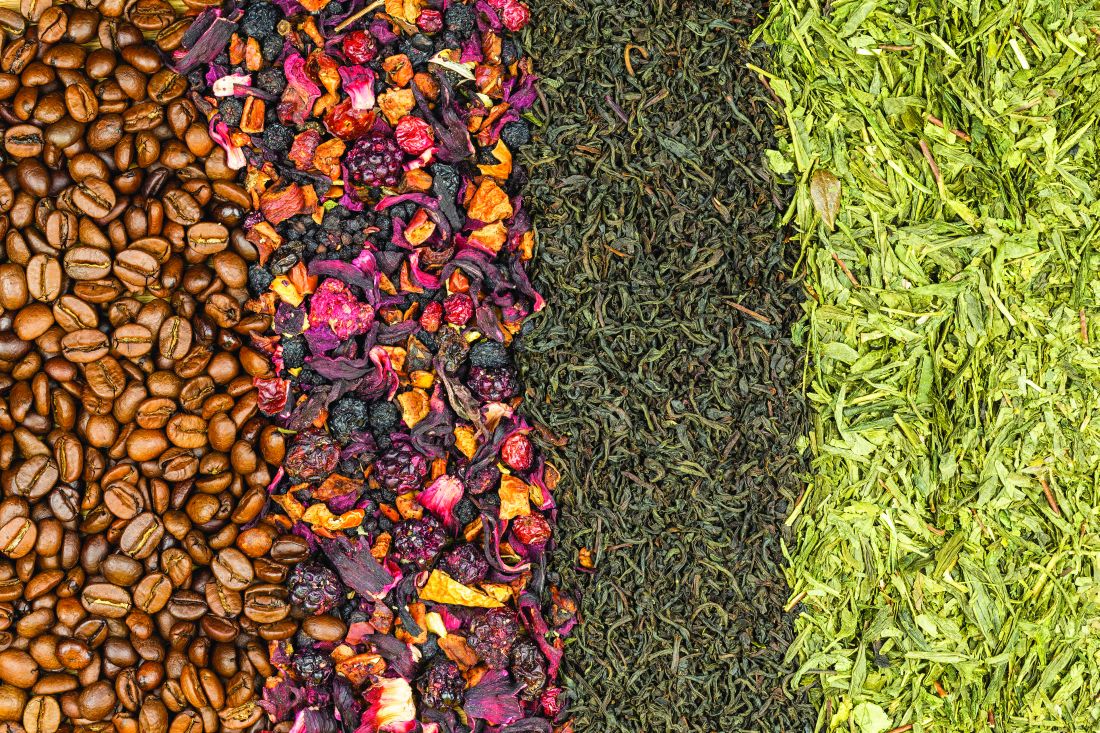
Chemistry and sources
Polyphenols share a common structural component: a phenol or an aromatic ring, usually two, with at least one hydroxyl, methyl, or acetyl group linked via a three-carbon bond to form a six-unit heterocyclic ring.8,9 When the “parent polyphenol” known as cinnamic acid is further catalytically transformed, scores of polyphenolic compounds result. These substances are divided into classes: glycosylated phenylpropanoids, flavonoids, isoflavonoids, stilbenoids, coumarins, curcuminoids, as well as phenolic polymers such as tannins, proanthocyanidins, suberin, lignins, and lignans. The flavonoids, which are the largest and most varied phenolic substances in plants, can be further divided into several categories: flavones (based on the 2-phenylchromen-4-one skeleton, such as apigenin and luteolin); flavonols (based on the 3-hydroxy-2-phenylchromen-4-one skeleton and functional group, such as quercetin, kaempferol, myricetin, and fisetin); flavanones (based on the 2,3-dihydro-2-phenylchromen-4-one skeleton and functional group, such as naringenin, hesperidin, and eriodictyol); isoflavones (based on the 3-phenylchromen-4-one skeleton, such as genistein and daidzein); flavanols – also known as flavan-3-ols or catechins – (based on the 2-phenyl-3,4-dihydro-2H-chromen-3-ol skeleton and functional groups, such as epicatechin, epicatechin 3-gallate, epigallocatechin, epigallocatechin 3-gallate (EGCG), catechin, and gallocatechin); and anthocyanins (based on the 2-phenylchromenylium ion skeleton, e.g., cyanidin and pelargonidin).5,10
The broader category of nonflavonoid polyphenols is rich and diverse, but is particularly noted for comprising the tannins, phenolic polymers of high molecular weight, which are divided into three classes, hydrolyzable tannins (such as ellagic acid, found in pomegranate, raspberries, strawberries, cranberries, and walnuts), derived tannins (created during food handling and processing and present in, for example, black and oolong teas), and condensed tannins (or proanthocyanidins, which are polymer chains of flavanols, such as catechins, and include pycnogenol, leukocyanidin, and leucoanthocyanin).1,4,5,8,10 There are a plethora of other nonflavonoid polyphenols, many of which confer health benefits, including stilbenes (such as resveratrol, found in red wine), lignans (such as enterodiol, found in flaxseed and flaxseed oil), lignins (found in green beans, carrots, peas, and Brazil nuts), and phenolic acids, such as hydroxybenzoic and hydroxycinnamic acids, among which caffeic and ferulic acids are often present in foods. In fact, hydroxycinnamic acids, which are the most common phenolic acids present in plant tissues, are present in numerous foods, such as apples, pears, plums, cherries, apricots, peaches, black currant, blueberries, potatoes, spinach, lettuce, cabbage, broccoli, asparagus, wine, and coffee.9
Broad health benefits have been associated with hundreds of polyphenolic substances. Notably, some of the best-known research results on polyphenols have reported on the success of various topical applications of green tea catechins, ferulic acid, and resveratrol, and other related compounds. Antioxidant, anti-inflammatory, and antimicrobial activities are the most common biologic properties associated with polyphenols, and antiaging activity has been widely reported.10
Conclusion
While the classification system for the 8,000 polyphenolic compounds may seem intimidating, the same essential activity is conferred by these abundant substances. Further, it is important to note the significant health benefits potentially derived from the oral consumption as well as topical application of polyphenols. The next two columns will delve into the research findings of flavonoid and nonflavonoid polyphenols.
Dr. Baumann is a private practice dermatologist, researcher, author and entrepreneur who practices in Miami. She founded the Cosmetic Dermatology Center at the University of Miami in 1997. Dr. Baumann wrote two textbooks: “Cosmetic Dermatology: Principles and Practice” (New York: McGraw-Hill, 2002), and “Cosmeceuticals and Cosmetic Ingredients,” (New York: McGraw-Hill, 2014), and a New York Times Best Sellers book for consumers, “The Skin Type Solution” (New York: Bantam Dell, 2006). Dr. Baumann has received funding for advisory boards and/or clinical research trials from Allergan, Evolus, Galderma, and Revance. She is the founder and CEO of Skin Type Solutions Franchise Systems LLC.
References
1. J Am Diet Assoc. 1999 Feb;99(2):213-8.
2. Ann N Y Acad Sci. 2012 Jul;1259:77-86.
3. Biomed Pap Med Fac Univ Palacky Olomouc Czech Repub. 2003 Dec;147(2):137-45.
4. J Nutr. 2000 Aug;130(8S Suppl):2073S-85S.
5. Annu Rev Nutr. 2002;22:19-34.
6. Pharmacol Ther. 2001 May-Jun;90(2-3):157-77.
7. Free Radic Biol Med. 2001 Jun 1;30(11):1213-22.
8. J Nutr. 2003 Oct;133(10):3248S-3254S.
9. Int J Mol Sci. 2016 Feb 18;17(2):160.
10. Asia Pac J Clin Nutr. 2004;13(Suppl):S72, 2004.
Polyphenols are well known as the largest group of and most widely distributed phytochemicals among plants.1 These secondary plant metabolites, which are produced in response to environmental hazards that contribute to free-radical synthesis,2 are represented by more than 8,000 naturally occurring compounds. This family of widely divergent substances has gained increasing attention in recent years as polyphenols have been found – in vegetables, fruits, herbs, grains, tea, coffee beans, honey, and red wine – to be the most abundant sources of antioxidants in the human diet and are known to exert antioxidant, anti-inflammatory, and antimicrobial benefits to human health.3-7 The most prevalent and studied polyphenols are known as flavonoids, but nonflavonoid polyphenols are increasingly well investigated. This column will address the basic chemistry of these compounds. Subsequent columns will discuss the latest research on the cutaneous benefits of selected flavonoid and nonflavonoid polyphenols.
Chemistry and sources
Polyphenols share a common structural component: a phenol or an aromatic ring, usually two, with at least one hydroxyl, methyl, or acetyl group linked via a three-carbon bond to form a six-unit heterocyclic ring.8,9 When the “parent polyphenol” known as cinnamic acid is further catalytically transformed, scores of polyphenolic compounds result. These substances are divided into classes: glycosylated phenylpropanoids, flavonoids, isoflavonoids, stilbenoids, coumarins, curcuminoids, as well as phenolic polymers such as tannins, proanthocyanidins, suberin, lignins, and lignans. The flavonoids, which are the largest and most varied phenolic substances in plants, can be further divided into several categories: flavones (based on the 2-phenylchromen-4-one skeleton, such as apigenin and luteolin); flavonols (based on the 3-hydroxy-2-phenylchromen-4-one skeleton and functional group, such as quercetin, kaempferol, myricetin, and fisetin); flavanones (based on the 2,3-dihydro-2-phenylchromen-4-one skeleton and functional group, such as naringenin, hesperidin, and eriodictyol); isoflavones (based on the 3-phenylchromen-4-one skeleton, such as genistein and daidzein); flavanols – also known as flavan-3-ols or catechins – (based on the 2-phenyl-3,4-dihydro-2H-chromen-3-ol skeleton and functional groups, such as epicatechin, epicatechin 3-gallate, epigallocatechin, epigallocatechin 3-gallate (EGCG), catechin, and gallocatechin); and anthocyanins (based on the 2-phenylchromenylium ion skeleton, e.g., cyanidin and pelargonidin).5,10
The broader category of nonflavonoid polyphenols is rich and diverse, but is particularly noted for comprising the tannins, phenolic polymers of high molecular weight, which are divided into three classes, hydrolyzable tannins (such as ellagic acid, found in pomegranate, raspberries, strawberries, cranberries, and walnuts), derived tannins (created during food handling and processing and present in, for example, black and oolong teas), and condensed tannins (or proanthocyanidins, which are polymer chains of flavanols, such as catechins, and include pycnogenol, leukocyanidin, and leucoanthocyanin).1,4,5,8,10 There are a plethora of other nonflavonoid polyphenols, many of which confer health benefits, including stilbenes (such as resveratrol, found in red wine), lignans (such as enterodiol, found in flaxseed and flaxseed oil), lignins (found in green beans, carrots, peas, and Brazil nuts), and phenolic acids, such as hydroxybenzoic and hydroxycinnamic acids, among which caffeic and ferulic acids are often present in foods. In fact, hydroxycinnamic acids, which are the most common phenolic acids present in plant tissues, are present in numerous foods, such as apples, pears, plums, cherries, apricots, peaches, black currant, blueberries, potatoes, spinach, lettuce, cabbage, broccoli, asparagus, wine, and coffee.9
Broad health benefits have been associated with hundreds of polyphenolic substances. Notably, some of the best-known research results on polyphenols have reported on the success of various topical applications of green tea catechins, ferulic acid, and resveratrol, and other related compounds. Antioxidant, anti-inflammatory, and antimicrobial activities are the most common biologic properties associated with polyphenols, and antiaging activity has been widely reported.10
Conclusion
While the classification system for the 8,000 polyphenolic compounds may seem intimidating, the same essential activity is conferred by these abundant substances. Further, it is important to note the significant health benefits potentially derived from the oral consumption as well as topical application of polyphenols. The next two columns will delve into the research findings of flavonoid and nonflavonoid polyphenols.
Dr. Baumann is a private practice dermatologist, researcher, author and entrepreneur who practices in Miami. She founded the Cosmetic Dermatology Center at the University of Miami in 1997. Dr. Baumann wrote two textbooks: “Cosmetic Dermatology: Principles and Practice” (New York: McGraw-Hill, 2002), and “Cosmeceuticals and Cosmetic Ingredients,” (New York: McGraw-Hill, 2014), and a New York Times Best Sellers book for consumers, “The Skin Type Solution” (New York: Bantam Dell, 2006). Dr. Baumann has received funding for advisory boards and/or clinical research trials from Allergan, Evolus, Galderma, and Revance. She is the founder and CEO of Skin Type Solutions Franchise Systems LLC.
References
1. J Am Diet Assoc. 1999 Feb;99(2):213-8.
2. Ann N Y Acad Sci. 2012 Jul;1259:77-86.
3. Biomed Pap Med Fac Univ Palacky Olomouc Czech Repub. 2003 Dec;147(2):137-45.
4. J Nutr. 2000 Aug;130(8S Suppl):2073S-85S.
5. Annu Rev Nutr. 2002;22:19-34.
6. Pharmacol Ther. 2001 May-Jun;90(2-3):157-77.
7. Free Radic Biol Med. 2001 Jun 1;30(11):1213-22.
8. J Nutr. 2003 Oct;133(10):3248S-3254S.
9. Int J Mol Sci. 2016 Feb 18;17(2):160.
10. Asia Pac J Clin Nutr. 2004;13(Suppl):S72, 2004.
Skin care Q and A for your staff
in helping us be more efficient with our time. I asked my staff to put together a list of the most common patient skin care questions. This column can be used as a guide for your staff when answering questions about skin care.
Q: Should my sunscreen be applied first or last?
A: The best way to remember is that medications should be applied closest to the skin and sunscreen should be applied closest to the sun. I recommend that morning skin care be applied in the following order:
Step 1. Facial cleanser
Step 2. Eye products (protect the delicate eye area from the medication)
Step 3. Treatment product (medications, or the most important active ingredients)
Step 4. Moisturizer (if needed)
Step 5. Sunscreen
Q: When can I restart my normal skin care regimen after receiving dermal fillers?
A: The morning after receiving dermal fillers, you can resume your normal facial skin care regimen.

Q: How do I treat my skin after receiving cosmetic injections such as Botox?
A: After you are injected with dermal fillers, keep the Arnica gel, Arnica pads, or post procedure product on your face until bedtime. Prior to bed, wash your face with a gentle cleanser and lukewarm water. Do not use any scrubs, facial brushes, or hydroxy acids. Apply a soothing gel or soothing moisturizer. Do not use hot water on the face for 48 hours. Avoid hot showers, saunas, facial steaming, facial massage, and exercise for 48 hours. If you must exercise, try to keep the face cool with an ice filled bottle of water. Heat makes you more likely to bruise.
Q: Will retinol make my skin sun sensitive?
A: Retinol breaks down upon sun exposure so it should only be used at night. It is a myth that retinol makes your skin sun sensitive. Retinol is a form of vitamin A that helps protect your skin from sun damage by reducing levels of an enzyme called collagenase that is produced after sun exposure. For this and other reasons, retinol can help protect your skin from sun damage. However, if your skin gets irritated from the retinol and you go into the sun, you can develop postinflammatory hyperpigmentation – or dark patches on the skin. It is a good idea to always wear sunscreen in the morning every day whether you are using retinol or not.
Q: What do I do to treat the dark circles under my eyes?
A: Dark circles can be caused by sluggish blood flow around the eyes leading to deposition of a pigment found in blood called hemosiderin. Dark circles around the eyes also can be caused by an increased amount of the pigment melanin that occurs from sun exposure, heat, estrogen, and stress. Treatments include lifestyle habits that increase blood circulation, such as smoking cessation and exercise. Supplements such as horse chestnut may help improve circulation. Ingredients such as the tyrosinase inhibitors ascorbic acid, kojic acid, arbutin, and hydroquinone will help decrease melanin production, and may improve dark circles caused by increased melanin. Wearing a daily sunscreen around the eyes is a very important part of improving the appearance of dark circles under the eyes.
Q: Do I need an eye cream?
A: Most people need an eye product whether it is a serum, gel, or cream. Eye treatment products are usually one of the first skin care products that male patients purchase. The most common indications for eye treatment products are dark circles under the eyes, puffy eyes, dryness, fine lines, or redness. Each one of these indications requires very different ingredients; therefore, most people need more than one eye product depending on what eye issues they are experiencing. Eye treatment products can protect the delicate eye area from medications such as tretinoin that gets on the pillowcase and is transferred to the eye area at night. Eye products that have humectants, such as heparan sulfate or hyaluronic acid, should be used to treat fines lines and dryness, but these may make eyes puffy. For those with dry and puffy eyes, a second eye product with vasoconstrictive ingredients should be used in the daytime while the hydrating one is used at night. Antiaging eye products with retinol and hydroxy acids may irritate sensitive skin types so they can be paired with a soothing eye product. Consider using different eye products for the morning and evening depending on what eye issues need to be treated.
Q: Will moisturizer make my acne worse?
A: Noncomedogenic moisturizers can help improve acne and speed recovery. Using the proper cleanser and moisturizer will make acne medications more tolerable. Many patients cannot tolerate their acne medications every day. The bacteria that cause acne reproduce every 12 hours. For this reason, acne medications must be used consistently twice a day. Using the proper cleanser and moisturizer makes this possible in most patients. Avoid moisturizers with coconut oil, isopropyl myristate, and other ingredients known to cause acne.
Q: Why does my face get so red after washing it?
A: Rosacea-prone skin types get red from the friction of washing the face – even if just using water. If this occurs, use skin care products with anti-inflammatory ingredients such as argan oil, green tea, fever few, chamomile, caffeine, resveratrol, and niacinamide to sooth the skin. These can be paired with prescription rosacea medications such as Rhofade (oxymetazoline hydrochloride) to reduce facial redness. People with rosacea-prone skin types should avoid facial brushes, scrubs, and vigorous exfoliation.
Q: Why is my melasma not getting better on skin lighteners?
Educating our staff on the basics of skin care can make our office much more efficient. If you liked the format of this column or have questions that you hear often from patients, please email me at DrB@derm.net.
Dr. Baumann is a private practice dermatologist, researcher, author and entrepreneur who practices in Miami. She founded the Cosmetic Dermatology Center at the University of Miami in 1997. Dr. Baumann wrote two textbooks: “Cosmetic Dermatology: Principles and Practice” (New York: McGraw-Hill, 2002), and “Cosmeceuticals and Cosmetic Ingredients,” (New York: McGraw-Hill, 2014), and a New York Times Best Sellers book for consumers, “The Skin Type Solution” (New York: Bantam Dell, 2006). Dr. Baumann has received funding for advisory boards and/or clinical research trials from Allergan, Evolus, Galderma, Aclaris, and Revance. She is the founder and CEO of Skin Type Solutions Franchise Systems LLC.
in helping us be more efficient with our time. I asked my staff to put together a list of the most common patient skin care questions. This column can be used as a guide for your staff when answering questions about skin care.
Q: Should my sunscreen be applied first or last?
A: The best way to remember is that medications should be applied closest to the skin and sunscreen should be applied closest to the sun. I recommend that morning skin care be applied in the following order:
Step 1. Facial cleanser
Step 2. Eye products (protect the delicate eye area from the medication)
Step 3. Treatment product (medications, or the most important active ingredients)
Step 4. Moisturizer (if needed)
Step 5. Sunscreen
Q: When can I restart my normal skin care regimen after receiving dermal fillers?
A: The morning after receiving dermal fillers, you can resume your normal facial skin care regimen.
Q: How do I treat my skin after receiving cosmetic injections such as Botox?
A: After you are injected with dermal fillers, keep the Arnica gel, Arnica pads, or post procedure product on your face until bedtime. Prior to bed, wash your face with a gentle cleanser and lukewarm water. Do not use any scrubs, facial brushes, or hydroxy acids. Apply a soothing gel or soothing moisturizer. Do not use hot water on the face for 48 hours. Avoid hot showers, saunas, facial steaming, facial massage, and exercise for 48 hours. If you must exercise, try to keep the face cool with an ice filled bottle of water. Heat makes you more likely to bruise.
Q: Will retinol make my skin sun sensitive?
A: Retinol breaks down upon sun exposure so it should only be used at night. It is a myth that retinol makes your skin sun sensitive. Retinol is a form of vitamin A that helps protect your skin from sun damage by reducing levels of an enzyme called collagenase that is produced after sun exposure. For this and other reasons, retinol can help protect your skin from sun damage. However, if your skin gets irritated from the retinol and you go into the sun, you can develop postinflammatory hyperpigmentation – or dark patches on the skin. It is a good idea to always wear sunscreen in the morning every day whether you are using retinol or not.
Q: What do I do to treat the dark circles under my eyes?
A: Dark circles can be caused by sluggish blood flow around the eyes leading to deposition of a pigment found in blood called hemosiderin. Dark circles around the eyes also can be caused by an increased amount of the pigment melanin that occurs from sun exposure, heat, estrogen, and stress. Treatments include lifestyle habits that increase blood circulation, such as smoking cessation and exercise. Supplements such as horse chestnut may help improve circulation. Ingredients such as the tyrosinase inhibitors ascorbic acid, kojic acid, arbutin, and hydroquinone will help decrease melanin production, and may improve dark circles caused by increased melanin. Wearing a daily sunscreen around the eyes is a very important part of improving the appearance of dark circles under the eyes.
Q: Do I need an eye cream?
A: Most people need an eye product whether it is a serum, gel, or cream. Eye treatment products are usually one of the first skin care products that male patients purchase. The most common indications for eye treatment products are dark circles under the eyes, puffy eyes, dryness, fine lines, or redness. Each one of these indications requires very different ingredients; therefore, most people need more than one eye product depending on what eye issues they are experiencing. Eye treatment products can protect the delicate eye area from medications such as tretinoin that gets on the pillowcase and is transferred to the eye area at night. Eye products that have humectants, such as heparan sulfate or hyaluronic acid, should be used to treat fines lines and dryness, but these may make eyes puffy. For those with dry and puffy eyes, a second eye product with vasoconstrictive ingredients should be used in the daytime while the hydrating one is used at night. Antiaging eye products with retinol and hydroxy acids may irritate sensitive skin types so they can be paired with a soothing eye product. Consider using different eye products for the morning and evening depending on what eye issues need to be treated.
Q: Will moisturizer make my acne worse?
A: Noncomedogenic moisturizers can help improve acne and speed recovery. Using the proper cleanser and moisturizer will make acne medications more tolerable. Many patients cannot tolerate their acne medications every day. The bacteria that cause acne reproduce every 12 hours. For this reason, acne medications must be used consistently twice a day. Using the proper cleanser and moisturizer makes this possible in most patients. Avoid moisturizers with coconut oil, isopropyl myristate, and other ingredients known to cause acne.
Q: Why does my face get so red after washing it?
A: Rosacea-prone skin types get red from the friction of washing the face – even if just using water. If this occurs, use skin care products with anti-inflammatory ingredients such as argan oil, green tea, fever few, chamomile, caffeine, resveratrol, and niacinamide to sooth the skin. These can be paired with prescription rosacea medications such as Rhofade (oxymetazoline hydrochloride) to reduce facial redness. People with rosacea-prone skin types should avoid facial brushes, scrubs, and vigorous exfoliation.
Q: Why is my melasma not getting better on skin lighteners?
Educating our staff on the basics of skin care can make our office much more efficient. If you liked the format of this column or have questions that you hear often from patients, please email me at DrB@derm.net.
Dr. Baumann is a private practice dermatologist, researcher, author and entrepreneur who practices in Miami. She founded the Cosmetic Dermatology Center at the University of Miami in 1997. Dr. Baumann wrote two textbooks: “Cosmetic Dermatology: Principles and Practice” (New York: McGraw-Hill, 2002), and “Cosmeceuticals and Cosmetic Ingredients,” (New York: McGraw-Hill, 2014), and a New York Times Best Sellers book for consumers, “The Skin Type Solution” (New York: Bantam Dell, 2006). Dr. Baumann has received funding for advisory boards and/or clinical research trials from Allergan, Evolus, Galderma, Aclaris, and Revance. She is the founder and CEO of Skin Type Solutions Franchise Systems LLC.